Embed Size (px)
Citation preview

Parasite Causing Skin Disorders
Agnes Kurniawan, MD. PhDDepartment of Parasitology FMUI
AK/Skin module/Nov’07

Parasites causing Skin Disorders
■ Cutaneous Larva Migrans (hookworm, Strongyloides)
■ Onchocerca dermatitis ■ Cercarial dermatitis, Swimmer’s itch
( Schistosoma)■ Subcutaneous Cysticercosis■ Cutaneous Leishmaniasis ■ Cutaneous Amebiasis■ Arthropod Infections
- Scabies - Pediculosis- Myasis - Demodiosis- Dermatitis (House Dust Mites)

Dermatosis■ Various skin irritations are caused by
arthropods, either through bites or skin invasions.
■ Skin irritations commonly results from the bites of insects: mosquitoes, fleas, lice, bed bugs.
■ Various species of burrowing mites cause skin irritations known as acariasis eg. scabies mites, the follicle mites etc
AK/Skin module/Nov’07

Scabies■ Etiology: Sarcoptes scabiei ■ Distribution: cosmopolitan
- poor urban populations- crowded community- minimal facilities for bath & washing scabies is common in slum area,
jail, orphanage, dormitory.
AK/Skin module/Nov’07

Morphology■ small■ flattened & whitish■ female’s:250-350цm ■ male’s: 150-200 цm■ Adults: 4 pairs of
short legs■ Larvae: 3 pairs of
legs
AK/Skin module/Nov’07

Life Cycle
■ Metamorphosis: egg-larva-nymph-adult■ The female enters the skin burrows
itself 2-3 mm during the course of 24 hours, especially at night.
■ Warmth increases its activity■ The mite, burrows very superficially (in
stratum corneum)
AK/Skin module/Nov’07

AK/Skin module/Nov’07

Pathology■ Predilection: thin & wrinkled skin (eg.
between fingers & toes, on the wrist, extensor surface of the elbows, arm pit, under the breasts,on the penis, scrotum and feet
■ In adults, the presence of burrows on the face or neck is unusual
■ in children whose skin is thin, infection may occur anywhere including the face and neck.
AK/Skin module/Nov’07

■ The lesions appear as slightly reddish elevated tracts on the skin.
■ Minute, translucent, vesicular swellings, possibly produced by the irritating fecal deposits or excretions.
AK/Skin module/Nov’07

Pathology■ The intense itching scratch spreads
the parasite, irritates the lesions multiple papular, vesicular and pustular may be produced
■ Unless treated, a chronic condition may ensue, and severe infestations may prove a serious health hazard.
AK/Skin module/Nov’07

AK/Skin module/Nov’07

AK/Skin module/Nov’07

Diagnosis
■ Finding the tunnel by burrow ink test (but this method can not be used for finding the mites)
■ Confirmed diagnosis is based on finding the mite by needle, skin scraping, or superficial biopsy.
AK/Skin module/Nov’07

Scabies controlTreatment: 5% permethrin topical cream 1% gamma benzene hexachloride 3-10% sulfur ointment
Prevention: sterilization of garment and bedding personal hygiene.
AK/Skin module/Nov’07

MYIASIS
■ Myiasis : invasion of dipterous larvae into living tissues of man / mammals
■ Metamorphosis of fly: egg-larvae-pupae-adult
AK/Skin module/Nov’07

Classification of Myiasis
1. Cutaneous : the larvae live in or under the skin
2. Intestinal : the larvae are in the stomach or intestine
3. Atrial : the larvae invade the oral, nasal, aural, ocular, vaginal, urethral cavities
4. Wound : the larvae enter artificial lesions5. External : the larvae are bloodsuckers
AK/Skin module/Nov’07

Cutaneus Myiasis■ The egg: deposited on the wound ■ The larvae are able to burrow through
necrotic or healthy tissues with their chitinous mandibular hooks
■ Tissues destruction is facilitated by 2nd bacterial inf & proteolytic secretions.
AK/Skin module/Nov’07

Identification of larvae

Cutaneous myiasis
Chrysomyia megacephala

Diagnosis1. Characteristic of the posterior
spiracle.2. Identification of adult flies– take the larvae from the lesion
culture adult stage identify the species

Treatment
■ Paralyzed the larvae with chloroform and then extract by forceps.
■ Anthelmintic drug may be used for intestinal form.

Control of myiasis
1. Reduction of breeding places (eg. destruction of garbage disposal)
2. Fly trap (ultraviolet, glue)3. Insecticides

PEDICULOSIS

associated with people living crowded together with limited facilities for regular bathing and laundering
the condition is most common during stressful time such as war and in concentration camps, evacuation centers, schools and institutions.
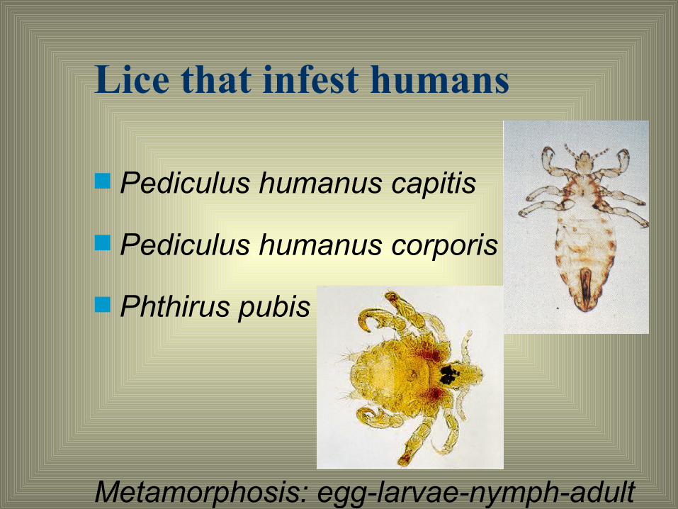
Lice that infest humans
■ Pediculus humanus capitis
■ Pediculus humanus corporis
■ Phthirus pubis
Metamorphosis: egg-larvae-nymph-adult

Phthirus pubis/Crab lice
■ small grayish-white insects ■ short abdomen: hairy lateral tufts ■ large second and third pairs of legs
which give a crab-like appearance. ■ most commonly found on pubic hairs■ may be found on hairy areas of the
chest, armpits, eyebrows, eyelashes.

Pathology– The lesions are due to the bites of young
and adult lice– The lesions due to the head louse occur
most frequently on the back of the head and neck, OR anywhere on the scalp
– The lesions due to body louse is present on the parts of the body in close contact with clothing

Pediculosis

PathologyThe irritating saliva, injected during feedingproduces a rosette elevated papule accompanied by severe itching. Individuals vary in sensitivity and in
chronic infestations, the reaction may be slight

Scratching increases the inflammation secondary bacterial infection with the sequelae of pustules, crusts, supurative processes, matted hair.
Severe infestations scarring, induration, skin pigmentation, ulceration
Pathology

Pathology
■ Infestation of the eyelashes through secondary infection conjunctivitis & keratitis.
■ Symptoms : related to cutaneous irritation, loss of sleep, depression.
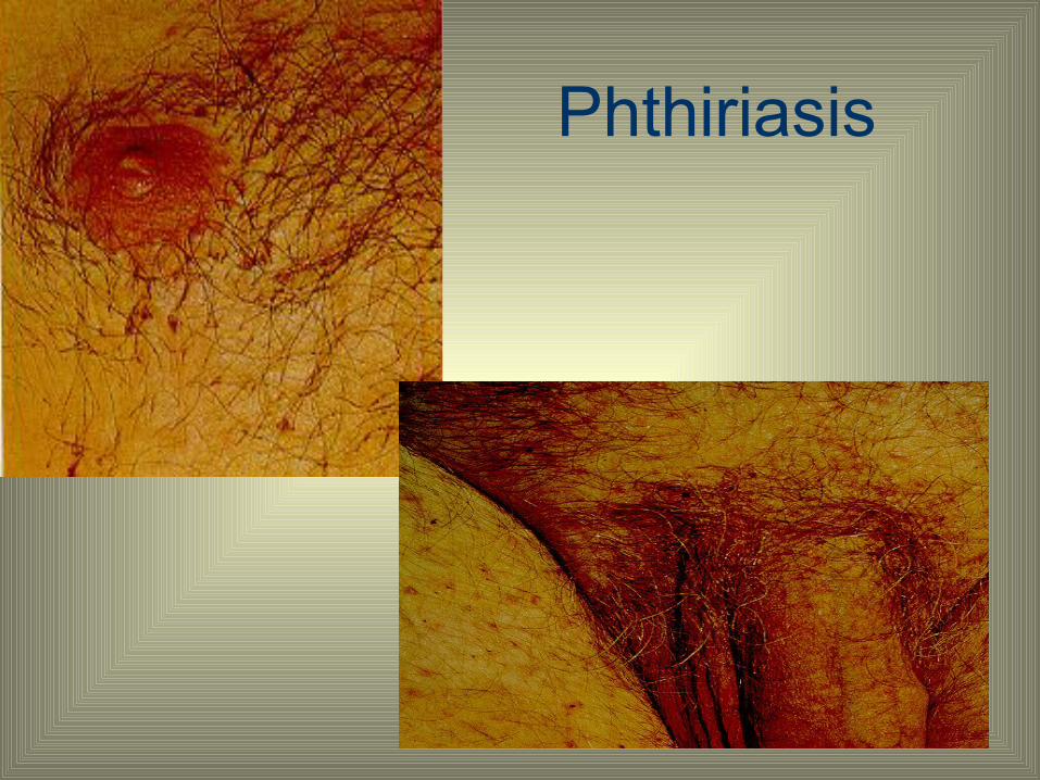
Phthiriasis


Control of body lice
■ Ordinary laundering with hot water will destroy all stages of lice on infested clothing & bedding
■ Dry cleaning may be used to destroy lice on wool garments
■ The solvent used in cleaning is toxic to lice, and the steam used in pressing makes certain that control is complete

Control of body lice
■ Pressing cloth at home is also satisfactory, but special attention must be given to the seams.
■ Insecticide powders 1% lindane or 1% malathion, have been extensively used to control body lice.

Control of head lice■ For school boys, men, prisoners, a very
close hair cut or even shaving the head to remove the eggs and lice
■ a simple treatment for crab louse control is shaving or cutting the infested hair to remove adults, immature stages, and eggs
■ The safest and best materials are 1% permethrin or 1% lindane

■ Comb the hair in the usual manner.■ After 10 minutes with the permethrin
emulsion, or after 24 hours with the lindane, shampoo the hair.
■ Dry, comb, and brush hair to remove dead lice and loosened eggs.

Dermatophagoides pteronyssinus
■ House dust mite■ Size 0.2 – 1.2 mm■ Adult & nymph: 4
pairs of legs■ Larvae: 3 pairs of
legs■ The legs are equal
in length and structure

Habitat
■ House dust- Carpets- Mattresses- Pillows- Furniture- Animals- Floor etc

Life cycle
■ Metamorphosis: egg-larva-nymph-adult■ The egg: 6 days■ The nymphal stage: 8-15 days■ Adult live: males 60-80 days
females 100-150 days

Factor influence the growth
■ Food: human scales■ Relative humidity: 80-90%■ Temperature: 24-26 C

Pathology
Mite produces allergen the allergen contaminate environment dust particles dust particles contaminated with the allergen are inhaled by the persons persons get sensitized dermatitis, asthma

Control
■ Expose mattresses, carpets etc to direct sunlight (6 hours)
■ Clean the house■ Insecticide: - 1% lindane or- benzyl benzoate

Cutaneous larva migrans (creeping eruption)
■ a dermatitis characterized by serpiginous, intracutaneous lesions caused by migration of nematode larvae that normally do not infect the human host
■ The hookworm of cats and dogs, Ancylostoma braziliense, is most commonly incriminated
■ other species of hookworms, such as A. caninum (rare)
■ Other cause : S. stercoralis infection
Cutaneous larva migrans
■ The Infective larvae of other nonhuman helminthic parasites can enter the human skin, or even be ingested, and fail to complete their development in this abnormal host can produce annoying skin eruptions by allergic sensitization.

Pathology and Symptomatology
■ At the points of larval invasion, indurated, reddish, itchy papules
■ in 2 to 3 days narrow, linear, slightly elevated, erythematous, serpiginous, intracutaneous tunnels, Ø1-2 mm, are produced by the migratory larvae
■ Speed : ±1 inch/day but rarely pass beyond a few inches from the original site of entry

(Courtesy of Kurniawan, A, 2006)
(Courtesy of Kurniawan, A, 2006)

■ Vesicles : along the tunnels, the surface becomes dry and crusty.
■ Local eosinophilia and round-cell infiltration may be present
■ The itching is intense, especially at night scratch secondary infection
■ The feet, legs and hands are most commonly involved, but infection on other of parts of the body may occur

Treatment
■ Mebendazole/albendazole, topical■ Light infections with only several larvae freezing the active area with ethyl chloride or CO2 snow
■ 2nd bacterial infection antibiotics.

Prevention
■ avoiding skin contact with soil■ keeping dogs and cats off beaches and
away from the space under houses ■ children’s sandboxes should be covered
when not in use. ■ anthelmintic treatment of dogs and cats
will prevent contamination of the soil

Onchocerca volvulusChronic skin manifestations : hipopigmentation,lichenification, decrease of skin elasticity, sowda, leopard skin, hanging groin

Enterobius vermicularis
Host : human Diseases : oxyuriasis, enterobiasis Distribution : cosmopolitan
prevalence in Indonesia is high especially in children

Enterobius vermicularisPathology and symptomatology
• pruritus ani • ectopic infection • enter into the vulva, vagina,
fallopian tubes, appendix • other symptoms

Enterobius vermicularisEpidemiology Infection may be effected by
1. hand (auto infection and infections to another person)
2. dressers and another tools 3. dust

SchistosomaCercaria : Release from the snail to the water skin penetration
schistosomula
Blood circulation
Adult worms in vein

Cercarial Dermatitis■ Schistosomiasis zoonotic disease ■ Infects - human + other mammal
(Reservoir hosts) Baboons, rats, horses, Water buffaloes, boars

Biologic Incubation Period :
Skin rash : ■ Cercariae : dermatitis ■ Shistosomulae/adult worms/protein of
the death worm :– Urticaria– Angioneurotic edema– Fever

Cystisercosis cellulose

Cysticercosis
■aetiology: cysticercus cellulosae (Larvae of the tapeworm T solium)
■Larvae are acquired by ingestion of T solium ova

T. solium: life cycle
Definitive host: man adult worm attached to wall proximal portion of small intestine
Intermediate host: pig, man, dog cyst in muscles /subcutaneous tissue, brain, heart, eye, etc.

Life Cycle
cysticercus evaginated in human small intestinal
scolex + neck
Immature proglotid
mature proglotid
Gravid proglotid expelled
Eggs in faeces
cysticercus
Taeniasis
Undercooked meat

SIGNS / SYMPTOMS /
■ TaeniasisGastro-intestinal complaints
Cysticercosis: Depend on infected organs: - brain seizures/epileptic, headache, changes in
behavior - subcutaneous nodules, eye, heart disorders etc.

Public Health Importance
Depend on location of the larvae:■ subcutaneous tumor
(subcutaneous cysticercosis)■ cerebral tumor■ eye■ muscle

DIAGNOSIS
– Detection of Ab against T. solium : Enzyme-linked immunotransfer blot (EITB) / ELISA
– Examine stools for ova and parasites ( 3 consecutive days )
– CT scanning shows the cyst (solitary or multiple and usually are 5-20 mm in diameter) and granuloma stages of neurocysticercosis. Most often in the cortex / gray-white junction.
– Magnetic resonance imaging
Cysticercosis
Treatment Albendazole / Praziquantel Prednisone

Evaluation of treatment
Cysticercosis: 3 months after treatment decreasing number /no nodules found
Neurocysticercosis: frequency of seizures decreased and after 2 years no seizures any more

Cutaneous Amebiasis
Aetiology : Entamoeba histolytica Host : man Mode of infection : ingestion of mature cyst Morphology :
– trophozoite form : histolytica form and minuta form
– cyst form (1-2-4 nuclei)


Cutaneous Amebiasis
■intestinal or hepatic fistula■mucosa bathed in fluids containing trophozoites– perianal ulcers– urogenital (eg, labia, vagina, penis)

Complications :
■ Perianal and perineal amoebiasis ■ Perforation■ Peritonitis■ Ulcers in skin and vagina

Diagnosis :
Histolytica forms in feces, abscess, ulcer Ab detection