Embed Size (px)
Citation preview

Parasites are divided into three main phylum:
1.Protozoa 2.Helminths 3.Arthropods
Protozoa divided into three classes
Apicomplexa
Sarcomastigophora
Ciliophora
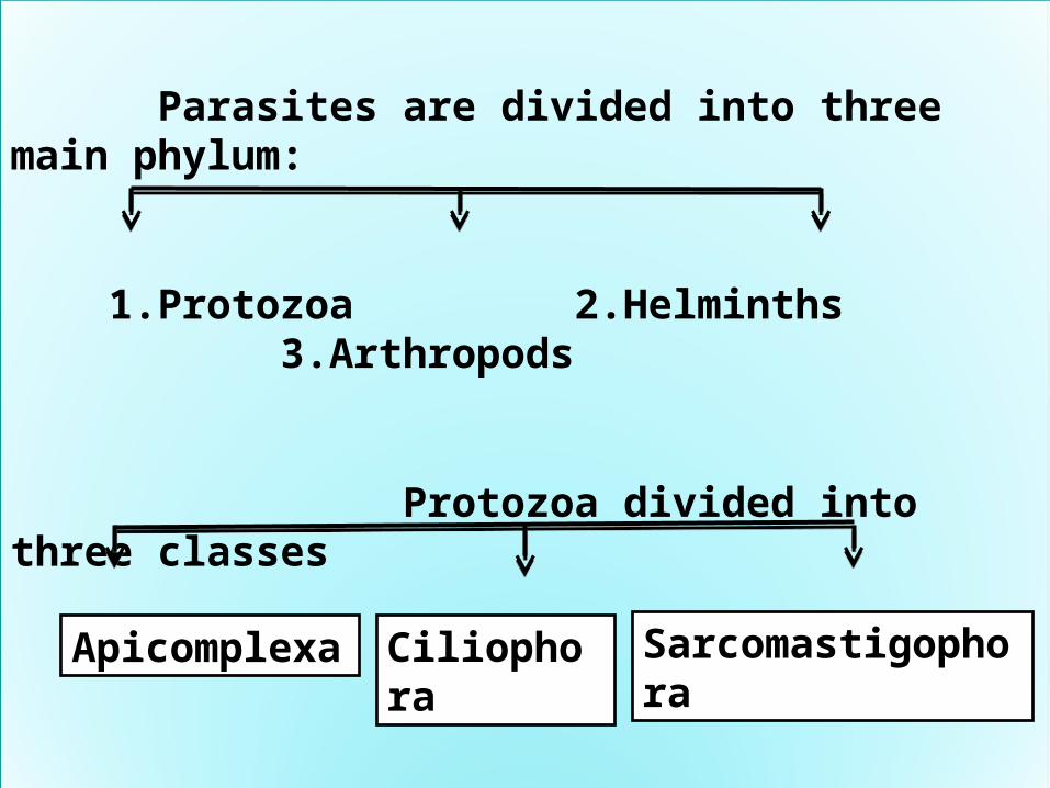
Phylum : Helminthes
Class:
Platyhelminthes
Nemathelminthes
Cestoda Trematoda
Nematoda
Taenia sp. and Echinococcus granulosis
Schistosoma sp. Entrobuis vermicularis and Ascaris lumbricoides
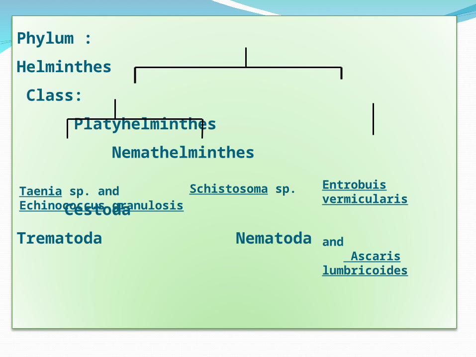
NEMATODES
Ascaris lumbricoides
Enterobius vermicularis
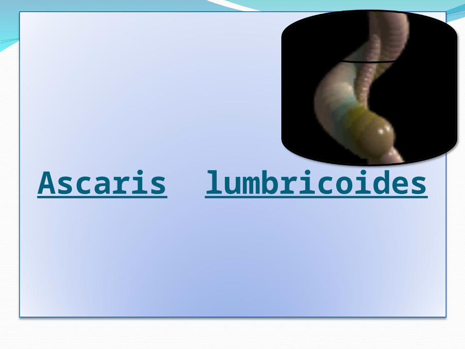
Ascaris lumbricoides

Common name:- Giant Intestinal round worms.
Disease:-Ascariaisis
Host :- The human is intermediate and final host.
Location in Definitive host :-
the adult worm: in small intestines .
larva: in lung .
Infective stage :- Ovum
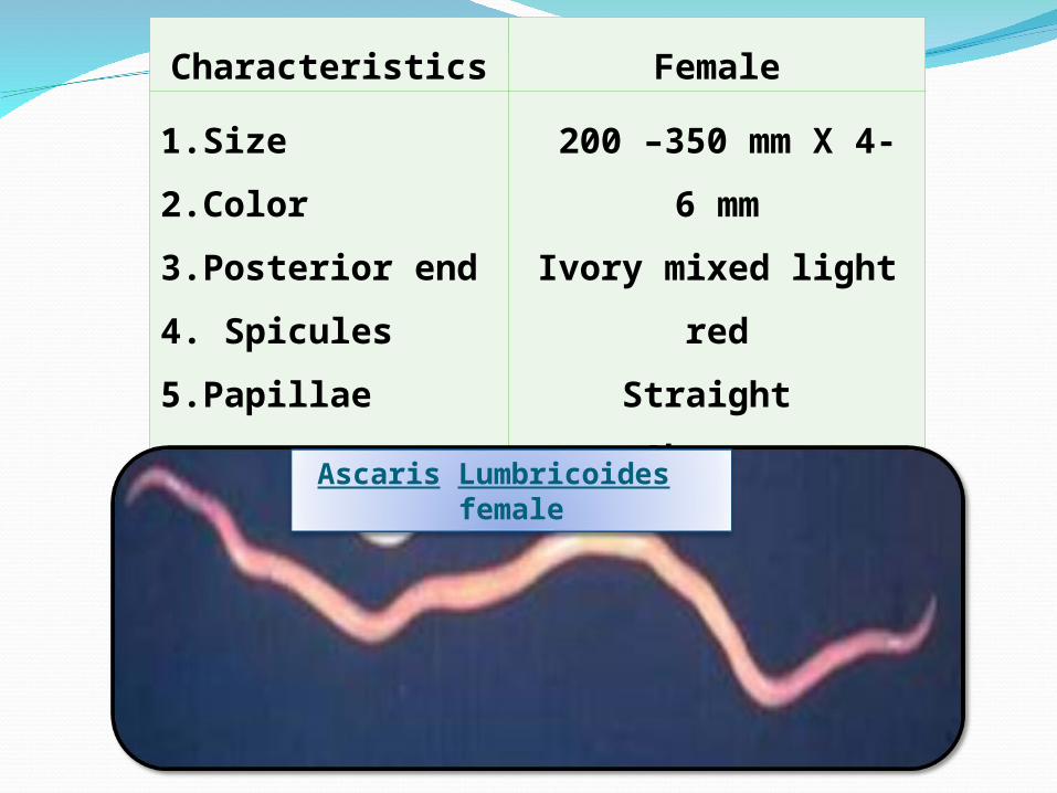
Characteristics Female
1.Size
2.Color
3.Posterior end
4. Spicules
5.Papillae
200 –350 mm X 4- 6 mm
Ivory mixed light red
Straight
Absent
Absent
Ascaris Lumbricoides female
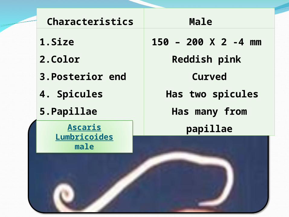
Characteristics Male
1.Size
2.Color
3.Posterior end
4. Spicules
5.Papillae
150 – 200 X 2 -4 mm
Reddish pink
Curved
Has two spicules
Has many from papillae
Ascaris Lumbricoides
male
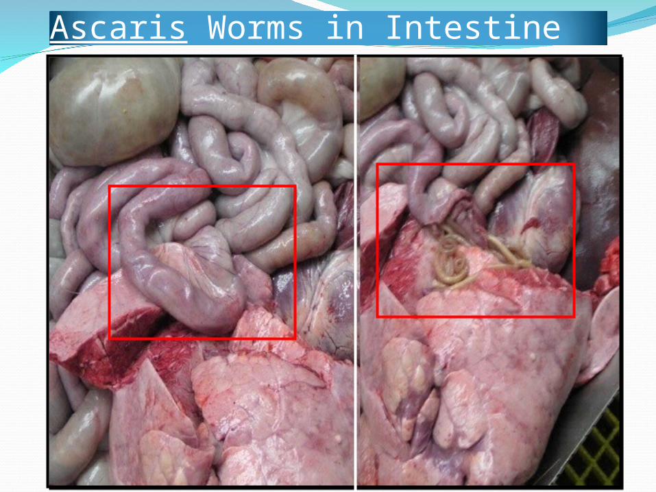
Ascaris Worms in Intestine
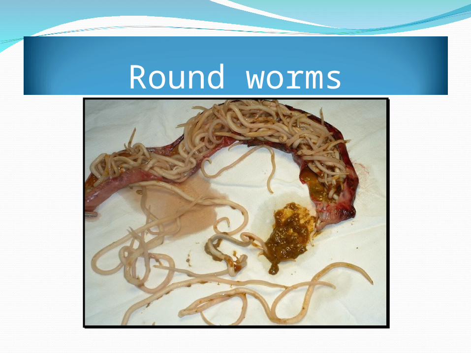
Round worms
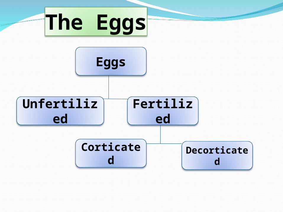
The Eggs
Eggs
Fertilized
Unfertilized
Decorticated
Corticated
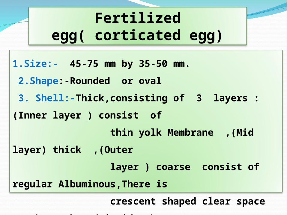
Fertilized egg( corticated egg)
1.Size:- 45-75 mm by 35-50 mm.
2.Shape:-Rounded or oval
3. Shell:-Thick,consisting of 3 layers : (Inner layer ) consist of
thin yolk Membrane ,(Mid layer) thick ,(Outer
layer ) coarse consist of regular Albuminous,There is
crescent shaped clear space at the each end inside the
shell.
4.Colour:-Golden brown.
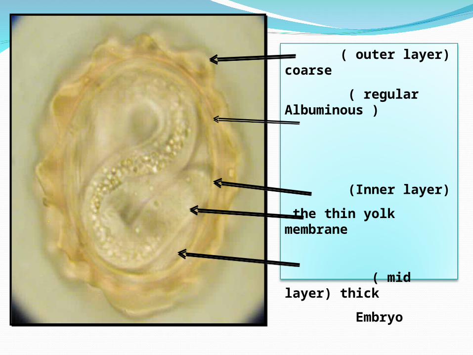
( outer layer) coarse
( regular Albuminous )
(Inner layer)
the thin yolk membrane
( mid layer) thick
Embryo
Crescent space
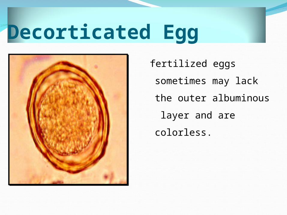
Decorticated Egg fertilized eggs sometimes
may lack the outer
albuminous layer and
are colorless.
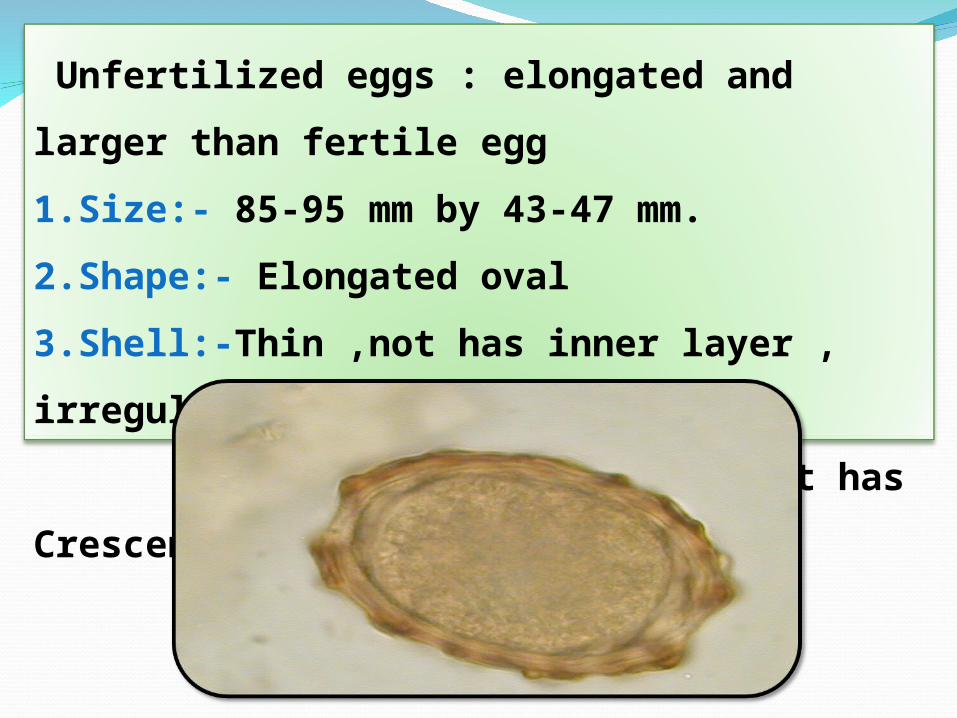
Unfertilized eggs : elongated and larger than fertile egg
1.Size:- 85-95 mm by 43-47 mm.
2.Shape:- Elongated oval
3.Shell:-Thin ,not has inner layer , irregular
albuminous layer , not has Crescent space.
Modes of transmission
Occurs mainly by ingestion of contaminated
food or water with eggs.
Occasionally by inhalation of contaminated dust
with eggs.
Children playing in contaminated soil may
acquire the parasite from their hands.
DiagnosisThe diagnosis depends on the Identify
worm or egg:-
The Stool examination : for seeing the
adult worm or egg .
The sputum examination :for seeing
larva during migration in the lung.
Eosinophilia as indicator on parasitic
infection .
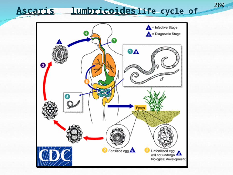
Ascaris lumbricoides life cycle of 280
Enterobius vermicularis
Known name:- pin worm
Disease:- Enterobiaisis
Host :- human is intermediate and final host.
Location in Definitive host :
The adult worms inhabit in the cecum and the
colon.
Infective stage :- Ovum
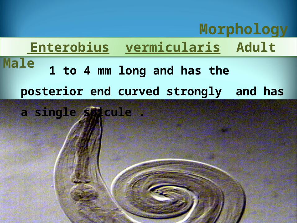
1 to 4 mm long and has the posterior end curved
strongly and has a single spicule .
Morphology Enterobius vermicularis Adult Male
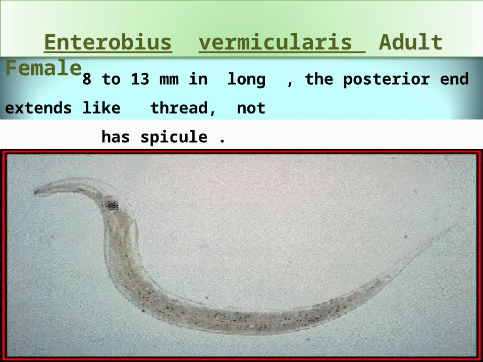
8 to 13 mm in long , the posterior end extends like thread, not
has spicule .
Enterobius vermicularis Adult Female
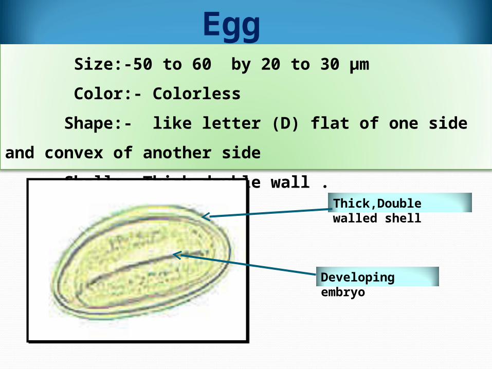
Size:-50 to 60 by 20 to 30 µm
Color:- Colorless
Shape:- like letter (D) flat of one side and convex of another side
Shell:- Thick double wall .
Egg
Developing embryo
Thick,Double walled shell
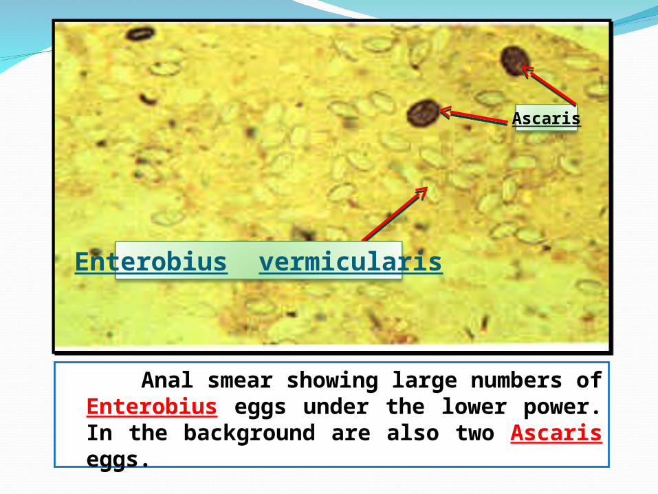
Anal smear showing large numbers of Enterobius eggs under the lower power. In the background are also two Ascaris eggs.
Enterobius vermicularis
Ascaris
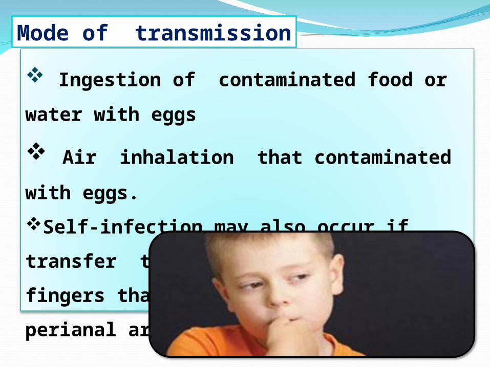
Ingestion of contaminated food or water with eggs
Air inhalation that contaminated with eggs.
Self-infection may also occur if
transfer the eggs to themouth by fingers
that have scratched the perianal area.
Mode of transmission

Diagnosis are often clinically by
observing the female worm (or many
worms) in the perianal region, but can
also be using the "scotch-tape" test, or
swab of stool.
Diagnosis