Embed Size (px)
Citation preview

Parasitic Copepods from the Gulfof Mexico and Caribbean Sea, I:Holobomolochus and Neobomolochus
ROGER CRESSEY
m
wtu.
SMITHSONIAN CONTRIBUTIONS TO ZOOLOGY • NUMBER 339

SERIES PUBLICATIONS OF THE SMITHSONIAN INSTITUTION
Emphasis upon publication as a means of "diffusing knowledge" was expressedby the first Secretary of the Smithsonian. In his formal plan for the Institution, JosephHenry outlined a program that included the following statement: "It is proposed topublish a series of reports, giving an account of the new discoveries in science, andof the changes made from year to year in all branches of knowledge." This themeof basic research has been adhered to through the years by thousands of titles issuedin series publications under the Smithsonian imprint, commencing with SmithsonianContributions to Knowledge in 1848 and continuing with the following active series:
Smithsonian Contributions to AnthropologySmithsonian Contributions to Astrophysics
Smithsonian Contributions to BotanySmithsonian Contributions to the Earth SciencesSmithsonian Contributions to the Marine Sciences
Smithsonian Contributions to PaleobiologySmithsonian Contributions to ZoologySmithsonian Studies in Air and Space
Smithsonian Studies in History and Technology
In these series, the Institution publishes small papers and full-scale monographsthat report the research and collections of its various museums and bureaux or ofprofessional colleagues in the world of science and scholarship. The publications aredistributed by mailing lists to libraries, universities, and similar institutions throughoutthe world.
Papers or monographs submitted for series publication are received by theSmithsonian Institution Press, subject to its own review for format and style, onlythrough departments of the various Smithsonian museums or bureaux, where themanuscripts are given substantive review. Press requirements for manuscript and artpreparation are outlined on the inside back cover.
S. Dillon RipleySecretarySmithsonian Institution

S M I T H S O N I A N C O N T R I B U T I O N S T O Z O O L O G Y • N U M B E R 3 3 9
Parasitic Copepods from the Gulfof Mexico and Caribbean Sea, I:Holobomolochus and Neobomolochus
Roger Cressey
SMITHSONIAN INSTITUTION PRESS
City of Washington
1981
A B S T R A C T
Cressey, Roger. Parasitic Copepods from the Gulf of Mexico and CaribbeanSea, I: Holobomolochus and Neobomolochus. Smithsonian Contributions to Zoology,number 339, 24 pages, 72 figures, 1981.—Four species of HolobomolochusVervoort are described: H. glyphisodontis (Kr0yer) from Abudefduf saxatilis; H.centropristis, new species, from Centropristis melana; H. crevalleus, new species,from Caranx hippos; H. serratus, new species, from Scorpaena brasiliensis; and anew genus and species, Neobomolochus elongatus, from Opisthonema oglinum. Thenew genus superficially resembles Pseudoeucanthus Brian but differs from it bythe presence of three modified setae on the first antennae of Neobomolochus.Bomolochus nothrus Wilson is placed in synonomy with Holobomolochus glyphiso-dontis (Kr0yer), and Bomolochus ardeolae Kr0yer is placed in synonomy withBomolochus belones Burmeister.
OFFICIAL PUBLICATION DATE is handstamped in a limited number of initial copies and is recordedin the Institution's annual report, Smithsonian Year. SERIES COVER DESIGN: The coral Montastreacavemosa (Linnaeus).
Library of Congress Cataloging in Publication DataCressey, Roger F., 1930-Parasitic copepods from the Gulf of Mexico and Caribbean Sea, I.(Smithsonian contributions to zoology ; no. 339)Bibliography: p.1. Holobomolochus—Classification. 2. Neobomolochus—Classification. 3. Crustacea—
Classification. 4. Crustacea—Mexico, Gulf of—Classification. 5. Crustacea—CaribbeanSea—Classification. I. Title. II. Series.
QL1.S54 no. 339 [QL444.C73] 591s [595.3'4] 81-9055 AACR2

Contents
Page
Introduction 1Acknowledgments 1
Holobomolochus Vervoort 1Holobomolochus glyphisodontis (Kr0yer) 1Holobomolochus centropristis, new species 3Holobomolochus crevalleus, new species 4Holobomolochus serratus, new species 6
Neobomolochus, new genus 7Neobomolochus elongatus, new species 7
Literature Cited 9Figures 10
in


Parasitic Gopepods from the Gulfof Mexico and Caribbean Sea, I:Holobomolochus and Neobomolochus
Roger Cressey
Introduction
This is the first in a series of reports concerningthe parasitic copepods of the Gulf of Mexico andCaribbean Sea. Between 1970 and 1976 I madeseveral trips to Charlotte Harbor, Florida, tocollect parasitic copepods from teleost fishes.Charlotte Harbor and adjacent waters comprisea major estuary on Florida's west coast and is ahabitat for several marine tropical and subtropi-cal species. The Mote Marine Laboratory, Sara-sota, Florida, made its Placida, Florida, fieldstation available to me throughout the study. Iam now engaged in a similar project at CarrieBow Cay, Belize. The collections made from thesetwo sites will be the basis for the series. Specimenswith USNM (for the former United States Na-tional Museum) numbers are deposited in theNational Museum of Natural History, Smithson-ian Institution.
ACKNOWLEDGMENTS.—I thank the staff of theMote Marine Laboratory for their continued co-operation during the Florida work. Special thanksare given to Dr. Perry Gilbert, Dr. Oliver Hewitt,and Mr. William Mote. The work in Belize wasfacilitated by Dr. Klaus Ruetzler (SmithsonianInstitution), director of the Investigations of Ma-rine Shallow Water Ecosystems project at Carrie
Roger Cressey, Department of Invertebrate Zoology, National Museumof Natural History, Smithsonian Institution, Washington, D.C.20560.
Bow Cay, Belize. I thank Dr. Brian Kensley andDr. Thomas Bowman for reviewing the manu-script and offering a number of helpful sugges-tions. The illustrations were done by my wife,Mrs. Hillary Boyle Cressey. This paper is contri-bution number 71 of the Investigations of MarineShallow Water Ecosystems Project, partly sup-ported by the Exxon Corporation.
Holobomolochus Vervoort
Holobomolochus glyphisodonds (Kr0yer)
FIGURES 1-22
Bomolochus glyphisodontis Kr0yer, 1863:297.Bomolochus nothrus Wilson, 1913:195.
MATERIAL EXAMINED.—229, 26* from the gillsof 14 Abudefduf saxatilis (Linnaeus) collected atCarrie Bow Cay, Belize, March, 1980, by theauthor.
FEMALE.—Body form as in Figure 1. Totallength 1.97 mm, greatest width 1.16 mm (mea-sured at widest part of cephalon). Cephalon short,comprising only about 20 percent of total bodylength and about twice as wide as long. Genitalsegment (Figure 2) slightly longer than wide (290X 260 /im). Abdomen (Figure 2) 3-segmented,segments measure 236 X 224 /Am, 118 X 212 /im,and 118 X 175 /un (length X width) respectively.Ventral surface of last abdominal segment (Fig-
1
SMITHSONIAN CONTRIBUTIONS TO ZOOLOGY
ure 3) with rows of fine spinules at outer distalcorner as in the figure. Caudal rami (Figure 3)about as long as wide (65 X 65 /*m) with lateralseta, 1 dorsal subterminal seta near inner distalcorner, and 4 terminal setae (inner 2 longest andabout equal in diameter at bases). First antenna(Figure 4) 7-segmented, no modified setae onbasal segment, plumose outer marginal setae offirst 4 segments relatively short (compared tomost bomolochids), second segment nearly form-ing right angle with basal segment (this attitudeprojects basal portion of antenna forward makingit easily visible from dorsal aspect). Second an-tenna (Figure 5) last segment with ill-definedrows of hooklike spinules (rows more apparentdistally), row of longer spinules near mid-innermargin, a subterminal inner process bearing rowof similar long spinules, 4 recurved terminal toinner hooklike spines, and 2 outer naked setae.
Mandible with short terminal blade fringed oninner margin and a subterminal fringed seta.Paragnath a simple lobe, finely fringed distally.First maxilla (Figure 6) with 3 plumose setae,innermost longest; outermost shortest, about halflength of innermost seta. Second maxilla (Figure6) with 2 equal-length terminal processes, eachwith spinules as in the figure, and short nakedsubterminal seta. Maxilliped (Figure 7) with ru-gose area on outer margin of second segment;terminal claw with accessory process bent atabout 90° angle. Legs 1-4 biramose. Leg 1 basi-pod with 2 large patches of spatulate spinules andthick hirsute seta at outer distal corner; exopod(Figure 8) first segment with fringed spine onouter distal corner and patch of hairs along inneredge, second segment partly fused with third,together they bear 3 prominent outer lateralspines, a more distal reduced spine (fringed attip), and 6 plumose setae; endopod as in H.centropristis. Leg 2 as in H. centropristis except exo-pod (Figure 9) with first 4 sclerotized outer spinesabout equal in length, each with terminal flagel-lum, last 2 outer spines not heavily sclerotized,each bearing inner fringe, no terminal flagellum(compare Figures 9 and 31). Leg 3 as in H.centropristis except exopod (Figure 10) with rela-tively longer spines, first 3 without fringe; termi-
nalmost 4 setae with short fringelike plumositieson outer margin rather than usual long plumosi-ties (same in H. centropristis). Leg 4 (Figure 11)exopod first segment with naked sclerotized spineon outer distal corner; second segment with outernaked sclerotized spine and inner seta; last seg-ment with heavily sclerotized naked outer spine.2 slightly sclerotized fringed spines and 5 setaewith very short plumosities on inner margin;endopod first 2 segments with short inner seta atinner distal corner, last segment with shortfringed spine on outer corner and 2 setae, termi-nalmost longest. Leg 5 (Figure 12) with well-developed lateral fringed seta and 3 terminalsetae, innermost with fine fringe, all setae aboutequal in length.
MALE.—Body form as in Figure 13. Totallength 900 Jim, greatest width 406 /tm (measuredat widest part of cephalon). Cephalon comprisingabout one-third total body length. Rostrum some-what produced. Genital segment (Figure 14)longer than wide. Abdomen 2-segmented, lastabdominal segment (Figure 15) with interruptedtransverse row of spinules on ventroproximal sur-face and 2 short rows on each ventrodistal corner.Caudal rami (Figure 15) each with a small ven-trodistal patch of spinules.
First antenna (Figure 16) 6-segmented withaesthete on each of last 2 segments. Maxilliped(Figure 17) with recurved claw; first segment withinner seta; second segment with 2 inner setae andprominent patch of stout spinules on inner mar-gin; last segment with inner basal seta, modifiedas a claw, inner margin with row of spinules.
Leg 1 (Figure 18) exopod with patch of hairson outer margin, long seta at inner corner androws and patches of spinules as in the figure;basipod with stout, hirsute seta at outer cornerand 2 patches of stout spinules; exopod first 2segments each with spine on outer distal cornerand a row of spinules at base of each, secondsegment with inner seta, last segment with outerspine and 5 setae, outer spines on all segmentseach with terminal fringed flagellum; endopodfirst 2 segments each with inner seta, last segmentwith a very short outer spine and 5 setae, eachsegment with spinules on distal margin. Leg 2

NUMBER 339
(Figure 19) basipod with outer naked seta; exopodfirst 2 segments each with outer spine with row ofspinules at base of each, second segment withinner seta, last segment with 2 outer spines, longterminal spine, and 5 setae, all spines with ter-minal flagellum; endopod first segment with in-ner seta, second segment with 2 inner setae, lastsegment with 2 outer spines and 3 inner setae,each segment with spinules on distal margin. Leg3 as in leg 2 except only 2 setae on last endopodsegment. Leg 4 (Figure 20) basipod arid exopodas in leg 2 except last exopod segment with only4 setae; endopod first segment with inner seta,second and last segment occasionally incom-pletely divided (see Figures 20, 21), last segmentwith outer fringed spine, long terminal seta andshorter inner seta, both setae with short plumos-ities. Leg 5 (Figure 22) with 3 subequal terminalsetae, innermost and outermost with spinules,middle seta naked, row of spinules near base ofinner seta.
REMARKS.—Kr0yer (1863:297) described Bom-olochus glyphisodontis from Glyphisodon saxatilis (=Abudefduf saxatilis) from Nicaragua. In the samepaper (p. 294) he described Bomolochus ardeolaefrom Be lone ardeola (= Platybelone argalus) LeSueurfrom New Orleans. The illustrations of these twospecies appear together on plate 11 in Kr0yer's1863 paper.
Wilson (1908) described a collection of bomo-lochids from Hypsypops rubicunda (Girard) fromCalifornia. He determined that his material wasthe same as Kr0yer's Bomolochus ardeolae, and hecreated the genus Artacolax to include it.
I have collected, and describe here, specimensfrom Abudefduf saxatilis from Belize and have alsoexamined specimens of bomolochids from Hypsy-pops rubincunda from California. The recent collec-tions from Abudefduf are unquestionably the sameas Kr0yer's Bomolochus glyphisodontis (— Holobomo-lochus glyphysodontis). The material from Hypsypopsrepresents a new, but closely related, species ofHolobomolochus. Both species are characterized bya prominent first antenna, clearly visible in dorsalaspect. Kr0yer's figure of Bomolochus glyphisodontisshows this feature clearly; the figure of Bomolochusardeolae, with which Wilson synonymized his Hyp-
sypops parasites, does not. Dr. Torben Wolffkindly lent me the type-specimen of B. ardeolae,and examination of it reveals that it is the sameas Bomolochus belones Burmeister and hence shouldbe considered a synonym of B. belones. ApparentlyWilson confused the illustrations and mistook thefigures of glyphisodontis for those of ardeolae. Thegenus Artacolax, based on B. ardeolae, has beencorrectly synonymized with Bomolochus. (Ver-voort, 1962:18.)
In conclusion, Wilson apparently erroneouslybased Artacolax on Kr0yer's B. ardeolae. It is diffi-cult to imagine how he could have compared hisCalifornia Hypsypops parasite with the two Kr0yer1863 species on plate 11 and conclude that theywere the same as B. ardeolae. The prominent firstantenna, clearly shown in Kr0yer's figure of gly-phisodontis and obvious in the Hypsypops parasites,could not have been overlooked. Wilson musthave misread the legend for plate 11.
Holobomolochus centropristis, new species
FIGURES 23-34
MATERIAL EXAMINED.—Holotype 9 (USNM181721) and 15 paratype 9 (USNM 181722) col-lected from the gill chambers of 27 Centropristismelana Ginsburg at Charlotte Harbor, Florida, bythe author July, 1972. Additional material (49$) collected from 169 specimens of the same hostand same locality at various times by the authorbetween 1970 and 1975.
FEMALE.—Body form as in Figure 23. Totallength 1.27 mm, greatest width 0.75 mm (mea-sured at widest part of cephalon). Cephalonabout one-third total body length (0.43 mm),nearly twice as wide as long. Rostrum with ven-tral hooks. Genital segment (Figure 24) widerthan long (153.4 X 112.1 /im). Abdomen 3-seg-mented; segments measure 47.2 X 106.2 /im, 35.4X 82.6 urn, and 53.1 X 64.9 /im (length X width)respectively. Ventral surface of last abdominalsegment unarmed (Figure 25). Caudal rami (Fig-ure 25) about 2.5 times longer than wide (59.0X 23.6 /im) each bearing outer lateral seta, 2subterminal setae (1 dorsal, 1 ventral), and 1

SMITHSONIAN CONTRIBUTIONS TO ZOOLOGY
short and 2 long terminal setae (longest 171 /un),ventral surface of rami without surface ornamen-tation.
First antenna (Figure 26) 6-segmented, first 3segments with 15 plumose outer setae and anaesthete on each of last 2 segments (first 3 seg-ments held in a relatively straight line, not bentas in H. glyphisodontis). Second antenna (Figure27) first segment with 1 distal seta, second seg-ment inner part bearing hooklike spinules notarranged in distinct rows, lateral palp with rowof prominent spinules along inner margin, secondsegment with 1 subterminal hook, 3 terminalarticulated hooks, and 3 naked setae. Mouthparts(Figure 28) of usual type, labrum with smallpatch of spinules at distal corners, first maxillawith 3 prominent setae each with lateral row ofspinules and 1 short naked seta. Maxilliped (Fig-ure 29) with terminal claw recurved nearly at aright angle and bearing a prominent accessoryprocess.
Legs 1-4 biramose. Leg 1 (Figure 30) basipodwith 2 large patches of scalelike spinules andouter, sparsely plumose seta; exopod first segmentwith spine at outer distal corner, second and thirdsegments partly fused, second bearing outer distalspine, third with 3 outer spines (middle longest)and 6 setae; endopod first and second segmentseach with outer seta, third segment with shortouter spine and 5 setae. Leg 2 (Figure 31) exopodwith row of spines at outer distal corner and innerplumose seta; basipod with small patch of scale-like spinules and plumose seta near outer distalcorner; exopod first segment with heavily sclero-tized spine bearing subterminal flagellum onouter distal corner, second segment with similarspine on outer distal corner and inner seta, thirdsegment with 4 outer spines, first short and weaklysclerotized, next 2 similar to those of first 2 seg-ments, last spine with membranous fringe onouter margin and long subterminal flagellum, 5setae on terminal to inner margin (first 4 withshort plumosities on outer margin); endopod firstsegment with inner seta, second segment with 3inner setae, last segment with 2 short outer spines(weakly sclerotized with short plumosities) and 3terminal setae. Leg 3 (Figure 32) armed as in leg
2 except exopod last segment lacking first shortouter spine, endopod last segment with only 2setae. Leg 4 (Figure 33) exopod without innerseta, basipod with seta near outer distal corner,exopod as in leg 3 except fringe on outer spinewith smooth edge (not serrate), endopod first 2segments each with short inner seta, last segmentwith inner and outer spine and terminal seta withshort plumosities. Leg 5 (Figure 34) second seg-ment with a row of 10-12 stout spinules on inneredge, subterminal inner seta with short plumosi-ties and 3 naked terminal setae, all setae of aboutequal length. Leg 6 represented by 3 long setaeat area of egg sac attachment (Figure 24).
MALE.—Unknown.ETYMOLOGY.—The specific epithet centropristis
alludes to the genus of the host Centropristis melana.REMARKS.—This species can be separated from
all known Holobomolochus by the lack of ornamen-tation on the ventral surface of the last abdominalsegment and caudal rami. All previously knownmembers of the genus are ornamented on both oreither sites. The unusual nature of leg 5 with onlya short row of 10-12 spinules on the inner edgeand the four nearly equal setae make this specieseasy to recognize.
Holobomolochus crevalleus, new species
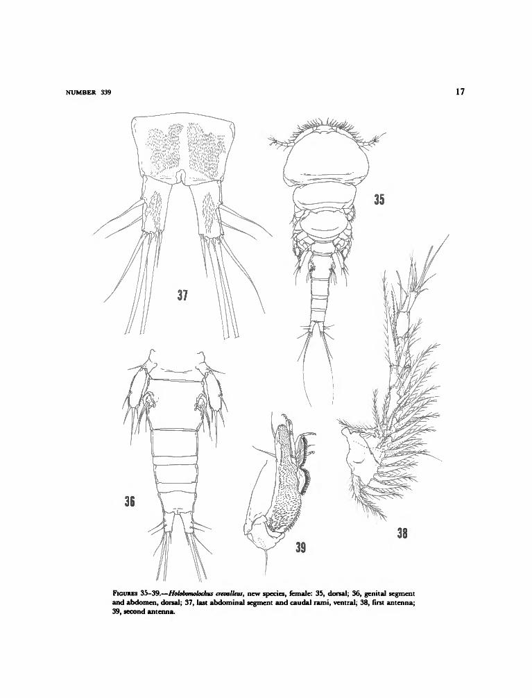
FIGURES 35-46
MATERIAL EXAMINED.—Holotype $ (USNM181723) and 4 $ paratypes (USNM 181724) fromthe gill chamber and nasal sinuses of 4 Caranxhippos (Linnaeus) collected by the author in Char-lotte Harbor, Florida, 5 Jun 1972. Other speci-mens (14 9) from the same host and area atvarious times from 1970 to 1972.
FEMALE.—Body form as in Figure 35. Totallength 1.53 mm, greatest width 0.65 mm (mea-sured at widest part of cephalon). Cephalon com-prising about one-fourth total body length (0.38mm), nearly twice as wide as long. Genital seg-ment (Figure 36) wider than long (206 X 182fim). Abdomen 3-segmented, segments measure100 X 129 /an, 59 X 123 /im, and 82 X 118 /*m(length X width) respectively; last abdominal

NUMBER 339
segment with 2 ventral patches of spinules (Figure37). Caudal rami (Figure 37) about twice as longas wide (76 X 35 fun), each armed with 2 nakedlateral setae and 4 naked terminal setae (longestseta 590 /im); ventral surface of each ramus withpatch of spinules (heavier than those on abdo-men).
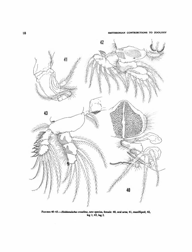
First antenna (Figure 38) with 5 distinct seg-ments, 15 plumose setae on basal part and aes-thete on each of last 2 segments. Rostrum withventral hooks. Second antenna (Figure 39) lastsegment with fine hooklike spinules not in distinctrows, palp with row of longer hooklike spinulesalong margin, 3 setae, and 4 articulated terminaland subterminal spines. Labrum (see Figure 40)with 2 patches of stout spinules. Oral area (Figure40) similar to H. divaricatus Cressey and Cressey;paragnath with long hairs on distal third, firstmaxilla with 3 long plumose setae and 1 shortnaked seta, labium represented by a row of stoutspinules and 2 lateral hirsute palps posterior tooral region. Maxilliped (Figure 41) claw withsmall accessory process (not easily seen in somespecimens).
Legs 1-4 biramose. Leg 1 (Figure 42) basipodwith 2 large patches of spinules and short, thickseta at outer distal corner; exopod 2-segmented,first segment with thumblike spinulose spine onouter distal corner, second second segment with2 small outer spines and 6 setae; endopod 3 -segmented, first 2 segments each with patch ofspinules on outer half and an inner seta, lastsegment with 5 setae. Leg 2 (Figure 43) basipodwith plumose seta on outer margin; exopod firstsegment with patch of bristles on outer marginand on outer distal corner a fringed spine bearingterminal flagellum, second segment with similarouter spine and inner seta, last segment with 3outer, equally long, similar spines and 6 setae, 5outermost setae with pectinate outer borders andshort plumosites on inner border; endopod firstsegment with patch of spinules on outer half andinner seta, second segment with patch of spinuleson outer half and 2 inner setae, third segmentwith small patch of spinules on outer margin, 2short plumose outer spines and 3 setae. Leg 3(Figure 44) basipod as in leg 2, exopod first
segment with heavily sclerotized spine on outerdistal corner, second segment with prominentheavily sclerotized spine on outer distal cornerand inner seta, last segment with 2 prominentheavily sclerotized outer spines and 6 setae (out-ermost with pectinate outer margin); endopod asin leg 2 except last segment with only 2 setae. Leg4 (Figure 45) basipod similar to that of legs 2 and3; exopod first segment with spines on outer distalcorner, second segment with outer spine and innerseta, last segment with outer short spine, longerweakly sclerotized outer terminal spines and 6setae (outermost seta with pectinate outer mar-gin); endopod first and second segments eachwith distal patch of spinules and inner spinuloseseta, last segment with short row of stout spinulesat each distal corner, outer and inner spines, andlong median seta (spines and setae with spinulosemargins). Leg 5 (Figure 46) basal segment withsmall patch of spinules and naked seta on outerdistal corner; free segment with inner and outerpatches of spinules, outer lateral spine, 2 terminalspines and inner subterminal spine, innermostterminal spine about twice length of other 3spines, innermost 2 spines with more prominentlyspinulose margins than outer 2 spines. Leg 6represented by 3 well-developed setae at area ofegg sac attachment (see Figure 36).
MALE.—Unknown.ETYMOLOGY.—The specific epithet crevalleus re-
fers to the common name of the host, "CrevalleJack."
REMARKS.—This new species can be separatedfrom all known species of Holobomolochus exceptH. asperatus Cressey and Cressey, H. divaricatusCressey and Cressey, and H. nudiusculus Cresseyand Cressey by the prominent, heavily sclerotizedspines on the exopod of leg 3. It can be separatedfrom H. nudiusculus and H. divaricatus in that thecaudal rami of those species are without ventralsurface ornamentation. It can be further sepa-rated from H. divaricatus because the distalmostexopod spine of leg 3 of H. divaricatus is shorterthan the preceding two. The new species can beseparated from H. asperatus by the large patchesof spinules on the endopod segments of legs 2 and3 of the new species, the fewer hooklike spinules

SMITHSONIAN CONTRIBUTIONS TO ZOOLOGY
on the second antenna of H. asperatus, and by thespinulose surface of the paragnath of H. asperatus(hirsute in H. crevalleus). Together these four spe-cies of Holobomolochus comprise a group character-ized by the heavily sclerotized long spines on theexopod of leg 3 and so far known only from thewestern Atlantic and eastern Pacific. The previ-ously described three species of this group areparasitic in the nasal sinuses of the New Worldspecies of Scomberomorus.
Holobomolochus serratus, new species
FIGURES 47-58
MATERIAL EXAMINED.—Holotype 9 (USNM181725) and 25 paratype $ (USNM 181726) col-lected from the gill chambers of 1 Scorpaena brasi-liensis Cuvier (220 mm FL) at Charlotte Harbor,Florida, by the author 27 Aug 1973. Additionalmaterial (12 $) collected from 81 specimens of thesame host and locality at various times by theauthor between 1972 and 1975.
FEMALE.—Body form as in Figure 47. Totallength 1.27 mm, greatest width 0.61 mm (mea-sured at widest part of cephalon). Cephalon com-prises about one-third total body length (413 /an).Rostrum with ventral hooks. Genital segment(Figure 48) wider than long (171 X 141 pirn).Abdomen 3-segmented, segments with patch offine spinules near origin of each caudal ramus(Figure 49). Caudal rami (Figure 49) nearly 3times longer than wide (82 X 30 jum), each with1 lateral seta, 1 subterminal dorsal seta, and 4terminal setae, innermost sparsely plumose.
First antenna (Figure 50) 6-segmented, first 3segments with 15 plumose outer setae, 1 aestheteon each of last 2 segments (first 3 segments heldin a relatively straight line). Second antenna(Figure 51) second segment with hooklike spinulesnot in rows, lateral palp with row of finer spinulesalong other edge, second segment with subter-minal heavily sclerotized recurved spine, 3 weakerdistal spines, and 2 naked setae (usual third setanot seen). Mouthparts as in Figure 52; the 3plumose setae on first maxilla not as long as inmost other species of genus, paragnath with only
fringe of small spinules distally. Maxilliped (Fig-ure 53) hook with prominent accessory spine,hook recurved at about a right angle, 2 lateralplumose setae shorter than in most other species.
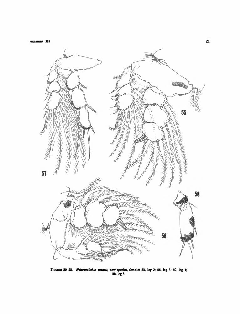
Legs 1-4 biramose. Leg 1 (Figure 54) basipodwith 2 small patches of spatulate spinules; exopodfirst segment with 2 spines on outer distal corner,segments 2-3 partly fused, bearing 2 outer spinesand 6 setae; endopod first 2 segments, each withinner seta and patch or row of spinules alongdistal margin, last segment with small outer spine,5 setae, and small group of spinules near base ofsmall spine. Leg 2 (Figure 55) basipod with patchof spatulate spinules near inner margin; exopodfirst segment with spinulose spine at outer distalcorner, second segment with outer spinulose spineand inner seta, last segment with 4 outer spines(first 2 spinulose, last 2 prominently serrate onouter margin) and 5 setae; endopod first segmentwith inner seta and row of spinules along distalmargin, second segment with 2 inner setae anddistal row of spinules, last segment with 2 spinu-lose outer spines and 3 setae. Leg 3 (Figure 56)basipod armed as in leg 2 except with row of finespinules near middle; exopod first segment withlong plumose outer spine and inner seta, thirdsegment with 3 long plumose outer spines and 5setae, outermost seta with short plumosities onouter edge; endopod as in leg 2 except last seg-ment with only 2 setae. Leg 4 (Figure 57) basipodunarmed except for usual seta at outer distalcorner; exopod first segments with outer nakedspine, second segment with similar outer spineand inner seta, last segment with 2 outer spines(first naked, second with short spinules on innermargin) and 6 setae, outermost seta with shortplumosities along outer edge; endopod first 2segments each with short plumose inner seta andhairs, and row of conspicuous spinules along outerdistal margin, last segment with 3 setae with shortplumosities, middle seta 3 times length of other2, and patch of conspicuous spinules distally. Leg5 (Figure 58) first segment with large patch ofspinules on distal half and a naked seta at outerdistal corner; free segment with 2 patches ofprominent spinules (1 at mid-outer margin andother distal) and short lateral seta and 3 terminal

NUMBER 339
setae, setae with short plumosities except midter-minal seta naked. Leg 6 represented by 6 longsetae at area of egg sac attachment.
MALE.—Unknown.ETYMOLOGY.—The Latin serratus (toothed like
a saw) alludes to the ornamentation on the spineson the exopod of leg 2.
REMARKS.—This new species can be separatedfrom all known species of Holobomolochus exceptH. spinulus (Cressey) and H. attenuatus (Wilson) bythe prominent lateral spine on the second an-tenna. It should be noted that H. spinulus wasdescribed from Scorpaena guttata Giard from Cali-fornia, and H. attenuatus described from Scorpaenaplumieri Bloch from Jamaica. The new species canbe separated from both of these species by thearrangement of the spinules in discrete rows onthe second antennae of each of these previouslyknown species. It can be further separated fromH. spinulus because the caudal rami of H. spinuluseach bear a ventral patch of spinules (naked inH. serratus). It can be separated from H. attenuatusas well as H. spinulus by the heavy serrations onthe last two spines of the last exopod segment ofleg 2 of the new species.
As in H. crevalleus this new species may be amember of a subgroup of Holobomolochus, in thiscase characterized by the presence of the promi-nent lateral spine on the second antenna. Al-though H. spinulus has been reported from hostsother than scorpaenids, scorpion fishes may bethe preferred hosts.
Neobomolochus, new genus
DIAGNOSIS.—Bomolochidae. Body form typicalof family. Thoracic segments bearing legs 2-5free. Abdomen indistinctly 3-segmented. Caudalrami with 5 minor setae and one major terminalseta. Rostrum without hooks. First antenna 5-segmented; first segment with sclerotized processbearing 3 modified setae. Other cephalic append-ages typical of family. Maxilliped hook withoutprominent accessory process. Legs 1-4 biramous.Leg 2 middle endopod segment with 2 innersetae. Leg 3 middle endopod segment with 1inner seta. Leg 4 middle endopod segment with-
out setae. Leg 5 free segment with 4 lateral toterminal setae.
Diagnosis based on female. Male known onlyfrom immature specimens.
ETYMOLOGY.—The Greek neo (new) plus thegeneric name bomolochus refers to the taxon asrepresenting a new bomolochid genus.
TYPE-SPECIES.—Neobomolochus elongatus, newspecies.
REMARKS.—Neobomolochus is separated from Ho-lobomolochus, Acanthocolax, Ceratocolax, Bomolochus,
Boylea, Unicolax, and Tegobomolochus by havingonly one major terminal seta on the caudal ramus,whereas the aforementioned genera all have twomajor terminal setae on each caudal ramus. Fromthe remaining genera with one major seta, thenew species can be separated by its lacking aninner seta on the mid-endopod segment of leg 4;all others with a 3-segmented endopod of leg 4bear a single seta on the middle segment. (Theendopods of legs 2-4 are 2-segmented in Pumiliopesand Pumiliopsis.)
Superficially the new genus resembles Pseudoeu-canthus Brian. The two species described in thatgenus (P. alosae Brian and P. uniseriatus Wilson)are poorly known. Both authors show a seta onthe mid-endopod segment of leg 4. Neither authormentions any modified setae on the first antenna,which should be easily seen if present (I examinedthe holotype of P. uniseriatus and did not see anymodified setae). The modified setae on the firstantenna of Neobomolochus are similar to thosefound in Nothobomolochus, but the new genus differsfrom Nothobomolochus by presence of two innersetae on the mid-endopod segment of leg 3 andone seta on the same segment of leg 4 of Nothbom-olochus.
The diagnosis is based on the female. Twoimmature males are present in the collections,but I do not feel it appropriate to base a genericdiagnosis on immature features.
Neobomolochus elongatus, new species
FIGURES 59-72
MATERIAL EXAMINED.—Holotype 9 (USNM181727) and 5 paratype $ (USNM 181728) col-

8 SMITHSONIAN CONTRIBUTIONS TO ZOOLOGY
lected from the eyes of 9 Opisthonema oglinum (Le-sueur) (USNM 108355) from "West Indies" col-lected by the author (1 fish negative). Additionalmaterial (7 9, 1 immature 6*) collected from 5specimens of the same host from Charlotte Har-bor, Florida, and 1 immature 6* from Epinephalusmorio (Valenciennes).
FEMALE.—Body form as in Figure 59. Totallength 4.20 mm, greatest width 1.16 mm (mea-sured at widest part of cephalon). Cephalon com-prises about 20 percent of total length (0.84 mm).Thoracic segment bearing leg 1 fused withcephalon. Thoracic segments bearing legs 2-5narrow and together about twice length ofcephalon. Rostrum without hooks. Genital seg-ment (Figures 60, 61) longer than wide (472 X265 jtim,) posterior corners inflated laterally. Ab-domen indistinctly 3-segmented, segments mea-sure 324 X 342 jam, 295 X 306 fim, and 365 X253 |im (length X width) respectively; last ab-dominal segment with 2 patches of spatulatespinules (see Figure 62). Caudal rami (Figure 62)longer than wide (141 X 100 ftm), each bearing1 lateral seta, 1 dorsal subterminal seta and 4terminal setae (3 short, 1 long), longest seta 413jum bearing short plumosities; each ramus withventral patch of spatulate spinules.
First antenna (Figure 63) 5-segmented; firstsegment bearing 2 plumose setae and 3 shortsclerotized spines at outer distal corner (Figure64); 1 aesthete on each of last 2 segments.Second antenna (Figure 65) last segment withhooklike spinules arranged in rows (rows morediscrete distally), outer edge with 2 short rows ofhooklike spinules and 4 weak hooks and 2 setaedistally. Labrum with 2 patches of spinules andshort row of spinules on outer posterior corner.Mouthparts (Figure 60) typically bomolochid.Mandible bladelike; paragnath with distal fringe;first maxilla with 4 setae, inner 2 with shortspinules; second maxilla with 2 terminal bladelike
processes, each bearing spinules on margins. Max-illiped (Figure 67) hook recurved nearly at rightangle and bearing 3 very small accessory spineson outer margin. Legs 1-4 biramose. Leg 1 (Fig-ure 68) basipod with 2 patches of spinules; exopodfirst segment with thumblike spine on outer distalcorner, second and third segments fused, bearing9 setae; endopod first segment with large patchof spinules and inner seta, second segment withinner seta, last segment with 5 setae. Leg 2 (Figure69) basipod with small patch of setules and largepatch of spinules between bases of rami; exopodfirst segment with rows of scalelike spinules alongouter margin and a long spine with hyaline tipon outer distal corner; second segment with stoutspine on outer distal corner and an inner seta, lastsegment with 4 outer spines and 4 setae (termi-nalmost spine longest); endopod first segmentwith patch of scalelike spinules on outer distalcorner and inner seta, second segment as in firstexcept with 2 inner setae, last segment with 2short, finely plumose outer spines and 3 setae,outer margins of setae finely plumose. Leg 3(Figure 70) as in leg 2 except no patch of pointedsetules on basipod, endopod with only 3 spineson last segment, only 1 inner seta on middleendopod segment, and only 2 setae on last endo-pod segment. Leg 4 (Figure 71) as in leg 3 exceptmiddle endopod segment without setae and lastsegment with outer, finely plumose spine, longspinulose midseta, and short, naked, inner seta.Leg 5 (Figure 72) free segment with scalelikespinules distally and 4 lateral to terminal setae,terminalmost longest and sparsely plumose, oth-ers finely plumose. Leg 6 represented by 3 setaeat area of egg sac attachment (see Figures 60,61).
Egg sacs long, extending well beyond caudalrami, about as long as body.
MALE.—Known only from two immature spec-imens, not described.
ETYMOLOGY.—The Latin elongatus (prolonged)alludes to the elongate body of the female.

Literature Cited
Kr0yer, H. Those Found upon the Fishes of the Pacific Coast,1863. Bidrag til Kundskab om Snylterkrebsene. Natur- w h h Descriptions of New Genera and Species.
historisk Tidsskdrifi, 2(3): 75-320. „ M. r L r r . J „ „ . f w
V rt W Proceedings of the United States National Museum, 35:1962. A Review of the Genera and Species of the Bom- 431-481
olochidae (Crustacea, Copepoda), Including the 1913. Crustacean Parasites of West Indian Fishes andDescription of Some Old and New Species. Zoolo- L a n f J C r a b s w h h D ^ t i o M o f N c w ^ ^ a n d
gische Verhandelingen, 5 6 : 111 p a g e s .Wilson C B Species. Proceedings of the United States National Mu-
1908. North American Parasitic Copepods: A List of sewn, 44:189-277

10 SMITHSONIAN CONTRIBUTIONS TO ZOOLOGY
FIGURES 1-6.—Holobomolochus glyphisodontis (Kr0yer), female: 1, dorsal; 2, genital segment andabdomen, dorsal; 3, last abdominal segment and caudal rami, ventral; 4, first antenna; 5,second antenna; 6, first and second maxillae.

NUMBER 339 11
11
FIGURES 7-12.—Holobomolochus glyphisodontu (Kr0ycr), female: 7, maxilliped; 8, leg 1 exopod; 9,leg 2 exopod; 10, leg 3 exopod; 11, leg 4; 12, leg 5.

12 SMITHSONIAN CONTRIBUTIONS TO ZOOLOGY
FIGURES 13-17.—Holobomolochus glyphisodontis (Kr0yer), male: 13, dorsal; 14, genital segment andabdomen, dorsal; 15, last abdominal segment and caudal rami, ventral; 16, first antenna; 17,maxilliped.

NUMBER 339 13
19
FIGURES 18, 19.—Holobomolochus glyphisodontis (Kr0yer), male: 18, leg 2; 19, leg 3.

14 SMITHSONIAN CONTRIBUTIONS TO ZOOLOGY
24
FIGURES 20-24.—Holobomolochus glyphisodonlis (Kr0yer), male: 20, leg 4; 21, leg 4 endopod; 22,leg 5. Holobomolochus centropnstis, new species, female: 23, dorsal; 24, genital segment andabdomen, dorsal.

NUMBER 339 15
30
FIGURES 25-30.—Holobomolochus centropristis, new species, female: 25, last abdominal segmentand caudal rami, ventral; 26, first antenna; 27, second antenna; 28, mandible, paragnath, firstand second maxillae; 29, maxilliped; 30, leg 1.

16 SMITHSONIAN CONTRIBUTIONS TO ZOOLOGY
FIGURES 31 -34.—Holobamolochus centropristis, new species, female: 31, leg 2; 32, leg 3; 33, leg 4;34, leg 5.
NUMBER 339 17
36
38
FIGURES 35-39.—Holobomolochus crevalleus, new species, female: 35, dorsal; 36, genital segmentand abdomen, dorsal; 37, last abdominal segment and caudal rami, ventral; 38, first antenna;39, second antenna.
18 SMITHSONIAN CONTRIBUTIONS TO ZOOLOGY
FIGURES 40-43.—Holobomolochus crevalleus, new species, female: 40, oral area; 41, maxilliped; 42,leg 1; 43, leg 2.

NUMBER 339 19
45
FIGURES 44-48.—Holobomolochus crevalleus, new species, female: 44, leg 3; 45, leg 4; 46, leg 5.Holobomolochus senatus, new species, female: 47, dorsal; 48, genital segment and abdomen, dorsal.

20 SMITHSONIAN CONTRIBUTIONS TO ZOOLOGY
54 50
FIGURES 49-54.—Holobomolochus serratus, new species, female: 49, last abdominal segment andcaudal rami, ventral; 50, first antenna; 51, second antenna; 52, mandible, paragnath, first andsecond maxillae; 53, maxilliped; 54, leg 1.
NUMBER 339 21
FIGURES 55-58.—Holobomolochus senates, new species, female: 55, leg 2; 56, leg 3; 57, leg 4;58, leg 5.

22 SMITHSONIAN CONTRIBUTIONS TO ZOOLOGY
59
FIGURES 59-63.—Neobomolochus elongatus, new species, female: 59, dorsal; 60, genital segment,ventral; 61, area of egg sac attachment, dorsal; 62, last abdominal segment and caudal rami,ventral; 63, first antenna.

NUMBER 339 23
64
FIGURES 64-68.—Neobomolochus elongates, new species, female: 64, base of first antenna; 65,second antenna; 66, mandible, paragnath, first and second maxillae; 67, maxilliped; 68, leg 1.

24 SMITHSONIAN CONTRIBUTIONS TO ZOOLOGY
71
FIGURES 69-72.—Neobomolochus elongatus, new species, female: 69, leg 2; 70, leg 3; 71 leg 4;72, leg 5.

REQUIREMENTS FOR SMITHSONIAN SERIES PUBLICATION
Manuscripts intended for series publication receive substantive review within theiroriginating Smithsonian museums or offices and are submitted to the Smithsonian InstitutionPress with approval of the appropriate museum authority on Form SI-36. Requests forspecial treatment—use of color, foldouts, casebound covers, etc.—require, on the sameform, the added approval of designated committees or museum directors.
Review of manuscripts and art by the Press for requirements of series format and style,completeness and clarity of copy, and arrangement of all material, as outlined below, willgovern, within the judgment of the Press, acceptance or rejection of the manuscripts and art.
Copy must be typewritten, double-spaced, on one side of standard white bond paper,with 1V4" margins, submitted as ribbon copy (not carbon or xerox), in loose sheets (notstapled or bound), and accompanied by original art. Minimum acceptable length is 30 pages.
Front matter (preceding the text) should include: title page with only title and authorand no other information, abstract page with author/title/series/etc., following the establish-ed format, table of contents with indents reflecting the heads and structure of the paper.
First page of text should carry the title and author at the top of the page and an unnum-bered footnote at the bottom consisting of author's name and professional mailing address.
Center heads of whatever level should be typed with initial caps of major words, withextra space above and below the head, but with no other preparation (such as all caps orunderline). Run-in paragraph heads should use period/dashes or colons as necessary.
Tabulations within text (lists of data, often in parallel columns) can be typed on the textpage where they occur, but they should not contain rules or formal, numbered table heads.
Formal tables (numbered, with table heads, boxheads, stubs, rules) should be sub-mitted as camera copy, but the author must contact the series section of the Press for edito-rial attention and preparation assistance before final typing of this matter.
Taxonomic keys in natural history papers should use the alined-couplet form in thezoology and paleobiology series and the multi-level indent form in the botany series. Ifcross-referencing is required between key and text, do not include page references within thekey, but number the keyed-out taxa with their corresponding heads in the text.
Synonymy in the zoology and paleobiology series must use the short form (taxon,author, yearpage), with a full reference at the end of the paper under "Literature Cited."For the botany series, the long form (taxon, author, abbreviated journal or book title, volume,page, year, with no reference in the "Literature Cited") is optional.
Footnotes, when few in number, whether annotative or bibliographic, should be typedat the bottom of the text page on which the reference occurs. Extensive notes must appear atthe end of the text in a notes section. If bibliographic footnotes are required, use the shortform (author/brief title/page) with the full reference in the bibliography.
Text-reference system (author/year/page within the text, with the full reference in a"Literature Cited" at the end of the text) must be used in place of bibliographic footnotes inall scientific series and is strongly recommended in the history and technology series:"(Jones, 1910:122)" or ".. . . Jones (1910:122)."
Bibliography, depending upon use, is termed "References," "Selected References," or"Literature Cited." Spell out book, journal, and article titles, using initial caps in all majorwords. For capitalization of titles in foreign languages, follow the national practice of eachlanguage. Underline (for italics) book and journal titles. Use the colon-parentheses systemfor volume/number/page citations: "10(2):5-9." For alinement and arrangement ofelements, follow the format of the series for which the manuscript is intended.
Legends for illustrations must not be attached to the art nor included within the text butmust be submitted at the end of the manuscript—with as many legends typed, double-spaced, to a page as convenient.
Illustrations must not be included within the manuscript but must be submitted sepa-rately as original art (not copies). All illustrations (photographs, line drawings, maps, etc.)can be intermixed throughout the printed text. They should be termed Figures and shouldbe numbered consecutively. If several "figures" are treated as components of a single largerfigure, they should be designated by lowercase italic letters (underlined in copy) on the illus-tration, in the legend, and in text references: "Figure 9b_." If illustrations are intended to beprinted separately on coated stock following the text, they should be termed Plates and anycomponents should be lettered as in figures: "Plate 9.b_." Keys to any symbols within anillustration should appear on the art and not in the legend.
A few points of style: (1) Do not use periods after such abbreviations as "mm, ft,yds, USNM, NNE, AM, BC." (2) Use hyphens in spelled-out fractions: "two-Thirds." (3)Spell out numbers "one" through "nine" in expository text, but use numerals in all othercases if possible. (4) Use the metric system of measurement, where possible, instead ofthe English system. (5) Use the decimal system, where possible, in place of fractions.(6) Use day/month/year sequence for dates: "9 April 1976." (7) For months in tabular listings or data sections, use three-letter abbreviations with no periods: "Jan, Mar, Jun," etc.
Arrange and paginate sequentially EVERY sheet of manuscript—including ALL frontmatter and ALL legends, etc., at the back of the text—in the following order: (1) title page,(2) abstract, (3) table of contents, (4) foreword and/or preface. (5) text, (6) appendixes,(7) notes, (8) glossary, (9) bibliography, (10) index, (11) legends.

i