Embed Size (px)
Citation preview
Parathyroid glands


History
1849: Sir Richard Owen provided the 1st accurate description of normal parathyroid glands after examining an Indian Rhinoceros in London zoo.
1879: Anton Wölfer described tetany in a patient after total thyroidectomy.
1880: Ivar Sandström a Swedish medical student grossly and microscopically described parathyroid glands.

1909:Calcium measurement was possible in and association with parathyroids established.
1925: the 1st successful parathyroidectomy on 38 yr old man with severe bone pain secondary to osteitis fibrosa cystica.
The parathyroid glands develop at 6 weeks and migrate caudally at 8 weeks
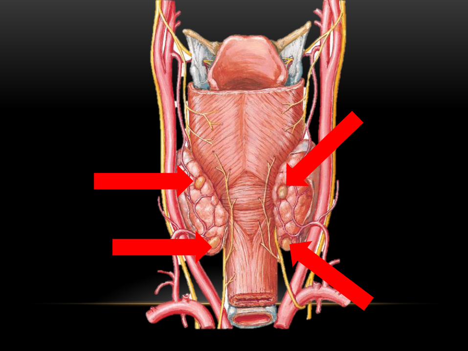
The paired superior parathyroid glands develop with the thyroid gland from the fourth branchial pouch and are generally consistent in position, residing lateral and posterior to the upper pole of the thyroid at the level of the cricothyroid cartilage.
The paired inferior glands descend with the thymus from the third branchial pouch and occasionally migrate to the level of the aortic arch or, rarely, fail to migrate, remaining in the high neck.
These glands are small (average total weight is about 130 mg) but essential for life.
Each parathyroid gland is surrounded by a thin connective tissue capsule.
Parenchymal cells are arranged in anastomosing cords surrounded by delicate connective tissue septa.
Capillaries are abundant (fenestrated).
A considerable number of fat cells infiltrate the gland (beginning around puberty) and may account for about half the weight of the parathyroid glands in adults.
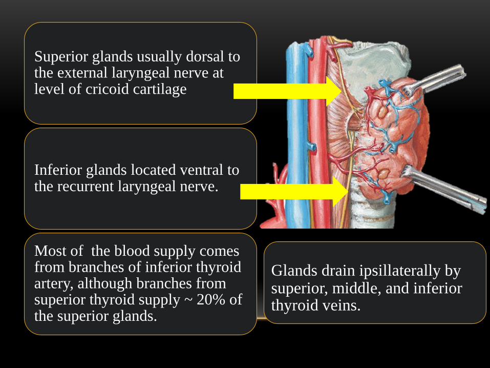
Superior glands usually dorsal to the external laryngeal nerve at level of cricoid cartilage
Inferior glands located ventral to the recurrent laryngeal nerve.
Most of the blood supply comes from branches of inferior thyroid artery, although branches from superior thyroid supply ~ 20% of the superior glands.
Glands drain ipsillaterally by superior, middle, and inferior thyroid veins.
Histology
Composed mostly of chief cells and oxyphil cells within an adipose stroma.
Oxyphil cells derived from chief cells and increase as one ages
Both types make Parathyroid hormone????
Chief (principal) Parathyroid cells
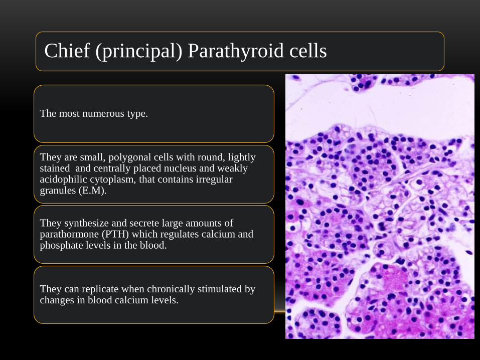
The most numerous type.
They are small, polygonal cells with round, lightly stained and centrally placed nucleus and weakly acidophilic cytoplasm, that contains irregular granules (E.M).
They synthesize and secrete large amounts of parathormone (PTH) which regulates calcium and phosphate levels in the blood.
They can replicate when chronically stimulated by changes in blood calcium levels.

Effects of Parathormone
Decreases kidney excretion of calcium.
Increases urinary phosphate excretion.
Regulates conversion of 25-OH vitamin D3 to hormonally 1,25 –(OH)2 Vitamin D3.
Increases intestinal absorption of Calcium.

Oxyphil Cells
They start to appear in the gland about 4-7 years of age and increase in number after puberty.
Their cytoplasm is strongly acidophilic, the nucleus is small and uniformly intense basophilic. They contain large amounts of abnormally shaped mitochondria.
As they some cells show low level of PTH activity, they are believed to be derivatives from chief cells.


Intra-operative parathyroid hormone testing
• introduced 1993 • Used to determine the adequacy of parathyroid
resection. • When the PTH falls by 50% or more in 10 minutes
after removal of a parathyroid tumor, as compared to the highest pre-removal value, the test is considered positive and the operation is terminated.