Embed Size (px)
DESCRIPTION
Pareidolias: complex visual illusions in dementiawith Lewy bodiesMakoto Uchiyama,1 Yoshiyuki Nishio,1 Kayoko Yokoi,1 Kazumi Hirayama,2 Toru Imamura,3Tatsuo Shimomura4 and Etsuro Mori1Patients rarely experience visual hallucinations while being observed by clinicians. Therefore, instruments to detect visualhallucinations directly from patients are needed. Pareidolias, which are complex visual illusions involving ambiguous formsthat are perceived as meaningful objects, are analogous to visual hallucinations and have the potential to be a surrogateindicator of visual hallucinations. In this study, we explored the clinical utility of a newly developed instrument for evokingpareidolic illusions, the Pareidolia test, in patients with dementia with Lewy bodies—one of the most common causes of visualhallucinations in the elderly. Thirty-four patients with dementia with Lewy bodies, 34 patients with Alzheimer’s disease and 26healthy controls were given the Pareidolia test. Patients with dementia with Lewy bodies produced a much greater number ofpareidolic illusions compared with those with Alzheimer’s disease or controls. A receiver operating characteristic analysisdemonstrated that the number of pareidolias differentiated dementia with Lewy bodies from Alzheimer’s disease with a sensitivityof 100% and a specificity of 88%. Full-length figures and faces of people and animals accounted for 480% of thecontents of pareidolias. Pareidolias were observed in patients with dementia with Lewy bodies who had visual hallucinations aswell as those who did not have visual hallucinations, suggesting that pareidolias do not reflect visual hallucinations themselvesbut may reflect susceptibility to visual hallucinations. A sub-analysis of patients with dementia with Lewy bodies who were orwere not treated with donepzil demonstrated that the numbers of pareidolias were correlated with visuoperceptual abilities inthe former and with indices of hallucinations and delusional misidentifications in the latter. Arousal and attentional deficitsmediated by abnormal cholinergic mechanisms and visuoperceptual dysfunctions are likely to contribute to the development ofvisual hallucinations and pareidolias in dementia with Lewy bodies.Abbreviations: MMSE = Mini-Mental State Examination; NPI = Neuropsychiatric Inventorydoi:10.1093/brain/aws126 Brain 2012: 135; 2458–2469 | 2458ReceivedAdvance Access publication May 30, 2012
Citation preview

BRAINA JOURNAL OF NEUROLOGY
Pareidolias: complex visual illusions in dementiawith Lewy bodiesMakoto Uchiyama,1 Yoshiyuki Nishio,1 Kayoko Yokoi,1 Kazumi Hirayama,2 Toru Imamura,3
Tatsuo Shimomura4 and Etsuro Mori1
1 Department of Behavioural Neurology and Cognitive Neuroscience, Tohoku University Graduate School of Medicine, Sendai 980-8575, Japan
2 Department of Occupational Therapy, Yamagata Prefectural University of Health Sciences, Yamagata 990-2212, Japan
3 Department of Speech, Language and Hearing Sciences, Niigata University of Health and Welfare, Niigata 950-3198, Japan
4 Department of Rehabilitation Medicine, Akita Prefectural Centre of Rehabilitation and Psychiatric Medicine, Daisen 019-2413, Japan
Correspondence to: Yoshiyuki Nishio,
Department of Behavioural Neurology and Cognitive Neuroscience,
Tohoku University Graduate School of Medicine,
2-1 Seiryo-machi,
Aoba-ku,
Sendai 980-8575, Japan
E-mail: [email protected]
Patients rarely experience visual hallucinations while being observed by clinicians. Therefore, instruments to detect visual
hallucinations directly from patients are needed. Pareidolias, which are complex visual illusions involving ambiguous forms
that are perceived as meaningful objects, are analogous to visual hallucinations and have the potential to be a surrogate
indicator of visual hallucinations. In this study, we explored the clinical utility of a newly developed instrument for evoking
pareidolic illusions, the Pareidolia test, in patients with dementia with Lewy bodies—one of the most common causes of visual
hallucinations in the elderly. Thirty-four patients with dementia with Lewy bodies, 34 patients with Alzheimer’s disease and 26
healthy controls were given the Pareidolia test. Patients with dementia with Lewy bodies produced a much greater number of
pareidolic illusions compared with those with Alzheimer’s disease or controls. A receiver operating characteristic analysis
demonstrated that the number of pareidolias differentiated dementia with Lewy bodies from Alzheimer’s disease with a sen-
sitivity of 100% and a specificity of 88%. Full-length figures and faces of people and animals accounted for 480% of the
contents of pareidolias. Pareidolias were observed in patients with dementia with Lewy bodies who had visual hallucinations as
well as those who did not have visual hallucinations, suggesting that pareidolias do not reflect visual hallucinations themselves
but may reflect susceptibility to visual hallucinations. A sub-analysis of patients with dementia with Lewy bodies who were or
were not treated with donepzil demonstrated that the numbers of pareidolias were correlated with visuoperceptual abilities in
the former and with indices of hallucinations and delusional misidentifications in the latter. Arousal and attentional deficits
mediated by abnormal cholinergic mechanisms and visuoperceptual dysfunctions are likely to contribute to the development of
visual hallucinations and pareidolias in dementia with Lewy bodies.
Keywords: acetylcholine; dementia with Lewy bodies; visual hallucinations; visual illusions
Abbreviations: MMSE = Mini-Mental State Examination; NPI = Neuropsychiatric Inventory
doi:10.1093/brain/aws126 Brain 2012: 135; 2458–2469 | 2458
Received October 6, 2011. Revised March 23, 2012. Accepted March 26, 2012. Advance Access publication May 30, 2012� The Author (2012). Published by Oxford University Press on behalf of the Guarantors of Brain.This is an Open Access article distributed under the terms of the Creative Commons Attribution Non-Commercial License (http://creativecommons.org/licenses/by-nc/3.0),which permits unrestricted non-commercial use, distribution, and reproduction in any medium, provided the original work is properly cited.

Introduction‘A perception without an object’ is the classic definition of a hal-
lucination and has been in use since the end of the 19th century
(Ey, 1973). According to this definition, a visual hallucination is
experienced when one sees something where nothing actually
exists. However, it is inevitable that when patients are awake
and have their eyes open, they see things. It is difficult to deter-
mine whether false perceptions arise independently of real visual
scenes or as a result of their distortion.
Dementia with Lewy bodies is arguably the most common cause
of visual hallucinations in the elderly. Although visual hallucin-
ations are one of the core clinical features differentiating dementia
with Lewy bodies from other dementias, clinicians rarely witness
patients as they experience them (McKeith et al., 1996, 2005).
Although questionnaires or interviews with patients or caregivers
are usually used to assess visual hallucinations, visual hallucinations
are commonly under-reported by patients and are often not dis-
covered by caregivers and health professionals (Mosimann et al.,
2008). Tests that detect visual hallucinations directly from the pa-
tients would thus be a helpful clinical tool.
Patients with dementia with Lewy bodies frequently experience
a variety of visual illusions as well as visual hallucinations (Iseki
et al., 2002; Nagahama et al., 2007). Pareidolias, which are illu-
sions of meaningful objects such as faces and animals, are thought
to arise from ambiguous forms embedded in visual scenes and
have a striking phenomenological resemblance to visual hallucin-
ations. For instance, patients with dementia with Lewy bodies may
incorrectly see a person in a curtain or perceive blobs on the wall
as faces. In this study, we attempted to evoke and measure par-
eidolic illusions using a simple experimental paradigm, the
Pareidolia test, and we explored the clinical utility of this test as
a surrogate indicator of visual hallucinations in dementia with
Lewy bodies.
Materials and methods
ParticipantsWe recruited 34 patients with probable dementia with Lewy bodies
and 34 with probable Alzheimer’s disease from the dementia clinics at
the Tohoku University Hospital, the Akita Prefectural Centre of
Rehabilitation and Psychiatric Medicine and the Niigata Rehabilitation
Hospital. Additionally, 26 healthy controls were recruited though an
advertisement. The three groups were comparable in age, sex, educa-
tional level and visual acuity. The severity of cognitive impairment,
which was assessed by the Mini-Mental State Examination (MMSE)
(Folstein et al., 1975), was matched between the dementia with Lewy
bodies and Alzheimer’s disease groups (Table 1). All patients under-
went an examination by experienced behavioural neurologists as well
as MRI and routine laboratory investigations. Probable dementia with
Lewy bodies and probable Alzheimer’s disease were diagnosed accord-
ing to the international workshop criteria of dementia with Lewy
bodies (McKeith et al., 2005) and the National Institute of
Neurological and Communicative Disorders and Stroke and the
Alzheimer’s Disease and Related Disorders Association criteria
(McKhann et al., 1984), respectively. The exclusion criteria were as
follows: (i) a history of other neurological, psychiatric or severe ocular
diseases; (ii) a best corrected visual acuity of 520/70; and (iii) lan-
guage deficits hindering task executions. At the time of examination,
12 patients with dementia with Lewy bodies were treated with done-
pezil, one was treated with levodopa, four were treated with quetia-
pine and two were treated with fluvoxamine. In the Alzheimer’s
disease group, eight patients were treated with donepezil and one
was treated with fluvoxamine. The demographic and clinical charac-
teristics of the participants are summarized in Table 1 and
Supplementary Table 1.
All procedures in this study were approved by the ethical committee
of the Tohoku University Graduate School of Medicine. All participants
provided written informed consent after receiving a detailed explan-
ation of the study.
NeuropsychologyThe Digit Span subtest from the Wechsler Memory Scale-Revised
(Sugishita, 2001) and the Object Naming subtest of the Western
Aphasia Battery (Sugishita, 1986) were used to assess attention/work-
ing memory and language, respectively. Visuoperceptual and visuo-
spatial functions were assessed using the Shape Detection Screening
and Position Discrimination subtests of the Visual Object and Space
Perception battery (Warrington and James, 1991), the Object Decision
subtest (Easy B) of the Birmingham Object Recognition Battery
(Riddoch and Humphreys, 1992) and the Face Recognition subtests
(face-to-face matching of unknown faces, same/different judgement
of unknown faces in different views, gender and age judgements of
unknown faces) of the Visual Perception Test for Agnosia (Japan
Society for Higher Brain Dysfunction, 1997).
Behavioural assessmentThe Neuropsychiatric Inventory (NPI) (Cummings et al., 1994) was
administered to the caregivers of the patients. We made some modi-
fications to the original NPI as follows. First, the ‘delusion’ domain was
separated into two different categories: persecutory delusions and de-
lusional misidentifications. The questions regarding the former included
‘believing that others are planning to hurt him/her’ and ‘believing that
others are stealing from him/her’; the questions regarding the latter
included ‘believing that unwelcome guests are living in the house’,
‘believing that television or magazine figures are actually present in
the home’ and ‘believing that patient’s family is an imposter’. Second,
we employed an additional domain for fluctuations in cognition, which
included questions such as ‘does the patient sometimes show a lack of
attention or a slow reaction to others’ call than usual?’ and ‘does the
patient sometimes show a poor understanding of things that he can
usually understand?’ (Mori et al., 2006). The frequency (range 1–4),
the severity (1–3) and the domain total scores (the product of the
frequency score multiplied by the severity score) were recorded for
each behaviour.
The Pareidolia test
Stimuli and task administration
Twenty-five coloured scenery pictures (25 � 19 cm2) containing ani-
mals, plants and artefacts were used. The pictures were selected on
the basis of preliminary experiments in patients with dementia with
Lewy bodies, Alzheimer’s disease and healthy controls. Each picture
was filtered by Gaussian blurring (0.17 mm full-width at
half-maximum) (Fig. 3B). Immediately before the administration of
Pareidolias in dementia with Lewy bodies Brain 2012: 135; 2458–2469 | 2459

the test, a detailed explanation and three training trials were given.
Subjects were instructed to point to and describe in as much detail as
possible the objects shown in each picture. When the subjects pro-
vided few responses, they were prompted to describe as many things
as possible. When subjects responded with such comments as, ‘It looks
like X’, we determined whether the object (X) was actually in the
picture or whether the subject saw something that looked like X.
Each picture was presented for 60 s. No feedback was given to sub-
jects, regardless of correct or incorrect responses. Subjects’ responses
were recorded by video camera for later analysis.
Scoring
Subjects’ responses were classified into three types: (i) correct re-
sponses; (ii) illusory responses, in which subjects falsely identified ob-
jects that were not in the pictures; and (iii) other responses, in which
subjects provided no response or said ‘I don’t know’. On the basis of
the preliminary experiments in healthy subjects, correct answers were
defined in advance. We calculated the sum of correct responses and
illusory responses in the 25 images for each subject. In some cases,
subjects provided two or more responses to a single picture.
Accordingly, the total numbers of correct responses and of illusory
responses were more than 25 in some cases.
Patients with dementia with Lewy bodies frequently see hallucin-
atory images in places where people do not usually direct their
attention, such as the corners of rooms, trees in the background or
the roofs of neighbouring houses. To assess where subjects saw
pareidolias, we categorized the objects in the pictures into the ‘gist’
and the ‘detail/background’ (Adolphs et al., 2001). The ‘gist’ is
the general information or major photographic subject that is im-
portant for the meaning of the image, and the ‘detail/background’
is the information that is of little importance for the meaning of the
image.
To conduct a phenomenological analysis of pareidolias, the content
of the illusory responses was classified into four categories: people,
animals (e.g. animals, birds and fishes), objects (man-made objects,
vegetables and flowers) and others (flash, stream of water, flame).
Table 1 Demographic and clinical profiles of the participants
Variables DLB (n = 34) AD (n = 34) HC (n = 26) P-value
Age, years 81.0 (3.9) 80 (3.6) 79.2 (4.9) 0.238
Sex (female/male)a 19/15 10/24 8/18 0.385
Education, years 9.7 (2.5) 9.3 (1.8) 10.2 (1.8) 0.265
Visual acuityb 20/25 (20/30–20/25) 20/25 (20/30–20/25) 20/25 (20/30–20/25) 0.357
Neuropsychology
MMSE [30] 18.9 (4.0) 19.2 (3.1) 29.0 (1.0) 50.001c,d
Digit span [24] 9.0 (2.3) 8.8 (2.2) 10.6 (1.7) 50.001c,d
Object naming [60] 54.9 (4.3) 55.3 (2.3) 58.7 (1.5) 50.001c,d
Shape detection [20] 18.1 (1.5) 19.3 (1.0) 19.7 (0.5) 50.001c,e
Position discrimination [20] 18.2 (1.7) 19.1 (1.1) 19.9 (0.4) 50.001c,d,e
Object decision [32] 24.3 (2.8) 27.7 (2.3) 29.8 (1.5) 50.001c,d,e
Face recognition [30] 23.9 (3.0) 26.8 (2.1) 28.6 (1.3) 50.001c,d,e
NPIf
Persecutory delusions 1.3 (2.8) 0.9 (2.5) 0.379
Delusional misidentifications 2.9 (4.1) 0.1 (0.5) 50.001
Hallucinations 3.0 (3.8) 0.1 (0.3) 50.001
Agitation/aggression 1.4 (2.6) 0.9 (1.5) 0.231
Dysphoria 1.1 (2.3) 0.6 (1.6) 0.117
Anxiety 2.4 (3.1) 0.9 (1.7) 0.088
Euphoria 0.3 (0.8) 0.0 (0.0) 0.021
Apathy 5.0 (3.5) 2.9 (3.3) 0.007
Disinhibition 0.7 (2.1) 1.2 (2.9) 0.731
Irritability/lability 0.7 (2.1) 1.2 (2.9) 0.819
Aberrant motor behaviour 1.4 (2.6) 1.3 (2.8) 0.476
Fluctuations in cognition 3.9 (3.3) 0.1 (0.2) 50.001
Prevalence [/34]a
Delusional misidentifications 22 2 50.001
Visual hallucinations 25 3 50.001
Fluctuations in cognition 31 2 50.001
The values in the second to fourth columns indicate the mean (standard deviation). The visual acuity is indicated as the median (IQR). The full scores for the neuropsy-chological tests are indicated in square brackets. The prevalence of behavioural symptoms are indicated as number of patients.
a �2 test.b Kruskal–Wallis test.c Scheffe test (P50.05): DLB5AD.d Scheffe test (P50.05): DLB5HC.e Scheffe test (P5 0.05): AD5HC.f Mann–Whitney U-test.
The remaining variables were tested using a one-way analysis of variance (ANOVA) and post hoc Scheffe tests.DLB = dementia with Lewy bodies; AD = Alzheimer’s disease; HC = healthy controls.
2460 | Brain 2012: 135; 2458–2469 M. Uchiyama et al.

Test–retest and inter-rater reliabilities
Ten patients with dementia with Lewy bodies and 10 patients
with Alzheimer’s disease underwent the test twice with a 2-week
interval. The scoring was performed by two independent raters who
were not informed about the diagnosis or other conditions of the
subjects.
Statistical analysesA Kruskal–Wallis test and a post hoc Mann–Whitney U-test with a
Bonferroni correction (P5 0.05/3) were used for between-group com-
parisons of the Pareidolia test. To evaluate the ability of the test to
differentiate dementia with Lewy bodies from Alzheimer’s disease, a
receiver operating characteristic analysis was performed on the neuro-
psychological tests, the NPI domain total scores and the Pareidolia test.
Relationships between performances on the Pareidolia test and other
neuropsychological and behavioural variables were assessed using
Pearson’s correlation coefficient or Spearman’s rank correlation
coefficient.
Results
Neuropsychology and behavioursThe results are summarized in Table 1. There were no significant
differences between the dementia with Lewy bodies and
Alzheimer’s disease groups in the MMSE, Digit Span or the
Object Naming tests. On all visuoperceptual and visuospatial
tests, the performance of the dementia with Lewy bodies group
was worse than the performance of the Alzheimer’s disease group.
The dementia with Lewy bodies group had significantly higher
scores for delusional misidentifications, hallucinations, euphoria,
apathy and fluctuations in cognition than did the Alzheimer’s dis-
ease group on the NPI. All of the patients with dementia with
Lewy bodies with a positive NPI hallucination score had visual
hallucinations and none had hallucinations in other modalities.
The Pareidolia test
Reliability
The inter-class correlation coefficients were 0.98 for illusory re-
sponses, 0.97 for correct responses and 0.89 for other responses,
indicating a good inter-rater reliability. The intra-class correlation
coefficients were 0.98 for illusory responses, 0.98 for correct re-
sponses and 0.95 for other responses, indicating good test–retest
reliability.
Group comparisons
The median [interquartile range (IQR)] numbers of illusory re-
sponses were 15.5 (11.0) for dementia with Lewy bodies, 1.0
(3.0) for Alzheimer’s disease and 0 (0) for healthy controls
(Fig. 1A). A Kruskal–Wallis test demonstrated a significant group
difference (�2 = 46.1, P5 0.001). The number of illusory re-
sponses in the dementia with Lewy bodies group was significantly
larger than that in the Alzheimer’s disease (U = 28.5, P50.001)
and the healthy control groups (U50.001, P50.001). The pa-
tients with Alzheimer’s disease gave more illusory responses than
the healthy control subjects (U = 252.0, P50.001). The numbers
of correct responses was 28.5 (13.0) for the dementia with Lewy
bodies group, 39.0 (5.0) for the Alzheimer’s disease group and
47.0 (6.0) for the healthy control group (Fig. 1A). There was a
significant difference among the three groups (�2 = 23.8,
P50.001). Post hoc tests showed significant differences in all of
the pairwise comparisons (P5 0.001).
When we classified the patients with dementia with Lewy
bodies on the basis of the use or non-use of donepezil therapy,
the median (IQR) numbers of illusory responses were 13.0 (9.0) in
the patients with dementia with Lewy bodies with donepezil and
16.0 (11.0) in those without donepezil. There was no significant
difference in illusory responses between the patients with and
without donepezil (U = 123.0, P = 0.75; Supplementary Table 1).
The numbers of correct responses was 21.0 (12.5) in the patients
with dementia with Lewy bodies with donepezil and 32.0 (9.75) in
those without donepezil. There was a significant difference in the
number of correct responses between the two groups (U = 65.0,
P = 0.002; Supplementary Table 1).
Next, we examined the impact of the presence of delusional
misidentifications and hallucinations on the number of illusory re-
sponses (Fig. 1B). The median (IQR) numbers of illusory responses
was 16.0 (10.0) in patients with dementia with Lewy bodies with
delusional misidentifications (n = 22) and 13.0 (9.0) in patients
without (n = 12). This difference in illusory responses did not
reach the level of statistical significance (U = 81.0, P = 0.065).
The numbers of illusory responses in patients with (n = 25) and
without hallucinations (n = 9) were 16.0 (12.0) and 13.0 (8.0),
respectively. There was no significant difference between these
two groups (U = 83.5, P = 0.256).
We further stratified the patients with dementia with Lewy
bodies according to both their medication status and the presence
of delusional misidentifications and hallucinations (Fig. 1C). In the
patients without donepezil, the median (IQR) numbers of illusory
response in patients with (n = 16) and without (n = 6) delusional
misidentifications were 17.0 (5.0) and 6.5 (4.0), respectively; and
the numbers of illusory response in patients with (n = 18) and
without hallucinations (n = 4) were 16.5 (12.0) and 8.5 (9.0), re-
spectively. In the patients with donepezil, the numbers of illusory
response in the patients with (n = 6) and without delusional mis-
identifications (n = 6) were 10.5 (12.0) and 14.5 (9.0), respect-
ively; and the numbers of illusory response in patients with
(n = 7) and without hallucinations (n = 5) were 13.0 (17.0) and
13.0 (5.0), respectively. In the absence of donepezil, although
we were unable to perform formal statistical comparisons because
of the small sample size, the number of illusory responses ap-
peared to be larger in the patients with delusional misidentifica-
tions than those without; however, the difference was less clear
between those with hallucinations and those without. In the pa-
tients treated with donepezil, there were no significant differences
in the number of illusory responses between the patients
with delusional misidentifications and hallucinations and those
without.
The relationships among the number of correct responses, the
presence of delusional misidentifications and hallucinations and
medication status are shown in Supplementary Fig. 1.
Pareidolias in dementia with Lewy bodies Brain 2012: 135; 2458–2469 | 2461

Locations of illusory responses
In the dementia with Lewy bodies group, the median (IQR) num-
bers of illusory responses were 4.0 (3.0) for the gist and 10.5 (8.0)
for the detail/background (Fig. 2A). In the Alzheimer’s disease
group, the median (IQR) numbers of illusory responses were
0 (1.0) for the gist and 0 (2.0) for the detail/background.
As the items in the detail/background outnumbers that in the
gist in the Pareidolia test, the number of illusory responses in the
detail/background is inevitably larger than that in the gist even if
one attends equally to the gist and the detail/background. In order
to clarify whether the patients saw pareidolic illusions in the detail/
background than in the gist, we performed the following analyses:
(i) a comparison of weighted numbers of illusory responses be-
tween the location categories (gist and detail/background), which
were calculated as: number of illusory responses in each location
category was divided by the total number of items in each location
category; and (ii) a comparison of the numbers of first illusory
responses that were made in the gist and those that were made
in the detail/background. In the dementia with Lewy bodies
group, the median (IQR) of the weighted numbers of illusory re-
sponse were 0.18 (0.15) for the gist and 0.52 (0.42) for the detail/
background. In the Alzheimer’s disease group, the medians of the
weighted numbers of illusory responses were 0 (0.05) for the gist
and 0 (0.09) for the detail/background. First illusory responses
were made for the gist in 3.0 (3.0) pictures and for the detail/
background in 7.0 (5.0) pictures in the dementia with Lewy bodies
group. In Alzheimer’s disease group, first illusory responses were
made for the gist in 0 (1.0) pictures and for the detail/background
in 0 (2.0) pictures (Fig. 2B).
In sum, the patients with dementia with Lewy bodies had a
tendency to see pareidolic illusions more in the detail/background
than in the gist.
Content of illusory responses and visual hallucinations
The content of illusory responses on the Pareidolia test are shown
in Fig. 3A. Illusions of people and animals accounted for 480% of
illusory responses. Faces and full-length figures of people and ani-
mals dominated whereas other body parts and man-made arte-
facts were infrequent. These characteristics were common to the
dementia with Lewy bodies group and to a subset of patients with
Alzheimer’s disease who gave illusory responses. In general, illu-
sory images were not familiar to subjects. In the dementia with
A
B C
Figure 1 (A) Numbers of illusory responses and correct responses on the Pareidolia test. Significance is denoted by an asterisk
(P50.05/3). (B) Numbers of illusory responses in patients with dementia with Lewy bodies with and without delusional misidentifications
and hallucinations. (C) Numbers of illusory responses in patients with dementia with Lewy bodies with and without delusional misiden-
tifications and hallucinations, stratified by medication status.
2462 | Brain 2012: 135; 2458–2469 M. Uchiyama et al.
Lewy bodies group, familiar people, such as parents and spouses,
and familiar animals, such as pets, accounted for only 2.1 and
2.6% of the total illusory responses, respectively. In the patients
with Alzheimer’s disease, the illusory responses of familiar people
and those of familiar animals accounted for 0 and 2.6% of all
illusory responses, respectively. Illusory responses often appeared
in bizarre and paradoxical contexts. For instance, patients often
saw faces in flowers and found a man in a tie after they correctly
identified the flowers and the tie (Fig. 3B). Even though they ac-
cepted the examiner’s suggestions that this was impossible, the
patients failed to correct their illusory descriptions.
We determined the contents of visual hallucinations in each of
25 patients with dementia with Lewy bodies with visual hallucin-
ations using medical records. Visual hallucinations of people, ani-
mals, plants and man-made objects were found in 21 (84%), 12
(48%), 2 (8%) and 2 patients (8%), respectively. Overall, the
dominant contents of illusory responses in the Pareidolia test
were consistent with those of visual hallucinations, and in both
of these, people and animals accounted for 480% of contents.
When comparing the contents of visual hallucinations and those of
illusory responses on a patient-by-patient basis, they did not
always agree with each other. The contents of illusory responses
depended on the contents of the pictorial stimuli; for instance,
patients with dementia with Lewy bodies frequently saw human
faces but rarely found animals in petals of flowers and they tended
to find animals but hardly saw people around the feet of a leopard
(Fig. 3B).
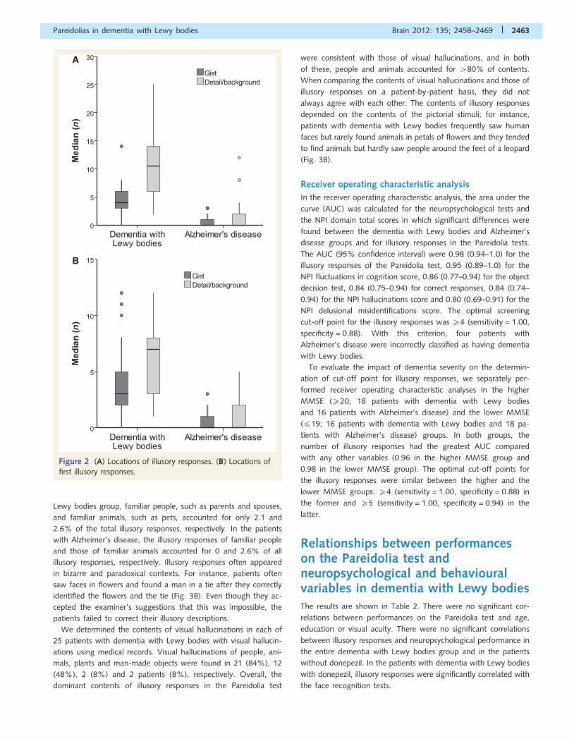
Receiver operating characteristic analysis
In the receiver operating characteristic analysis, the area under the
curve (AUC) was calculated for the neuropsychological tests and
the NPI domain total scores in which significant differences were
found between the dementia with Lewy bodies and Alzheimer’s
disease groups and for illusory responses in the Pareidolia tests.
The AUC (95% confidence interval) were 0.98 (0.94–1.0) for the
illusory responses of the Pareidolia test, 0.95 (0.89–1.0) for the
NPI fluctuations in cognition score, 0.86 (0.77–0.94) for the object
decision test, 0.84 (0.75–0.94) for correct responses, 0.84 (0.74–
0.94) for the NPI hallucinations score and 0.80 (0.69–0.91) for the
NPI delusional misidentifications score. The optimal screening
cut-off point for the illusory responses was 54 (sensitivity = 1.00,
specificity = 0.88). With this criterion, four patients with
Alzheimer’s disease were incorrectly classified as having dementia
with Lewy bodies.
To evaluate the impact of dementia severity on the determin-
ation of cut-off point for illusory responses, we separately per-
formed receiver operating characteristic analyses in the higher
MMSE (520; 18 patients with dementia with Lewy bodies
and 16 patients with Alzheimer’s disease) and the lower MMSE
(419; 16 patients with dementia with Lewy bodies and 18 pa-
tients with Alzheimer’s disease) groups. In both groups, the
number of illusory responses had the greatest AUC compared
with any other variables (0.96 in the higher MMSE group and
0.98 in the lower MMSE group). The optimal cut-off points for
the illusory responses were similar between the higher and the
lower MMSE groups: 54 (sensitivity = 1.00, specificity = 0.88) in
the former and 55 (sensitivity = 1.00, specificity = 0.94) in the
latter.
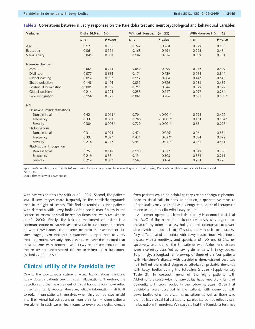
Relationships between performanceson the Pareidolia test andneuropsychological and behaviouralvariables in dementia with Lewy bodiesThe results are shown in Table 2. There were no significant cor-
relations between performances on the Pareidolia test and age,
education or visual acuity. There were no significant correlations
between illusory responses and neuropsychological performance in
the entire dementia with Lewy bodies group and in the patients
without donepezil. In the patients with dementia with Lewy bodies
with donepezil, illusory responses were significantly correlated with
the face recognition tests.
A
B
Figure 2 (A) Locations of illusory responses. (B) Locations of
first illusory responses.
Pareidolias in dementia with Lewy bodies Brain 2012: 135; 2458–2469 | 2463

In the entire dementia with Lewy bodies group, the number of
illusory responses was significantly correlated with the domain
total score of the NPI delusional misidentifications domain and
the frequency score of the hallucination domain. In the patients
with dementia with Lewy bodies without donepezil, the number of
illusory responses was significantly correlated with the domain
total, severity and frequency scores of the NPI delusional misiden-
tifications and hallucination domains.
Discussion
Phenomenological similarities betweenvisual hallucinations and pareidolias indementia with Lewy bodiesAlthough it is conceptually easy to distinguish between hallucin-
ations and illusions, the phenomenological distinction between the
two conditions is ambiguous and difficult to determine. There
have been longstanding criticisms that hallucinations should be
defined not by the presence or absence of objects but as ‘percep-
tions without objects which are to be perceived’ (Ey, 1973;
Oyebode, 2008). The following findings in this study support
the view that visual hallucinations and illusions are continuous
conditions. First, faces and whole bodies of people and animals
constituted a large portion of the content of pareidolic illusions,
whereas man-made objects were considerably less frequent. The
pareidolic illusions often appeared in bizarre and paradoxical con-
texts, such as a man in a tie or human faces in flowers. These
characteristics are similar to those of visual hallucinations in de-
mentia with Lewy bodies. Patients with dementia with Lewy
bodies experience visual hallucinations of meaningful objects
(complex visual hallucinations) far more frequently than dots,
flashes or amorphous forms (simple visual hallucinations)
(Collerton et al., 2005; Mosimann et al., 2006). Their contents
are dominated by people and animals (Mosimann et al., 2006;
Nagahama et al., 2007) and they often develop into delusions
A
B
Figure 3 (A) Contents of illusory responses on the Pareidolia test. Numbers denote a percentage of the total illusory responses in each
patient group. (B) Examples of illusory responses. Patients with dementia with Lewy bodies often misidentified objects or patterns in the
picture as real faces (yellow triangle) or as figures of people and animals (white triangle).
2464 | Brain 2012: 135; 2458–2469 M. Uchiyama et al.
with bizarre contents (McKeith et al., 1996). Second, the patients
saw illusory images more frequently in the details/backgrounds
than in the gist of scenes. This finding reminds us that patients
with dementia with Lewy bodies often see human figures in the
corners of rooms or small insects on floors and walls (Mosimann
et al., 2006). Finally, the lack or impairment of insight is a
common feature of pareidolias and visual hallucinations in demen-
tia with Lewy bodies. The patients maintain the existence of illu-
sory images, even though the examiner prompts them to verify
their judgement. Similarly, previous studies have documented that
most patients with dementia with Lewy bodies are convinced of
the reality (or unconvinced of the unreality) of hallucinations
(Ballard et al., 1997).
Clinical utility of the Pareidolia testDue to the spontaneous nature of visual hallucinations, clinicians
rarely observe patients seeing visual hallucinations. Therefore, the
detection and the measurement of visual hallucinations have relied
on self and family reports. However, reliable information is difficult
to obtain from patients themselves when they do not have insight
into their visual hallucinations or from their family when patients
live alone. In such cases, techniques to evoke pareidolias directly
from patients would be helpful as they are an analogous phenom-
enon to visual hallucinations. In addition, a quantitative measure
of pareidolias may be useful as a surrogate indicator of therapeutic
responses in dementia with Lewy bodies.
A receiver operating characteristic analysis demonstrated that
the AUC of the number of illusory responses was larger than
those of any other neuropsychological and neuropsychiatric vari-
ables. With the optimal cut-off score, the Pareidolia test success-
fully differentiated dementia with Lewy bodies from Alzheimer’s
disease with a sensitivity and specificity of 100 and 88.2%, re-
spectively, and four of the 34 patients with Alzheimer’s disease
were incorrectly classified as having dementia with Lewy bodies.
Surprisingly, a longitudinal follow-up of three of the four patients
with Alzheimer’s disease with pareidolias demonstrated that two
had fulfilled the clinical diagnostic criteria for probable dementia
with Lewy bodies during the following 2 years (Supplementary
Table 2). In contrast, none of the eight patients with
Alzheimer’s disease with no pareidolias have met the criteria of
dementia with Lewy bodies in the following years. Given that
pareidolias were observed in the patients with dementia with
Lewy bodies who had visual hallucinations as well as those who
did not have visual hallucinations, pareidolias do not reflect visual
hallucinations themselves. We suggest that the Pareidolia test may
Table 2 Correlations between illusory responses on the Pareidolia test and neuropsychological and behavioural variables
Variables Entire DLB (n = 34) Without donepezil (n = 22) With donepezil (n = 12)
r, rs P-value r, rs P-value r, rs P-value
Age 0.17 0.335 0.247 0.268 0.079 0.808
Education 0.061 0.551 0.168 0.454 �0.225 0.48
Visual acuity �0.045 0.801 �0.107 0.636 0.099 0.761
Neuropsychology
MMSE �0.065 0.713 0.059 0.795 �0.252 0.429
Digit span 0.077 0.664 0.174 0.439 �0.064 0.844
Object naming 0.014 0.937 0.117 0.604 �0.447 0.145
Shape detection �0.148 0.404 0.035 0.625 �0.233 0.465
Position discrimination 50.001 0.999 0.211 0.346 �0.529 0.077
Object decision �0.214 0.224 �0.258 0.247 �0.097 0.764
Face recognition �0.156 0.379 0.061 0.786 �0.601 0.039*
NPI
Delusional misidentifications
Domain total 0.42 0.013* 0.704 50.001* �0.256 0.422
Frequency 0.337 0.051 0.706 50.001* �0.163 0.034*
Severity 0.304 0.008* 0.729 50.001* �0.63 0.028*
Hallucinations
Domain total 0.311 0.074 0.474 0.026* �0.06 0.854
Frequency 0.397 0.02* 0.471 0.027* 0.094 0.072
Severity 0.218 0.217 0.44 0.041* �0.231 0.471
Fluctuations in cognition
Domain total 0.253 0.149 0.198 0.377 0.349 0.266
Frequency 0.219 0.33 0.13 0.308 0.389 0.211
Severity 0.214 0.057 0.565 0.164 0.253 0.428
Spearman’s correlation coefficients (rs) were used for visual acuity and behavioural symptoms; otherwise, Pearson’s correlation coefficients (r) were used.*P5 0.05.DLB = dementia with Lewy bodies.
Pareidolias in dementia with Lewy bodies Brain 2012: 135; 2458–2469 | 2465

be able to identify patients with subclinical hallucinations or a
predisposition to visual hallucinations.
The contribution of visuoperceptualimpairment to visual hallucinationsand pareidoliasCharles Bonnet syndrome in patients with ocular diseases and
visual hallucinations in the hemianopic field in patients with
damage to the central visual pathway have led to the idea that
visuoperceptual impairment plays a critical role in the emergence
of visual hallucinations (Kolmel, 1988; Ffytche, 2005, 2009). In
accord with this idea, visuoperceptual dysfunction is reportedly
more pronounced in patients who have visual hallucinations with
dementia with Lewy bodies and its related disorder, Parkinson’s
disease, than in those without visual hallucinations (Mori et al.,
2000; Mosimann et al., 2004). The contribution of defective
visuoperceptual processing is also supported by neuroimaging stu-
dies showing dysfunction of the occipitotemporal visual association
cortices in patients with dementia with Lewy bodies and patients
with Parkinson’s disease with visual hallucinations (Oishi et al.,
2005; Perneczky et al., 2008; Meppelink et al., 2009).
In this study, there was a negative correlation between the
number of illusory responses in the Pareidolia test and scores on
the face recognition tests in patients with dementia with Lewy
bodies with donepezil. This finding suggests that visuoperceptual
mechanisms are involved in pareidolias. However, the Pareidolia
test may be no more than a test of visuoperceptual function itself
rather than a measure of pathological processes associated with
visual hallucinations. To address this possibility, we performed a
supplementary experiment on two patients with visual agnosia
arising from cerebral infarction (Supplementary Fig. 2). Although
the performance of the two agnosics on the visuoperceptual tests
was similar to that of the patients with dementia with Lewy
bodies, one agnosic provided no illusory responses and the other
provided only a small number of illusory responses that was below
the cut-off score. These findings indicate that visuoperceptual im-
pairment alone is insufficient to cause the emergence of
pareidolias.
The roles of arousal and attention:relevance to acetylcholineneurotransmissionThe deafferentation hypothesis, in which visual hallucinations are
understood as release phenomena of the visual association cortices
that arise because of defective visual input, does not explain why
most patients with visual impairment do not develop visual hallu-
cinations (Ffytche, 2009). In fact, there should be additional
non-visual mechanisms in the pathogenesis of visual hallucin-
ations. Evidence suggests that the alteration of arousal and
abnormalities of the modulatory neurotransmitter systems, particu-
larly cholinergic systems, contribute to the emergence of visual
hallucinations. Visual hallucinations usually occur during
sleep-wake transitions (hypnagogic or hypnopompic hallucin-
ations) in narcolepsy and in healthy people (Manni, 2005).
Recent studies indicate that the arousal deficits in narcolepsy are
associated with the activation failure of the basal forebrain cho-
linergic systems resulting from orexinergic deficiencies (Arrigoni
et al., 2010). Furthermore, damage to the upper brainstem teg-
mentum occasionally results in visual hallucinations (referred to as
peduncular hallucinosis) (Manford and Andermann, 1998; Nishio
et al., 2007). Cholinergic, dopaminergic, serotoninergic and nora-
drenergic systems are densely packed in this brain region and play
critical roles in generating sleep-wake cycles. Finally, sleep
abnormalities and electroencephalographic slowing, a hallmark of
the low-arousal state, are prevalent in dementia with Lewy bodies
and Parkinson’s disease (Diederich et al., 2005; McKeith et al.,
2005; Bonanni et al., 2008). The amelioration of visual hallucin-
ations and fluctuating arousal/attention by cholinesterase inhibi-
tors has been repeatedly documented in dementia with Lewy
bodies and Parkinson’s disease (McKeith et al., 2005; Diederich
et al., 2009).
In their proposition of the perception and attention deficit
model, Collerton et al. (2005) proposed that visual hallucinations
arise from the combination of visuoperceptual deficits and top–
down attentional impairment. According to this model, in normal
scene perception, an external visual input activates a number of
potentially seen visual object representations, which they called
‘proto-objects’. Then, top–down attentional processes allow only
one proto-object to enter conscious visual awareness to accom-
plish visual object recognition. In the case of visual hallucinations,
top–down attentional processes are defective, and therefore, pa-
tients aberrantly recognize incorrect proto-objects as real objects.
Accordingly, the current observation suggests that abnormal top–
down attentional orientation is involved in the pathomechanisms
of pareidolias in dementia with Lewy bodies; the patients with
dementia with Lewy bodies saw pareidolias more in the detail/
background, which is usually of little semantic importance and
less noticeable, than in the gist of scenes, which carries semantic-
ally important information and has attentional priority.
The perception and attention deficit model proposes that atten-
tional impairment in visual hallucinations is associated with cholin-
ergic insufficiency (Collerton et al., 2005). This view is constructed
on the grounds of the following clinical and neuroscientific find-
ings. First, it has long been known that anticholinergic agents
produce a hypoarousal state (also referred to as a confusional
state) associated with severe attentional deficits and visual hallu-
cinations (Collerton et al., 2005). There is also abundant evidence
suggesting that cholinomimetic drugs improve attentional deficits
and visual hallucinations (McKeith et al., 2000; Mori et al., 2006).
Second, acetylcholine facilitates local cortical processing of exter-
nal input and at the same time suppresses internal information
processing, consequently enhancing the neuronal signal-to-noise
ratio for external information (Perry et al., 1999; Hasselmo and
McGaughy, 2004). Under low acetylcholine states, information
processing is susceptible to internal ‘noise’ and thereby attentional
control processes are disrupted. In the current study, the NPI hal-
lucination score and the number of illusory responses were smaller
in the patients with dementia with Lewy bodies with donepezil
than in those without (Supplementary Table 1). Moreover, the
number of illusory responses was not correlated with the NPI hal-
lucination score but with performance on the visual perception
2466 | Brain 2012: 135; 2458–2469 M. Uchiyama et al.

tests in the patients treated with donepezil (Table 2). These results
suggest that the role of visuoperceptual deficits is larger in parei-
dolias than in visual hallucinations, whereas the role of attentional
deficits is larger in visual hallucinations than in pareidolias.
Accordingly, pareidolias may tend to persist after cholinomimetic
drugs alleviate attentional deficits and improve visual hallucin-
ations. When patients are treated with cholinomimetic drugs, par-
eidolias may primarily reflect residual visuoperceptual impairment.
Lack of insight into visual hallucinationsand pareidoliasInsight into visual hallucinations is lacking or impaired in most
patients with dementia with Lewy bodies (Ballard et al., 1997).
These patients often develop secondary delusions of the real ex-
istence of persons and animals that appear in visual hallucinations
(McKeith et al., 2005). Similarly, the current study demonstrated
that insight into pareidolias is lacking in dementia with Lewy
bodies. The contexts in which pareidolias occur were bizarre and
paradoxical, e.g. a man appearing in a tie and human faces ap-
pearing in flowers. These findings suggest that abnormal insight is
a key feature of both visual hallucinations and pareidolias in pa-
tients with dementia with Lewy bodies. Thus, how do these pa-
tients falsely accept the bizarre and paradoxical contents of visual
hallucinations and pareidolias as reality? A similar question is found
in research on psychosis in schizophrenia: how do patients acquire
delusional beliefs and false convictions of reality of hallucinatory
perceptions? Kapur (2003) proposed that the initial step in the
development of psychosis is heightened awareness or aberrant
assignment of motivational salience to unimportant perceptual ex-
periences. In a similar way, we could consider a model in which
the aberrant assignment of salience to illusory perceptual informa-
tion (or incorrect proto-objects in the terminology of the percep-
tion and attention deficit model) leads to abnormal insight into
visual hallucinations and pareidolias in dementia with Lewy
bodies. We suggest that abnormal insight may be a common
underlying pathology of visual hallucinations, pareidolias and de-
lusions in dementia with Lewy bodies. In fact, in the current study,
the number of illusory responses was significantly correlated with
both hallucination and delusional misidentifications scores on the
NPI.
Some patients with dementia with Lewy bodies treated with
cholinesterase inhibitors report their experiences in ways such as
‘I had thought it was real before, but now I realize that that was
just an illusion.’ Such an introspective description suggests a rela-
tionship between cholinergic insufficiency and abnormal insight.
Other evidence for cholinergic mechanisms in abnormal insights
comes from peduncular hallucinosis, a condition of recurrent visual
hallucinations after damage to the upper brainstem tegmentum in
which the visual pathways are spared (Manford and Andermann,
1998; Nishio et al., 2007). Hypoarousal or attentional impairment
due to ascending modulatory neurotransmitter systems is a pri-
mary pathomechanism of visual hallucinations in this condition.
Insight into visual hallucinations is usually lacking or impaired in
most cases of peduncular hallucinosis (Feinberg and Rapcsak,
1989; Kolmel, 1991; Furuta et al., 2002; Benke, 2006;
Nishio et al., 2007) (for a counter view, see Manford and
Andermann, 1998). Moreover, there have been previous reports
of the concomitance of visual hallucinations and delusional mis-
identifications in peduncular hallucinosis and those of delusions
subsequent to upper brainstem lesions (Drake et al., 1990;
Mitsuhata and Tsukagoshi, 1992; Benke, 2006). The roles of cho-
linergic insufficiency in abnormal insight into visual hallucinations
and pareidolias and the pathomechanisms of delusional misidenti-
fications in dementia with Lewy bodies should be addressed in
future pharmacological intervention studies.
LimitationsSeveral limitations of this study should be noted. First, the high
sensitivity and moderate specificity of the Pareidolia test in differ-
entiating between dementia with Lewy bodies and Alzheimer’s
disease may be associated with an inclusion bias. Since the current
diagnostic criteria for dementia with Lewy bodies emphasize the
existence of visual hallucinations, the differential diagnosis of de-
mentia with Lewy bodies and Alzheimer’s disease depends heavily
on whether patients have visual hallucinations. In addition, given
the low sensitivity of the criteria for dementia with Lewy bodies,
there could be cases in which pathological dementia with Lewy
bodies is misdiagnosed as Alzheimer’s disease (McKeith et al.,
2005). Second, a cross-sectional comparison between patients
with and without donepezil does not provide definitive evidence
for cholinergic mechanisms. Longitudinal investigations are needed
to elucidate the relationship between pareidolias and acetylcholine
and to examine the usefulness of the Pareidolia test in measuring
the therapeutic effects of cholinergic agents. Third, although there
has been a substantial interest in the dopaminergic and prefrontal
mechanisms of psychosis, we did not address these issues in our
study (Kapur, 2003; Coltheart et al., 2011; Feinberg, 2011).
Finally, illusory responses in the Pareidolia test may not purely
reflect visual illusions and may be contaminated by confabulatory
responses. Doubleday et al. (2002) reported that confabulatory
responses on memory tests were observed in 17% of patients
with dementia with Lewy bodies. Although the memory load is
fairly low in the Pareidolia test, the prompts requesting more de-
scription may evoke confabulatory responses. To diminish the pos-
sibilities of contamination of confabulation, we are currently
developing a new task to evoke pareidolias in which subjects are
not required to give detailed descriptions.
Conclusion and futuredirectionsUsing a simple task, we successfully evoked pareidolias, which are
complex visual illusions phenomenologically analogous to visual
hallucinations, in patients with dementia with Lewy bodies.
Compared with patients with Alzheimer’s disease and healthy con-
trols, patients with dementia with Lewy bodies experienced a
much larger number of pareidolias. A sub-analysis of patients
with or without donepezil suggested that cholinergic mechanisms
are involved in visual hallucinations and in pareidolias. Our results
Pareidolias in dementia with Lewy bodies Brain 2012: 135; 2458–2469 | 2467

suggest that tests that quantify pareidolias may be beneficial in the
differential diagnosis of dementing disorders and may be a useful
indicator of therapeutic response in dementia with Lewy bodies. In
addition, the pathomechanisms of visual hallucinations in
Parkinson’s disease have been considered similar to those observed
in dementia with Lewy bodies (Ffytche, 2007). Visual illusions and
minor hallucinations, such as presence and passage hallucinations,
are reportedly common in Parkinson’s disease without visual hal-
lucinations (Fenelon et al., 2000). We suggest that pareidolias may
be a phenomenon bridging visual illusions, minor hallucinations
and visual hallucinations. We are currently involved in a study
on this topic.
AcknowledgementsWe thank the patients who participated in this study. We also
thank Akiko Hayashi and Mayumi Shinohara for their help in
data acquisition.
FundingGrant-in-Aid for Scientific Research for Young Scientists from the
Ministry of Education, Culture, Sports, Science and Technology of
Japan (90451591 to Y.N.) and Grant-in-Aid for Scientific Research
on Innovative Areas (Face Perception and Recognition) from the
Ministry of Education, Culture, Sports, Science and Technology of
Japan (30368477 to E.M.).
Supplementary materialSupplementary material is available at Brain online.
ReferencesAdolphs R, Denburg NL, Tranel D. The amygdala’s role in long-term
declarative memory for gist and detail. Behav Neurosci 2001; 115:
983–92.Arrigoni E, Mochizuki T, Scammell TE. Activation of the basal forebrain
by the orexin/hypocretin neurones. Acta Physiol 2010; 198: 223–35.
Ballard C, McKeith I, Harrison R, O’Brien J, Thompson P, Lowery K, et al.
A detailed phenomenological comparison of complex visual hallucin-
ations in dementia with Lewy bodies and Alzheimer’s disease. Int
Psychogeriatr 1997; 9: 381–8.
Benke T. Peduncular hallucinosis: a syndrome of impaired reality moni-
toring. J Neurol 2006; 253: 1561–71.
Bonanni L, Thomas A, Tiraboschi P, Perfetti B, Varanese S, Onofrj M.
EEG comparisons in early Alzheimer’s disease, dementia with Lewy
bodies and Parkinson’s disease with dementia patients with a 2-year
follow-up. Brain 2008; 131: 690–705.Collerton D, Perry E, McKeith I. Why people see things that are not
there: a novel Perception and Attention Deficit model for recurrent
complex visual hallucinations. Behav Brain Sci 2005; 28: 737–57; dis-
cussion 757–94.
Coltheart M, Langdon R, McKay R. Delusional belief. Annu Rev Psychol
2011; 62: 271–98.
Cummings JL, Mega M, Gray K, Rosenberg-Thompson S, Carusi DA,
Gornbein J. The Neuropsychiatric Inventory: comprehensive assess-
ment of psychopathology in dementia. Neurology 1994; 44: 2308–14.
Diederich NJ, Fenelon G, Stebbins G, Goetz CG. Hallucinations in
Parkinson disease. Nat Rev Neurol 2009; 5: 331–42.
Diederich NJ, Goetz CG, Stebbins GT. Repeated visual hallucinations in
Parkinson’s disease as disturbed external/internal perceptions: focused
review and a new integrative model. Mov Disord 2005; 20: 130–40.Doubleday EK, Snowden JS, Varma AR, Neary D. Qualitative perform-
ance characteristics differentiate dementia with Lewy bodies and
Alzheimer’s disease. J Neurol Neurosurg Psychiatry 2002; 72: 602–7.
Drake ME Jr, Pakalnis A, Phillips B. Secondary mania after ventral pon-
tine infarction. J Neuropsychiatry Clin Neurosci 1990; 2: 322–5.
Ey H. Traite des hallucinations. Paris: Masson; 1973.
Feinberg TE. Neuropathologies of the self: clinical and anatomical fea-
tures. Conscious Cogn 2011; 20: 75–81.
Feinberg WM, Rapcsak SZ. ‘Peduncular hallucinosis’ following parame-
dian thalamic infarction. Neurology 1989; 39: 1535–6.
Fenelon G, Mahieux F, Huon R, Ziegler M. Hallucinations in Parkinson’s
disease: prevalence, phenomenology and risk factors. Brain 2000;
123(Pt 4): 733–45.
Ffytche DH. Visual hallucinations and the Charles Bonnet syndrome. Curr
Psychiatry Rep 2005; 7: 168–79.
Ffytche DH. Visual hallucinatory syndromes: past, present, and future.
Dialogues Clin Neurosci 2007; 9: 173–89.
Ffytche DH. Visual hallucinations in eye disease. Curr Opin Neurol 2009;
22: 28–35.
Folstein MF, Folstein SE, McHugh PR. "Mini-mental state". A practical
method for grading the cognitive state of patients for the clinician. J
Psychiatr Res 1975; 12: 189–98.Furuta T, Nobutou H, Nobutou F. [Peduncular hallucinosis due to a small
hemorrhage around the substantia nigra: a case report]. No To Shinkei
2002; 54: 423–6.
Hasselmo ME, McGaughy J. High acetylcholine levels set circuit dynamics
for attention and encoding and low acetylcholine levels set dynamics
for consolidation. Prog Brain Res 2004; 145: 207–31.
Iseki E, Marui W, Nihashi N, Kosaka K. Psychiatric symptoms typical of
patients with dementia with Lewy bodies - similarity to those of
levodopa-induced psychosis. Acta Neuropsychiatr 2002; 14: 237–41.
Japan Society for Higher Brain Dysfunction. Visual perception test for
agnosia. Tokyo: Shinko Igaku Shuppan; 1997.
Kapur S. Psychosis as a state of aberrant salience: a framework linking
biology, phenomenology, and pharmacology in schizophrenia. Am J
Psychiatry 2003; 160: 13–23.
Kolmel HW. Peduncular hallucinations. J Neurol 1991; 238: 457–9.Kolmel HW. Die homonymen Hemianopsien - Klinik und
Pathophysiologie zentraler Sehstorungen. Berlin: Springer; 1988.
Manford M, Andermann F. Complex visual hallucinations. Clinical and
neurobiological insights. Brain 1998; 121(Pt 10): 1819–40.
Manni R. Rapid eye movement sleep, non-rapid eye movement sleep,
dreams, and hallucinations. Curr Psychiatry Rep 2005; 7: 196–200.
McKeith I, Del Ser T, Spano P, Emre M, Wesnes K, Anand R, et al.
Efficacy of rivastigmine in dementia with Lewy bodies: a randomised,
double-blind, placebo-controlled international study. Lancet 2000;
356: 2031–6.
McKeith IG, Dickson DW, Lowe J, Emre M, O’Brien JT, Feldman H, et al.
Diagnosis and management of dementia with Lewy bodies: third
report of the DLB Consortium. Neurology 2005; 65: 1863–72.McKeith IG, Galasko D, Kosaka K, Perry EK, Dickson DW, Hansen LA,
et al. Consensus guidelines for the clinical and pathologic diagnosis of
dementia with Lewy bodies (DLB): report of the consortium on DLB
international workshop. Neurology 1996; 47: 1113–24.McKhann G, Drachman D, Folstein M, Katzman R, Price D, Stadlan EM.
Clinical diagnosis of Alzheimer’s disease: report of the
NINCDS-ADRDA Work Group under the auspices of Department of
Health and Human Services Task Force on Alzheimer’s Disease.
Neurology 1984; 34: 939–44.
Meppelink AM, de Jong BM, Renken R, Leenders KL, Cornelissen FW,
van Laar T. Impaired visual processing preceding image recognition in
Parkinson’s disease patients with visual hallucinations. Brain 2009; 132:
2980–93.
2468 | Brain 2012: 135; 2458–2469 M. Uchiyama et al.
Mitsuhata Y, Tsukagoshi H. [Cerebellar infarction presenting erotic delu-sion and delusion of jealousy in the acute phase]. Rinsho Shinkeigaku
1992; 32: 1256–60.
Mori S, Mori E, Iseki E, Kosaka K. Efficacy and safety of donepezil in
patients with dementia with Lewy bodies: preliminary findings from anopen-label study. Psychiatry Clin Neurosci 2006; 60: 190–5.
Mori E, Shimomura T, Fujimori M, Hirono N, Imamura T, Hashimoto M,
et al. Visuoperceptual impairment in dementia with Lewy bodies. Arch
Neurol 2000; 57: 489–93.Mosimann UP, Collerton D, Dudley R, Meyer TD, Graham G, Dean JL,
et al. A semi-structured interview to assess visual hallucinations in
older people. Int J Geriatr Psychiatry 2008; 23: 712–8.Mosimann UP, Mather G, Wesnes KA, O’Brien JT, Burn DJ, McKeith IG.
Visual perception in Parkinson disease dementia and dementia with
Lewy bodies. Neurology 2004; 63: 2091–6.
Mosimann UP, Rowan EN, Partington CE, Collerton D, Littlewood E,O’Brien JT, et al. Characteristics of visual hallucinations in Parkinson
disease dementia and dementia with lewy bodies. Am J Geriatr
Psychiatry 2006; 14: 153–60.
Nagahama Y, Okina T, Suzuki N, Matsuda M, Fukao K, Murai T.Classification of psychotic symptoms in dementia with Lewy bodies.
Am J Geriatr Psychiatry 2007; 15: 961–7.
Nishio Y, Ishii K, Kazui H, Hosokai Y, Mori E. Frontal-lobe syndrome andpsychosis after damage to the brainstem dopaminergic nuclei. J Neurol
Sci 2007; 260: 271–4.
Oishi N, Udaka F, Kameyama M, Sawamoto N, Hashikawa K,
Fukuyama H. Regional cerebral blood flow in Parkinson disease withnonpsychotic visual hallucinations. Neurology 2005; 65: 1708–15.
Oyebode F. Sim’s symptoms in the mind. Philadelphia: Elsevier; 2008.
Perneczky R, Drzezga A, Boecker H, Forstl H, Kurz A, Haussermann P.
Cerebral metabolic dysfunction in patients with dementia with Lewybodies and visual hallucinations. Dement Geriatr Cogn Disord 2008;
25: 531–8.
Perry E, Walker M, Grace J, Perry R. Acetylcholine in mind: a neuro-transmitter correlate of consciousness? Trends Neurosci 1999; 22:
273–80.
Riddoch M, Humphreys G. Birmingham Object Recognition Battery.
Hove (UK): Lawrence Erlbaum; 1992.Sugishita M. The Western Aphasia Battery [Japanese edition]. Tokyo:
Igakushoin; 1986.
Sugishita M. Japanese Wechsler Memory Scale-Revised. Tokyo:
Nihonbunkakagakusha; 2001.Warrington EK, James M. Visual object and space perception battery.
Bury St Edmunds (UK): Thames Valley Test Company; 1991.
Pareidolias in dementia with Lewy bodies Brain 2012: 135; 2458–2469 | 2469