Embed Size (px)
Citation preview
PARP activation regulates the RNA-binding proteinNONO in the DNA damage response to DNAdouble-strand breaksJana Krietsch1,2, Marie-Christine Caron2, Jean-Philippe Gagne1, Chantal Ethier1,
Julien Vignard2, Michel Vincent3, Michele Rouleau1, Michael J. Hendzel4,
Guy G. Poirier1,* and Jean-Yves Masson2,*
1Cancer Research Unit, Laval University Medical Research Center, CHUQ-CRCHUL, Quebec, QC, Canada G1V4G2, 2Genome Stability Laboratory, Laval University Cancer Research Center, Hotel-Dieu de Quebec, QC,Canada G1R 2J6, 3Faculty of Medicine, Laval University, Quebec, QC, Canada G1V 0A6 and 4Department ofOncology, Faculty of Medicine and Dentistry, University of Alberta, 11560 University Avenue, Edmonton,Alberta, Canada T6G 1Z2
Received May 7, 2012; Revised July 26, 2012; Accepted July 30, 2012
ABSTRACT
After the generation of DNA double-strand breaks(DSBs), poly(ADP-ribose) polymerase-1 (PARP-1) isone of the first proteins to be recruited andactivated through its binding to the free DNA ends.Upon activation, PARP-1 uses NAD+ to generatelarge amounts of poly(ADP-ribose) (PAR), which fa-cilitates the recruitment of DNA repair factors. Here,we identify the RNA-binding protein NONO, apartner protein of SFPQ, as a novel PAR-bindingprotein. The protein motif being primarily respon-sible for PAR-binding is the RNA recognition motif1 (RRM1), which is also crucial for RNA-binding,highlighting a competition between RNA and PARas they share the same binding site. Strikingly, thein vivo recruitment of NONO to DNA damage sitescompletely depends on PAR, generated by activatedPARP-1. Furthermore, we show that upon PAR-dependent recruitment, NONO stimulates nonho-mologous end joining (NHEJ) and represseshomologous recombination (HR) in vivo. Ourresults therefore place NONO after PARP activationin the context of DNA DSB repair pathway decision.Understanding the mechanism of action of proteinsthat act in the same pathway as PARP-1 is crucial toshed more light onto the effect of interference onPAR-mediated pathways with PARP inhibitors,which have already reached phase III clinical trialsbut are until date poorly understood.
INTRODUCTION
Each day, the cells genome is confronted with up to 50endogenous DNA double-strand breaks (DSBs). Theseare extremely hazardous for a cell, as they do not leavean intact complementary strand to serve as a template forrepair (1). If left unrepaired, DSBs can have consequencessuch as cell death or carcinogenesis. Hence, understandingthe mechanisms that lead to successful repair of DSBs willfurther increase the knowledge of cancer progression andtreatments. The DNA damage response (DDR) to DSBs isa multilayered process, initiated with sensing and signalingDNA damage, subsequent recruitment of repair proteinsand execution of repair (2).Poly(ADP-ribose) polymerase-1 (PARP-1) is an
abundant and ubiquitous nuclear protein that usesNAD+ to synthesize a negatively charged polymer,called poly(ADP-ribose) (PAR), onto a variety of targetproteins, such as histones, DSB repair factors andPARP-1 itself. The latter post-translational protein modi-fication has an impact on cellular processes as diverse astranscription (3), cell death (4) and especially DNA repair(5). PARP-1 acts as a strong sensor for DNA damage andrapidly produces PAR at newly generated DNA DSBs,provoking therewith local chromatin relaxation due toits negative charge (3) and facilitating the recruitment ofrepair factors, such as MRE11 (2,6). The dynamic turn-over of PAR within seconds to minutes is executed bypoly(ADP-ribose) glycohydrolase (PARG), that possessesendo- and exoglycosidic activities, hence enabling a newround of DNA damage signaling (7).For subsequent repair, two major DSB repair pathways
have evolved, namely nonhomologous end joining
*To whom correspondence should be addressed. Tel:+1 418 525 4444 (ext 15154); Fax:+1 418 691 5439; Email: [email protected] may also be addressed to Guy G. Poirier. Tel: +1 418 654 2267; Fax: +1 418 654 2159; Email: [email protected]
Published online 31 August 2012 Nucleic Acids Research, 2012, Vol. 40, No. 20 10287–10301doi:10.1093/nar/gks798
� The Author(s) 2012. Published by Oxford University Press.This is an Open Access article distributed under the terms of the Creative Commons Attribution Non-Commercial License (http://creativecommons.org/licenses/by-nc/3.0), which permits unrestricted non-commercial use, distribution, and reproduction in any medium, provided the original work is properly cited.
Downloaded from https://academic.oup.com/nar/article-abstract/40/20/10287/2414786by gueston 09 February 2018
(NHEJ) and homologous recombination (HR). WhereasHR is considered as error-free and restricted to the S/G2-phase (8) by its necessity for a homologous template,error-prone NHEJ functions throughout the cell cycleand represents the major pathway for DSB repair in multi-cellular eukaryotes. Although the NHEJ pathway is highlyflexible in terms of substrate ends used for repair,participating repair proteins and possible outcomes, anumber of key proteins are indispensable to accomplishclassical NHEJ (cNHEJ): Initially, the heterodimericKu70/Ku80 complex binds to both ends of the brokenDNA molecule (9). Interestingly, Ku has an affinity forPAR (10) and is also a direct target for PARylation (11).The Ku–DNA complex is further bound by the catalyticsubunit of DNA–PK (DNA–PKcs) to assemble theend-bridging DNA–PK complex (12). If the two endsare not directly ligatable they have to be processed priorto the final ligation step. A variety of proteins (such asArtemis, PNK, APLF nucleases, TdT, polymerases �and m) have been implicated in the end-processing step,emphasizing the mechanistic flexibility of the NHEJreaction (13–16). The final ligation step is carried out byX4-L4 complex, composed of XRCC4, DNA ligase IVand XLF (17).Within the last years, growing attention has been drawn
to proteins with dual roles in RNA biology and DNADSB repair. Examples include the Ku protein, which iscrucial for the NHEJ pathway but interestingly also forthe control of mRNA expression (18,19), the TFHIIcomplex that acts in nucleotide excision repair as well asin transcriptional initiation mediated by RNA polymeraseII (20), and recently the RNA-binding protein RBMX andthe RNA-splicing factor THRAP3 were implied in theDDR (21–23). About twenty years ago the group ofHarris Busch purified and characterized a heterodimerconsisting of a 52 and a 100 kDa subunit, most certainlycorresponding to what is nowadays known as the 54 kDanuclear RNA-binding protein (p54nrb/NONO) and thepolypyrimidine tract-binding protein-associated splicingfactor (PSF/SFPQ). NONO and SFPQ show 71%sequence identity and, together with paraspeckle compo-nent 1 (PSPC1), belong to a subfamily of RNA recogni-tion motif (RRM) proteins defined by tandem RRMmotifs, flanked by an additional region of sequence simi-larity predicted to promote formation of heteromericcomplexes between each of the proteins (24). NONOand SFPQ have been implicated in nuclear retention ofA- to I-edited RNA as paraspeckle components (25),pre-mRNA 30-end formation (26), cAMP cycling (27)and transcriptional activation (28–30). Interestingly,apart from their functions in RNA biogenesis, NONOand SFPQ were reported to interact with DNA in vitro,which lead to an investigation of their function in thecontext of DNA repair. Both proteins are transiently re-cruited with the same kinetics to DNA damage induced bya laser track in human cells (31). Interestingly, a proteincomplex containing NONO and SFPQ stimulates NHEJabout 10-fold in vitro (32). Furthermore, it has beendemonstrated that the attenuation of NONO protein ex-pression, independent of its partner protein SFPQ, delaysthe resolution of g-H2AX foci after ionizing irradiation
and leads to an accumulation of chromosomal aberrations(33). However, the exact mechanism by which NONO isrecruited to DNA damage sites and regulates DSB repairis unclear. Interestingly, a bioinformatics screen from ourgroup for proteins that potentially bind PAR, which isgenerated within seconds at a new DSB, identifiedNONO/SFPQ among a variety of NHEJ factors (10,34),leading to the hypothesis that PARP and its associatedpolymer regulates NONO. In this manuscript, we dissectthe role of NONO in DSB repair in the context of PARPactivation. We suggest here that NONO is directlyimplicated in NHEJ, and that its recruitment to DNAdamage sites is strictly dependent on activated PARP-1.These results highlight the emerging concept of RNA-binding proteins in DSB repair.
MATERIALS AND METHODS
Cell lines, cell culture, and DNA constructs
HeLa cells and mouse embryonic fibroblasts (MEFs) pro-ficient for PARP-1 and PARP-2 [wild type (WT)], or de-ficient for either PARP-1 (PARP-1�/�) or PARP-2(PARP-2�/�) were cultured in DMEM, while MCF-7cells were cultured in MEM-alpha (air/CO2, 19:1, 37
�C).Both media were supplemented with 10% fetal bovineserum and 1% penicillin/streptomycin.
The NHEJ reporter construct ‘sGEJ’ was kindlyprovided by Dr. Ralph Scully (35) and stably integratedinto the genomic DNA of MCF-7 cells by using G418disulfate salt (400mg/ml; Sigma) as a selection marker.The HR reporter construct ‘DR-GFP’ [kindly providedby Dr. Maria Jasin; (36)] was integrated into thegenomic DNA of MCF-7 cells by hygromycin selection(400 mg/ml; Invitrogen).
The GFP-NONO construct is a generous gift fromDr. James Patton (Vanderbilt University, Nashville,TN). NONO was cloned for protein purification fromthe pEGFP vector into a pET-16 b (Novagen) vectorusing the primers shown in Supplementary Table S1.
Site-directed mutagenesis on the His-NONO and GFP-NONO constructs was carried out with the QuikChange
TM
Site-Directed Mutagenesis Kit (Stratagene) using theoligos shown in Supplementary Table S1.
Antibodies and siRNAs
For Western blotting analysis and chromatin-immuno-precipitation (ChIP) experiments, polyclonal antibodiesfor NONO and SFPQ were obtained from Bethyllaboratories. The monoclonal antibody against GAPDH(6C5) was obtained from Fitzgerald Industries. Polyclonalantibodies for RAD51 and PSPC1 were purchased fromSanta Cruz. PARP-1 (C2–10) monoclonal antibody wasproduced in house as described (37).
Gene silencing was performed using siRNA directedagainst the following target sequences: 50-GGAAGCCAGCUGCUCGGAAAGCUCU-30 against NONO, 50-GCCAGCAGCAAGAAAGGCAUUUGAA-30 againstSFPQ (Invitrogen). A scrambled siRNA (50-GACGTCATATACCAAGCTAGTTT-30) from Dharmacon was usedas a negative control. Transfection of 5 nM siRNA per
10288 Nucleic Acids Research, 2012, Vol. 40, No. 20
Downloaded from https://academic.oup.com/nar/article-abstract/40/20/10287/2414786by gueston 09 February 2018
condition was performed for 48 hr using HiPerfect trans-fection reagent (Qiagen) according to the manufacturer’sprotocol. For the siRNA directed against NONO, asecond round of transfection (�36 hr after the first trans-fection) was performed for another 24 hr.
Colony forming assays
Long-term cell viability of HeLa cells transfected with theindicated siRNAs was assessed by colony forming assays.Briefly, a total of 200 cells per condition were plated into35-mm dishes. Cells were then exposed to ionizing radi-ation of 0, 0.5 or 2 Gray using a g-irradiator (Gammacell-40; MDS Nordion). After 7 to 10 days, colonies were fixedwith methanol, stained using a 4 g/L solution of methyleneblue in methanol, extensively washed with PBS andcounted.
Protein purification
Recombinant wild-type human NONO (NONO-WT) andthe RRM1-deletion mutant (NONO�RRM1) proteinswere purified from an Escherichia coli BL-21 straincarrying pET16b-10XHis-NONO or pET16b-10XHis-NONO�RRM1 expression constructs, grown in 4 L ofLB media supplemented with 100 mg/ml ampicillin and25 mg/ml chloramphenicol. Protein expression wasinduced for 16 hr at 16�C with 0.1mM IPTG added tothe culture at an OD600=0.4. Cells were then harvestedby centrifugation and resuspended in 40ml lysis buffer A(20mM Tris-HCl pH 8.0, 10% glycerol, 2mMb-mercapthoethanol, 500mM NaCl, 5mM imidazole,1mM PMSF, 1 mg/ml leupeptin, 0.019 TIU/ml aprotinin).Samples were lysed with a Dounce homogenizer (10strokes with the tight pestle), sonicated using a sonicator(Bioruptor; Diagenode) (10min at the ‘high’ setting, 30 sON and 30 s OFF) and returned to the Dounce for asecond round of lysis. Insoluble material was removedby centrifugation at 40 000 rpm for 1 hr at 4�C and thesupernatant subsequently loaded on a 5 ml cobalt-basedimmobilized metal affinity chromatography resinTalon column (BD Biosciences, Palo Alto, CA). Thecolumn was washed and eluted with a linear gradient ofimidazole ranging from 5 to 1000mM prepared in bufferA. Fractions containing His-tagged NONO-WT orNONO�RRM1 were identified by sodium dodecylsulphate-polyacrylamide gel electrophoresis (SDS-PAGE), carefully selected, pooled and dialyzed for 1 hragainst 20mM Tris-HCl pH 8.0, 375mM NaCl, 10%glycerol and 0.05% Tween-20 buffer.
FACS analysis of the cell cycle
Cells were collected by trypsinization, centrifuged and re-suspended at 106 cells per 300 ml of PBS and fixed with700 ml of ice-cold ethanol (100%) while vortexing. Oncefixed, cells were washed with PBS and stained with pro-pidium iodide (0.1% sodium citrate, 0.3% Nonidet P40,propidium iodide 50mg/ml and RNAse A 20mg/ml). Cellcycle analysis was performed on a Beckman Coulter EpicsElite model ESP by using the Expo2 analysis software.
Pulse-field gel electrophoresis
HeLa cells treated with the indicated siRNA wereincubated for 2 hr at 37�C in the presence of 500 ng/mlNeocarzinostatin (NCS). After treatment, cells werereleased for the indicated time points and trypsinized.One percent agarose plugs containing 5� 106 cells wereprepared with a CHEF-disposable plug mold (Bio-Rad).Cells were lysed by incubation of the gel blocks for 72 hr at45�C in 1mg/mL proteinase K, 100mM ethylenediamine-tetraacetic acid (EDTA), 0.2% sodium deoxycholate, 1%N-laurylsarcosyl. Samples were then washed three timesfor 1 hr each in 20mM Tris pH 8.0, 50mM EDTA andembedded into an agarose gel (0.9% agarose in 0.5Xfiltered TBE). DNA separation was performed at 14�Cfor 24 hr with a two block pulse linear program (block1: 0.1 s at 30 s, 5.8V/cm, 14�C, angle 120�, TBE 0.5X,12 hr; block 2: 0,1 s at 5 s, 3.6V/cm, 14�C, angle 110�,TBE 0.5X, 12 hr) in a CHEF-DR III Pulsed FieldElectrophoresis System (Bio-Rad). The gel was thendried for 30min at 55�C and for additional 30min atroom temperature, stained overnight with SYBR green(Molecular Probes) and visualized using a UV lamp. Ayeast chromosome PFG marker (NEB 345) served as aladder for molecular weight.
Nuclear extract preparation
Up to 107 HeLa cells per condition were washed threetimes with PBS, resuspended and incubated for 15minon ice in 250ml hypotonic buffer (10mM Tris pH 7.4,10mM MgCl2, 10mM KCl and 1mM DTT). Thesamples were then passed 5 times through a 1ml syringewith a 27G needle and centrifuged for 15min at 3300� gat 4�C. Pellets were resuspended in 200 ml high salt buffer(hypotonic buffer A with 350mM NaCl and protease in-hibitors) and incubated for 1 hr on ice. After centrifuga-tion for 30min at 13 000rpm at 4�C, the supernatants weretransferred to a clean tube and adjusted to 10% glycerol(v/v) and 10 mM of b-mercapthoethanol.
Cell fractionation and western blot analysis
Cell fractionation was carried out as described in (38) withslight modifications. Briefly, 3� 106 HeLa cells per condi-tion were collected and resuspended in 200 mL of buffer A(10mM HEPES pH 8.0, 10mM KCl, 1.5mM MgCl2,0.34M sucrose, 10% glycerol, 1mM DTT, 1mM PMSF,0.1% Triton-X-100, 10mMNaF, 1mMNa2VO3, proteaseinhibitors) and kept for 5min on ice. The soluble cytoplas-mic fraction (S1) was separated from the nuclei (P2) bycentrifugation for 4min at 1300� g at 4�C. The nuclearfraction P2 was washed twice with 300 mL buffer A thenresuspended in 200 mL buffer B (3mM EDTA, 0.2mMEGTA, 1mM DTT, 1mM PMSF, 10mM NaF, 1mMNa2VO3, protease inhibitors) and kept for 30min on ice.The insoluble chromatin fraction (P3) was separated fromnuclear soluble proteins (S3) by centrifugation for 4min at1700� g at 4 �C. S1 was cleared from insoluble proteinsby centrifugation at 14 000rpm for 15min at 4�C andthe supernatant (S2) was kept for analysis. Cell fractions
Nucleic Acids Research, 2012, Vol. 40, No. 20 10289
Downloaded from https://academic.oup.com/nar/article-abstract/40/20/10287/2414786by gueston 09 February 2018
were subsequently analysed by western blotting asdescribed in (39).
ChIP and quantitative polymerase chain reaction
A unique DSB in MCF-7 cells was introduced by electro-porating the I-SceI expression vector (pCBASce) intoMCF-7 DR-GFP (carrying a chromosomally integratedhomology-directed repair site) cells using the GenePulser Xcell apparatus (Bio Rad). A total of 2� 106 cellsper electroporation, resuspended in 650 ml PBS, weremixed with 50 mg of circular plasmid and pulsed at0.25 kV and 1000mF in 4-mm cuvettes. Cells were thenplated onto 10-cm dishes containing fresh medium andkept at 37�C for 12 hr. To crosslink proteins to DNA,cells were treated for 10min with a 1% formaldehydesolution in PBS. Subsequently, glycine to a final concen-tration of 0.125M was added to quench the reaction. Cellswere collected in ice cold PBS using a cell scraper, washedtwice in cold PBS containing 1mM PMSF, washed for10min in solution I (10mM HEPES, pH 7.5, 10mMEDTA, 0.5mM EGTA, 0.75% Triton X-100) and10min in solution II (10mM HEPES, pH 7.5, 200mMNaCl, 1mM EDTA, 0.5mM EGTA). Cells were resus-pended in lysis buffer (25mM Tris–HCl, pH 7.5,150mM NaCl, 1% Triton X-100, 0.1% SDS, 0.5%deoxycholate) and kept for 45min on ice. To shear chro-matin to an average size of 0.5 kb, cells were sonicatedwith a Bioruptor sonicator (Diagenode) for 10min (high,30 s ON, 30 s OFF). Samples were then centrifuged atmaximum speed in a benchtop centrifuge until clear andthe lysate precleared overnight with Sepharose CL-6Bbeads. Immunoprecipitation was performed for 2 hr inlysis buffer with polyclonal antibodies against NONO.Rabbit anti-human IgG (H+L) antibody (JacksonImmunoresearch Laboratories) was used as a negativecontrol. Protein–antibody complexes were subsequentlyincubated with protein A/G beads for 1 hr. Complexeswere washed twice with RIPA buffer (150mM NaCl,50mM Tris-HCl pH 8.0, 0.1% SDS, 0.5% deoxycholate,1% NP-40, 1mM EDTA), once in high salt buffer (50mMTris–Cl, pH 8.0, 500mM NaCl, 0.1% SDS, 0.5%deoxycholate, 1% NP-40, 1mM EDTA), once in LiClbuffer (50mM Tris–HCl, pH 8.0, 250mM LiCl, 1%NP-40, 0.5% deoxycholate, 1mM EDTA) and twice inTE buffer (10mM Tris–HCl, pH 8.0, 1mM EDTA, pH8.0). Beads were resuspended in TE containing 50 mg/mlRNase A and incubated for 30min at 37�C. Beads werewashed with deionized water and incubated for 15min inelution buffer (1% SDS, 0.1 M NaHCO3). Crosslinks werereversed by adding 200mM NaCl followed by an incuba-tion for 6 hr at 65�C. Samples were deproteinized over-night with 300 mg/ml proteinase K and DNA wasextracted with phenol–chloroform followed by ethanolprecipitation.Immunoprecipitated DNA was quantified by quantita-
tive polymerase chain reaction (q-PCR) using the LightCycler Fast Start DNA Master SYBR Green I (RocheApplied Sciences), which is composed of Fast Start TaqDNA polymerase and SYBR Green Dye. Oligonucleo-tides [Supplementary Table S1; (40)] flanking the break
site were designed and optimized for linearity range andefficiency using a light cycler (Roche). Immunopre-cipitated DNA samples were amplified in triplicate andvalues calculated as fold-enrichment compared with theIgG ChIP control and versus GAPDH as a control locus.
PAR-binding assay
PAR-binding properties of purified proteins were analysedas described in (34). Briefly, 500 ng of the indicated proteinwere either spotted onto a 0.2 -mm pore size nitrocellulosemembrane using a slot blot manifold (Bio Rad) ortransferred onto a nitrocellulose membrane followingseparation on an 8% SDS-PAGE. For both conditions,the membranes were washed three times in TBS-T (10mMTris-HCl pH 7.4, 150mM NaCl, 0.05% Tween) andincubated for 1 hr at room temperature in TBS-T toallow proper refolding of the protein. Subsequently, themembrane was incubated with 250 nM [32P]-PAR[synthesized as described in (41)] in TBS-T with orwithout 100-fold of unlabeled competitor RNA (yeastRNA mix, Ambion). The membrane was then washed ex-tensively in TBS-T, air-dried and subjected toautoradiography.
Surface plasmon resonance spectroscopy
Interaction of 10X-His-tagged NONO with PAR wasinvestigated using surface plasmon resonance (SPR) spec-troscopy. The binding experiments were carried out on aProteOn XPR36 (Bio-Rad) biosensor at 25�C using theHTE sensor chip (Bio-Rad). The flow cells of the sensorchip were loaded with a nickel solution to saturate theTris–NTA surface with Ni2+-ions. Purified His-taggedwild-type NONO diluted in 10mM MOPS [pH 8.0] wasinjected in one of six channels of the chip at a flow rate of30 ml/min, until approximately a 5000 resonance unit (RU)level was reached. After a wash with running buffer (PBS[pH 7.4] with 0.005% (v/v) Tween-20), PAR binding to theimmobilized substrates was monitored by injecting a rangeof concentrations of PAR (500, 250 and 125 nM) alongwith a blank at a flow rate of 50 ml/min. When the injec-tion of PAR was completed, running buffer was allowedto flow over the immobilized substrates for PAR to dis-sociate with an association and dissociation phase of 300and 600 s, respectively. Following dissociation of PAR,the chip surface was regenerated with an injection of 1M NaCl at a flow rate of 100ml/ml followed by 100mMHCl and 300mM EDTA at a flow rate of 30 ml/min.Interspot channel reference was used for non-specificbinding corrections and the blank channel used witheach analyte injection served as a double reference tocorrect for possible baseline drift. Data were analysedusing ProteOn Manager Software version 3.1. TheLangmuir 1:1 binding model was used to determine theKD values.
Live-cell microscopy and laser micro-irradiation
Recruitment experiments were carried out as describedin (6). Briefly, cells were grown on glass-bottomdishes (MatTek Corp.) and transfected using Effectenereagent (Invitrogen) with the indicated constructs.
10290 Nucleic Acids Research, 2012, Vol. 40, No. 20
Downloaded from https://academic.oup.com/nar/article-abstract/40/20/10287/2414786by gueston 09 February 2018
Twelve hours post-transfection with GFP-NONO, GFP-NONO�RRM1 and mCherry-PARG, cells were placed infresh medium, treated with 10 mM ABT-888 (Enzo LifeSciences; 5mM stock solution prepared in H2O) for 2 hrand sensitized with 1 mg/ml Hoechst 33 342 for 30minprior to irradiation and live cell analysis of recruitmentto DNA damage sites. A 37�C preheated stage with 5%CO2 perfusion was used for the time-lapse on a ZeissLSM-510 META NLO laser-scanning confocal micro-scope. Localized DNA damage was generated along adefined region across the nucleus of a single living cellby using a bi-photonic excitation of the Hoechst 33 342dye, generated with a near-infrared 750-nm titanium:sap-phire laser line (Chameleon Ultra, Coherent Inc.). Thelaser output was set to 3%, and we used 10 iterations togenerate localized DSB clearly traceable with a 40� ob-jective. Protein accumulation within the laser path wascompared with an undamaged region within the samemicroirradiated cell. We generally selected cells with lowexpression levels and normalized the fluorescence intensityin the microirradiated area to the initial fluorescence in thewhole nucleus to compensate for photobleaching duringacquisition. The average accumulation±S.E. of fluo-rescently tagged proteins from at least 10 cells fromthree independent experiments was plotted.
Immunofluorescence
Laser-irradiated HeLa cells from earlier process wereanalysed by immunofluorescence (IF) for protein-co-localization with PAR as recently published by ourgroup (42). Briefly, cells were washed three times withice-cold PBS, fixed for 15min at room temperature in4% formaldehyde diluted in PBS, washed five times withPBS prior to permeabilization with 0.5% Triton X-100 inPBS for 5min. After three washes with PBS, cells wereincubated with the first antibody diluted in PBS contain-ing 2% FBS for 90min at room temperature. Followingone wash with 0.1% Triton-X in PBS and four washeswith PBS, cells were incubated with a secondaryantibody diluted in PBS containing 2% FBS for 45min.Subsequently, cells were washed once with 0.1% TritonX-100 in PBS, four times with PBS and then mounted inFluoromount-G mounting media (Southern Biotech,Birmingham, AL). Images were acquired using a Leica6000 microscope. Volocity software v5.5 (Perkin-ElmerImprovision) was used for image acquisition.
NHEJ/HR in vivo reporter assays
To analyse I-SceI induced GFP+-expression in NHEJ orHR reporter MCF7 cells, cell lines were plated ontocover-slips, treated with the indicated siRNAs for 36 hrand subsequently infected with an adenovirus coding forI-SceI. Cells were fixed 24 hr post-infection with 4%paraformaldehyde for 30min. To enhance the GFPsignal-to-noise ratio and therewith enhance the differencein signal intensity between GFP+ and GFP� cells, im-munofluorescence was conducted as follows. Cells werepermeabilized for 5min with 0.5% Triton-X/PBS,washed twice with 0.1% Triton-X/PBS and incubatedwith 1% goat serum/PBS for 1 hr to block unspecific
antibody binding. Cells were incubated for 1 hr with apolyclonal GFP antibody (Abcam ab290). The percentageof GFP+ cells per condition was calculated by countingthe GFP+ cells over the total number of cells (2500 cellswere counted based on DAPI nuclear staining). The per-centage was expressed as fold-change normalized to thecontrol siRNA condition.
RESULTS
NONO knockdown leads to a decrease in survival ofIR-treated cells and deficient NHEJ repair
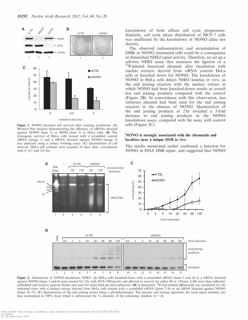
It has been previously shown that miRNA-mediatedknockdown of NONO in HTC 116 cells left cell survivalunaffected but sensitized the latter cells to ionizing irradi-ation (33). Here, we verified the necessity of NONO forcell proliferation by measuring the impact of attenuatedNONO on the long-term survival of HeLa cells with andwithout ionizing irradiation. We used siRNA-mediatedknockdown to attenuate the NONO protein expressionlevel in HeLa cells. Immunoblotting confirmed that theexpression level of NONO was reduced by more than90%, whereas the attenuation of NONO did not affectthe expression level of its partner protein SFPQ andvice versa (Figure 1A). A knockdown of NONO had noeffect on long-term survival (Figure 1B). However,attenuated NONO sensitizes HeLa cells to ionizingirradiation at low (0.5 Gray) and intermediate doses(2.0 Gray), strongly suggesting a defect in DNA DSBrepair (Figure 1C).These results suggest that NONO is crucial for survival
after ionizing radiation. We therefore analysed the abilityof NONO attenuated cells to repair DSBs. Hence, weoptimized an assay to assess the sensitivity of these cellsto the radiomimetic antibiotic NCS as a means to measureDSB repair kinetics in HeLa cells. NCS consists of anenediyne chromophore, which is tightly bound to a 113amino acid single chain protein, the active compound re-sponsible for tandem DNA cleavage and highly potent inthe induction of DNA single and especially DSBs (43,44).Pulse-field gel electrophoresis (PFGE) was accomplishedwith HeLa cells 48 hr following transfection with scrambleor NONO siRNA and treated for 2 hr with 500 ng/mlNCS to introduce DSBs. Cells were then released for 60or 120min and DSB repair kinetics indirectly surveyed byanalysing the accumulation of DSBs. We observed thatNONO protein knockdown by siRNA impairs therecovery from DNA damage as persistent accumulationof DNA DSBs following a 2-hr NCS treatment isdetected by PFGE (Figure 2A). The slower recoverykinetics observed in the context of NONO depletionprovides strong indication for the involvement ofNONO in DSB repair. However, this observation couldalso be explained by an effect on cell cycle checkpointsthat occurred in NONO knockdown cells. To rule outthe possibility that NONO plays in indirect role inrepair by affecting cell cycle progression, we analysedthe cell cycle phase distribution of siCTRL and siNONOHeLa cells (Supplementary Figure S1). Neither theknockdown of NONO, nor SFPQ, nor the combined
Nucleic Acids Research, 2012, Vol. 40, No. 20 10291
Downloaded from https://academic.oup.com/nar/article-abstract/40/20/10287/2414786by gueston 09 February 2018
knockdown of both affects cell cycle progression.Similarly, cell cycle phase distribution of MCF-7 cellswas unaffected by the knockdown of NONO (data notshown).
The observed radiosensitivity and accumulation ofDSBs in NONO attenuated cells could be a consequenceof diminished NHEJ repair activity. Therefore, we set up acell-free NHEJ assay that measures the ligation of a32P-labeled linearized plasmid, after incubation withnuclear extracts derived from siRNA control HeLacells or knocked down for NONO. The knockdown ofNONO in HeLa cells delays NHEJ kinetics in vitro, asthe end joining reaction with the nuclear extract inwhich NONO had been knocked-down results in overallless end joining products compared with the control(Figure 2B). In concordance with this observation, lesssubstrate plasmid had been used for the end joiningreaction in the absence of NONO. Quantitation ofthe end joining products at 2 hr revealed a 5-folddecrease in end joining products in the NONOknockdown assay, compared with the assay with controlcells (Figure 2C).
NONO is strongly associated with the chromatin andlocalizes near a unique DSB in vivo
The results mentioned earlier confirmed a function forNONO in DNA DSB repair, and suggested that NONO
Figure 2. Attenuation of NONO decelerates NHEJ. (A) HeLa cells knocked-down with a scrambled siRNA (lanes 3 and 4) or a siRNA directedagainst NONO (lanes 5 and 6) were treated for 2 hr with NCS (500 ng/ml) and allowed to recover for either 60 or 120min. Cells were then collected,embedded and lysed in agarose blocks and used for pulse-field gel electrophoresis. (B) A linearized, 32P-end-labeled pBluescript was incubated for theindicated times with a nuclear extract derived from HeLa cells treated with a scrambled siRNA (lanes 2–9) or an siRNA directed against NONO(lanes 10–17). (C) Quantitation of the end joining events using a phosphorimager: The percent end joining represents the total signal intensity perlane normalized to 100% from which is substracted the % intensity of the remaining template (n=4).
Figure 1. NONO increases cell survival after ionizing irradiation. (A)Western blot analysis demonstrating the efficiency of siRNAs directedagainst NONO (lane 2) or SFPQ (lane 3) in HeLa cells. (B) Theclonogenic survival of HeLa cells treated with a scrambled controlsiRNA (image 1) and a siRNA directed against NONO (image 2)was analysed using a colony forming assay. (C) Quantitation of cellsurvival. HeLa cell colonies were counted 10 days after g-irradiationwith 0, 0.5 and 2.0 Gy.
10292 Nucleic Acids Research, 2012, Vol. 40, No. 20
Downloaded from https://academic.oup.com/nar/article-abstract/40/20/10287/2414786by gueston 09 February 2018
might play a direct role in DNA repair rather than havingan indirect effect through RNA biogenesis. One predictionof such a direct role would be to observe physical associ-ation of NONO with DNA damage sites. Following thisidea, we used ChIP combined with q-PCR using oligo-nucleotides flanking a unique I-SceI restriction site inMCF-7 cells to monitor the distribution of NONOrelative to a DSB.
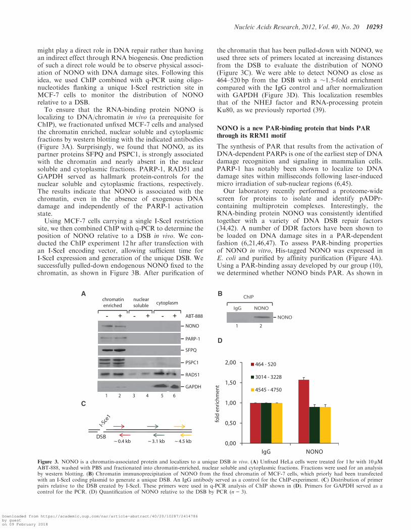
To ensure that the RNA-binding protein NONO islocalizing to DNA/chromatin in vivo (a prerequisite forChIP), we fractionated unfixed MCF-7 cells and analysedthe chromatin enriched, nuclear soluble and cytoplasmicfractions by western blotting with the indicated antibodies(Figure 3A). Surprisingly, we found that NONO, as itspartner proteins SFPQ and PSPC1, is strongly associatedwith the chromatin and nearly absent in the nuclearsoluble and cytoplasmic fractions. PARP-1, RAD51 andGAPDH served as hallmark protein-controls for thenuclear soluble and cytoplasmic fractions, respectively.The results indicate that NONO is associated with thechromatin, even in the absence of exogenous DNAdamage and independently of the PARP-1 activationstate.
Using MCF-7 cells carrying a single I-SceI restrictionsite, we then combined ChIP with q-PCR to determine theposition of NONO relative to a DSB in vivo. We con-ducted the ChIP experiment 12 hr after transfection withan I-SceI encoding vector, allowing sufficient time forI-SceI expression and generation of the unique DSB. Wesuccessfully pulled-down endogenous NONO fixed to thechromatin, as shown in Figure 3B. After purification of
the chromatin that has been pulled-down with NONO, weused three sets of primers located at increasing distancesfrom the DSB to evaluate the distribution of NONO(Figure 3C). We were able to detect NONO as close as464–520 bp from the DSB with a �1.5-fold enrichmentcompared with the IgG control and after normalizationwith GAPDH (Figure 3D). This localization resemblesthat of the NHEJ factor and RNA-processing proteinKu80, as we previously reported (39).
NONO is a new PAR-binding protein that binds PARthrough its RRM1 motif
The synthesis of PAR that results from the activation ofDNA-dependent PARPs is one of the earliest step of DNAdamage recognition and signaling in mammalian cells.PARP-1 has notably been shown to localize to DNAdamage sites within milliseconds following laser-inducedmicro irradiation of sub-nuclear regions (6,45).Our laboratory recently performed a proteome-wide
screen for proteins to isolate and identify pADPr-containing multiprotein complexes. Interestingly, theRNA-binding protein NONO was consistently identifiedtogether with a variety of DNA DSB repair factors(34,42). A number of DDR factors have been shown tobe loaded on DNA damage sites in a PAR-dependentfashion (6,21,46,47). To assess PAR-binding propertiesof NONO in vitro, His-tagged NONO was expressed inE. coli and purified by affinity purification (Figure 4A).Using a PAR-binding assay developed by our group (10),we determined whether NONO binds PAR. As shown in
Figure 3. NONO is a chromatin-associated protein and localizes to a unique DSB in vivo. (A) Unfixed HeLa cells were treated for 1 hr with 10 mMABT-888, washed with PBS and fractionated into chromatin-enriched, nuclear soluble and cytoplasmic fractions. Fractions were used for an analysisby western blotting. (B) Chromatin immunoprecipitation of NONO from the fixed chromatin of MCF-7 cells, which priorly had been transfectedwith an I-SceI coding plasmid to generate a unique DSB. An IgG antibody served as a control for the ChIP-experiment. (C) Distribution of primerpairs relative to the DSB created by I-SceI. These primers were used in q-PCR analysis of ChIP shown in (D). Primers for GAPDH served as acontrol for the PCR. (D) Quantification of NONO relative to the DSB by PCR (n=3).
Nucleic Acids Research, 2012, Vol. 40, No. 20 10293
Downloaded from https://academic.oup.com/nar/article-abstract/40/20/10287/2414786by gueston 09 February 2018
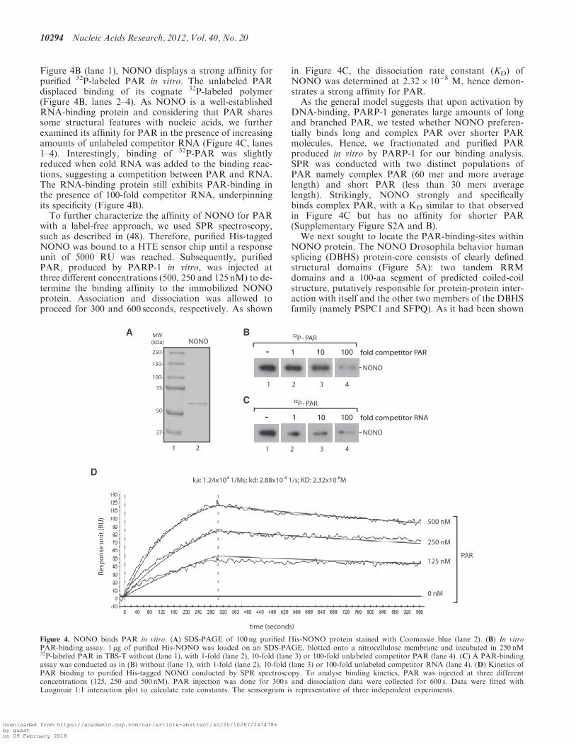
Figure 4B (lane 1), NONO displays a strong affinity forpurified 32P-labeled PAR in vitro. The unlabeled PARdisplaced binding of its cognate 32P-labeled polymer(Figure 4B, lanes 2–4). As NONO is a well-establishedRNA-binding protein and considering that PAR sharessome structural features with nucleic acids, we furtherexamined its affinity for PAR in the presence of increasingamounts of unlabeled competitor RNA (Figure 4C, lanes1–4). Interestingly, binding of 32P-PAR was slightlyreduced when cold RNA was added to the binding reac-tions, suggesting a competition between PAR and RNA.The RNA-binding protein still exhibits PAR-binding inthe presence of 100-fold competitor RNA, underpinningits specificity (Figure 4B).To further characterize the affinity of NONO for PAR
with a label-free approach, we used SPR spectroscopy,such as described in (48). Therefore, purified His-taggedNONO was bound to a HTE sensor chip until a responseunit of 5000 RU was reached. Subsequently, purifiedPAR, produced by PARP-1 in vitro, was injected atthree different concentrations (500, 250 and 125 nM) to de-termine the binding affinity to the immobilized NONOprotein. Association and dissociation was allowed toproceed for 300 and 600 seconds, respectively. As shown
in Figure 4C, the dissociation rate constant (KD) ofNONO was determined at 2.32� 10�8 M, hence demon-strates a strong affinity for PAR.
As the general model suggests that upon activation byDNA-binding, PARP-1 generates large amounts of longand branched PAR, we tested whether NONO preferen-tially binds long and complex PAR over shorter PARmolecules. Hence, we fractionated and purified PARproduced in vitro by PARP-1 for our binding analysis.SPR was conducted with two distinct populations ofPAR namely complex PAR (60 mer and more averagelength) and short PAR (less than 30 mers averagelength). Strikingly, NONO strongly and specificallybinds complex PAR, with a KD similar to that observedin Figure 4C but has no affinity for shorter PAR(Supplementary Figure S2A and B).
We next sought to locate the PAR-binding-sites withinNONO protein. The NONO Drosophila behavior humansplicing (DBHS) protein-core consists of clearly definedstructural domains (Figure 5A): two tandem RRMdomains and a 100-aa segment of predicted coiled-coilstructure, putatively responsible for protein-protein inter-action with itself and the other two members of the DBHSfamily (namely PSPC1 and SFPQ). As it had been shown
Figure 4. NONO binds PAR in vitro. (A) SDS-PAGE of 100 ng purified His-NONO protein stained with Coomassie blue (lane 2). (B) In vitroPAR-binding assay. 1 mg of purified His-NONO was loaded on an SDS-PAGE, blotted onto a nitrocellulose membrane and incubated in 250 nM32P-labeled PAR in TBS-T without (lane 1), with 1-fold (lane 2), 10-fold (lane 3) or 100-fold unlabeled competitor PAR (lane 4). (C) A PAR-bindingassay was conducted as in (B) without (lane 1), with 1-fold (lane 2), 10-fold (lane 3) or 100-fold unlabeled competitor RNA (lane 4). (D) Kinetics ofPAR binding to purified His-tagged NONO conducted by SPR spectroscopy. To analyse binding kinetics, PAR was injected at three differentconcentrations (125, 250 and 500 nM). PAR injection was done for 300 s and dissociation data were collected for 600 s. Data were fitted withLangmuir 1:1 interaction plot to calculate rate constants. The sensorgram is representative of three independent experiments.
10294 Nucleic Acids Research, 2012, Vol. 40, No. 20
Downloaded from https://academic.oup.com/nar/article-abstract/40/20/10287/2414786by gueston 09 February 2018
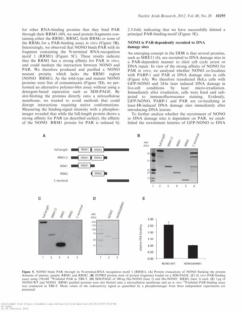
for other RNA-binding proteins that they bind PARthrough their RRM1 (49), we used protein fragments con-taining either the RRM1, RRM2, both RRMs or none ofthe RRMs for a PAR-binding assay in vitro (Figure 5B).Interestingly, we observed that NONO binds PAR with itsfragment containing the N-terminal RNA-recognitionmotif 1 (RRM1) (Figure 5C). These results indicatethat the RRM1 has a strong affinity for PAR in vitro,and could mediate the interaction between NONO andPAR. We therefore produced and purified a NONOmutant protein, which lacks the RRM1 region(NONO�RRM1). As the wild-type and mutant NONOproteins were free of contaminants (Figure 5D), we per-formed an alternative polymer-blot assay without using adetergent-based separation such as SDS-PAGE. Byslot-blotting the proteins directly onto a nitrocellulosemembrane, we wanted to avoid methods that coulddisrupt interactions requiring native conformations.Measuring the binding-signal intensity with a phosphor-imager revealed that while the full-length protein shows astrong affinity for PAR (as described earlier), the affinityof the NONO�RRM1 protein for PAR is reduced by
2.5-fold, indicating that we have successfully deleted aprincipal PAR-binding-motif (Figure 5E).
NONO is PAR-dependently recruited to DNAdamage sites
An emerging concept in the DDR is that several proteins,such as MRE11 (6), are recruited to DNA damage sites ina PAR-dependent manner to elicit cell cycle arrest orDNA repair. In view of the strong affinity of NONO forPAR in vitro, we analysed whether NONO co-localizeswith PARP-1 and PAR at DNA damage sites in cells(Figure 6A). We therefore transfected HeLa cells withGFP-NONO and 24 hr later induced DNA damage inlive-cell conditions by laser micro-irradiation.Immediately after irradiation, cells were fixed and sub-jected to immunofluorescence staining. Evidently,GFP-NONO, PARP-1 and PAR are co-localizing atlaser-IR-induced DNA damage sites immediately afterintroducing DNA lesions.To further analyse whether the recruitment of NONO
to DNA damage sites is dependent on PAR, we estab-lished the recruitment kinetics of GFP-NONO to DNA
Figure 5. NONO binds PAR through its N-terminal/RNA recognition motif 1 (RRM1). (A) Protein truncations of NONO flanking the proteindomains of interest, namely RRM1 and RRM2. (B) SYPRO protein stain of protein fragments loaded on a SDS-PAGE. (C) In vitro PAR-bindingassay using 250 nM 32P-labeled PAR in TBS-T. (D) SDS-PAGE of 500 ng His-NONO (lane 2) and His-NONO�RRM1 (lane 3) each. (E) 1 mg ofNONO-WT and NONO�RRM1 purified proteins were slot blotted onto a nitrocellulose membrane and an in vitro 32P-labeled PAR-binding assaywas conducted in TBS-T. Mean values of the radioactivity signal as quantified by a phosphorimager from three independent experiments arepresented.
Nucleic Acids Research, 2012, Vol. 40, No. 20 10295
Downloaded from https://academic.oup.com/nar/article-abstract/40/20/10287/2414786by gueston 09 February 2018
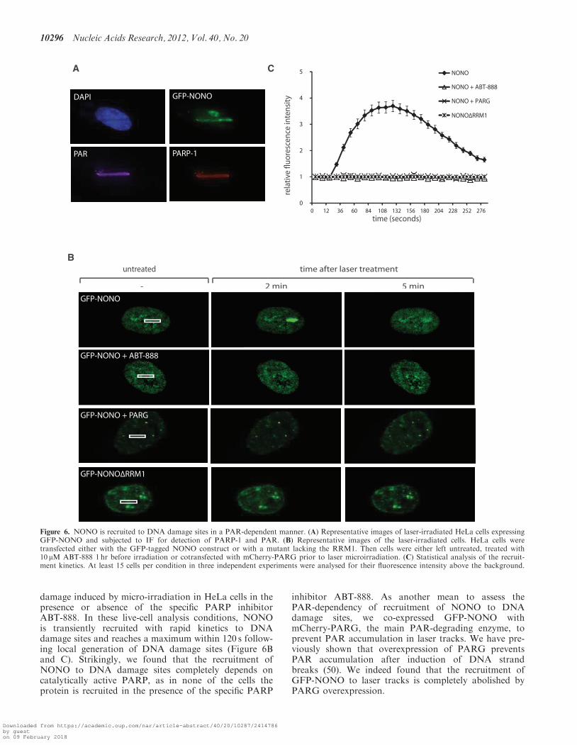
damage induced by micro-irradiation in HeLa cells in thepresence or absence of the specific PARP inhibitorABT-888. In these live-cell analysis conditions, NONOis transiently recruited with rapid kinetics to DNAdamage sites and reaches a maximum within 120 s follow-ing local generation of DNA damage sites (Figure 6Band C). Strikingly, we found that the recruitment ofNONO to DNA damage sites completely depends oncatalytically active PARP, as in none of the cells theprotein is recruited in the presence of the specific PARP
inhibitor ABT-888. As another mean to assess thePAR-dependency of recruitment of NONO to DNAdamage sites, we co-expressed GFP-NONO withmCherry-PARG, the main PAR-degrading enzyme, toprevent PAR accumulation in laser tracks. We have pre-viously shown that overexpression of PARG preventsPAR accumulation after induction of DNA strandbreaks (50). We indeed found that the recruitment ofGFP-NONO to laser tracks is completely abolished byPARG overexpression.
2 min 5 min
time after laser treatment
-GFP-NONO
GFP-NONO + ABT-888
GFP-NONO + PARG
GFP-NONOΔRRM1
untreated
A
time (seconds)
0
1
2
3
4
5
0 12 36 60 84 108 132 156 180 204 228 252 276
NONO
NONO + ABT-888
NONO + PARG
NONOΔRRM1
B
C
DAPI GFP-NONO
PAR PARP-1
Figure 6. NONO is recruited to DNA damage sites in a PAR-dependent manner. (A) Representative images of laser-irradiated HeLa cells expressingGFP-NONO and subjected to IF for detection of PARP-1 and PAR. (B) Representative images of the laser-irradiated cells. HeLa cells weretransfected either with the GFP-tagged NONO construct or with a mutant lacking the RRM1. Then cells were either left untreated, treated with10 mM ABT-888 1 hr before irradiation or cotransfected with mCherry-PARG prior to laser microirradiation. (C) Statistical analysis of the recruit-ment kinetics. At least 15 cells per condition in three independent experiments were analysed for their fluorescence intensity above the background.
10296 Nucleic Acids Research, 2012, Vol. 40, No. 20
Downloaded from https://academic.oup.com/nar/article-abstract/40/20/10287/2414786by gueston 09 February 2018
This observation is consistent with the finding thatPARP inhibition abrogates the recruitment of GFP-NONO and confirms a strict requirement for PAR-binding for its relocation to DNA damage sites. We thensought to define the domain mediating NONO interactionwith PAR. Hence, we tested if interaction with PAR occursthrough interaction with the RRM1 domain of NONO. Asshown in Figure 6A and B, a deletion mutant lacking theRRM1 domain (GFP-NONO�RRM1) is not recruited toDNA damage sites. This result strongly implicates theRRM1 domain in regulating the interaction with PAR.
Although our results underscore the importance ofPAR for NONO dynamics in the DDR, they leave openthe question which PARP family member generates thePAR that mediates the recruitment of NONO to DNAdamage sites. It is well accepted, that PARP-1 is respon-sible for �95% of all PARylation events after DNAdamage, whereas PARP-2 carries out almost all of the re-maining 5%. Therefore, we overexpressed GFP-NONO inwild-type and PARP-1�/� MEFs. Recruitment of GFP-NONO was detected in the PARP-1-proficient MEFswith similar kinetics to those in HeLa cells, whereasGFP-NONO was not recruited to the laser track inPARP-1�/� cells, highlighting the necessity of PARP-1to generate PAR at the DNA damage sites (Figure 7).The specificity for PARP-1 is further highlighted by theobservation that GFP-NONO is recruited with fast andtransient kinetics in PARP-2�/� MEFs similar to that inthe WT-MEFs and HeLa cells (Figure 7). Hence, PARP-1is required to recruit NONO to DNA damage sites,whereas PARP-2 is rather dispensable. Collectively,these results show that the recruitment of NONO isPARP-1 and PAR-dependent, and mediated by theRRM1 region of NONO.
NONO promotes NHEJ and represses HR in vivo in thesame pathway as PARP-1
As a consequence of the results described above, we hy-pothesize that NONO plays important regulatory role inthe DDR by stimulating DSB repair. Indeed, we showedthat NONO promotes cell survival and DSB repairthrough NHEJ, localizes near a unique DSB site and ac-cumulates to sites of DNA damage in a pADPr-dependentfashion. However, a direct implication of NONO inNHEJ has not been shown in vivo and the question aswhether NONO also influences the other DSB repairpathway, namely HR, has not been answered yet.
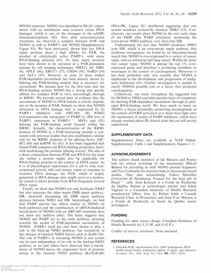
To address these two key questions, we generated twostable reporter cell lines enabling us to monitor both,NHEJ and HR repair (Figure 8A and B). Each of thesecells lines has an integrated cassette comprising an I-SceIcleavage site that, upon repair by either NHEJ or HR,restores GFP expression, as previously described (35,36).Cells with normal or knocked down expression of NONOwere assessed for each repair mechanism as indicated bythe percentage of cells that express GFP. In the NHEJreporter system assay, we found that the knockdown ofNONO decreases NHEJ by more than 50% (Figure 8C).In this same assay, PARP inhibition, with the potent andspecific PARP inhibitor ABT-888 also significantly
reduced NHEJ repair. Knowing that NONO is PAR-dependently recruited to DNA DSBs, we combined thesiRNA directed against NONO with PARP inhibitor toconfirm our findings above. As expected, the siRNA-mediated knockdown of NONO combined with theinhibition of PARP does not have an additive effect ininhibiting NHEJ, indicating that PARP and NONOfunction in the same pathway and hence supporting theidea of PAR-dependent recruitment. Interestingly, an at-tenuation of NONO does not only decrease NHEJ butalso facilitates repair by HR �40% (Figure 8D). Againhere, when combining siRNA directed against NONOwith the PARP-1 inhibitor ABT-888, no additive effectwas observed, supporting the same conclusion regardingPAR-dependent recruitment.
DISCUSSION
Although the RNA binding properties of NONO relatedto RNA biogenesis and the architecture of paraspeckleshave been subject of an abundant literature, [reviewed in(51)], little is known on the functions of NONO in thecontext of DNA DSB repair. We have conducted adetailed molecular and cellular analysis of NONO in thecontext of the DDR and our data establish NONO as aPARP-1-dependent regulator of DSB repair by facilitatingNHEJ and promoting cell survival after irradiation.In the past few years, the list of proteins that possess
dual roles in gene regulation and genomic stabilitythrough RNA biology and DNA repair, respectively,has largely expanded. Examples include the catalyticsubunit of DNA-PK, a core complex of NHEJ that isnecessary to arrest RNA-polymerase II transcriptionafter the induction of DSBs (52) and the Ku proteinthat has dual roles in transcriptional reinitiation andNHEJ (19). In addition, the heterogeneous nuclearribonucleoprotein (hnRNP) RBMX acts in alternativesplicing and accumulates at DNA damage sites in aPARP-dependent manner (21). Also, the heterogeneousnuclear ribonucleoprotein hnRNPU influences end resec-tion (53). Another study highlights the role of thesplicing-associated protein THRAP3 in the DNAdamage signaling network (22). Even PARP-1 itself func-tions in promoter/enhancer regulation (54), single-strandbreak repair and the alternative NHEJ pathway (55,56).Because of its possible role in RNA biogenesis, it came
as a surprise to find that NONO is mostly associated tothe chromatin. Moreover, we show here for the first timethat NONO is localized with close proximity to a uniqueDSB in vivo. In an earlier study (39), we have detected theNHEJ-related protein Ku80 within the same distance tothe break-site as NONO (� 400 bps), suggesting a directimplication for NONO in DNA DSB repair. In line withthese findings, the Shiloh group has detected NONO in aprotein complex composed of Ku70, Ku80 and Ligase IV(31). Here, we are giving further evidence for a direct im-plication of NONO in DSB repair by showing thatdown-regulation of NONO protein expression by siRNAsensitizes HeLa cells to ionizing irradiation and decreasesNHEJ in vitro and in vivo. Hence, the data presented
Nucleic Acids Research, 2012, Vol. 40, No. 20 10297
Downloaded from https://academic.oup.com/nar/article-abstract/40/20/10287/2414786by gueston 09 February 2018
complement the recent findings that attenuation ofNONO delays the resolution of g-H2AX foci and resultsin an increase of chromosomal aberrations following ra-diation exposure (33). The fact that cells with attenuatedNONO are still viable and capable of NHEJ might beexplained by a possible backup through its homologousprotein partner PSPC1. The expression level of PSPC1 inthe presence of NONO in HeLa cells is very low and in-creases upon siRNA-mediated knock-down of NONO(data not shown).
PARP-1 is an abundant nuclear chromatin-associatedprotein, well characterized for its high DNA damagesensing ability. Once encountering free DNA ends,PARP-1 is catalytically activated and generates largeamounts of PAR serving as a scaffold for the recruitmentof a variety of DNA repair proteins. We performed a largescale analysis of proteins bound to PAR following
Figure 8. Attenuation of NONO decreases NHEJ and increases HR. (A) Schematic representation of the I-SceI-based NHEJ in vivo reporter system.(B) Schematic representation of the I-SceI-based HR in vivo reporter system. (C) NHEJ repair rates in percent with siCTRL or siNONO and with orwithout 10 mM of the PARP-inhibitor ABT-888. The siCTRL condition was normalized to 100% (n=3). (D) Diagram of the HR repair rates aftertreatment with siCTRL or siNONO and with or without 10 mM ABT-888. The siCTRL condition was normalized to 100% (n=3).
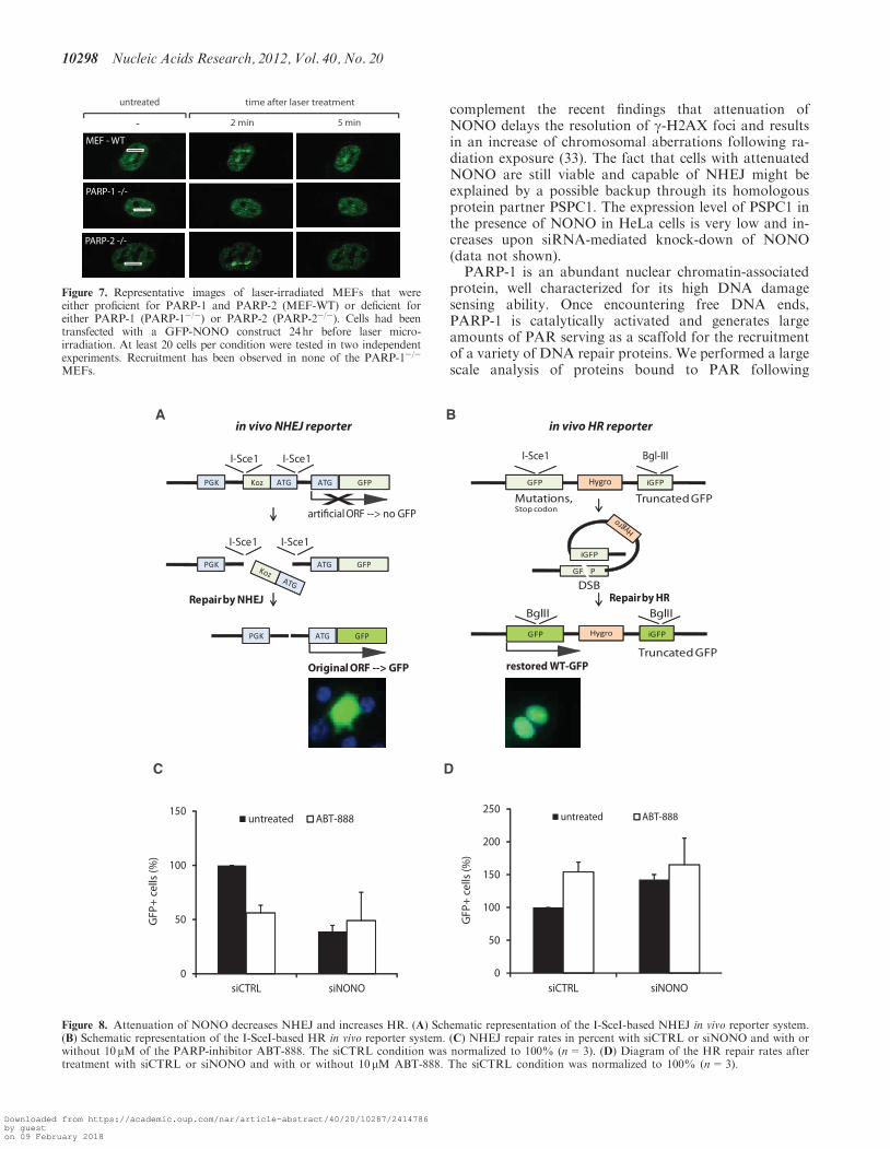
Figure 7. Representative images of laser-irradiated MEFs that wereeither proficient for PARP-1 and PARP-2 (MEF-WT) or deficient foreither PARP-1 (PARP-1�/�) or PARP-2 (PARP-2�/�). Cells had beentransfected with a GFP-NONO construct 24hr before laser micro-irradiation. At least 20 cells per condition were tested in two independentexperiments. Recruitment has been observed in none of the PARP-1�/�
MEFs.
10298 Nucleic Acids Research, 2012, Vol. 40, No. 20
Downloaded from https://academic.oup.com/nar/article-abstract/40/20/10287/2414786by gueston 09 February 2018
MNNG exposure. NONO was identified in SILAC experi-ments with an enrichment ratio (control versus DNAdamage), which is one of the strongest in the pADPrimmunoprecipitates (42). Also after neocarzinostatintreatment, we observed a complex between PAR andNONO as well as PARP-1 and NONO (SupplementaryFigure S3). We have previously shown that key DNArepair proteins share a high affinity for PAR, theproduct of catalytically active PARP-1, with manyRNA-binding proteins (41). To date, many proteinshave been shown to be recruited in a PAR-dependentmanner by cell imaging techniques: MRE11 (6), NBS1(6), APLF (46), XRCC1 (57), CHD4 (58), NuRD (59)and ALC1 (47). However, in none of these studiesPAR-dependent recruitment has been directly shown bydeleting the PAR-binding module that is necessary forrecruitment. We present here for the first time that theRNA-binding protein NONO has a strong and specificaffinity for complex PAR in vitro, interestingly throughits RRM1. We provide several lines of evidence that therecruitment of NONO to DNA lesions is strictly depend-ent on the presence of PAR. Indeed, we show that NONOrelocation to DNA damage sites is suppressed by (I)PARP-1 inhibition with ABT-888; (II) PARGoverexpression (the antagonist of PARP-1); (III) loss ofPARP-1 expression in PARP-1�/� MEFs and (IV)deleting the PAR-binding motif located within theRRM1. Actually, the characterization of the RRM1domain of NONO as a PAR-interacting module is con-sistent with previous studies that also established a similarrole for the RRM1 domains of the splicing factor ASF/SF2 (49) and hnRNP A1 (41). It has been suggested thatbound PAR competes out RNA-binding properties, there-with modulating the proteins splicing activity. The idea ofa direct competition between PAR and RNA for the samesite within a protein might also be applicable forRNA-binding proteins in the context of DNA repair. Asit is of physiological importance for a cell to stop tran-scriptional activity (60) and initiate repair in response toexcessive DNA damage, the PAR, which is largelygenerated at DNA damage sites, might serve as a molecu-lar switch to direct proteins from RNA biogenesis towardDNA repair.
Finally, we show that NONO not only facilitates NHEJbut also represses the other major DSB repair pathway,HR, therewith channeling the DSB repair pathwaydecision between NHEJ and HR. Interestingly, we findthat PARP activity has effects similar to NONO onboth pathways and the combination of siRNA-mediatedknock-down of NONO with an ABT-888 treatment doesnot show any additive effect. The latter suggests thatNONO and PARP act in the same pathway, pointingtowards the model of PAR-dependent recruitment ofNONO. PARP-1 itself has also been shown to play arole in the back-up NHEJ pathway, but exclusively inthe absence of classical NHEJ factors such as Ku80 (56).The role of PARP-1 in recruiting NONO in our systemcan be seen independent of its role in the backup-NHEJpathway as we and others have observed that a knock-down of NONO leaves the expression level of proteinsacting in the classical NHEJ pathway (Ku70/Ku80,
DNA-PK, Ligase IV) unaffected suggesting that oursystem monitors exclusively classical NHEJ (33). Con-clusively, our results place NONO in the very early stepsof the DDR after PARP activation, promoting theerror-prone NHEJ pathway over error-free HR.Underpinning the fact that NONO promotes NHEJ
over HR, which is an error-prone repair pathway thatfacilitates mutagenesis, we found by an Oncomine-basedsearch that NONO is over-expressed in a variety of cancertypes, such as colorectal and lung cancer. Within the lattertwo cancer types, NONO is among the top 1% over-expressed genes and therefore a promising candidate toinvestigate in the context of carcinogenesis. Moreover ithas been published only very recently that NONO isimplicated in the development and progression of malig-nant melanoma (61). Further investigation is needed toclarify NONOs possible role as a factor that promotescarcinogenesis.Collectively, our study strengthens the suggested role
for NONO in NHEJ and adds another layer of complexityby showing PAR-dependent recruitment through its prin-cipal RNA-binding motif. We have much to learn onNONO, a factor potentially promoting carcinogenesis inthe context of PARP-activation as it sheds more light ontothe mechanism of action of PARP inhibitors, which havealready reached phase III clinical trials but are still poorlyunderstood.
SUPPLEMENTARY DATA
Supplementary Data are available at NAR Online:Supplementary Table 1 and Supplementary Figures 1–3.
ACKNOWLEDGEMENTS
The authors thank members of the Masson and Poirierlabs for critical reviewing of the manuscript, MikaelBedard for providing us with NONO protein fragmentsand Yan Coulombe for precious help in microscopy-basedstudies. They also acknowledge Valerie Schreiber(Universite de Strasbourg, France) for the kind gift ofParp2�/� cells. Jana Krietsch is a Fonds de Recherchedu Quebec Nature et technologies scholar and JulienVignard is a Canadian Institutes of Health Researchpostdoctoral fellow. Guy G. Poirier holds a CanadaResearch Chair in Proteomics and Jean-Yves Masson isa Fonds de Recherche en Sante du Quebec seniorinvestigator.
FUNDING
Funding for open access charge: Canadian Institutes ofHealth Research (to J.-Y.M. and G.G.P.).
Conflict of interest statement. None declared.
REFERENCES
1. Vilenchik,M.M. and Knudson,A.G. (2003) Endogenous DNAdouble-strand breaks: production, fidelity of repair, and inductionof cancer. Proc. Natl. Acad. Sci. USA, 100, 12871–12876.
Nucleic Acids Research, 2012, Vol. 40, No. 20 10299
Downloaded from https://academic.oup.com/nar/article-abstract/40/20/10287/2414786by gueston 09 February 2018
2. Ciccia,A. and Elledge,S.J. (2010) The DNA damage response:making it safe to play with knives. Mol. Cell, 40, 179–204.
3. Wacker,D.A., Frizzell,K.M., Zhang,T. and Kraus,W.L. (2007)Regulation of chromatin structure and chromatin-dependenttranscription by poly(ADP-ribose) polymerase-1: possible targetsfor drug-based therapies. Subcell Biochem., 41, 45–69.
4. Bouchard,V.J., Rouleau,M. and Poirier,G.G. (2003) PARP-1, adeterminant of cell survival in response to DNA damage. Exp.Hematol., 31, 446–454.
5. Krishnakumar,R. and Kraus,W.L. (2010) The PARP side of thenucleus: molecular actions, physiological outcomes, and clinicaltargets. Mol. Cell., 39, 8–24.
6. Haince,J.F., McDonald,D., Rodrigue,A., Dery,U., Masson,J.Y.,Hendzel,M.J. and Poirier,G.G. (2008) PARP1-dependent kineticsof recruitment of MRE11 and NBS1 proteins to multiple DNAdamage sites. J. Biol. Chem., 283, 1197–1208.
7. Slade,D., Dunstan,M.S., Barkauskaite,E., Weston,R., Lafite,P.,Dixon,N., Ahel,M., Leys,D. and Ahel,I. (2011) The structure andcatalytic mechanism of a poly(ADP-ribose) glycohydrolase.Nature, 477, 616–620.
8. Takata,M., Sasaki,M.S., Sonoda,E., Morrison,C., Hashimoto,M.,Utsumi,H., Yamaguchi-Iwai,Y., Shinohara,A. and Takeda,S.(1998) Homologous recombination and non-homologousend-joining pathways of DNA double-strand break repair haveoverlapping roles in the maintenance of chromosomal integrity invertebrate cells. EMBO J., 17, 5497–5508.
9. Lieber,M.R. (2010) The mechanism of double-strand DNA breakrepair by the nonhomologous DNA end-joining pathway. AnnuRev. Biochem., 79, 181–211.
10. Gagne,J.P., Isabelle,M., Lo,K.S., Bourassa,S., Hendzel,M.J.,Dawson,V.L., Dawson,T.M. and Poirier,G.G. (2008)Proteome-wide identification of poly(ADP-ribose) binding proteinsand poly(ADP-ribose)-associated protein complexes. Nucleic AcidsRes., 36, 6959–6976.
11. Li,B., Navarro,S., Kasahara,N. and Comai,L. (2004)Identification and biochemical characterization of a Werner’ssyndrome protein complex with Ku70/80 and poly(ADP-ribose)polymerase-1. J. Biol. Chem., 279, 13659–13667.
12. Meek,K., Dang,V. and Lees-Miller,S.P. (2008) DNA-PK: themeans to justify the ends? Adv. Immunol., 99, 33–58.
13. Wang,J., Pluth,J.M., Cooper,P.K., Cowan,M.J., Chen,D.J. andYannone,S.M. (2005) Artemis deficiency confers a DNAdouble-strand break repair defect and Artemis phosphorylationstatus is altered by DNA damage and cell cycle progression.DNA Repair (Amst), 4, 556–570.
14. Chappell,C., Hanakahi,L.A., Karimi-Busheri,F., Weinfeld,M. andWest,S.C. (2002) Involvement of human polynucleotide kinase indouble-strand break repair by non-homologous end joining.EMBO J., 21, 2827–2832.
15. Capp,J.P., Boudsocq,F., Bertrand,P., Laroche-Clary,A.,Pourquier,P., Lopez,B.S., Cazaux,C., Hoffmann,J.S. andCanitrot,Y. (2006) The DNA polymerase lambda is required forthe repair of non-compatible DNA double strand breaks byNHEJ in mammalian cells. Nucleic Acids Res., 34, 2998–3007.
16. Capp,J.P., Boudsocq,F., Besnard,A.G., Lopez,B.S., Cazaux,C.,Hoffmann,J.S. and Canitrot,Y. (2007) Involvement of DNApolymerase mu in the repair of a specific subset of DNAdouble-strand breaks in mammalian cells. Nucleic Acids Res., 35,3551–3560.
17. Ahnesorg,P., Smith,P. and Jackson,S.P. (2006) XLF interacts withthe XRCC4-DNA ligase IV complex to promote DNAnonhomologous end-joining. Cell, 124, 301–313.
18. Giffin,W., Torrance,H., Rodda,D.J., Prefontaine,G.G., Pope,L.and Hache,R.J. (1996) Sequence-specific DNA binding by Kuautoantigen and its effects on transcription. Nature, 380, 265–268.
19. Woodard,R.L., Lee,K.J., Huang,J. and Dynan,W.S. (2001)Distinct roles for Ku protein in transcriptional reinitiation andDNA repair. J. Biol. Chem., 276, 15423–15433.
20. Beck,B.D., Hah,D.S. and Lee,S.H. (2008) XPB and XPD betweentranscription and DNA repair. Adv. Exp. Med. Biol., 637, 39–46.
21. Adamson,B., Smogorzewska,A., Sigoillot,F.D., King,R.W. andElledge,S.J. (2012) A genome-wide homologous recombinationscreen identifies the RNA-binding protein RBMX as a componentof the DNA-damage response. Nat. Cell Biol., 14, 318–328.
22. Beli,P., Lukashchuk,N., Wagner,S.A., Weinert,B.T., Olsen,J.V.,Baskcomb,L., Mann,M., Jackson,S.P. and Choudhary,C. (2012)Proteomic investigations reveal a role for RNA processing factorTHRAP3 in the DNA damage response. Mol. Cell, 46, 212–225.
23. Paulsen,R.D., Soni,D.V., Wollman,R., Hahn,A.T., Yee,M.C.,Guan,A., Hesley,J.A., Miller,S.C., Cromwell,E.F., Solow-Cordero,D.E. et al. (2009) A genome-wide siRNA screen revealsdiverse cellular processes and pathways that mediate genomestability. Mol. Cell, 35, 228–239.
24. Peng,R., Dye,B.T., Perez,I., Barnard,D.C., Thompson,A.B. andPatton,J.G. (2002) PSF and p54nrb bind a conserved stem in U5snRNA. RNA, 8, 1334–1347.
25. Zhang,Z. and Carmichael,G.G. (2001) The fate of dsRNA in thenucleus: a p54(nrb)-containing complex mediates the nuclearretention of promiscuously A-to-I edited RNAs. Cell, 106,465–475.
26. Kaneko,S., Rozenblatt-Rosen,O., Meyerson,M. and Manley,J.L.(2007) The multifunctional protein p54nrb/PSF recruits theexonuclease XRN2 to facilitate pre-mRNA 30 processing andtranscription termination. Genes Dev., 21, 1779–1789.
27. Amelio,A.L., Miraglia,L.J., Conkright,J.J., Mercer,B.A.,Batalov,S., Cavett,V., Orth,A.P., Busby,J., Hogenesch,J.B. andConkright,M.D. (2007) A coactivator trap identifies NONO(p54nrb) as a component of the cAMP-signaling pathway. Proc.Natl. Acad. Sci. USA, 104, 20314–20319.
28. Mathur,M., Tucker,P.W. and Samuels,H.H. (2001) PSF is a novelcorepressor that mediates its effect through Sin3A and the DNAbinding domain of nuclear hormone receptors. Mol. Cell Biol.,21, 2298–2311.
29. Dong,X., Sweet,J., Challis,J.R., Brown,T. and Lye,S.J. (2007)Transcriptional activity of androgen receptor is modulated by twoRNA splicing factors, PSF and p54nrb. Mol. Cell Biol., 27,4863–4875.
30. Ishitani,K., Yoshida,T., Kitagawa,H., Ohta,H., Nozawa,S. andKato,S. (2003) p54nrb acts as a transcriptional coactivator foractivation function 1 of the human androgen receptor. Biochem.Biophys. Res. Commun., 306, 660–665.
31. Salton,M., Lerenthal,Y., Wang,S.Y., Chen,D.J. and Shiloh,Y.(2010) Involvement of Matrin 3 and SFPQ/NONO in the DNAdamage response. Cell Cycle, 9, 1568–1576.
32. Bladen,C.L., Udayakumar,D., Takeda,Y. and Dynan,W.S. (2005)Identification of the polypyrimidine tract bindingprotein-associated splicing factor.p54(nrb) complex as a candidateDNA double-strand break rejoining factor. J. Biol. Chem., 280,5205–5210.
33. Li,S., Kuhne,W.W., Kulharya,A., Hudson,F.Z., Ha,K., Cao,Z.and Dynan,W.S. (2009) Involvement of p54(nrb), a PSF partnerprotein, in DNA double-strand break repair and radioresistance.Nucleic Acids Res., 37, 6746–6753.
34. Gagne,J.P., Haince,J.F., Pic,E. and Poirier,G.G. (2011)Affinity-based assays for the identification and quantitativeevaluation of noncovalent poly(ADP-ribose)-binding proteins.Methods Mol. Biol., 780, 93–115.
35. Xie,A., Kwok,A. and Scully,R. (2009) Role of mammalian Mre11in classical and alternative nonhomologous end joining. Nat.Struct. Mol. Biol., 16, 814–818.
36. Pierce,A.J., Johnson,R.D., Thompson,L.H. and Jasin,M. (1999)XRCC3 promotes homology-directed repair of DNA damage inmammalian cells. Genes Dev., 13, 2633–2638.
37. Duriez,P.J., Desnoyers,S., Hoflack,J.C., Shah,G.M., Morelle,B.,Bourassa,S., Poirier,G.G. and Talbot,B. (1997) Characterizationof anti-peptide antibodies directed towards the automodificationdomain and apoptotic fragment of poly (ADP-ribose) polymerase.Biochim. Biophys. Acta, 1334, 65–72.
38. Zou,L., Cortez,D. and Elledge,S.J. (2002) Regulation of ATRsubstrate selection by Rad17-dependent loading of Rad9complexes onto chromatin. Genes Dev., 16, 198–208.
39. Rodrigue,A., Lafrance,M., Gauthier,M.C., McDonald,D.,Hendzel,M., West,S.C., Jasin,M. and Masson,J.Y. (2006)Interplay between human DNA repair proteins at a uniquedouble-strand break in vivo. EMBO J., 25, 222–231.
40. Ismail,I.H., Gagne,J.P., Caron,M.C., McDonald,D., Xu,Z.,Masson,J.Y., Poirier,G.G. and Hendzel,M.J. (2012)
10300 Nucleic Acids Research, 2012, Vol. 40, No. 20
Downloaded from https://academic.oup.com/nar/article-abstract/40/20/10287/2414786by gueston 09 February 2018
CBX4-mediated SUMO modification regulates BMI1 recruitmentat sites of DNA damage. Nucleic Acids Res., 40, 5497–5510.
41. Gagne,J.P., Hunter,J.M., Labrecque,B., Chabot,B. andPoirier,G.G. (2003) A proteomic approach to the identification ofheterogeneous nuclear ribonucleoproteins as a new family ofpoly(ADP-ribose)-binding proteins. Biochem. J., 371, 331–340.
42. Gagne,J.P., Pic,E., Isabelle,M., Krietsch,J., Ethier,C., Paquet,E.,Kelly,I., Boutin,M., Moon,K.M., Foster,L.J. et al. (2012)Quantitative proteomics profiling of the poly(ADP-ribose)-relatedresponse to genotoxic stress. Nucleic Acids Res, 40, 7788–7805.
43. Smith,B.L., Bauer,G.B. and Povirk,L.F. (1994) DNA damageinduced by bleomycin, neocarzinostatin, and melphalan in aprecisely positioned nucleosome. Asymmetry in protection at theperiphery of nucleosome-bound DNA. J. Biol. Chem., 269,30587–30594.
44. Povirk,L.F. (1996) DNA damage and mutagenesis byradiomimetic DNA-cleaving agents: bleomycin, neocarzinostatinand other enediynes. Mutat. Res., 355, 71–89.
45. Tartier,L., Spenlehauer,C., Newman,H.C., Folkard,M.,Prise,K.M., Michael,B.D., Menissier-de Murcia,J. and deMurcia,G. (2003) Local DNA damage by proton microbeamirradiation induces poly(ADP-ribose) synthesis in mammaliancells. Mutagenesis, 18, 411–416.
46. Rulten,S.L., Cortes-Ledesma,F., Guo,L., Iles,N.J. andCaldecott,K.W. (2008 Jul) APLF (C2orf13) is a novel componentof poly(ADP-ribose) signaling in mammalian cells. Mol Cell Biol.,28, 4620–4628.
47. Ahel,D., Horejsi,Z., Wiechens,N., Polo,S.E., Garcia-Wilson,E.,Ahel,I., Flynn,H., Skehel,M., West,S.C., Jackson,S.P. et al. (2009)Poly(ADP-ribose)-dependent regulation of DNA repair by thechromatin remodeling enzyme ALC1. Science, 325, 1240–1243.
48. Fahrer,J., Kranaster,R., Altmeyer,M., Marx,A. and Burkle,A.(2007) Quantitative analysis of the binding affinity ofpoly(ADP-ribose) to specific binding proteins as a function ofchain length. Nucleic Acids Res., 35, e143.
49. Malanga,M., Czubaty,A., Girstun,A., Staron,K. and Althaus,F.R.(2008) Poly(ADP-ribose) binds to the splicing factor ASF/SF2and regulates its phosphorylation by DNA topoisomerase I.J. Biol. Chem., 283, 19991–19998.
50. Haince,J.F., Ouellet,M.E., McDonald,D., Hendzel,M.J. andPoirier,G.G. (2006) Dynamic relocation of poly(ADP-ribose)glycohydrolase isoforms during radiation-induced DNA damage.Biochim. Biophys. Acta, 1763, 226–237.
51. Shav-Tal,Y. and Zipori,D. (2002) PSF and p54(nrb)/NonO—multi-functional nuclear proteins. FEBS Lett., 531,109–114.
52. Pankotai,T., Bonhomme,C., Chen,D. and Soutoglou,E. (2012)DNAPKcs-dependent arrest of RNA polymerase II transcriptionin the presence of DNA breaks. Nat. Struct. Mol. Biol., 19,276–282.
53. Polo,S.E., Blackford,A.N., Chapman,J.R., Baskcomb,L.,Gravel,S., Rusch,A., Thomas,A., Blundred,R., Smith,P.,Kzhyshkowska,J. et al. (2012) Regulation of DNA-end resectionby hnRNPU-like proteins promotes DNA double-strand breaksignaling and repair. Mol. Cell, 45, 505–516.
54. Kraus,W.L. (2008) Transcriptional control by PARP-1: chromatinmodulation, enhancer-binding, coregulation, and insulation. Curr.Opin. Cell. Biol., 20, 294–302.
55. Wang,M., Wu,W., Rosidi,B., Zhang,L., Wang,H. and Iliakis,G.(2006) PARP-1 and Ku compete for repair of DNA doublestrand breaks by distinct NHEJ pathways. Nucleic Acids Res., 34,6170–6182.
56. Mansour,W.Y., Rhein,T. and Dahm-Daphi,J. (2010) Thealternative end-joining pathway for repair of DNA double-strandbreaks requires PARP1 but is not dependent uponmicrohomologies. Nucleic Acids Res., 38, 6065–6077.
57. El-Khamisy,S.F., Masutani,M., Suzuki,H. and Caldecott,K.W.(2003) A requirement for PARP-1 for the assembly or stability ofXRCC1 nuclear foci at sites of oxidative DNA damage. NucleicAcids Res., 31, 5526–5533.
58. Polo,S.E., Kaidi,A., Baskcomb,L., Galanty,Y. and Jackson,S.P.(2010) Regulation of DNA-damage responses and cell-cycleprogression by the chromatin remodelling factor CHD4. EMBOJ., 29, 3130–3139.
59. Chou,D.M., Adamson,B., Dephoure,N.E., Tan,X., Nottke,A.C.,Hurov,K.E., Gygi,S.P., Colaiacovo,M.P. and Elledge,S.J. (2010) Achromatin localization screen reveals poly(ADP ribose)-regulatedrecruitment of the repressive polycomb and NuRD complexes tosites of DNA damage. Proc. Natl. Acad. Sci. USA, 107,18475–18480.
60. Shanbhag,N.M., Rafalska-Metcalf,I.U., Balane-Bolivar,C.,Janicki,S.M. and Greenberg,R.A. (2010) ATM-dependentchromatin changes silence transcription in cis to DNAdouble-strand breaks. Cell, 141, 970–981.
61. Schiffner,S., Zimara,N., Schmid,R. and Bosserhoff,A.K. (2011)p54nrb is a new regulator of progression of malignant melanoma.Carcinogenesis, 32, 1176–1182.
Nucleic Acids Research, 2012, Vol. 40, No. 20 10301
Downloaded from https://academic.oup.com/nar/article-abstract/40/20/10287/2414786by gueston 09 February 2018


![· 2014-02-24 · E o o O > o o > o o e s a [ul wmossv nono tul wzrnossv nono . O O c: > o ; vzrnossv nono [w] nmossw nono . E o o 2 o o 3 o o —1 vzrnossv nono vurnossv nono](https://img.pdfslide.net/doc/110x75/5e9725c7cfabc70e7024435b/2014-02-24-e-o-o-o-o-o-o-o-e-s-a-ul-wmossv-nono-tul-wzrnossv-nono-.jpg)