Embed Size (px)
Citation preview
Part 1 X-ray Crystallography
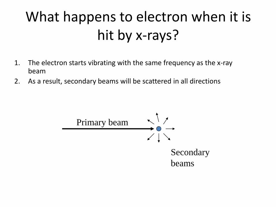
What happens to electron when it is hit by x-rays?
1. The electron starts vibrating with the same frequency as the x-ray beam
2. As a result, secondary beams will be scattered in all directions
Primary beam
Secondary
beams
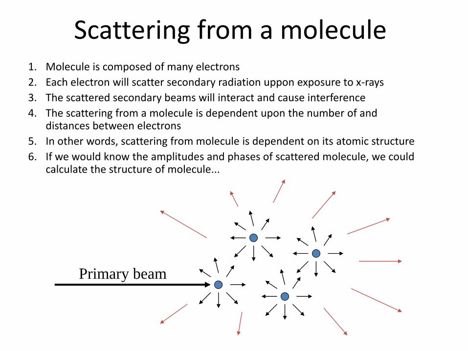
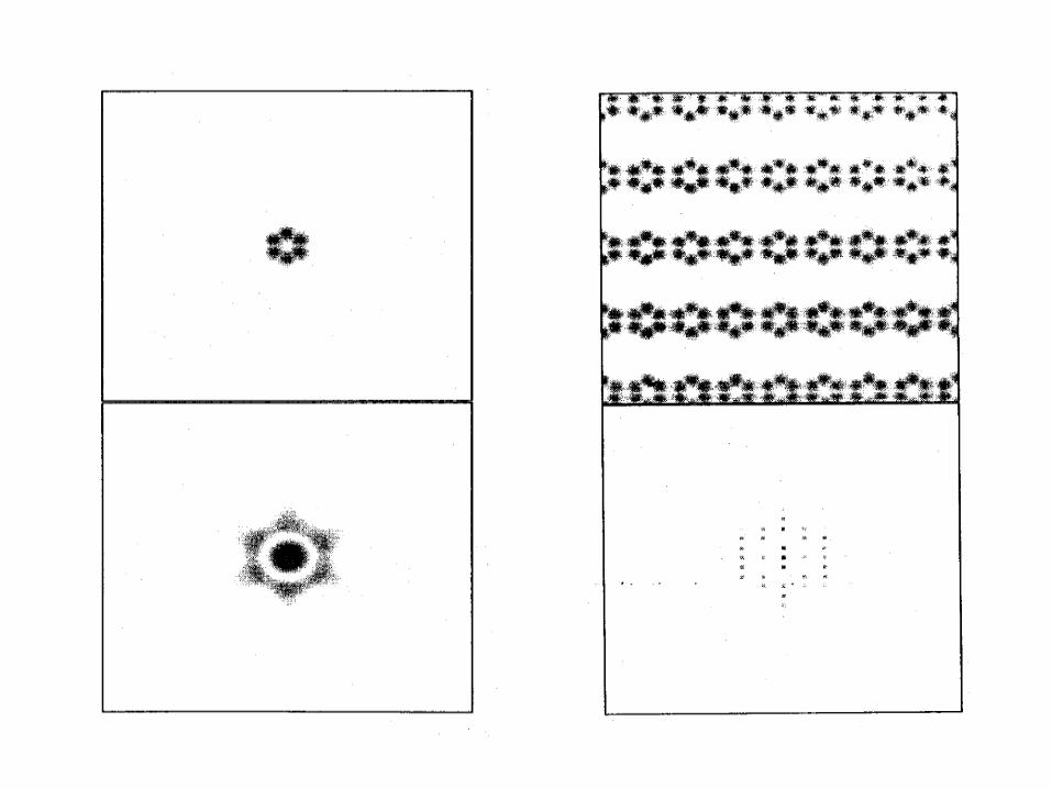
Scattering from a molecule 1. Molecule is composed of many electrons
2. Each electron will scatter secondary radiation uppon exposure to x-rays
3. The scattered secondary beams will interact and cause interference
4. The scattering from a molecule is dependent upon the number of and distances between electrons
5. In other words, scattering from molecule is dependent on its atomic structure
6. If we would know the amplitudes and phases of scattered molecule, we could calculate the structure of molecule...
Primary beam
In practice...
• Scattering from a single molecule is far too weak to be observed but possible.
• If molecules are all oriented in the same way (like in crystal), the scattering from individual molecules will be multiplied in certain directions
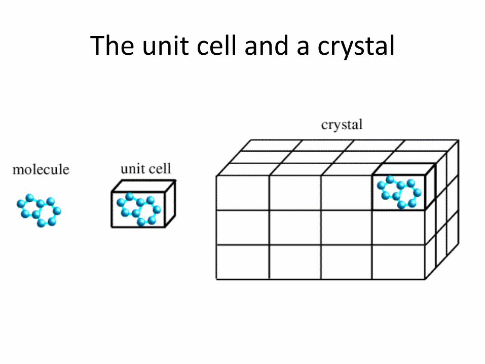
The unit cell and a crystal
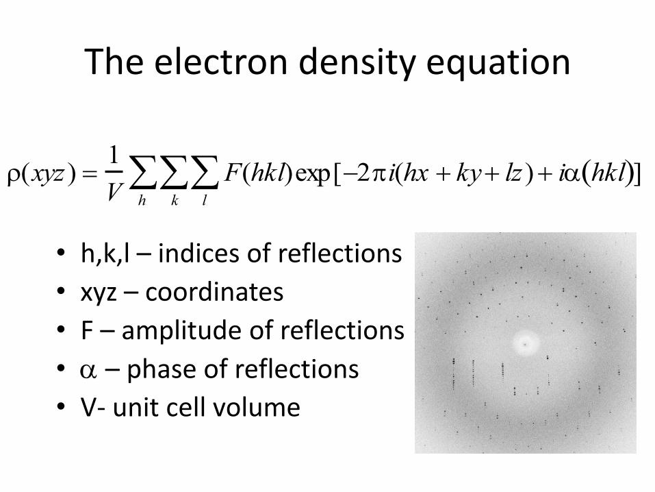
The electron density equation
• h,k,l – indices of reflections
• xyz – coordinates
• F – amplitude of reflections
• a – phase of reflections
• V- unit cell volume
(xyz) 1
VF(hkl)
l
k
h
exp[2i(hx ky lz) ia hkl ]
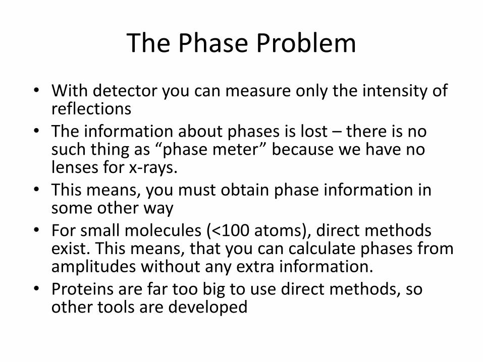
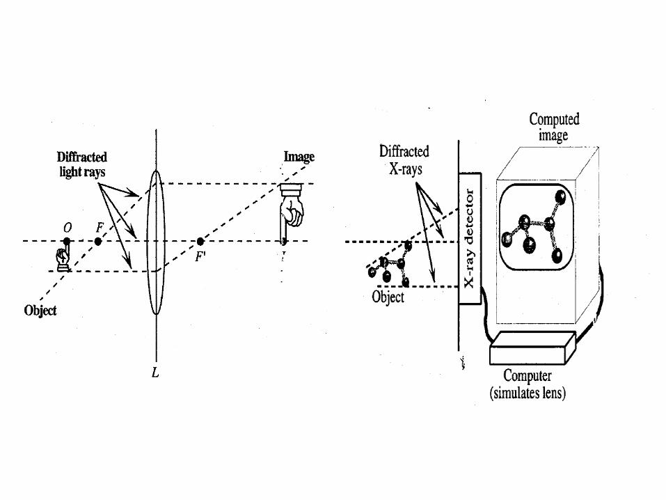
The Phase Problem
• With detector you can measure only the intensity of reflections
• The information about phases is lost – there is no such thing as “phase meter” because we have no lenses for x-rays.
• This means, you must obtain phase information in some other way
• For small molecules (<100 atoms), direct methods exist. This means, that you can calculate phases from amplitudes without any extra information.
• Proteins are far too big to use direct methods, so other tools are developed
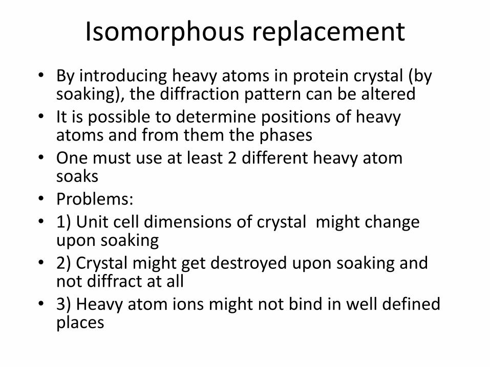
Isomorphous replacement
• By introducing heavy atoms in protein crystal (by soaking), the diffraction pattern can be altered
• It is possible to determine positions of heavy atoms and from them the phases
• One must use at least 2 different heavy atom soaks
• Problems: • 1) Unit cell dimensions of crystal might change
upon soaking • 2) Crystal might get destroyed upon soaking and
not diffract at all • 3) Heavy atom ions might not bind in well defined
places
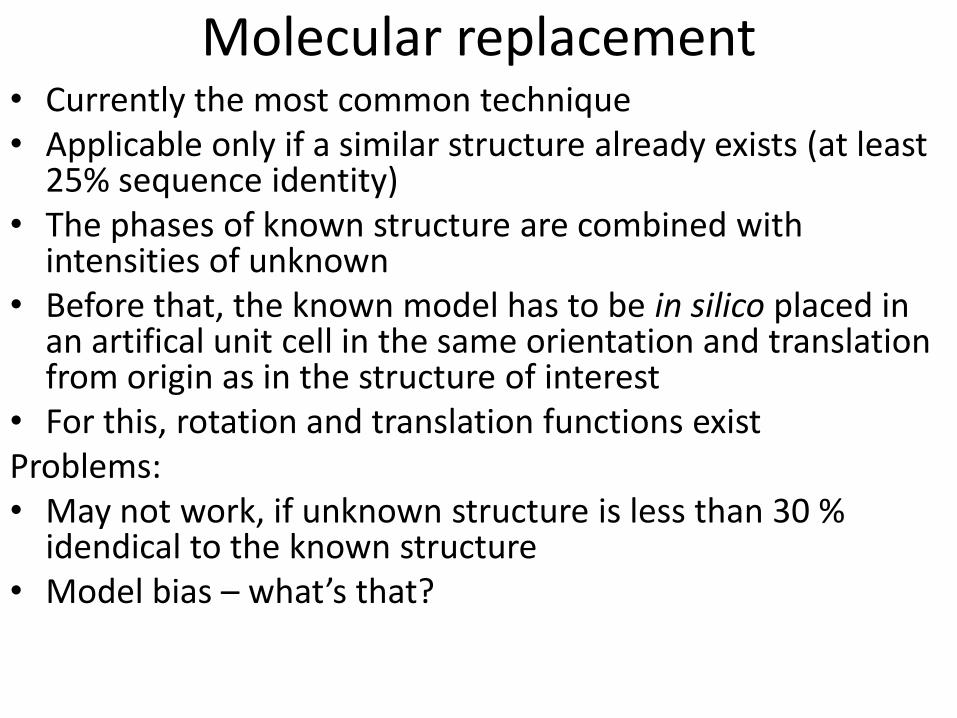
Molecular replacement • Currently the most common technique • Applicable only if a similar structure already exists (at least
25% sequence identity) • The phases of known structure are combined with
intensities of unknown • Before that, the known model has to be in silico placed in
an artifical unit cell in the same orientation and translation from origin as in the structure of interest
• For this, rotation and translation functions exist Problems: • May not work, if unknown structure is less than 30 %
idendical to the known structure • Model bias – what’s that?
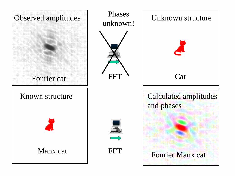
Observed amplitudes Unknown structure
Known structure Calculated amplitudes
and phases
Phases
unknown!
Fourier cat Cat
Manx cat Fourier Manx cat
FFT
FFT
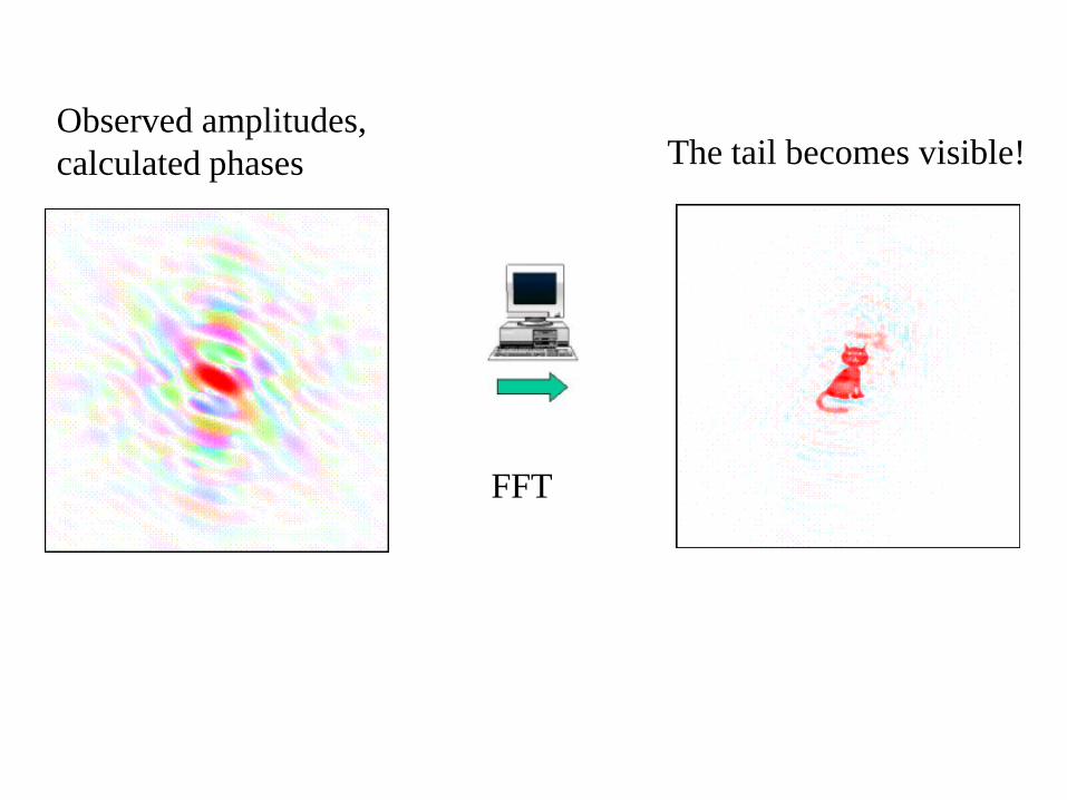
Observed amplitudes,
calculated phases The tail becomes visible!
FFT
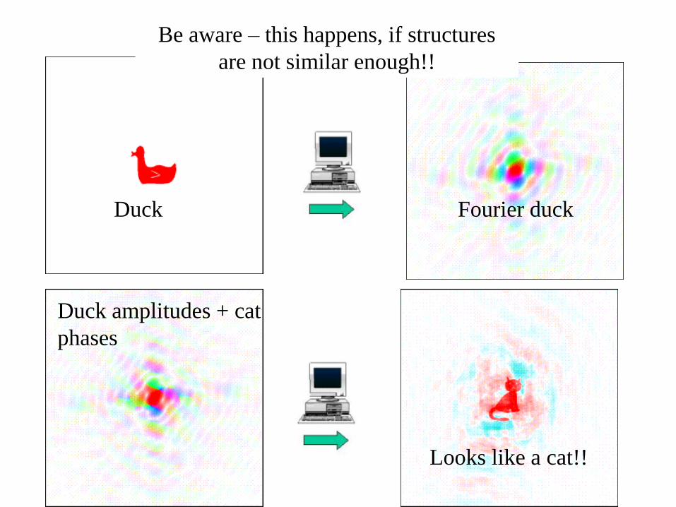
Duck amplitudes + cat
phases
Duck Fourier duck
Looks like a cat!!
Be aware – this happens, if structures
are not similar enough!!
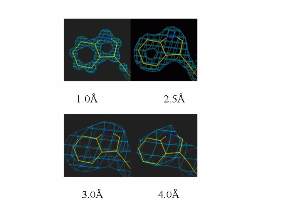
Model building
• Fitting of protein sequence in the electron density
• Easy in molecular replacement
• More difficult if no initial model is available
• Unambiquous if resolution is high enough (better than 3.0 Å)
• Can be automated, if resolution is close to 2Å or better
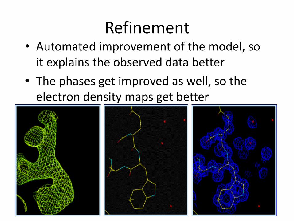
Refinement • Automated improvement of the model, so
it explains the observed data better
• The phases get improved as well, so the electron density maps get better
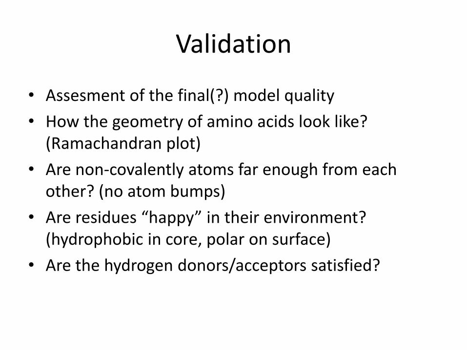
Validation
• Assesment of the final(?) model quality
• How the geometry of amino acids look like? (Ramachandran plot)
• Are non-covalently atoms far enough from each other? (no atom bumps)
• Are residues “happy” in their environment? (hydrophobic in core, polar on surface)
• Are the hydrogen donors/acceptors satisfied?

Part 2 NMR Structure Determination
4.1Å
2.9Å
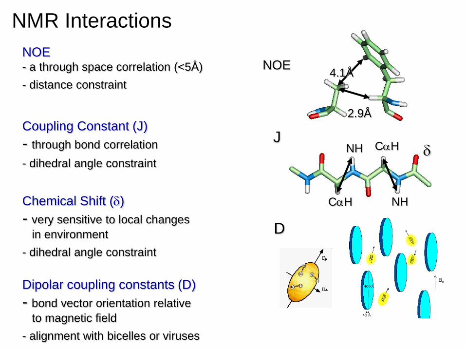
NOE
CaH
NH
NH
CaH J
NOE - a through space correlation (<5Å)
- distance constraint
Coupling Constant (J)
- through bond correlation
- dihedral angle constraint
Chemical Shift (d)
- very sensitive to local changes
in environment
- dihedral angle constraint
Dipolar coupling constants (D)
- bond vector orientation relative
to magnetic field
- alignment with bicelles or viruses
D
NMR Interactions
d
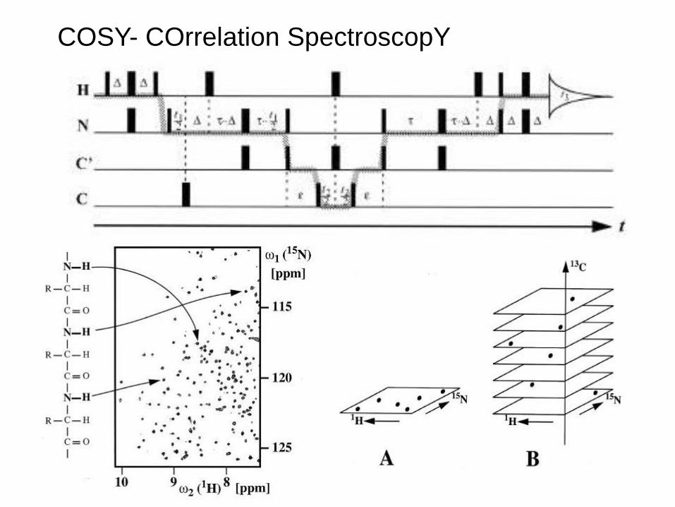
COSY- COrrelation SpectroscopY
NOE- Nuclear Overhauser Effect
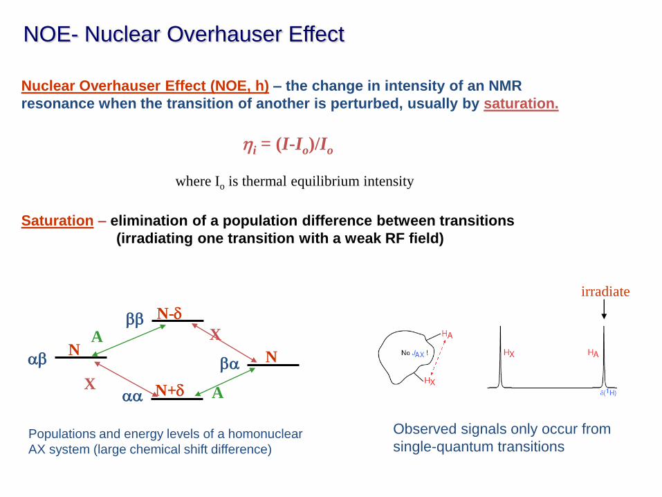
Nuclear Overhauser Effect (NOE, h) – the change in intensity of an NMR
resonance when the transition of another is perturbed, usually by saturation.
Saturation – elimination of a population difference between transitions
(irradiating one transition with a weak RF field)
hi = (I-Io)/Io
where Io is thermal equilibrium intensity
aa
ab ba
bb
N N
N+d
N-d
X
X A
A
irradiate
Populations and energy levels of a homonuclear
AX system (large chemical shift difference)
Observed signals only occur from
single-quantum transitions
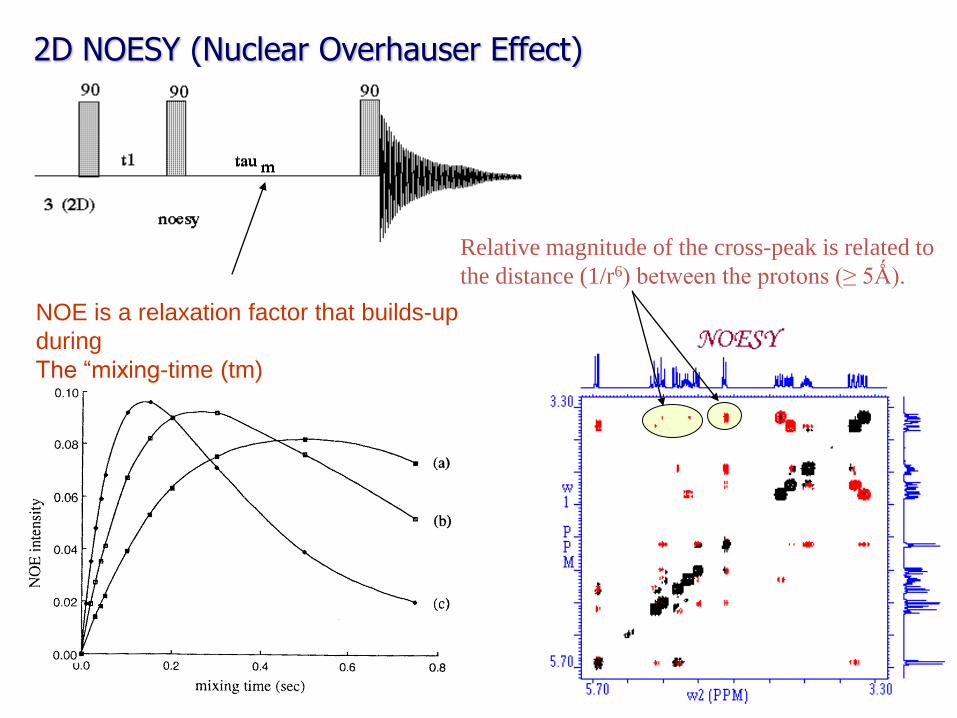
2D NOESY (Nuclear Overhauser Effect)
Relative magnitude of the cross-peak is related to
the distance (1/r6) between the protons (≥ 5Ǻ).
NOE is a relaxation factor that builds-up
during
The “mixing-time (tm)
2D NOESY Spectra at 900 MHz
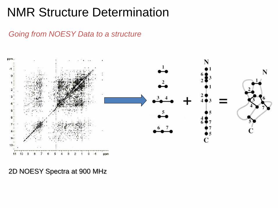
NMR Structure Determination
Going from NOESY Data to a structure
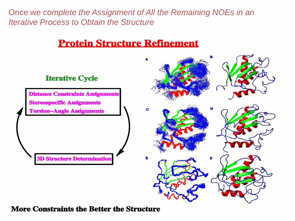
Once we complete the Assignment of All the Remaining NOEs in an
Iterative Process to Obtain the Structure
NMR Structure Determination
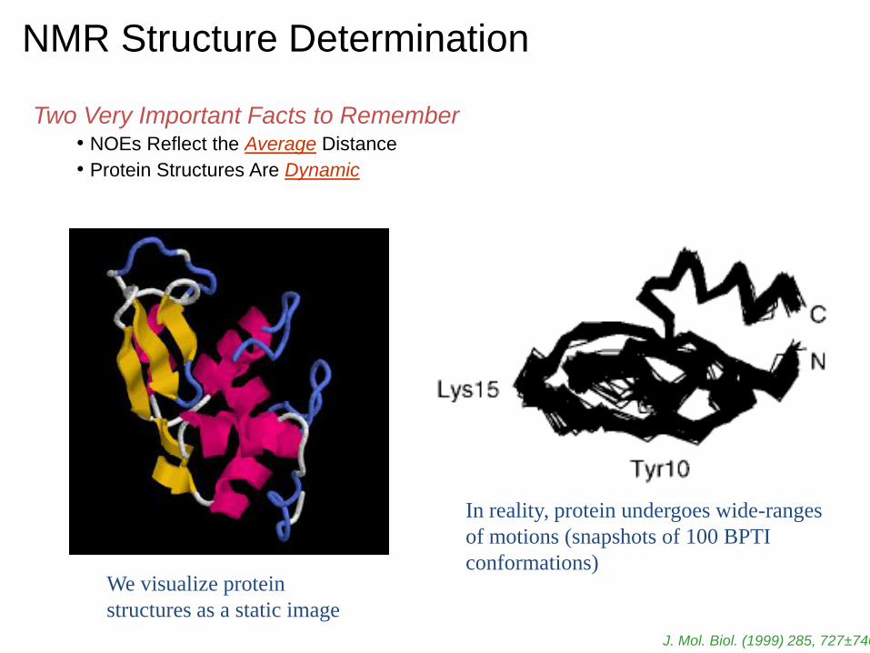
Two Very Important Facts to Remember • NOEs Reflect the Average Distance
• Protein Structures Are Dynamic
We visualize protein
structures as a static image
In reality, protein undergoes wide-ranges
of motions (snapshots of 100 BPTI
conformations)
J. Mol. Biol. (1999) 285, 727±740
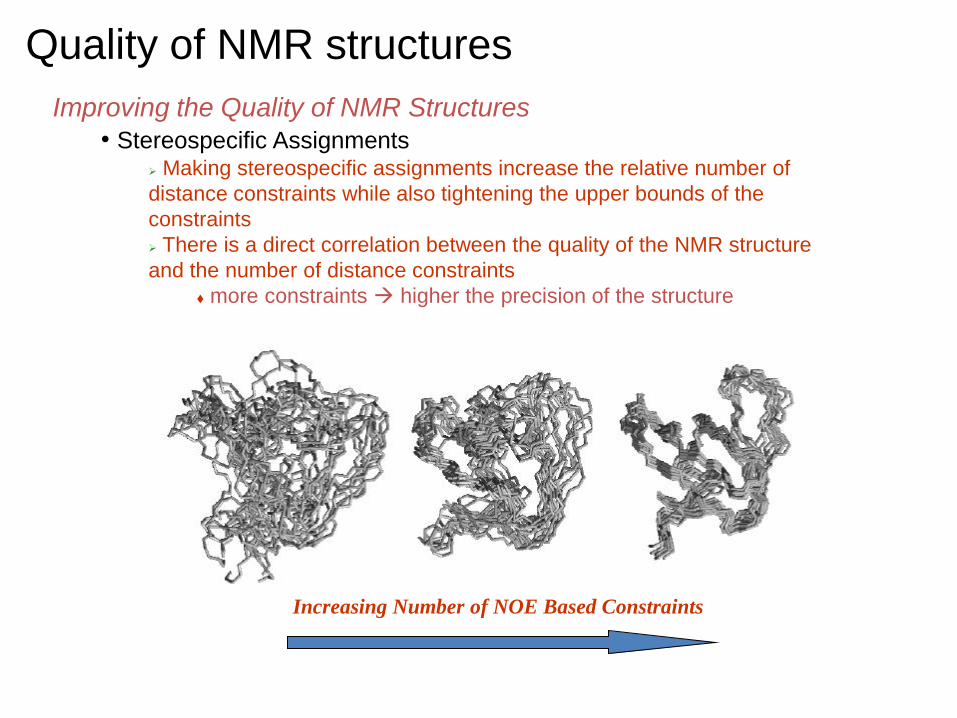
Quality of NMR structures
Improving the Quality of NMR Structures • Stereospecific Assignments
Making stereospecific assignments increase the relative number of
distance constraints while also tightening the upper bounds of the
constraints
There is a direct correlation between the quality of the NMR structure
and the number of distance constraints
more constraints higher the precision of the structure
Increasing Number of NOE Based Constraints
Part 3 Structure Determination by
Electron Microscopy
References and other useful material • Texts
– Biophysical Electron Microscopy: Basic Concepts and Modern Techniques by U. Valdre (Editor), Peter W. Hawkes (Editor)
– Three-Dimensional Electron Microscopy of Macromolecular Assemblies by Joachim Frank
– Negative Staining and Cryoelectron Microscopy: The Thin Film Techniques by Robin J. Harris, James R. Harris
• Reviews – Henderson, R. The potential and limitations of neutrons, electrons and X-rays
for atomic resolution microscopy of unstained biological molecules. Q Rev Biophys 28, 171-93 (1995).
– Glaeser, R. M. Review: electron crystallography: present excitement, a nod to the past, anticipating the future. J Struct Biol 128, 3-14 (1999).
– Stowell, M. H., Miyazawa, A. & Unwin, N. Macromolecular structure determination by electron microscopy: new advances and recent results. Curr Opin Struct Biol 8, 595-600 (1998).
• Web – http://ncmi.bcm.tmc.edu/%7Estevel/spintro/siframes.htm – http://cryoem.berkeley.edu/~nieder/em_for_dummies/
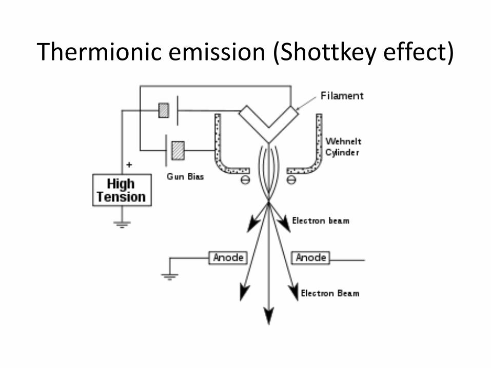
Thermionic emission (Shottkey effect)
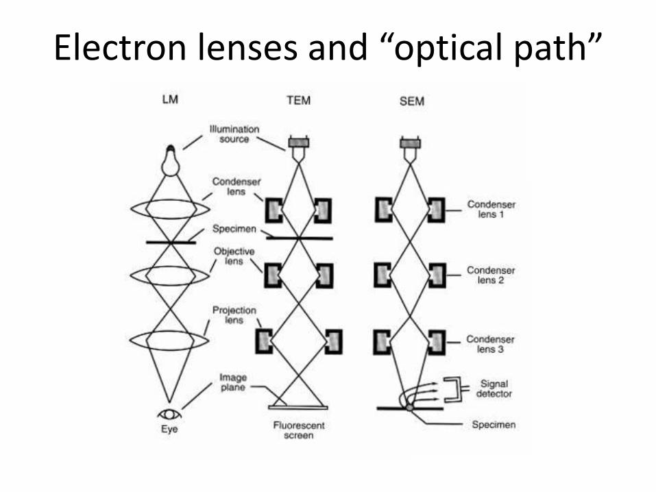
Electron lenses and “optical path”
Why use electrons….Part 1
?

Why use electrons…Part 2 Electrons
80-500keV
X-rays
1.5 A
Ratio inelastic/elastic 3 10
Mechanism of damage 2nd e-
emission
Photoelectric
e- emission
Energy per inelastic event 20 eV 8 keV
Energy per elastic event 60 eV 80 eV
Energy relative to electrons
inelastic (Compton) 1 400
elastic (Rayleigh) 1 1000
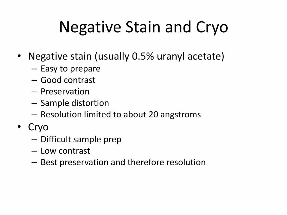
Negative Stain and Cryo
• Negative stain (usually 0.5% uranyl acetate) – Easy to prepare – Good contrast – Preservation – Sample distortion – Resolution limited to about 20 angstroms
• Cryo – Difficult sample prep – Low contrast – Best preservation and therefore resolution
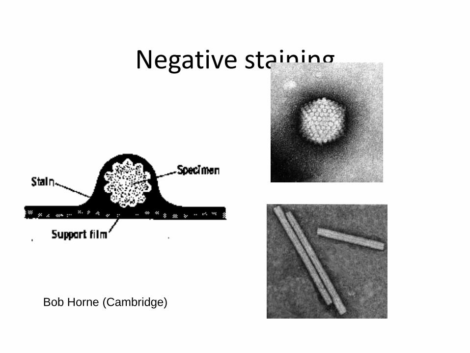
Negative staining
Bob Horne (Cambridge)
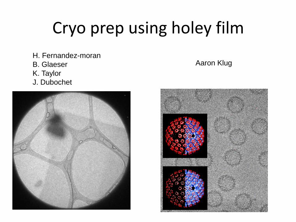
Cryo prep using holey film
H. Fernandez-moran
B. Glaeser
K. Taylor
J. Dubochet
Aaron Klug
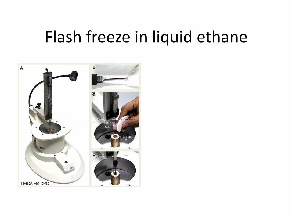
Flash freeze in liquid ethane
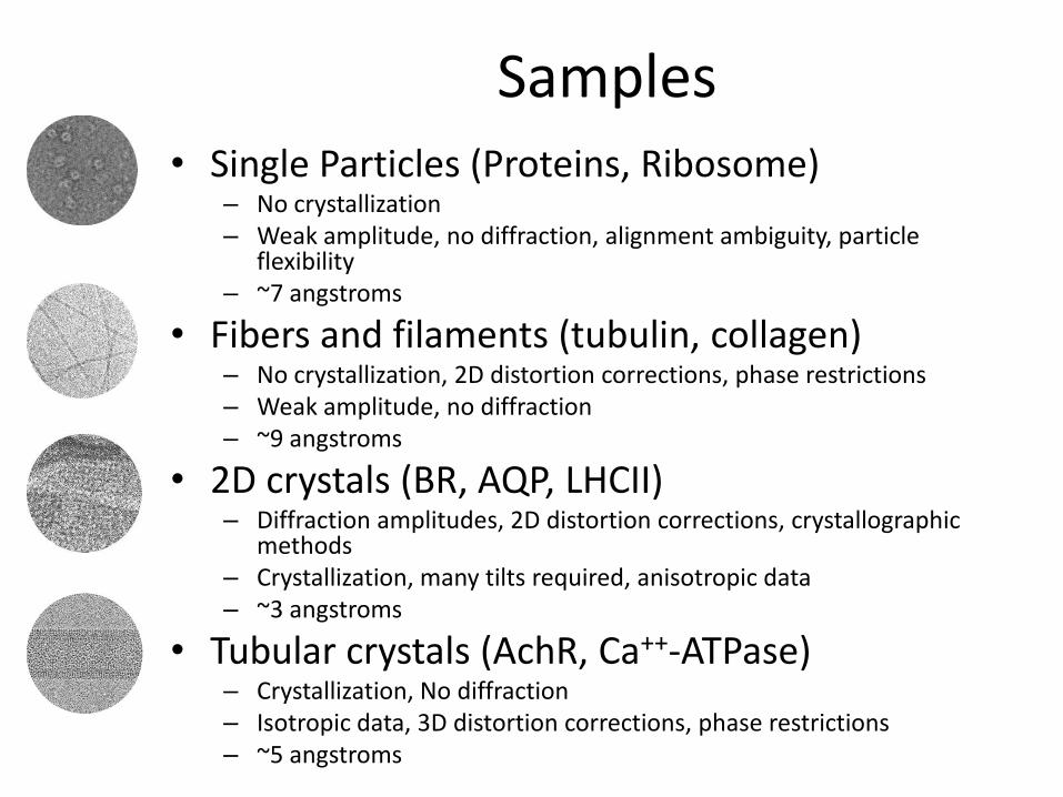
Samples • Single Particles (Proteins, Ribosome)
– No crystallization – Weak amplitude, no diffraction, alignment ambiguity, particle
flexibility – ~7 angstroms
• Fibers and filaments (tubulin, collagen) – No crystallization, 2D distortion corrections, phase restrictions – Weak amplitude, no diffraction – ~9 angstroms
• 2D crystals (BR, AQP, LHCII) – Diffraction amplitudes, 2D distortion corrections, crystallographic
methods – Crystallization, many tilts required, anisotropic data – ~3 angstroms
• Tubular crystals (AchR, Ca++-ATPase) – Crystallization, No diffraction – Isotropic data, 3D distortion corrections, phase restrictions – ~5 angstroms
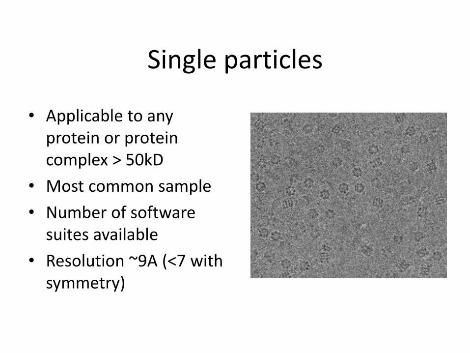
Single particles
• Applicable to any protein or protein complex > 50kD
• Most common sample
• Number of software suites available
• Resolution ~9A (<7 with symmetry)
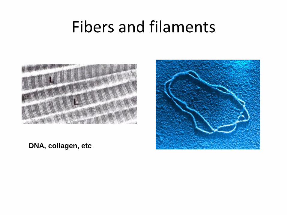
Fibers and filaments
DNA, collagen, etc
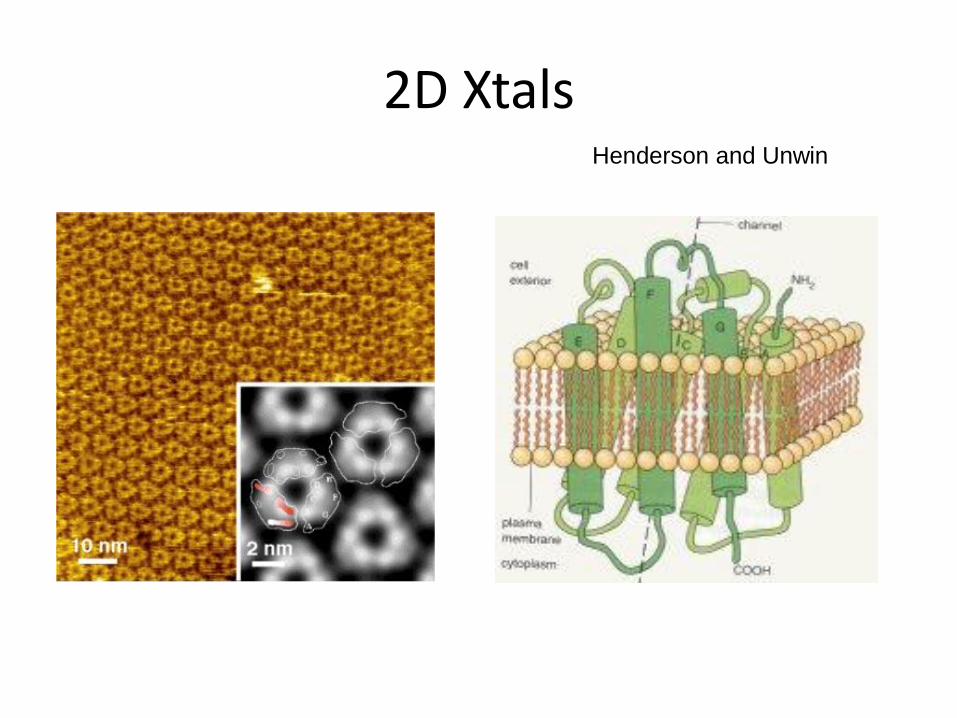
2D Xtals Henderson and Unwin
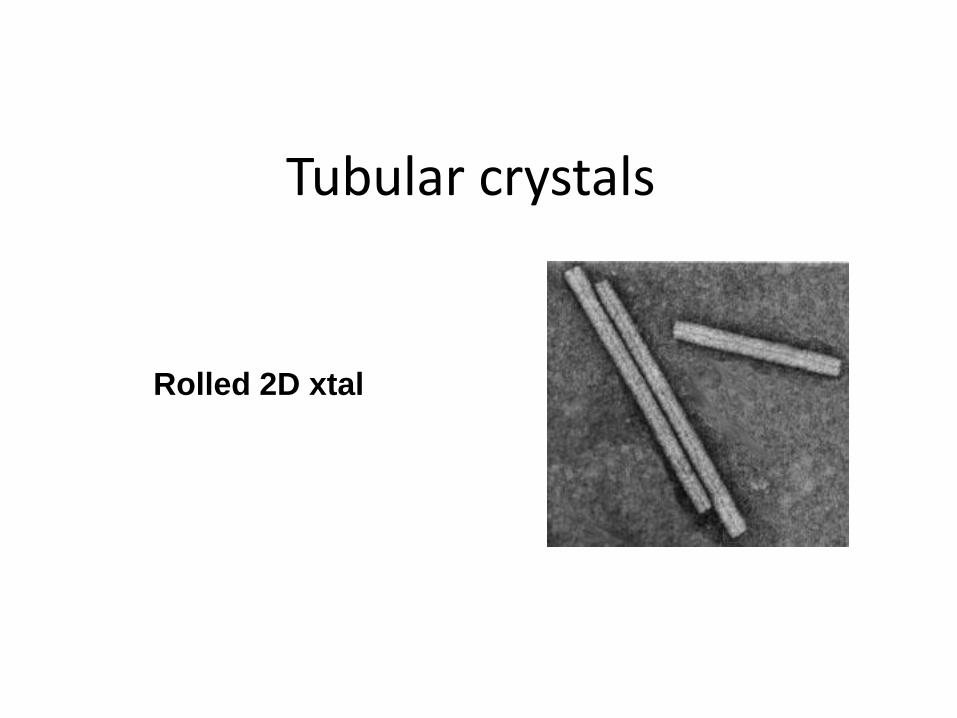
Tubular crystals
Rolled 2D xtal
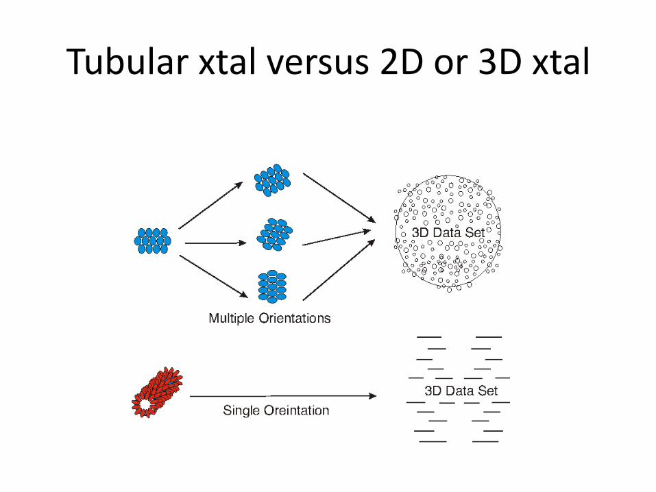
Tubular xtal versus 2D or 3D xtal
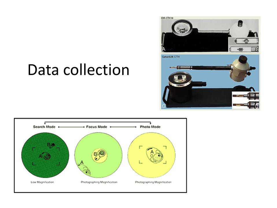
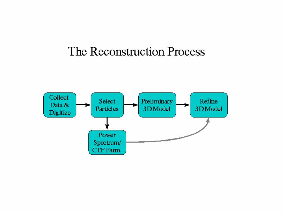
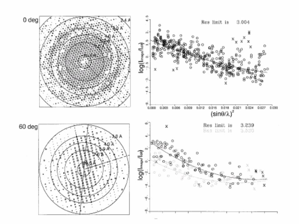
Data collection
Image recording
• Film – High density content (~20kx16k pixels)
– Slow (development time, drying)
– Requires digitization (scanning takes hours)
• CCD – Low density content (4kx4k pixels)
– Fast (ms to sec)
– Direct digital
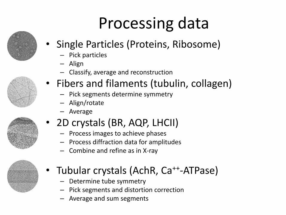
Processing data • Single Particles (Proteins, Ribosome)
– Pick particles – Align – Classify, average and reconstruction
• Fibers and filaments (tubulin, collagen) – Pick segments determine symmetry – Align/rotate – Average
• 2D crystals (BR, AQP, LHCII) – Process images to achieve phases – Process diffraction data for amplitudes – Combine and refine as in X-ray
• Tubular crystals (AchR, Ca++-ATPase) – Determine tube symmetry – Pick segments and distortion correction – Average and sum segments
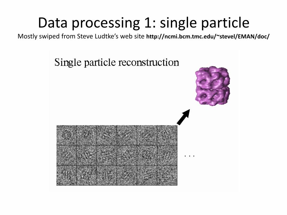
Data processing 1: single particle Mostly swiped from Steve Ludtke’s web site http://ncmi.bcm.tmc.edu/~stevel/EMAN/doc/

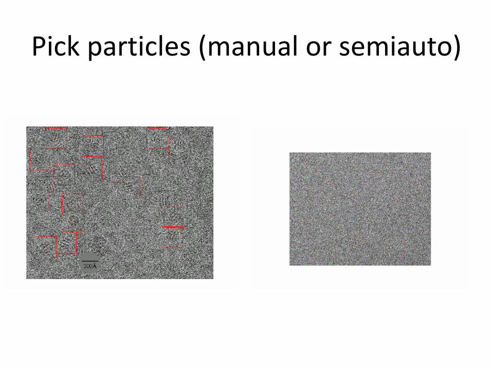
Pick particles (manual or semiauto)
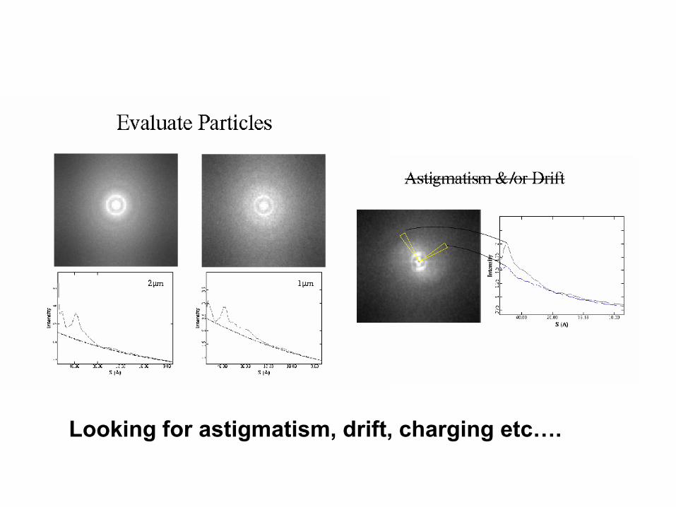
Looking for astigmatism, drift, charging etc….
Now on to the first model
• First rule of thumb…be cautious…
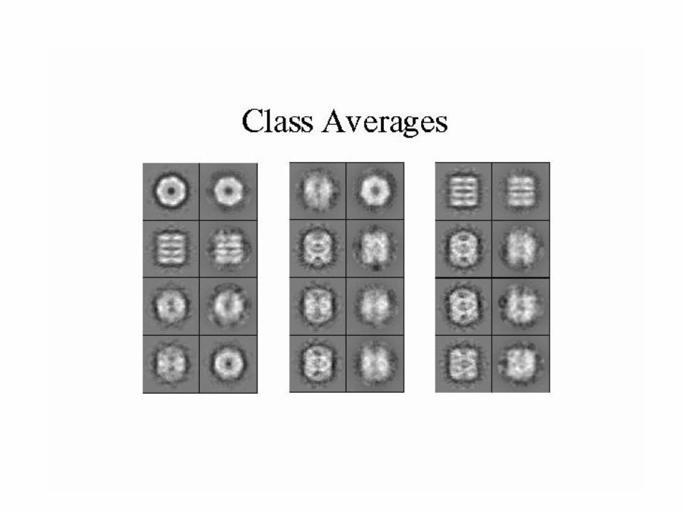
• How to classify particles
– Reference free classification and alignment
– MSA
• Application of symmetry
• Random conical tilt
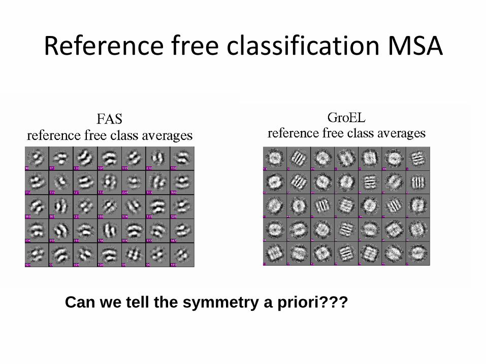
Reference free classification MSA
Can we tell the symmetry a priori???
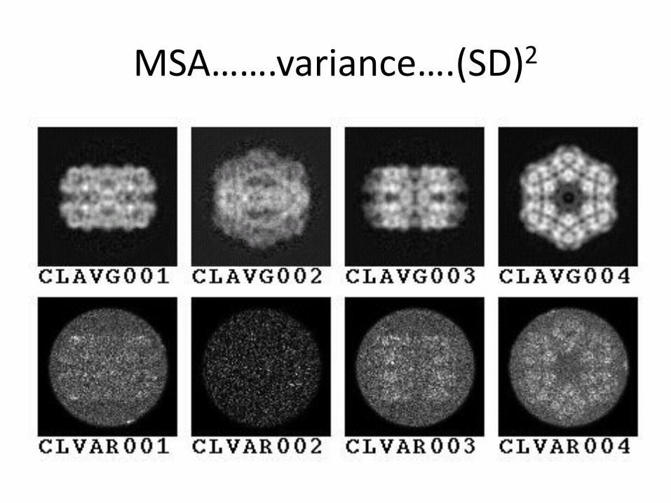
MSA…….variance….(SD)2
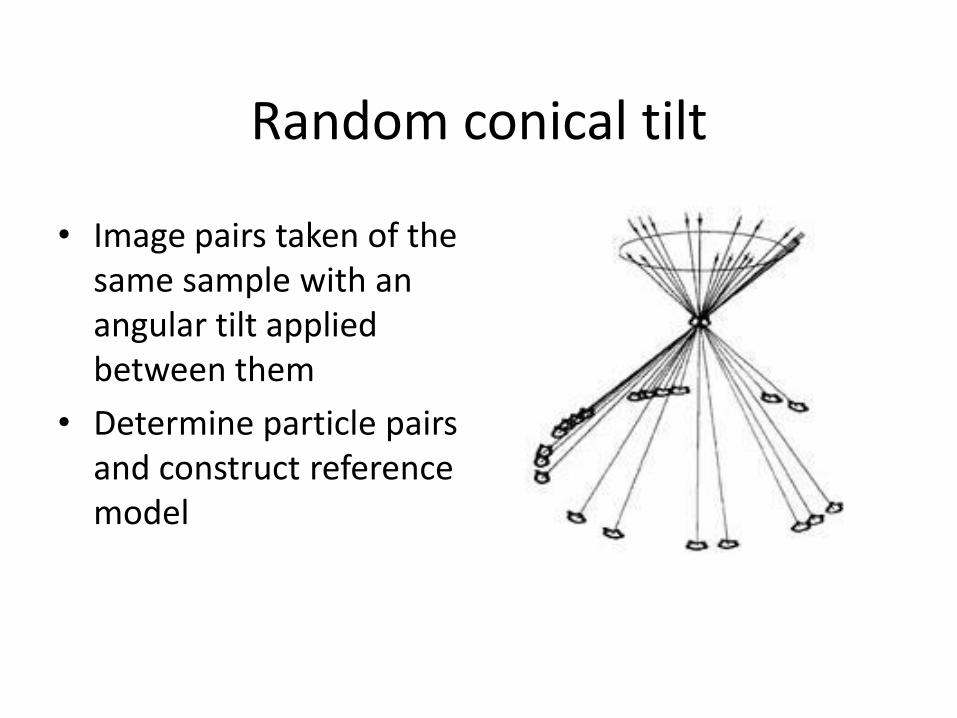
Random conical tilt
• Image pairs taken of the same sample with an angular tilt applied between them
• Determine particle pairs and construct reference model
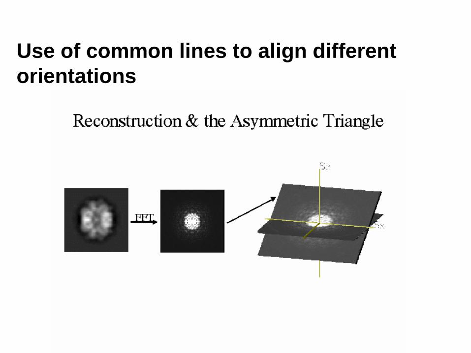
Use of common lines to align different
orientations
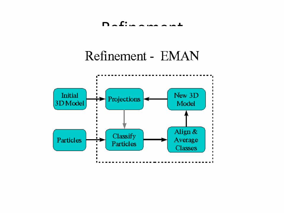
Refinement
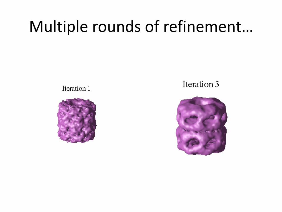
Multiple rounds of refinement…
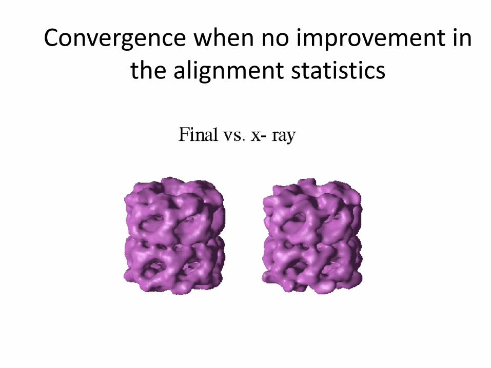
Convergence when no improvement in the alignment statistics
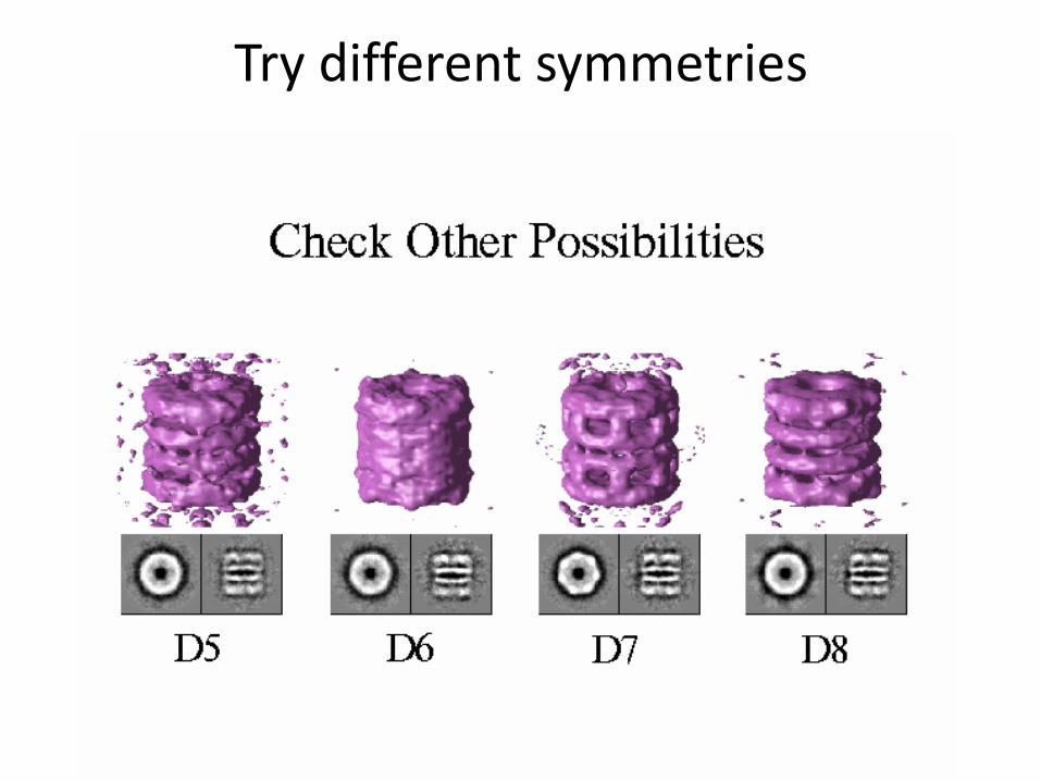
Try different symmetries
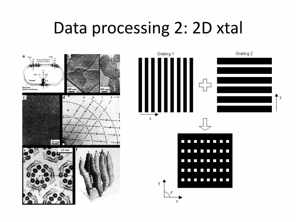
Data processing 2: 2D xtal
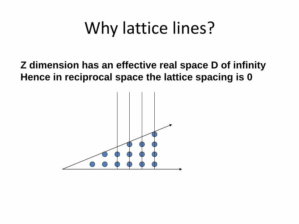
Why lattice lines?
Z dimension has an effective real space D of infinity
Hence in reciprocal space the lattice spacing is 0
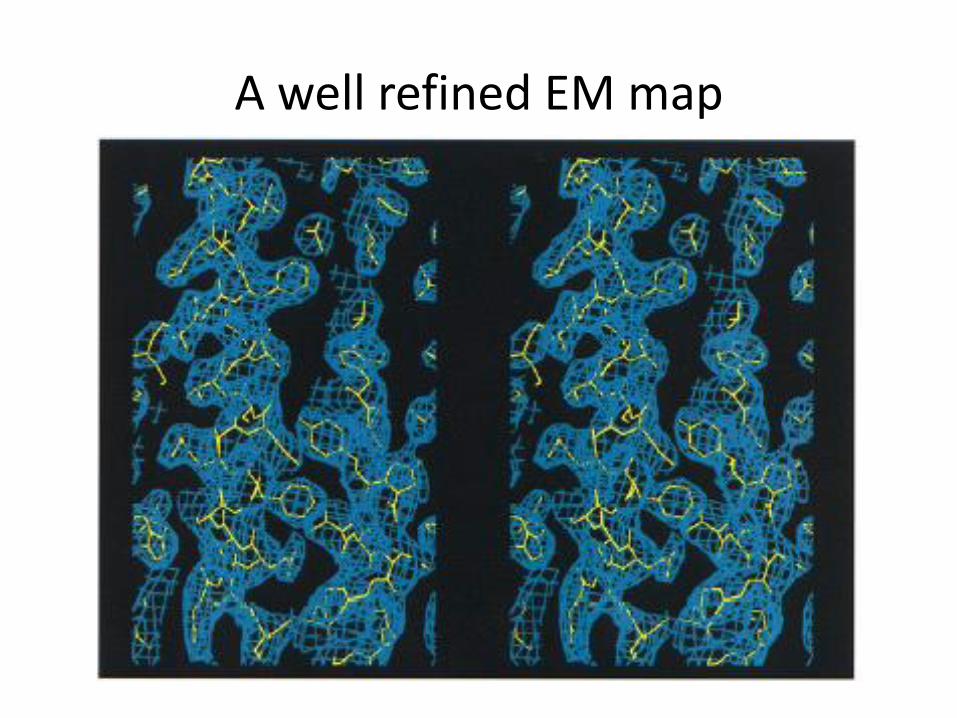
A well refined EM map
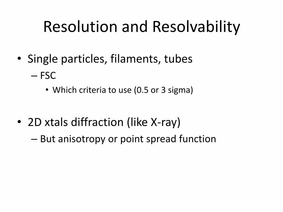
Resolution and Resolvability
• Single particles, filaments, tubes
– FSC
• Which criteria to use (0.5 or 3 sigma)
• 2D xtals diffraction (like X-ray)
– But anisotropy or point spread function
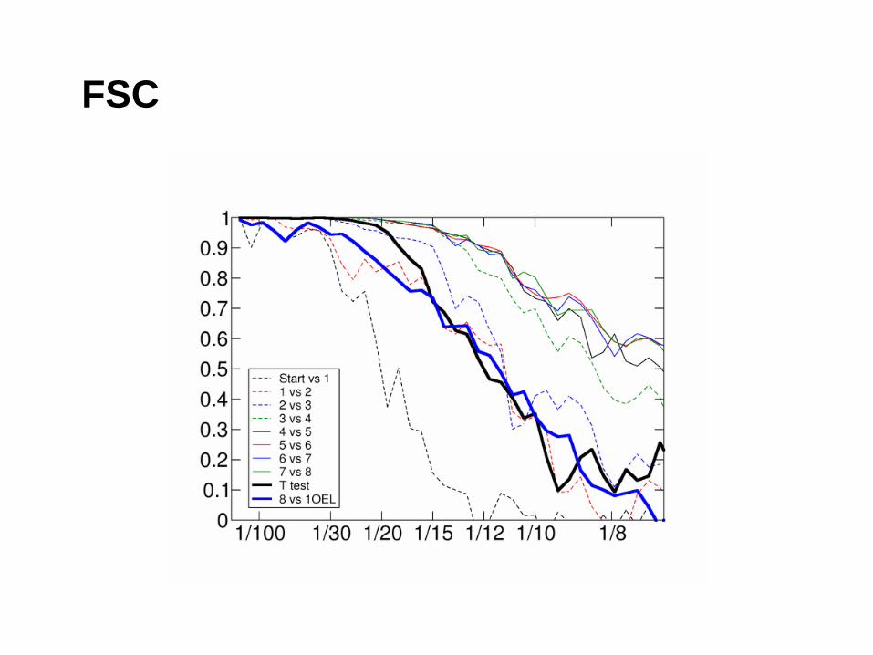
FSC
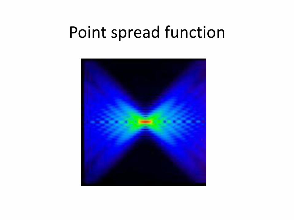
Point spread function
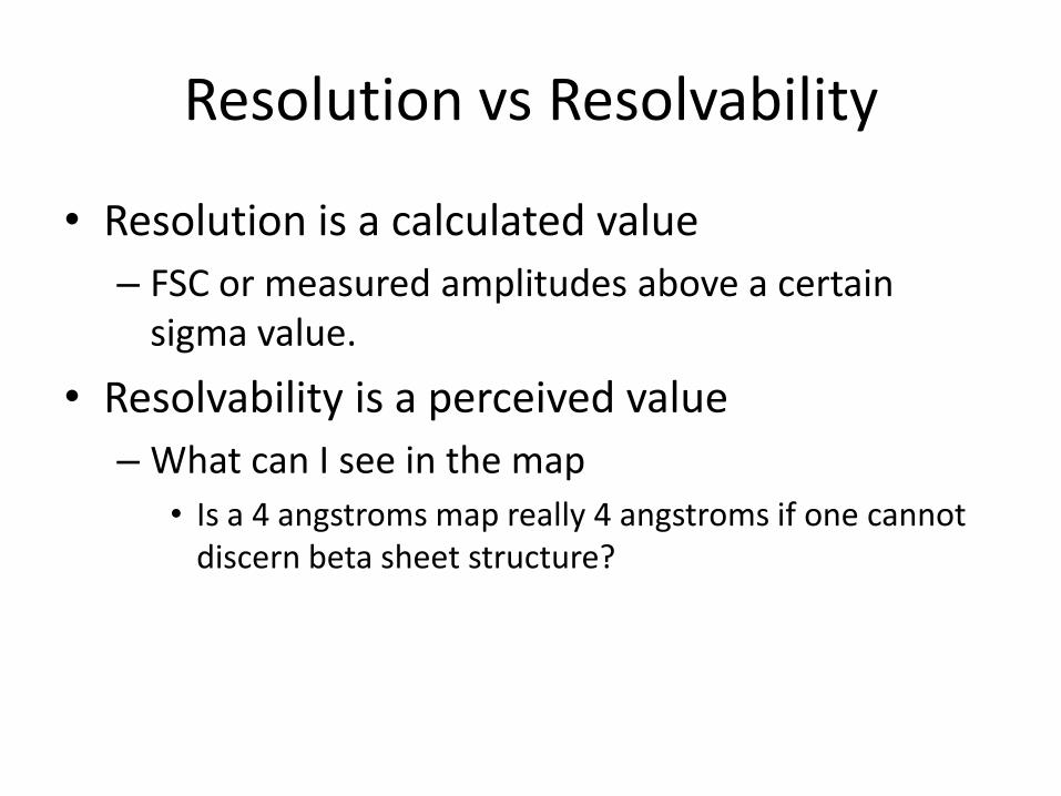
Resolution vs Resolvability
• Resolution is a calculated value
– FSC or measured amplitudes above a certain sigma value.
• Resolvability is a perceived value
– What can I see in the map
• Is a 4 angstroms map really 4 angstroms if one cannot discern beta sheet structure?