Embed Size (px)
Citation preview
PART -IV
ANTI BACTERIAL STUDIES

217
CHAPTER I
INTRODUCTION
The true masters of life on earth are not humans but microbes.
Animals could not live in the absence of micro-organisms. Germ free
animals are usually more susceptible to pathogens. We humans have lots of
microbial friends that with in our intestine.
The normal microbiota use space, resources, nutrients and may
produce chemicals that repel invading pathogens and possible diseases
through bacterial interference. The relationship between the body and its
normal microflora is an example of symbiosis (Dubey and Maheswari,
1999). The microbial symbiotic associations are essential to the livelihood of
both the microorganism and its partner. Without microbial symbiotants
most animals and plants could not survive in natural communities. In fact
human kind enjoys a peaceful co-existence with a majority of micro
organisms. (Harwood and Greenberg, 1999)
Humanbeings harbour a wide variety of micro organisms both on
and in their bodies. The normal microflora are more or less constant and are
broadly divided into residents and transients. The residents constitute a

218
constant population which cannot be completely removed while transients
vary from time to time. The residents prevent permanent colonisation of the
body by other organisms (Alcomo E, 2001).
The normal and constant microflora of human body have adapted
themselves to life in certain parts of the body. Besides these some microbes
reside as temporary microflora. The pathogens among the microflora may
cause disease, when the body’s defence mechanism fails. Their abnormal
multiplication can cause disease such as enteritis and entotoxic shock.
Normal microflora is mainly found of E.coli, Aerobactor aerugenes, Clostridiun
sp., Staphylococcus sp., Streptococcus sp., Fungi, Diphtheroids and certain
pathogenic and non pathogenic bacteria. They survive by receiving nutrition
from the body (Carpenter PL, 1977).
The immune system provides protection against potential pathogens.
Both commensalistic and mutualistic relationships are found between man
and microbes. The frequency of microbial infection in humans has increased
dramatically because of multidrug resistant microbial isolates like fungi and
bacteria. The increasing clinical significance of drug resistant bacterial
pathogens has lend additional urgency to microbiological and antibacterial
research.
219
Many natural antimicrobial compounds can be used in food
preservation systems but only a few have been exploited. Microbial control
in foods could be assured by suppressing one or more essential factors for
microbial survival (Horace DG, 1982). It could be possible by adding
suitable substances (weak organic acids, hydrogen peroxide, chelators,
organic biomolecules) and applying physical (temperature, packa-ging)
and/or chemical procedures (pH, oxide-reduction potential, osmotic
pressure) (Ray, 1996; Brull S, 1999). These procedures could kill or make
some unviable microorganisms. There has been increasing concern of the
consumers about foods free or with lower level of chemical preservatives
because these could be toxic for humans
Lactic acid bacteria based cultures have been traditionally used to
preserve fermented food in the tropics. In USA and other developed
countries such fermented foods as yoghurt, buttermilk and fermented
sausages are increasing in popularity as they are considered natural and
healthy. Works were carried out to study the effectiveness of different
antimicrobial packaging systems on the microbial quality decay kinetics
during storage of Mozzarella cheese was evaluated. Lemon extract, at
3 different concentrations, was used as active agent, in combination with
brine and with a gel solution made of sodium alginate. Results show an
220
increase in the shelf life of all active packaged Mozzarella cheeses,
confirming that the investigated substance may exert an inhibitory effect on
the microorganisms responsible for spoilage phenomena without affecting
the functional microbiota of the product (Conte A, 2007).
Bacterial Strains
Bacteria have been one of the most studied test systems. They will
thrive under a wide range of temperature. Some of them may grow
throughout the range of 6-50oC. Usually these micro organisms are divided
into two groups. One is Gram positive and the other is Gram negative. The
Gram reaction is a procedure which was developed in 1884 by a Danish
physician, Christian Gram, who detected the disease producing bacteria in
animal tissues. The Gram stain reaction is determined by microscopic
examinations of cells that have been successfully stained with Crystal Violet
dye, treated with iodine solution and rinsed with acetone or alcohol. The
Gram positive cells retain the violet stain while the Gram negative ones
decolourise the solvent (Glem LJ, 1966). The bacterial strains studied during
this research work are both Gram positive and Gram negative.
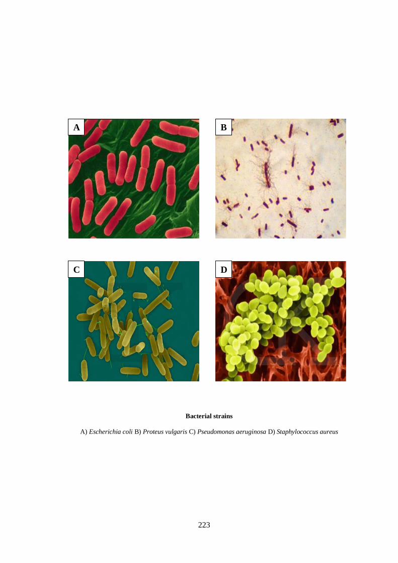
The major clinical pathogens among the normal microflora associated
with the human body are Escherichia coli, Proteus vulgaris, Pseudomonas
221
aeruginosa and Staphylococcus aureus (Boman HG, 2002). Among these
Escherichia coli, Proteus vulgaris, Pseudomonas aeruginosa are Gram negative
organisms while staphylococcus aureus belongs to gram positive category.
Escherichia coli (E.coli)
E.coli is an enteric organism and a part of normal flora which is a
large heterogeneous group of Gram negative rods, whose natural habitat is
the intestinal tract of human and animals (Donnenberg, 2002). In the
intestine they generally do not cause disease and may even contribute to
normal functions and nutrition. The bacteria become pathogenic only when
they reach tissues outside the intestinal tract, particularly the urinary and
billary tract, lungs causing inflammation at these sites. E. coli can generally
cause intestinal and extra intestinal infections such as urinary tract
infections, meningitis, peritonitis, septicemia and Gram negative pneumonia
(Feng P, 2007; Pearson H, 2007).
Proteus vulgaris
P. vulgaris is a rod shaped Gram negative bacterium that inhabits the
intestinal tracts of animals and can be pathogenic. It can produce acid with
the fermentation of glucose and sucrose. Optimal growing temperature is
23oC in a facultative environment. In humans it can cause urinary tract
infections and wound infections.
222
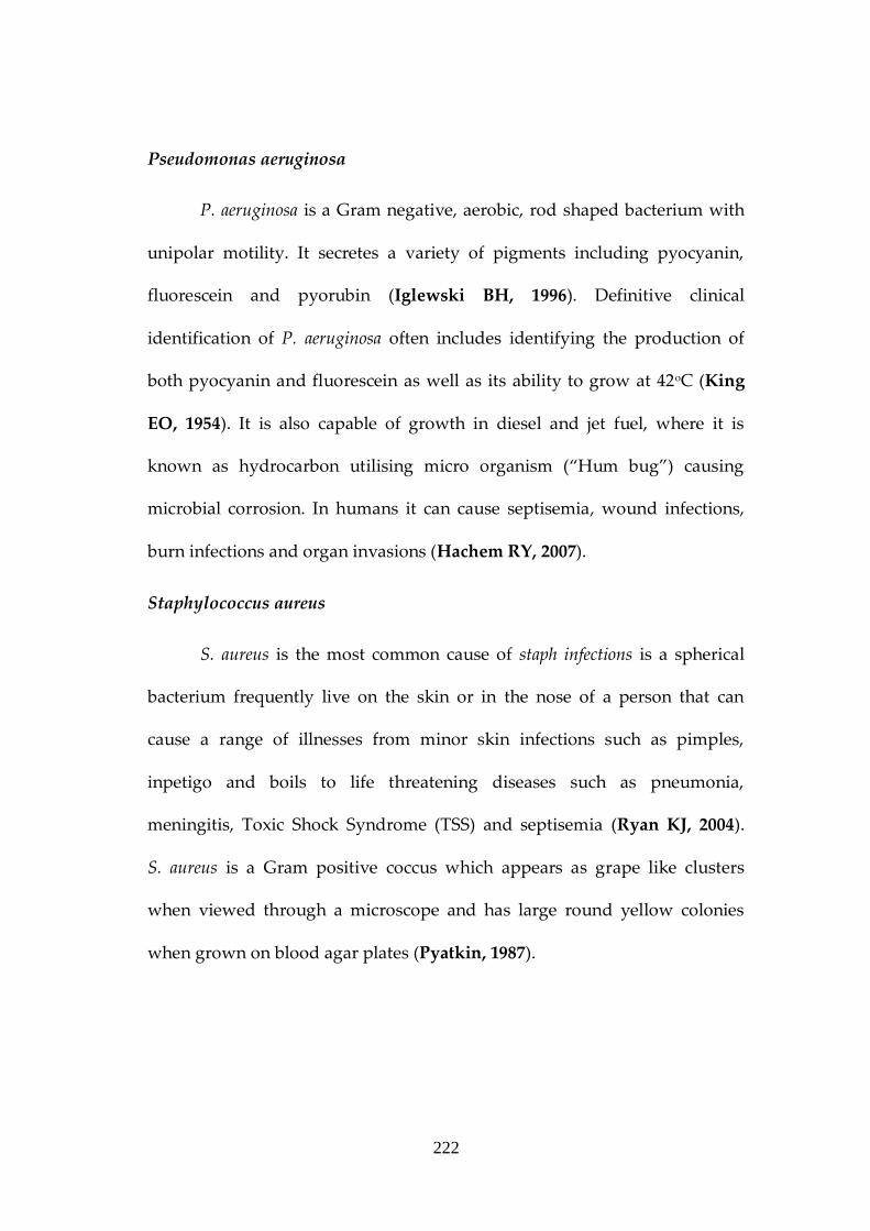
Pseudomonas aeruginosa
P. aeruginosa is a Gram negative, aerobic, rod shaped bacterium with
unipolar motility. It secretes a variety of pigments including pyocyanin,
fluorescein and pyorubin (Iglewski BH, 1996). Definitive clinical
identification of P. aeruginosa often includes identifying the production of
both pyocyanin and fluorescein as well as its ability to grow at 42oC (King
EO, 1954). It is also capable of growth in diesel and jet fuel, where it is
known as hydrocarbon utilising micro organism (“Hum bug”) causing
microbial corrosion. In humans it can cause septisemia, wound infections,
burn infections and organ invasions (Hachem RY, 2007).
Staphylococcus aureus
S. aureus is the most common cause of staph infections is a spherical
bacterium frequently live on the skin or in the nose of a person that can
cause a range of illnesses from minor skin infections such as pimples,
inpetigo and boils to life threatening diseases such as pneumonia,
meningitis, Toxic Shock Syndrome (TSS) and septisemia (Ryan KJ, 2004).
S. aureus is a Gram positive coccus which appears as grape like clusters
when viewed through a microscope and has large round yellow colonies
when grown on blood agar plates (Pyatkin, 1987).
223
A
D C
B
Bacterial strains
A) Escherichia coli B) Proteus vulgaris C) Pseudomonas aeruginosa D) Staphylococcus aureus

224
Growth of Bacteria
Bacteria can be found in all natural environments, often in extremely
large numbers. As a group, they display exceedingly diverse metabolic
capabilities and use almost any organic compound, and even some inorganic
salts, as a food source. Some bacteria cause disease in humans, animals, or
plants, but most are harmless or beneficial ecological agents whose
metabolic activities sustain higher life-forms. Without bacteria, soil would
not be fertile, and dead organic material would decay much more slowly.
Some bacteria are widely used in the preparation of foods, chemicals, and
antibiotics (Gorbach SL, 1990).
Bacterial Nutrition
All bacterial life has the same basic nutritional requirements which
include:
1. Source of Energy. This may be light (the sun or lamps) or inorganic
substances like sulfur, carbon monoxide or ammonia, or preformed
organic matter like sugar, protein, fats etc. Without energy life can not
exist and quickly dies or becomes inactive.
225
2. Source of Nitrogen. This may be nitrogen gas, ammonia,
nitrate/nitrite, or a nitrogenous organic compound like protein or
nucleic acid.
3. Source Of Carbon. This can be carbon dioxide or monoxide,
methane, carbon monoxide, or complex organic material
4. Source Of Oxygen. All cells use oxygen in a bound form and many
require gaseous oxygen (air), but oxygen is lethal to many microbes.
5. Source Of Water. All life requires liquid water in order to grow and
reproduce
6. Source of Minerals Like Iron, Zinc, Cobalt etc. These are called
Trace metals that are required by some enzymes to function.
The most common environmental conditions that a microbiologist
considers are temperature, pH, oxygen, light, salt/sugar concentration and
special nutrients. Each bacterium has an optimum range of these conditions
within which it grows at a maximum rate. In some cases this may be a fairly
broad range such as bacteria that can grow maximally over a 5 to 10 degree
temperature span. The hydrogen ion concentration of the growth medium
plays a very important role in bacterial growth. Most of the bacteria prefer

226
to grow in neutral or near neutral pH (6.5-7.5). Highly acidic and alkaline
pH is not suitable for bacterial growth (Janocha R, 1995).
The gaseous environment of the growth medium will affect bacterial
growth. The important gases which influence bacterial growth are oxygen
and carbon dioxide. Based on their requirements for oxygen, bacteria are
generally classified into three groups.
1. Aerobic bacteria; growing in presence of free oxygen or air
2. Anaerobic bacteria; growing in absence of free oxygen or air
3. Facultative anaerobic bacteria; they are really aerobic but also have
the capacity to grow in the absence of air or oxygen.
Anti Microbial Therapy
The control of infectious micro organisms is critical for the prevention
and treatment of disease. Modern medicine is dependant on
chemotherapeutic agents and most of them are antibiotic. The drug may be
administered orally, intravenously or intramuscularly. All these affect, the
invading pathogens but high doses may affect the host seriously
(Kell, 1997).
227
Exposure time to an antimicrobial agent is the most important and
frequently overlooked factor in the control of micro organism. Killing a
microbial population is a gradual process except in incineration. Young,
actively growing cells are unusually susceptible to antimicrobial agents
whereas mature or dormant cells are very resistant. The nature of medium in
or upon which the organisms occur, influences the efficiency of the
antimicrobial drugs (Kadri MS, 2004). Micro organisms are usually more
resistant when suspended in a media of a reaction with a satisfactory pH for
growth and killing by chemicals occurs rapidly as the pH changes from this
value. A dry chemical placed in contact with bacteria is usually ineffective
because moisture is essential for the disinfection process.
The first stage of antimicrobial therapy began in 1980 when Van
Behring discovered that administration of immune serum could protect an
immunologically naive animal against tetanus and diphtheria. This stage
came to an end by the introduction of effective chemotherapy like
sulphonamide. The introduction of antibiotics into clinical medicine was
unquestionably one of the triumps of 20th century and physicians were able
to treat most of the bacterial diseases. Unfortunately drug resistance
inevitably developed (Katherine M, 2004).

228
Antibiotics are chemical substances excreted by some micro
organisms which inhibit the growth and development of other microbes
(Levy S, 1998). The study of antibiotics began in 1929 by experiment of
Alexander Fleming. Some of these drugs were obtained naturally. This
naturally occurring drugs were chemically modified to enhance beneficial
effects while minimising their toxic effects and the products thus obtained
are called semisynthetic antibiotics. e.g. ampicillin, carbenicillin,
methcilline,etc. Some drugs are synthesised by chemists artificially and are
called synthetic drugs. e.g. sulphanomide, chloramphenicol, etc.
One of the major effect that an antibiotic can bring is the development
of resistant varieties of microbes. The resistance developed by a microbe
against an antibiotic may be due to many factors. This may occur due to
inherent resistance, natural resistance, acquired resistance and vertical and
horizontal evolution. Bacteria are able to exchange genes in nature by three
processes - conjugation, transduction, and transformation. Since bacteria
developed genes for drug resistance on plasmids, they are able to spread
drug resistance to other strains and species during genetic exchange
processes. The fast growth rate, high concentration of cells, genetic process
of mutation and selection are responsible for the resistance and evolution of
the bacteria (Kfrisell W, 1982).
229
The use of live bacteria to induce an immune response to itself or to a
carried vaccine component is an attractive vaccine strategy. Advantages of
live bacterial vaccines include their mimicry of a natural infection, intrinsic
adjuvant properties and their possibility to be administered orally.
Derivatives of pathogenic and non-pathogenic food related bacteria are
currently being evaluated as live vaccines. However, administration of live
bacterial vaccines poses some risks. In addition, vaccination using
recombinant bacteria results in the release of live recombinant organisms
into nature. This places these vaccines in the debate on application of
genetically modified organisms. (Ann Detmer, 2006).
Antibacterial drugs exert their action by interfering with either the
structure or the metabolic pathways of bacteria. Important methods of
antibiotic action include interfering with metabolic pathways, binding to the
cytoplasmic membrane, inhibiting protein synthesis, inhibiting protein
biosynthesis, inhibiting nucleic acid biosynthesis and disrupting cell wall
biosynthesis.
Metals and Microbes
It is logical to observe that evolution has selected elements for tasks
that fully related with chemical experience (Bernsteen, 1977; Monod J, 1988;

230
Hanzlik 1976). For example Fe and Cu with stable oxidation states for
electrons transfer, binding and activation of O2, oxidation-reduction of
substrates, Mo with three stable oxidation states for oxygen atom transfer;
Zinc with flexible stereochemistry for non redox catalysis; Ni and Co for
catalysis involving formation and rupture of metal carbon bonds (Fiabane
AM, 1977). Thus it can be stated that nature has made extensive use of metal
ions in biological systems and their functions can be conveniently explained
on the basis of various principles of co-ordination chemistry (Melson GA,
1989; Hughes MN, 1981).
The inorganic pharmacology considered to be an important field with
more that 25 inorganic compounds being used in therapy as anti bacterial,
antifungal and anticancer agents (Louie AY, 1999; Walsh C, 2001; Bertini I,
1994). Most of the heavy metals either along or in certain compounds exert a
determinal effect upon micro organisms. The most effective are mercury,
copper and silver. This ability of extremely small amounts of certain metals
particularly silver, to exert a lethal effect upon bacteria is called oligo
dynamic action (Cotton FA, 1966). This phenomenon is designed to
demonstrate the zone of inhibition surrounding the metal after incubation.
The effectiveness of these small amounts of metallic ions is believed to be
due to high affinity of certain cellular proteins for the ion. Large amounts are

231
accumulated in the cell from a dilute solution. Oligo dynamically active
metals have been used in variety of applications such as treatments of water
supplies, preparation of antiseptic articles like bandages, ointments and in
the impregnation of various fabrics (David WM, 1972).
The vital role of transition metal complexes of Schiff base ligands in
anti microbial therapy has been witness for many years by the large number
of publications and reviews. The reason for this sustained interest in this
compounds are many but major among them must be their general easiness
of preparation, diverse properties and uses as biological models. (Mayor TJ,
2004; Cotton SA, 2005).
The metal ions coordinate to the biological ligands through nitrogen,
oxygen and sulphur atoms. The active sites of a large number of proteins
and enzymes contains one or more metal ions. Their structural properties
are often modulated by the co-ordination environment of metal ions (Dhar
SK, 1973; Hoare RJ, 1980; Schroedar A, 1975). It has been variously
estimated that approximately one third of all proteins and enzymes require
metal ions as cofactors for biological functions.
For metal complexes showing antibacterial activity the following five
principal factors have been considered: (i) The chelate effect: Ligand like

232
bipyridine, phenanthroline, o-phenyldiamine bound to metal ions in a
bidentate fashion show higher antimicrobial efficiency than complexes with
unidentate N-donor ligands e.g. pyridine.) (ii) The total charge of the
complex: Generally the antimicrobial efficiency decreases in the order
cationic,_neutral ,anionic complex. This behavior may be related to the
redox potential which is decreased in the same order. (iii) The nature of the
ion. (iv) The nature of the N-donor ligands, and (v) The nuclearity of the
metal center in the complex: Dinuclear complexes are more active than
uninuclear ones.
Antibacterial Activity of Transition Metal Complexes
Dwyer and Meller reported that some metal complexes are very
effective against certain microbial infections. It was found that metal
chelates are more potent than the metals and chelating agents themselves.
The early results on the antibacterial activity of transition metal ions and
their complexes have been summarized by Dwyer (Dwyer FP, 1964). They
found that Iron(II) and Ruthenium (II) Phenanthroline complexes have some
bacteriostatic activity against Gram-positive and Gram-negative bacteria.
A series of copper complexes of some amino acid displayed
antimicrobial activity against gram positive bacteria. Co(II), Cu(II), Ni(II)
233
and Zn(II) complexes of amino acid derived compounds were evaluated for
their antibacterial activity against bacterial species E.coli, S.pneumomiae and
S. typh. The screening studies showed that the metal complexes are more
antibacterial than the simple uncomplexed ligand (Chohan ZH, 1993).
Co(II), Ni(II), Cu(II) and Zn(II) complexes of acylhydrazine derived
pyrrolyl compounds were synthesised and evaluated for their antibacterial
activity against E. coli, P. aeruginosa and S. typh. Here also the complexes are
found to be more potent than their ligands (Perrvez H, 2002).
Some new transition metal complexes of ciprofloxacin-imines derived
from ciprofloxacin and p-substituted anilines were synthesized and
characterized. These ligands as well as their metal complexes were also
evaluated for their antibacterial activity against several bacterial strains,
such as Staphylococcus aureus, Bacillus subtilus, Salmonella typhae, and E. coli. It
was found that metal complexes are more antibacterial as compared to
uncomplexed ligands (Muhammad I, 2007).
Mixed ligand transition metal complexes of Cu(II), Ni(II) and Co(II)
ions with Schiff base ligands derived from the condensation of O-hydroxy
benzaldehyde with Amino phenols and nitrogen donor amine bases, e.g.
Ethylenediamine, 2-Aminopyridine, O-Phenylenediamine or Thiocyanate
234
have been synthesized. These complexes showed antibacterial and
antifungal activity (Saidul Islam M, 2001).
Mixed-ligand transition metal complexes of Co (II) ions were
synthesized, where, Maleicacid as a primary ligand and heterocyclic amine
bases as secondary ligands have been used, respectively. Their anti-bacterial
and anti-fungal activity has been evaluated. Disc diffusion methods were
employed for anti-microbial assays against 14 pathogenic bacteria (5 Gram
positive and 9 Gram negative) and 14 fungi. The complexes containing
8-Hyroxy-Quinoline as secondary ligand were having much more microbial
activity than the other complexes (Saidul Islam M, 2003).
New complex of Cu(II) ion with Schiff base derived from the
condensation of m-Aminophenol with o-Hydroxybenzaldehyde has been
synthesized. The complex has the formulae [Cu(L)(NN)]. Antineoplastic
activity of this complex has been carried out on Swiss Albino male mice
(Saidul Islam M, 2002).
A study was done to investigate the biological activity of seven new
chromium based coordination complexes against Gram-positive and Gram-
negative bacteria, fungi and brine shrimp nauplii. The complexes showed
good antibacterial activity at the concentration of 200g disc-1 and gave MIC
values between 16-64g ml-1 against the tested bacteria. The complexes gave

235
comparatively better antibacterial activity against the Gram-negatives
(Chanmyia Sheikh, 2004).
Preparation, characterisation and bioactivity of peroxo complexes of
Mo (VI) containing organic acid and amine bases were carried out. The
antimicrobial properties of the peroxo complexes of Mo (VI) indicated that
both the complexes were stronger antibacterial and antifungal agents.
However, the highest antifungal activity was shown by Mo-Alanine
complex (Nasrin J, 2007).
Co(II), Ni(II), Cu(II) and Zn(II) complexes with three potentially
tridentate ligands formed by coupling of diazotised anthranilic acid with
1,3-diketones have been synthesised and characterised. The Cu(II)
complexes have been screened for their antibacterial properties. It has been
observed that the metal complexes are more potent bactericides than the
ligands (Cheriayan M, 2007; Chandrappa GT, 1985).
A new Schiff base ligand has been synthesised by the condensation
of 2,4 dihydroxy benzaldehyde with p-benzyl oxy aniline in ethanol
solution. The ligand and its Co(II), Ni(II) and Co(II) complexes were
screened for their antibacterial and antifungal properties by agar well
diffusion method. Accordingly to the UV and IR Spectra, the ligand is
236
co-ordinated to the metal through the phenolic oxygen and the imino
nitrogen (Ispir E, Kurtoglu M, 2007).
Scope of the Present Investigation
A review of the literature showed that transition metal complexes can
be considered as an effective tool in antibacterial studies. It was seen that
biological active compounds become more bacteriostatic by chelation
process with metal ions. A plethora of literature exist on the antibacterial
effect of phenolic derivatives on all kind of bacteria. It is worthy to mention
that recently there are reports about the importance of hydrophobic effects
in chemical-biological interaction which were brought about by QSAR
analysis. (Quantitative Structure – Activity Relationships) (Thakur A, 2006;
Hansch C, 2001). Recent developments of transition metal complexes as
antibacterial agents look for a special attention in molecular and genetic
level also.
In the present work, complexes of Co(II), Ni(II) Cu(II) and Zn(II) with
two new Schiff bases i.e. 2-Hydroxyacetophenone2-Aminothiophenol
(HAPATP) and Benzil 2-Aminophenol (BAP) have been synthesised and
characterised as given in Part I. Further, their antibacterial activity towards
some clinically important bacteria such as Escherichia coli, Proteus vulgaris,
Pseudomonas aeruginosa and Staphylococcus aureus was evaluated.
237
CHAPTER II
MATERIALS, METHODS AND
INSTRUMENTS
All the complexes used in the study were synthesised and
characterised as discussed in Part I. Analar grade chemicals and
commercially available media were purchased from BDH, Glaxo and
E.Merck. The micro organisms used were supplied from the stock collections
of Department of Biotechnology, University of Kerala, Trivandrum.
All the glasswares used were of Borosil. They were washed
thoroughly and rinsed with double distilled water. All the tubes and
petridishes were sterilised at 121oC before preparing the samples.
Metals and ligands
Metals Ligands
M1 – Cobalt L1–2-Hydroxyacetophenone
M2 – Nickel 2-Aminothiophenol (HAPATP)
M3 – Copper L4 – Benzil-2-Aminophenol (BAP)
M4 – Zinc
238
General Methods of Determination of Antibacterial Activity
(1) Preparation of the Media
Nutrient Agar (NA) was used as the media for plate preparation and
Nutrient Broth (NB) was used for culturing the bacterial strains.
Compositions of the Medium
a) Nutrient Agar (NA)
Peptone 10g
Beef Extract 10g
NaCl 5g
Agar 15g
Distilled Water 1000ml
pH (25oC) 7.4±0.02
Dissolved ingredients in sufficient quantity of distilled water. Then it
is sealed with a sterilised cotton plug and autoclaved at 121oC for 15
minutes.
239
b) Nutrient Broth
Peptone 10g
Beef Extract 10g
NaCl 5g
Distilled Water 1000ml
pH (25oC) 7.4±0.02
Dissolve the ingredients by heating in distilled water. Distributed in
225ml quantities in 500ml conical flask, plugged with not absorption cottons.
Autoclaved at 121oC for 15 minutes.
(2) Preparation of Agar Plates
1000 ml of pH adjusted NA is prepared and autoclaved. After
autoclaving it is allowed to cool for some time at 42-45oC. Then it is poured
into sterilised petri plates inside the Laminar flowhood chamber. The plates
were allowed for solidification and dried and they kept for sterility checkup
for 24 hours.
(3) Preparation of Sample Discs
Stock solutions of the synthesised ligands and complexes were
prepared in DMSO. The compounds were suitably dissolved and diluted to

240
obtain the concentrations ranging from 50gdisc-1 to 500gdisc-1. These
samples were applied to paper disc having 5mm diameter (Whatman No:1)
with the help of a micropipett. The disc were kept in an incubator for 24
hours at 37oC.
(4) Antibiotic Standard
Commercially available standard Gentamycin discs were used as a
standard antibiotic, against all the bacterial strains studied.
(5) Antibacterial Screening
Antibacterial activity of the transition metal complexes and ligands
was determined by the microdilution method according to the National
Committee for Clinical Laboratory Standards (NCCLS) (Villanova PA, 1997;
Isenberg DH, 1998; Zgoda JR, 2001) and paper disc diffusion technique
(Wayne PA, 2001). 5-6 drops of sterilised water was taken in a test tube and
with the help of inoculation loop, test organisms were taken off from the
sland culture and diluted in the test tube. Using a sterile swab stick, the
activated bacterial strains were spread over the entire surface of the Nutrient
Agar plates in a uniform manner. Swab was used to obtain a continuous
layer of microbe on the culture medium. This was then allowed to dry for
15-30 minutes. The paper discs which contains the samples were placed on
241
Agar plates using sterile forceps. Each disc was placed with sufficient
distance from each other. The plates were incubated at 37oC for 24 hours.
The zone of inhibition (diameter in millimeter) was then measured around
the disc (Tkaczynski T, 1995; Pasternak K, 2006). A solvent control disc was
also kept along with the test sample discs.
Apparatus and Equipments used
1. Petridishes
2. Sample dishes
3. Pipette-1ml and 10 ml capacity
4. Test tubes - 10ml, 25ml, 50ml capacity
5. Durham’s tubes
6. Flasks-150ml, 250ml, 500ml, & 1litre
7. Microscopic slides
8. Microscope (Olympus Model K.H) Olympus India Pvt Limited
9. Incubator 37oC
10. Electronic balance
11. Serological water bath
12. Air oven
13. Autoclave
14. Platinum loop.
242
CHAPTER III
ANTIBACTERIAL STUDIES OF Co(II), Ni(II), Cu(II) and Zn(II)
COMPLEXES OF 2-HYDROXYACETOPHENONE
2-AMINOTHIOPHENOL (HAPATP) AND BENZIL
2-AMINOPHENOL (BAP)
Metal ions play a vital role in a wide variety of biological processes,
through co-enzymatic systems. The interaction of these ions with
biologically active ligands is matter of great interest. The use of
organometallic drugs as antibacterial agents cannot be overlooked, as
compared to antibiotics which now a days have became more prone to
bacterial resistance. It is also well known that the elements of first transition
series form biologically active transition metal complexes. Among these
complexes of Cu(II) and Zn(II) found many applications in antimicrobial
therapy. So the study of the antibacterial activity of complexes of Co(II),
Ni(II), Cu(II) and Zn(II) ions are highly relevant.
In the present investigation the title Schiff bases and their metal
complexes were evaluated for their antibacterial activity against some
clinically important Gram negative and Gram positive bacterial strains.

243
These include Escherichia coli, Proteus vulgaris, Pseudomonas aeruginosa and
Staphylococcus aureus.
Synthesis and Characterisation of the Ligands and Complexes
The details regarding the synthesis and characterisation of Schiff
bases HAPATP and BAP and their metal complexes [Co(II), Ni(II), Cu(II)
and Zn(II)] were explained in Part I.
Determination of Antibacterial Activity
The analysis of the antibacterial activity of the newly synthesised
ligands and their metal complexes were done by paper disc diffusion
method as explained in Chapter II.
The prepared preset antibiotic agar plates were dried at 56oC for 45
minutes and cooled to room temperature. The sterile filter paper discs were
dipped in various concentrations of the ligand and complex solutions
ranging from 50gdisc-1 to 500gdisc-1. These discs were placed at respective
quarter on the surface of the agar plate. These plates were incubated for 24
hours at 370C and examined for clear zones of inhibition around the discs
using a hard lens.

244
After 24 hours of incubation at 37oC, the zone of inhibition found
around each disc were measured as diameter in mm and the results of the
inhibition growth were recorded and interpreted. Both the ligands and their
eight metal complexes along with the solvent control were screened by this
method. The bacterial strains used for the study are, three-Gram negative
organisms (Escherichia coli, Proteus vulgaris, Pseudomonas aeruginosa) and one
gram positive organism (Staphylococcus aureus).
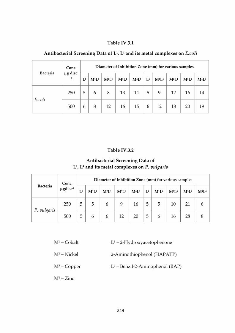
Results and Discussion
The Schiff bases and their metal chelates were evaluated for their
antibacterial activity against different bacterial strains at concentrations
ranging from 50g disc-1 to 500g disc-1. In testing the antibacterial activity of
these compound we used more than one test organism to increase the
chance of detecting the antibiotic potential of the tested materials. The
antibacterial screening data are presented in the Tables IV.3.1 – IV.3.4.
During the evaluation it was found that no inhibition zones were obtained
with test samples of concentrations ranging from 50g disc-1 to 100g disc-1.
But when the concentration was increased to 250gdisc-1 to 500gdisc-1 the
metal complexes and the ligands exhibit well defined inhibition zones. But it
is important to note that the complexes exhibit enhanced activity in contrast

245
to free ligands in all the screening tests. This shows that the metal chelates
are more antibacterial than the uncomplexed ligands.
In order to clarify any participating role of the solvent, DMSO in the
bacterial screening, separate studies were carried out with DMSO as solvent
control and it showed no meaningful activity against the bacterial strains
under study. The activity of metal chelates was also compared with a known
antibiotic, gentamycin (10g disc-1). The activities of the tested complexes
were found to be less than that of the standard antibacterial agent used. For
the standard drug, the exhibited inhibition zone diameter was in the range
of 15 – 25mm against all the bacterial strains used in this study.
The inhibition zone obtained for Co(II), Ni(II), Cu(II) and Zn(II)
chelates of HAPATP and BAP against E.coli was given in Table IV.3.1. In
both cases, the Cu(II) complexes showed considerable antibacterial activity
than other metal complexes in the given concentration of 250g disc-1 and
500g disc-1. Zn(II) and Ni(II) complexes with 500gdisc-1 also showed some
moderate activity than their corresponding ligands. Hence it can be
concluded that the metal ions in the complexes influence the antibacterial
activity. Chelation can considerably reduce the polarity of the metal ion
which in turn increases the lipophilic character of the chelate.
246
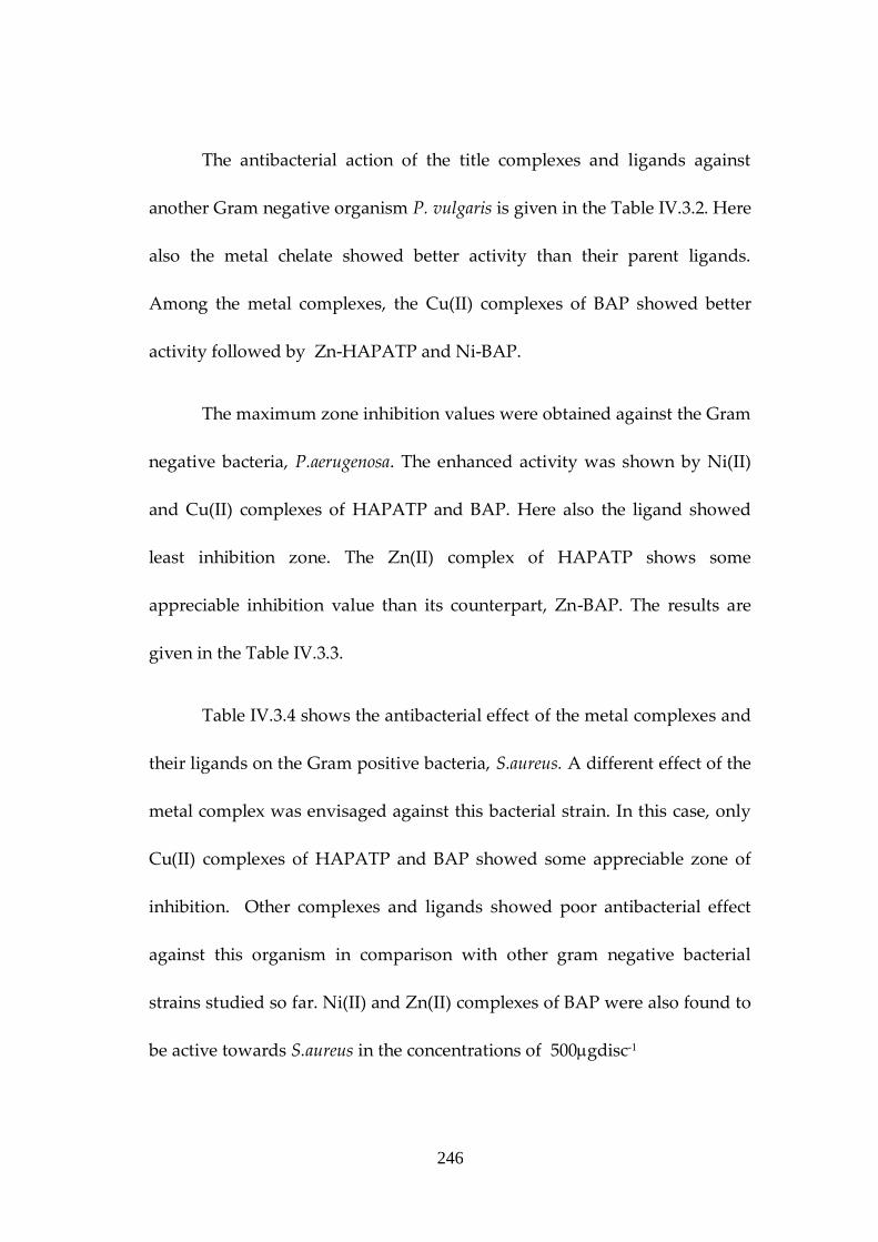
The antibacterial action of the title complexes and ligands against
another Gram negative organism P. vulgaris is given in the Table IV.3.2. Here
also the metal chelate showed better activity than their parent ligands.
Among the metal complexes, the Cu(II) complexes of BAP showed better
activity followed by Zn-HAPATP and Ni-BAP.
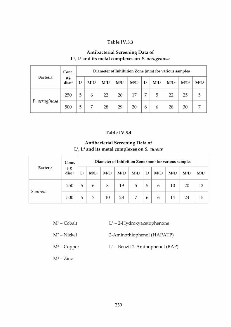
The maximum zone inhibition values were obtained against the Gram
negative bacteria, P.aerugenosa. The enhanced activity was shown by Ni(II)
and Cu(II) complexes of HAPATP and BAP. Here also the ligand showed
least inhibition zone. The Zn(II) complex of HAPATP shows some
appreciable inhibition value than its counterpart, Zn-BAP. The results are
given in the Table IV.3.3.
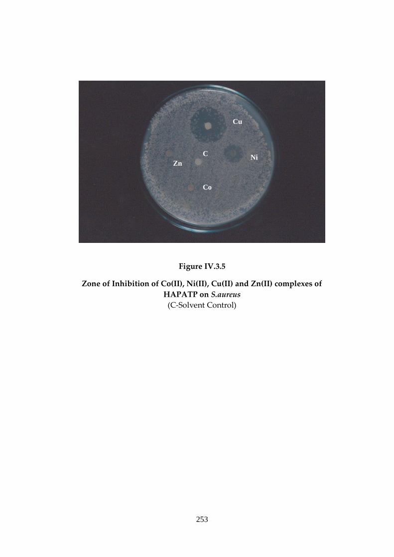
Table IV.3.4 shows the antibacterial effect of the metal complexes and
their ligands on the Gram positive bacteria, S.aureus. A different effect of the
metal complex was envisaged against this bacterial strain. In this case, only
Cu(II) complexes of HAPATP and BAP showed some appreciable zone of
inhibition. Other complexes and ligands showed poor antibacterial effect
against this organism in comparison with other gram negative bacterial
strains studied so far. Ni(II) and Zn(II) complexes of BAP were also found to
be active towards S.aureus in the concentrations of 500gdisc-1
247
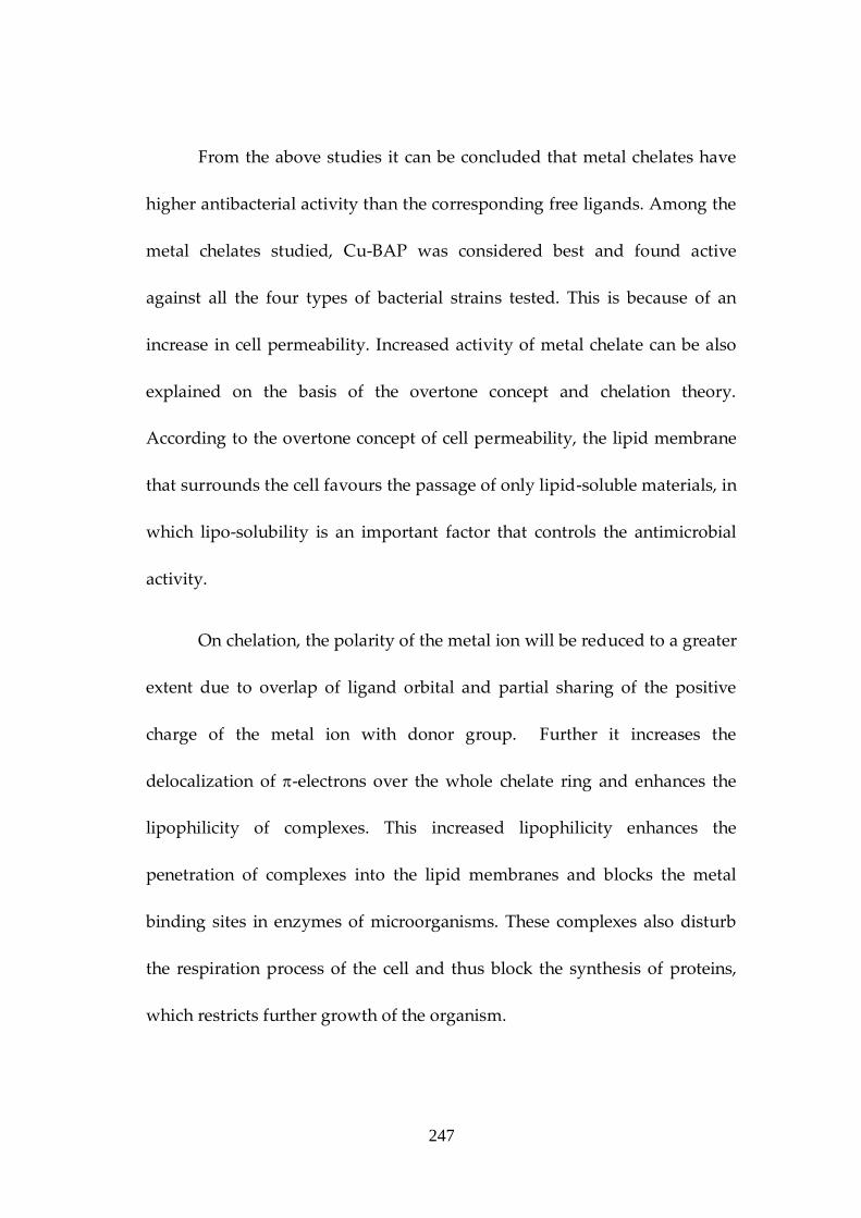
From the above studies it can be concluded that metal chelates have
higher antibacterial activity than the corresponding free ligands. Among the
metal chelates studied, Cu-BAP was considered best and found active
against all the four types of bacterial strains tested. This is because of an
increase in cell permeability. Increased activity of metal chelate can be also
explained on the basis of the overtone concept and chelation theory.
According to the overtone concept of cell permeability, the lipid membrane
that surrounds the cell favours the passage of only lipid-soluble materials, in
which lipo-solubility is an important factor that controls the antimicrobial
activity.
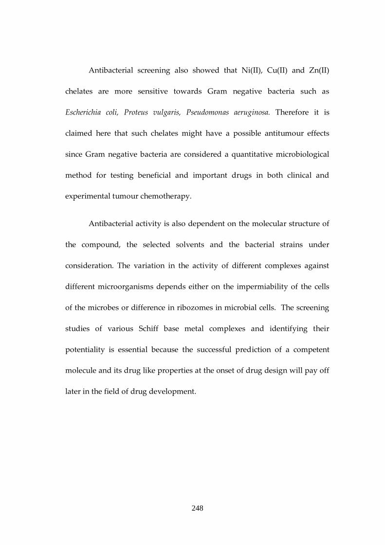
On chelation, the polarity of the metal ion will be reduced to a greater
extent due to overlap of ligand orbital and partial sharing of the positive
charge of the metal ion with donor group. Further it increases the
delocalization of -electrons over the whole chelate ring and enhances the
lipophilicity of complexes. This increased lipophilicity enhances the
penetration of complexes into the lipid membranes and blocks the metal
binding sites in enzymes of microorganisms. These complexes also disturb
the respiration process of the cell and thus block the synthesis of proteins,
which restricts further growth of the organism.
248
Antibacterial screening also showed that Ni(II), Cu(II) and Zn(II)
chelates are more sensitive towards Gram negative bacteria such as
Escherichia coli, Proteus vulgaris, Pseudomonas aeruginosa. Therefore it is
claimed here that such chelates might have a possible antitumour effects
since Gram negative bacteria are considered a quantitative microbiological
method for testing beneficial and important drugs in both clinical and
experimental tumour chemotherapy.
Antibacterial activity is also dependent on the molecular structure of
the compound, the selected solvents and the bacterial strains under
consideration. The variation in the activity of different complexes against
different microorganisms depends either on the impermiability of the cells
of the microbes or difference in ribozomes in microbial cells. The screening
studies of various Schiff base metal complexes and identifying their
potentiality is essential because the successful prediction of a competent
molecule and its drug like properties at the onset of drug design will pay off
later in the field of drug development.
249
Table IV.3.1
Antibacterial Screening Data of L1, L4 and its metal complexes on E.coli
Bacteria
Conc.
g disc-
1
Diameter of Inhibition Zone (mm) for various samples
L1 M1L1 M2L1 M3L1 M4L1 L4 M1L4 M2L4 M3L4 M4L4
E.coli
250 5 6 8 13 11 5 9 12 16 14
500 6 8 12 16 15 6 12 18 20 19
Table IV.3.2
Antibacterial Screening Data of
L1, L4 and its metal complexes on P. vulgaris
Bacteria Conc.
gdisc-1
Diameter of Inhibition Zone (mm) for various samples
L1 M1L1 M2L1 M3L1 M4L1 L4 M1L4 M2L4 M3L4 M4L4
P. vulgaris
250 5 5 6 9 16 5 5 10 21 6
500 5 6 6 12 20 5 6 16 28 8
M1 – Cobalt L1 – 2-Hydroxyacetophenone
M2 – Nickel 2-Aminothiophenol (HAPATP)
M3 – Copper L4 – Benzil-2-Aminophenol (BAP)
M4 – Zinc
250
Table IV.3.3
Antibacterial Screening Data of
L1, L4 and its metal complexes on P. aerugenosa
Bacteria
Conc.
g
disc-1
Diameter of Inhibition Zone (mm) for various samples
L1 M1L1 M2L1 M3L1 M4L1 L4 M1L4 M2L4 M3L4 M4L4
P. aeruginosa
250 5 6 22 26 17 7 5 22 25 5
500 5 7 28 29 20 8 6 28 30 7
Table IV.3.4
Antibacterial Screening Data of
L1, L4 and its metal complexes on S. aureus
Bacteria
Conc.
g
disc-1
Diameter of Inhibition Zone (mm) for various samples
L1 M1L1 M2L1 M3L1 M4L1 L4 M1L4 M2L4 M3L4 M4L4
S.aureus
250 5 6 8 19 5 5 6 10 20 12
500 5 7 10 23 7 6 6 14 24 15
M1 – Cobalt L1 – 2-Hydroxyacetophenone
M2 – Nickel 2-Aminothiophenol (HAPATP)
M3 – Copper L4 – Benzil-2-Aminophenol (BAP)
M4 – Zinc
251
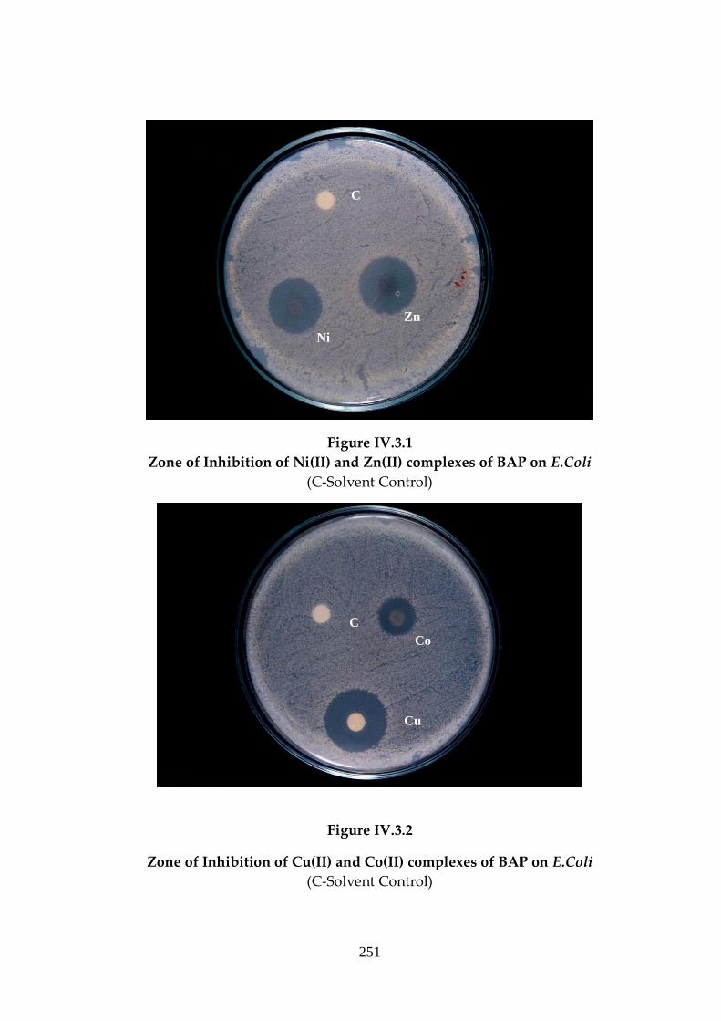
Figure IV.3.1
Zone of Inhibition of Ni(II) and Zn(II) complexes of BAP on E.Coli
(C-Solvent Control)
Figure IV.3.2
Zone of Inhibition of Cu(II) and Co(II) complexes of BAP on E.Coli
(C-Solvent Control)
Zn
Ni
C
Co
Cu
C
252
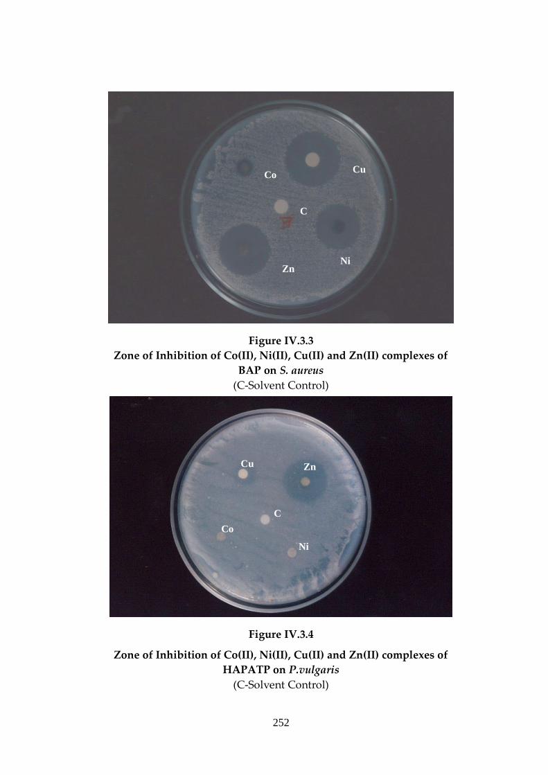
Figure IV.3.3
Zone of Inhibition of Co(II), Ni(II), Cu(II) and Zn(II) complexes of
BAP on S. aureus
(C-Solvent Control)
Figure IV.3.4
Zone of Inhibition of Co(II), Ni(II), Cu(II) and Zn(II) complexes of
HAPATP on P.vulgaris
(C-Solvent Control)
C
Cu
Zn Ni
Co
C
Co
Cu Zn
Ni
253
Figure IV.3.5
Zone of Inhibition of Co(II), Ni(II), Cu(II) and Zn(II) complexes of
HAPATP on S.aureus
(C-Solvent Control)
C
Cu
Ni Zn
Co

254
References
1. Alcomo E. Fundamentals of Microbiology 6th Ed: 745, 2001.
2. Ann D, Jacob G. Microbial Cell Factories, 23: 5, 2006.
3. Bernsteen T. Journal of Molecular Biology, 112: 535, 1977.
4. Bertini I, Gray HB. Bioinorganic chemsitry, Universal Science Books,
Mill Valley, 1994.
5. Boman HG, Immunological Reviews 173: 5-16, 2002.
6. Brull S, Coote P. International Journal of Food Microbiology, 50: 1-17.
1999.
7. Carpenter PL. Microbiology, Human Normal Microbiota: 329-330,
1977.
8. Chandrappa GT, Thimmaiah KN. Inorganic Chim Acta, 106: 81, 1985.
9. Chanmyia S, Shamim HM. Pakistan Journal of Biological Sciences, 7(3):
335-339, 2004.
10. Cheriyan M, Mohanan K. Asian Journal of Chemistry, 19(4): 283-288,
2007.
11. Chohan ZH and Humayun P. J Res (Sci), 4: 49, 1992.
12. Cotton SA, Chamic CR. 8: 129, 2005.
13. David WM, Martin PA, Victor WR, Daryl KG. Harpers Review of
Biochemistry, 20th Ed., Language Medical Publication, 1972.
14. Dhar SK. Metal Ions in Biological Systems, Plenum New York, 1973.
15. Donnenberg M. E.coli; Virulance mechanism of a versatile pathogen,
Science Encyclopaedia, Vol II, 2002.

255
16. Dubey RC, Maheswari DK. A Textbook of Microbiology, 551-552, 1999.
17. Dwyer FP. Chelating Agents and Metal Chelates, Academic Press, New
York, 1964.
18. Feng P, Weagant S. Enumeration of E.coli and the coliform bacteria,
Bacteriological analytical manual, 8th Ed., FDA/Center for food safety
and Applied nutrition, 2007.
19. Fibane AM, Williams DR. Principles of Bio inorganic chemistry, The
Chemical Society, London, 1977.
20. Glem LJ. Chemistry of Organic Medicinal Products, 4th Ed., John Willi &
Sons, New York, 1966
21. Gutkoshi SB, Wiest JM. 13: 26-29, 1996.
22. Hanzlik RP. Inorganic aspects of biological and organic chemistry.
Academic Press, New York, 1976.
23. Harwood CS, Greenberg EP. Megaroles of Microorganisms, Science,
1096, 1999.
24. Hoare RJ, Harisson PM. Metals in Biochemistry, Chapmen and Hall,
New York, 1980.
25. Horace DG. The safety foods, Connecticut Avi. Pub. Comp, 1982.
26. Hughes MN. The inorganic chemistry of biological processes, 2nd Ed.,
Willey Chichester, UK, 1981.
27. Iglewski BH. Pseudomonas in Baron’s Medical Microbiology, 4th Ed.,
Univ. of Texas, Medical Branch, 1996.
28. Isenberg DH, Essential Procedure for Clinical Microbiology. American
Society for Microbiology, Washington, 1998.
256
29. Kadrim MS, Gash B. Antibiotic sensitivity and resistance profile of
microorganism responsible for urinary tract infection observed in
Kashmir, India, Indian Journal of the Practising Doctors, Vol. I, No.1,
2004.
30. Katherine M Shea. Pediatrics., 114: 3, 2004.
31. Kell DB. Target Practice – Novel approaches to antimicrobial
chemotherapy TIBTECH 15: 334-336, 1997.
32. King EO, Ward MK. Two simple media for the demonstration of
Pyocyajmin and fluoresin, Laboratory Journal of Clinical Medicine, 44(2):
301-307, 1954.
33. Kurtoglu M, Ispir E. Asian Journal of Chemistry, 19(2): 1239-1245, 2007.
34. Levy S. The antibiotic paradox; How miracle drugs are destroying the
miracle, Plenum Publishers: 1-11, 1998.
35. Louie AY, Meate TJ. Metal complexes as enzyme inhibitors, Chemical
Rev, 99: 2711 -2734, 1999.
36. Mayer TJ, McCleverty JA. Comprehensive co-ordination chemistry,
Vol III. Pergamon Press, 2004.
37. Melson GA. Co-ordination chemistry of macro cyclic compounds,
Plenum Publishers, New York, 1989.
38. Monod J, Wyman J, Changeux P. Journal of Molecular Biology, 12: 965,
1988.
39. Muhammad I, Javed I, Shahid I, Nazia IT. Journal Biology, 31: 67-72,
2007.
40. Nasrin J, Saidul IM. Journal of Applied Sciences 7(4): 597-603, 2007.

257
41. Pasternak K, Sztanke K. Bioinorganic and Medicinal Chemistry,
ELSEVIER 14: 3635-3642, 2006.
42. Pearson H. The dark side of E.coli, Nature, 445-7123, 2007.
43. Pyatkin KD. Microbiology with vibrology and immunology, 121-122, 1987.
44. Rosenberg B, Van Camp L, Grimley EB, and Thomson AJ. J. Biol.
Chem., 242: 1347, 1967.
45. Ryan KJ, Ray CG. Sherris Medical Microbiology, 4th Ed., McGraw Hill,
2004.
46. Saidul IM, Akhter FM. Journal of Biological Sciences, 1(8): 711-713, 2001.
47. Saidul IM, Akhter FM. Pakistan Journal of Biological Sciences, 5(3): 335-
337, 2002.
48. Saidul IM, Belayet HM. Journal of Medical Sciences, 3(4): 289-293, 2003.
49. Schroedar A. Elements in Living Systems, Plenum Publishers, New
York, 1975.
50. Sinigaglia M, Delnobile MA. Journal of Diary Science., 2007.
51. Tkaczynski T, Janocha R. Acta Pol. Pharm. Drug Res. 52: 39, 1995.
52. Villanova PA. National Committee for clinical and Laboratory
Standards, Methods for dilution antimicrobial susceptibility tests for
bacteria, NCCLS Document M7-A4, 1997.
53. Walsh C. Enabling the science of life, Nature, 409: 226-231, 2001.
54. Wayne PA.NCCLS, DISC-Diffusion Eleventh International
Supplement, NCCLS Document, M100-S11, 2001.
55. Zgoda JR, Porter JR. Pharm. Biol. 39: 221, 2001.