Embed Size (px)
Citation preview
RESEARCH Open Access
Biological effects of carbon blacknanoparticles are changed by surfacecoating with polycyclic aromatichydrocarbonsKarina Lindner1, Michael Ströbele2, Sandra Schlick3, Sina Webering3, André Jenckel4, Johannes Kopf5, Olga Danov5,Katherina Sewald5, Christian Buj6, Otto Creutzenberg5, Thomas Tillmann5, Gerhard Pohlmann5, Heinrich Ernst5,Christina Ziemann5, Gereon Hüttmann6, Holger Heine4, Henning Bockhorn2, Tanja Hansen5†, Peter König1*†
and Heinz Fehrenbach3†
Abstract
Background: Carbon black nanoparticles (CBNP) are mainly composed of carbon, with a small amount of otherelements (including hydrogen and oxygen). The toxicity of CBNP has been attributed to their large surface area,and through adsorbing intrinsically toxic substances, such as polycyclic aromatic hydrocarbons (PAH). It is not clearwhether a PAH surface coating changes the toxicological properties of CBNP by influencing their physicochemicalproperties, through the specific toxicity of the surface-bound PAH, or by a combination of both.
Methods: Printex®90 (P90) was used as CBNP; the comparators were P90 coated with either benzo[a]pyrene (BaP)or 9-nitroanthracene (9NA), and soot from acetylene combustion that bears various PAHs on the surface (AS-PAH).Oxidative stress and IL-8/KC mRNA expression were determined in A549 and bronchial epithelial cells (16HBE14o-,Calu-3), mouse intrapulmonary airways and tracheal epithelial cells. Overall toxicity was tested in a rat inhalationstudy according to Organization for Economic Co-operation and Development (OECD) criteria. Effects on cytochromemonooxygenase (Cyp) mRNA expression, cell viability and mucociliary clearance were determined in acute exposuremodels using explanted murine trachea.
Results: All particles had similar primary particle size, shape, hydrodynamic diameter and ζ-potential. All PAH-containingparticles had a comparable specific surface area that was approximately one third that of P90. AS-PAH contained amixture of PAH with expected higher toxicity than BaP or 9NA. PAH-coating reduced some effects of P90 such as IL-8mRNA expression and oxidative stress in A549 cells, granulocyte influx in the in vivo OECD experiment, andagglomeration of P90 and mucus release in the murine trachea ex vivo. Furthermore, P90-BaP decreased particle transportspeed compared to P90 at 10 μg/ml. In contrast, PAH-coating induced IL-8 mRNA expression in bronchial epithelial celllines, and Cyp mRNA expression and apoptosis in tracheal epithelial cells. In line with the higher toxicity compared to P90-BaP and P90-9NA, AS-PAH had the strongest biological effects both ex vivo and in vivo.(Continued on next page)
* Correspondence: [email protected]†Equal contributors1Institut für Anatomie, Zentrum für medizinische Struktur- und Zellbiologie,Universität zu Lübeck (UzL), Airway Research Center North (ARCN), GermanCenter for Lung Research (DZL), Ratzeburger Allee 160, 23562 Lübeck,GermanyFull list of author information is available at the end of the article
© The Author(s). 2017 Open Access This article is distributed under the terms of the Creative Commons Attribution 4.0International License (http://creativecommons.org/licenses/by/4.0/), which permits unrestricted use, distribution, andreproduction in any medium, provided you give appropriate credit to the original author(s) and the source, provide a link tothe Creative Commons license, and indicate if changes were made. The Creative Commons Public Domain Dedication waiver(http://creativecommons.org/publicdomain/zero/1.0/) applies to the data made available in this article, unless otherwise stated.
Lindner et al. Particle and Fibre Toxicology (2017) 14:8 DOI 10.1186/s12989-017-0189-1
(Continued from previous page)
Conclusions: Our results demonstrate that the biological effect of CBNP is determined by a combination of specific surfacearea and surface-bound PAH, and varies in different target cells.
Keywords: Carbon black nanoparticles, Polycyclic aromatic hydrocarbons, Airway epithelial cells, Physicochemicalcharacteristics, Mucociliary clearance, Cytotoxicity
BackgroundThe toxicity of nanoparticles is thought to be deter-mined by their surface reactivity, influenced in turn bytheir chemical composition, physical properties andsurface chemistry [1–8]. Carbon black nanoparticles(CBNP) are composed mainly of carbon with a smallamount of other elements (including hydrogen and oxy-gen), and in themselves are nontoxic. However, CBNPare potentially harmful since their large surface area caninteract with biological systems [1, 9–14]. Furthermore,substances such as polycyclic aromatic hydrocarbons(PAHs) that are intrinsically toxic can be adsorbed dur-ing CBNP synthesis [15]. The theory that the toxicity ofCBNP can be increased by other substances has beenvalidated by the application of metal ions, which in-creased the toxicity due to autophagy and lysosomal dys-function [16]. However, it is still not clear how directbinding of chemical add-ons to CBNP modifies theirinteraction with biological systems [5, 17–19]. Severaloutcomes are theoretically possible. First, binding of che-micals to the particle surface can modify the surfaceproperties of CBNP, reducing the surface area, therebypotentially reducing toxicological effects [1]. Second, thetoxicity of the bound substances can determine the over-all toxicity of the resulting particles [20]. Third, themodification could lead to a new particle type, whosebiological activity might be a combination of changedphysicochemical properties and the toxicity of the boundsubstances.A common surface modification of CBNP is the adsorp-
tion of PAHs, occurring during incomplete combustion[21]. PAHs are thought to be mutagenic and carcinogenic,generally after toxification by cytochrome P450 enzymes(Cyp), and therefore are regarded as lung tumor inducingsubstances, although with only very limited acute toxicity[15, 22, 23]. Despite this limited acute toxicity, their effecton biological systems can be easily monitored by theirpotential to induce specific Cyp enzymes.Compared with unmodified CBNP, surface modification
of CBNP with PAHs has not shown increased tumor in-duction either following long-term exposure [24] or in celllines in terms of cytokine release, induction of single anddouble strand breaks, cell cycle arrest, or phosphorylationof p53 or histone [25–27]. This suggests that PAHs havelimited toxic effects when bound to CBNP, supporting thetheory that they are not biologically available in this state
[28] and that surface modification has a minor effect onthe overall toxicity of CBNP.However, diesel exhaust particles that contain PAHs
are associated with an increased risk of asthma, andexperiments involving exposure of a bronchial cellline to ultrafine particles implied a role of PAH intheir toxic effects [29–31]. Furthermore, removal ofPAHs from these exhaust particles resulted in reducedcarcinogenic potential [24]. These data suggests thatthe effect of PAHs on the behavior of CBNP in bio-logical systems is more complex.Since inhalation is the predominant route of exposure
to CBNP, we tested the toxicity of particles in differenttest systems including cell cultures, ex vivo studies onexcised airways, and an in vivo inhalation studyconducted according to Organization for Economic Co--operation and Development (OECD) criteria [32].Initially, we tested the CBNP in high-throughput cellculture systems that can predict the toxicity of particles,and which therefore allowed us to preselect CBNP for invivo testing. We then analyzed the in vivo effects of theselected CBNP in inhalation studies, and then finallytested the CBNP in ex vivo airway preparations to deter-mine their impact on ciliary beat frequency, particletransport speed and mucus appearance. In every testsystem we analyzed parameters that indicate CBNP-induced oxidative stress and IL-8 response.This allowed us to compare specific toxicological
effects in each system to gain a more comprehensiveinsight into the cellular effects of the particles.
MethodsTo understand how surface modification can changethe interaction of CBNP with biological systems weused PAHs to modify the surface of a toxicologicallywell-defined CBNP, Printex®90 (P90). This is charac-terized by a high surface area and has been widelyused in toxicological studies, resulting in only minortoxic effects [1, 33, 34].For modification of the P90 surface, we used ben-
zo[a]pyrene (BaP) and 9-nitroanthracene (9NA). BaPwas chosen because of the well characterized toxicityof its metabolites, which are known to induce ROSand DNA adducts [23, 35–38]. BaP is known to in-duce Cyp1A1 and 1B1, which then metabolize BaP totoxic metabolites; this therefore allows monitoring of
Lindner et al. Particle and Fibre Toxicology (2017) 14:8 Page 2 of 17

BaP activity and its biological effect [39, 40]. In con-trast, 9NA is a PAH that occurs during combustion,and is regarded as a low toxicity PAH, as predictedby the Ames test and human cell mutagenicity assay[41–43]. However, due to its nitro group other toxicmechanisms can occur induced by intermediatesresulting from reduction of the nitro group [41]. Ascoating of a particle does not necessarily representthe situation found in nanoparticles that acquire PAHduring synthesis, we also generated CBNP by acetyl-ene combustion [44]. The resulting acetylene soot(AS) had a mixture of PAHs on the surface (AS-PAH). Inthe suspensions we used, AS-PAH had a slightly largerspecific surface area, but similar aggregate size and ζ-po-tential compared to PAH-coated P90, The physicochemi-cal parameters of the different particles were evaluated bya variety of analytic test methods (see Tables 1, 2 in theResults section and Additional files 1, 2).
Particle synthesis and characterizationModification of CBNP80 g of unmodified P90 (Evonik Carbon Black GmbH,Frankfurt, Germany) together with 20 g of BaP (ThermoFisher Scientific-Alfa Aesar, Heysham, Lancashire, UK)or 9NA (Merck Schuchardt OHG, Hohenbrunn, Germany)were suspended in 100 ml dimethyl ether and incubated inan ultrasonic bath at room temperature for 15 min. Aftersubsequent vacuum desiccation, the coated P90 nano-particles were stored at room temperature.
Gas phase synthesis of AS-PAHA flat flame burner in a low pressure combustion cham-ber with acetylene as the fuel was used for the synthesisof AS-PAH. The experiments were carried out with acarbon to oxygen ratio of 1.1 to 1. The laminar massflow of acetylene was set via mass flow controllers(Brooks 5850E; Brooks Instruments, Hatfield, PA, USA)on 88 ln/h with the oxygen mass flow adjusted to 80 ln/h.Temperature and pressure were monitored by athermocouple and a pressure sensor, with the pres-sure set to 70 mbar during synthesis. The AS-PAHwas collected for 60 min on a filter (Gore MembraneFilter Bags) between the combustion chamber and therotary vane pump (TRIVAC D 65 B; Oerlikon LeyboldVakuum, Cologne, Germany).
Particle size distributionThe particle size distributions were measured by scan-ning electron microscopy (SEM; Leo 1530; LEO ElectronMicroscopy Inc., Thornwood, NY, USA) and transmis-sion electron microscopy (TEM; CM200 FEG/ST,Philips, Amsterdam, Netherlands). The PAHs on theparticle surface had to be extracted with toluene prior toelectron microscopy, as otherwise they would be cracked
by the beam and the particles would grow. RemainingCBNP were then suspended with acetone in an ultra-sonic bath, and the suspension was applied thinly usingan ultrasonic nebulizer, either to a silicon wafer (forSEM) or to a 400 mesh cooper grid (Ted Pella, Inc.,Redding, CA, USA) (for TEM). The diameter of 200 par-ticles of 5 images was determined with the programImageJ. A log normal curve was fitted on the particlesize distribution to get the average particle diameter.
Thermal analysisThe thermal release of coated PAHs from P90 and ofvolatile substances from AS-PAH was investigated usinga thermogravimetric analyzer (DuPont 951; DuPontCompany, Wilmington, DE, USA) connected to a quad-rupole mass spectrometer (QMG 420; Balzers AG, Bal-zers, Liechtenstein) through a differentially pumped inletsystem. The transfer line between the thermobalance andthe mass spectrometer was heated up to 400 °C to preventcondensation of the pyrolysis products. The experimentswere performed with sample sizes of 5–10 mg at a heatingrate of 10 °C/min up to a final temperature of 800 °C at ahelium flow rate of 100 ml/min. Each sample was mea-sured at least three times.
Identification of PAHs on AS-PAH surfaceTo characterize the PAHs on the particle surfaces,CBNP samples were heated under dynamic conditions.A sample of 100 mg was heated up in an oven in 10 °C/min increments to vaporize the PAHs, with the volatilePAHs collected in a cold trap. The condensed productswere dissolved in 5 ml toluene, and were characterizedby a standard gas chromatograph-mass spectrometer(GC/MS-QP 2010SE; Shimadzu Corporation, Tokyo,Japan). The chromatographic parameters for GC/MSwere: Phenomenex zb-5msi column (column length:30 m; I.D.: 0.25 mm; film thickness 0.25 mm); injectortemperature 250 °C; oven temperature program: 65 °C at5 min constant, 10 °C min to 350 °C at 10 min constant.
Specific surface areaThe surface area was determined by Brunauer, Emmettand Teller (BET) measurement using a BelSorp II device(BelJapan, Inc., Osaka, Japan), using the BelSorp analysisprogram.
Particle size and ζ-potential in suspensionSuspensions were prepared with 10 mg of sample plus100 ml of double-deionized water. To keep the suspen-sion stable, 500 mg of bovine serum albumin (BSA)(AppliChemInc., Maryland Heights, MO, USA) wasadded. The suspensions were treated with an ultrasonichomogenizer (Sonoplus, Rod: VS 70 D; Bandelin elec-tronic GmbH & Co. KG, Berlin, Germany) for 40 min in
Lindner et al. Particle and Fibre Toxicology (2017) 14:8 Page 3 of 17
pulse mode, changing 20 times between 30 s ultrasonicand 90 s resting interval. To avoid thermal denaturationof BSA, the suspensions were cooled in a beaker con-taining ice water during treatment. The particle sizemeasurements were carried out as soon as thesuspension had reached room temperature using aMalvern ZetaSizer Nano-ZS (Malvern InstrumentsLtd, Worcestershire, UK). Particle size was quantifiedby dynamic light scattering, and ζ-potential was mea-sured by laser Doppler microelectrophoresis using theSmoluchowski model. The index of refraction and theabsorption were preset at 2.0 in the measurementsoftware (DTS Nano; Malvern Instruments Ltd,Worcestershire, UK). The cuvettes to measure theparticle size distribution and the ζ-potential werewashed using double-deionized water and ethanol(70%) to remove potential impurities. Subsequently,the cuvettes were rinsed using approximately 2 ml ofthe sample, and were then filled with the sample andplaced in the Zetasizer Nano-ZS. The suspensionswere characterized in triplicates, and the means andstandard derivations were calculated.
Endotoxin testsCBNP suspensions were tested for endotoxins using theCROMO-LAL assay (Associates of Cape Cod Inc., EastFalmouth, MA, USA) according to the manufacturer’sprotocol. The limit of detection was 0.005 EU/ml (com-parable to 0.5 pg/ml endotoxin of E. coli Type 055:B5).The suspensions were only used for experiments if noendotoxins were detected.
Experiments with human pulmonary cell linesCell cultureWe used A549 cells (DSMZ, Braunschweig, Germany),16HBE14o- cells (University California, San Francisco,USA; described in [45]) and Calu-3 cells (AmericanType Culture Collection, Manassas, VA, USA). The epi-thelial cells were cultured in Dulbecco’s modified eagle’smedium (DMEM) with 10% fetal calf serum (FCS) and0.1% gentamicin for ROS and transepithelial electricalresistance (TEER) measurements. For mRNA expressionanalysis, A549 cells were cultured in DMEM (FisherScientific GmbH, Schwerte, Germany) with 10% FCSand 0.1% gentamicin, 16HBE14o- cells were cultured inDMEM (Biochrom AG, Berlin, Germany) with 10% FCS(PAA Laboratories GmbH, Cölbe, Germany) 1% L-glu-tamine (Gibco™, Life Technologies GmbH, Darmstadt,Germany) and 1% penicillin/streptomycin (Sigma AldrichChemie GmbH, Taufkirchen, Germany), and Calu-3 cellswere cultured in RPMI 1640 containing stable glutaminesupplemented with 10% FCS, 100 U/ml penicillin and100 μg/ml streptomycin (all from Biochrom AG, Berlin,
Germany). Cells were kept in a 37 °C humidified atmos-phere containing 5% CO2.
Flow cytometric measurements of reactive oxygenspecies (ROS)Cells were seeded in T25 flasks (PAA LaboratoriesGmbH, Cölbe, Germany), at 0.6 × 10⁶ for A549 cells andat 1 × 10⁶ for 16HBE14o- cells. Subconfluent cultureswere exposed to CBNP at a concentration of 10 μg/mlor 50 μg/ml for 24 h. The culture medium was subse-quently removed and the cell monolayers were washedtwice with 5 ml pre-warmed phosphate buffered saline(PBS). The fluorescent dye dichloro-dihydro-fluoresceindiacetate (DCFH-DA) was applied to the cells in 5 mlfresh medium, with the resulting mixture incubated for30 min at 37 °C. The culture medium was then removedand the cells were washed twice with 5 ml pre-warmedPBS and harvested by trypsinization. Flow cytometrymeasurements were done with a FACScan cytometer(Becton Dickinson Biosciences, San Jose, CA, USA)equipped with a 488 nm argon laser, with CELLQuest™software used for data acquisition. For each sample,10,000 events were analyzed and fluorescence emissionof dichlorofluorescein (DCF) was examined at 530 nm.
Quantitative RT-PCRA549 cells were seeded in T25 flasks at 0.6 × 10⁶. Calu-3cells and 16HBE14o- cells were seeded in 12-well plates(Costar, Corning, NY, USA) at 5 × 10⁵ cells and 1.6 × 10⁵
cells, respectively. After reaching a confluency of ≥90%,culture medium was exchanged and cells were treatedwith CBNP at 10 μg/ml, 50 μg/ml or medium/BSA alonefor 24 h. Subsequently, cells were washed twice withpre-warmed medium or PBS to remove particles, andtotal RNA was extracted from Calu-3 cells using aNucleoSpin RNA Kit (Macherey-Nagel, Düren, Germany)and from A549 and 16HBE14o- cells using a RNeasy Kit(Quiagen GmbH, Hilden, Germany) according to themanufacturers’ instructions.Reverse transcription was performed for RNA of A549
cells with the Omniscript Kit (Quiagen GmbH, Hilden,Germany), of 16HBE14o- cells using MaximaFirstStrandcDNA synthesis (Fermentas/Life Technologies GmbH,Darmstadt, Germany), and of Calu-3 cells usingOligo(dT)12–18 and Superscript III reverse transcriptase(Thermo Fisher Scientific, Waltham, USA), all accordingto the manufacturers’ protocols.To quantify IL-8 and hypoxanthine-guanine phosphor-
ibosyltransferase (HPRT) mRNA levels in A549 cellpreparations, we used Viia™7 (Life Technologies GmbH,Darmstadt, Germany); for preparations of 16HBE14o-and Calu-3 cells, we used LightCycler 480 SYBR green IMaster (Roche Applied Science, Mannheim, Germany).
Lindner et al. Particle and Fibre Toxicology (2017) 14:8 Page 4 of 17
The sequences of used IL-8 primers and housekeepinggene HRPT1 primers are shown in Additional file 3. Thehousekeeping gene was evaluated with GenEx software(MultiD Analyses AB, Gothenburg, Sweden) [46]. Thequality of amplificates was tested by melting curve ana-lysis. The n-fold change was calculated for A549 cellswith the program GenEx (GenEx Professional 5.4.3 Soft-ware), and by normalization of relative expression datato medium control data for 16HBE14o- and Calu-3 cells.
Measurement of the transepithelial electrical resistance(TEER)A total of 2 × 10⁵ Calu-3 cells were seeded per well onTranswell® filter inserts (polyester, 12-well plates, 12 mmdiameter, 0.4 μm pore size; Corning Costar, Bodenheim,Germany) and cultivated for 9 days. On day 10, the cellmonolayers were exposed to CBNP at 10 μg/ml or50 μg/ml for 24 h. The culture medium was changedand the cells were incubated for at least 15 min at 37 °C,before TEER was measured using an EVOM volt-ohm-meter (World Precision Instruments, Sarasota, Fl, USA).
Statistical analysisDifferences between untreated controls and treated cul-tures were considered statistically significant at p < 0.05.Flow cytometric and TEER data were analyzed by two-sided Student’s t-test for unpaired values using MicrosoftExcel. Relative mRNA expression results were analyzedby Mann Whitney U test.
Nose-only inhalation study in ratsAnimalsMale Wistar rats (strain Crl:WI (Han)) were purchasedfrom Charles River Deutschland (Sulzfeld, Germany).The age of the animals was 10 to 11 weeks and theweight 285 ± 24 g (mean ± SD) at the onset of exposure.Rats were exposed to the test item by nose-only inhal-ation. For 3 weeks prior to exposure, animals weretrained to become accustomed to nose-only tubes. TheOECD inhalation study on rats was approved by theNiedersächsisches Landesamt für Verbraucherschutz undLebensmittelsicherheit (33.14 42502-04013/1199).
Study design and dosing schemeThe CBNP were administered to the test animals bynose-only inhalation. P90, P90-BaP and AS-PAH weretested; P90-9NA was not administered, given the previ-ously described low toxicity [41–43] and the in vitrotests in this study. A nominal aerosol concentration of6 mg/m3 was used, with exposure of 6 h/day for twoweeks (no exposure on weekends) according to theOECD criteria for acute exposure [32]. CBNP-related ef-fects on innate immune cells in the bronchoalveolar lav-age (BAL) and histopathological changes in lung tissue
were analyzed on day 1 and day 14 after cessation of ex-posure. Potassium bromate (KBrO3) treated animals(250 mg/kg in physiological saline, intraperitoneal)served as positive controls for Comet-assay. The animalswere treated with KBrO3 on day 1 and day 14. BAL wascollected and analyzed 3 h after application.
Bronchoalveolar lavage and leukocyte determinationBAL was performed in five rats per group after the endof exposure (day 1) and following a 14-day recoveryperiod. Following preparation, lungs were lavaged withsaline using two lavages of 5 ml each. The lavage fluidwas collected in calibrated tubes and the harvested vol-ume was recorded. Leukocyte concentration was deter-mined using a counting chamber, and two cytoslideswere prepared with a cytocentrifuge (Shandon Co.,Frankfurt, Germany) for differential cell counts (macro-phages, granulocytes, lymphocytes). The slides were airdried, and the cells were stained with Diffquick solutions(Medion Grifols Diagnostics AG, Düdingen, Switzerland)and counted. The results are presented as percentage ofthe total number of BAL cells.
Statistical analysisDifferences between BAL analysis groups were consid-ered statistically significant at p < 0.05. Data were ana-lyzed using analysis of variance. If the group meansdiffered significantly by the analysis of variance themeans of the treated groups were compared with themeans of the control groups using Dunnett’s test.
Experiments with explanted mouse tracheaParticle suspension in cell culture mediumFor the ex vivo studies we used CBNP concentrations of10 and 30 μg/ml. The concentration of 30 μg/ml wascalculated by surface correction of the in vitroconcentration.The CBNP suspensions were diluted in DMEM (Gibco™,
Life Technologies GmbH, Darmstadt, Germany) with20 mM HEPES, 1% penicillin/streptomycin (Sigma AldrichChemie GmbH, Taufkirchen, Germany) and 2 mM L-glu-tamine (Life Technologies GmbH, Darmstadt, Germany)for incubation of tracheae.
AnimalsWe used 8 to 12 week old female Balb/c mice(Charles River Laboratories, Sulzfeld, Germany). Micewere euthanized with an overdose of isoflurane forexplantation of the trachea. The ex vivo studies onmouse tracheae were approved by the Ministeriumfür Landwirtschaft, Umwelt und ländliche Räume desLandes Schleswig-Holstein.
Lindner et al. Particle and Fibre Toxicology (2017) 14:8 Page 5 of 17
Preparation and culture conditions of explanted tracheaTracheae were explanted and transferred to culturedishes coated with a thin layer of Sylgard polymer andfilled with medium. Each trachea sample was fixed onthe bottom of a well with an insect needle, covered with1 ml of medium with or without suspended CBNP andincubated at 37 °C for 24 h.
Qualitative RT-PCRTo examine differences in mRNA expression levels, tra-cheae were cut into two pieces. One piece was incubatedfor 24 h with particle suspension; the other piece was in-cubated in medium only, serving as negative control.The epithelium was then removed using a sterile swab(Collection Swab H268A; Copan diagnostic, Inc., Murrieta,CA, USA). This isolation method was previously describedin [47]. The RNA was isolated with innuPREP RNA isola-tion Kit (Analytic Jena AG, Jena, Germany) according tothe manufacturer’s protocols. Any remaining DNA wasenzymatically degraded using Amplification Grade DNaseI kit AMPD-1 (Sigma-Aldrich Chemie GmbH, Steinheim,Germany). The quality and quantity of isolated mRNAwas analyzed with a NanoDrop spectrophotometer(Thermo Fisher Scientific, Waltham, MA, USA). ThemRNA was subsequently transcribed to cDNA usingRevertAid H Minus Reverse Transcriptase (FisherScientific-Germany GmbH, Schwerte, Germany) mixed intranscription buffer with dNTPs (0.6 mM) (FisherScientific-Germany GmbH, Schwerte, Germany), DTT(10 mM) (Life technologies GmbH, Darmstadt, Germany)and random hexamer primers (17.5 ng/μl) (Biomers.netGmbH, Ulm, Germany).Real time RT-PCR was performed in TaqMan Universal
PCR Master Mix (Life technologies GmbH, Darmstadt,Germany), with probes tagged with reporter 6-FAM andquencher Tamra. The TaqMan probes and primers usedfor quantitative real time RT-PCR are shown inAdditional file 3. The PCR program was: one cycle at95 °C for 10 min followed by 50 cycles at 95 °C for 45 sand at 60 °C for 1 min. Changes in the transcript levelswere determined by calculating the difference in cyclethreshold to the respective negative control with the ΔΔCt
method and represented as n-fold expression (2 −ΔΔCt).
Apoptosis and necrosis assaysTracheae were explanted, incubated in pre-warmedmedium, opened and cut in four equal parts. One partwas cultured in medium alone, two parts were incubatedfor 24 h with CBNP suspensions of 10 μg/ml and30 μg/ml, respectively, and the remaining parts wereincubated for 24 h with 1 μM staurosporine (RocheDiagnostics GmbH, Mannheim, Germany) or 1 mMparaquat (Sigma-Aldrich GmbH, Seelze, Germany), re-spectively as positive controls for the staining procedure.
For the apoptosis analysis, whole mount samples of tra-cheae were fixed with phosphate buffered 4% paraformal-dehyde (pH 7.4), followed by washing with PBS (pH 7.4)and permeabilization with ice cold acetone. The speci-mens were then incubated overnight at room temperaturewith rabbit anti-cleaved caspase-3 antibody (#9661, dilu-tion 1:300, Cell Signaling Technology Inc., Danvers, MA,USA). The primary antibody was detected usingAlexa555-conjugated donkey anti-rabbit IgG (dilution1:800, Life Technologies GmbH, Darmstadt, Germany).For membrane damage analysis, ethidium homodimer-1
(1 μg/ml, Life Technologies GmbH, Darmstadt, Germany)in medium was added to the trachea preparation, and wasthen incubated for 30 min at 37 °C. The samples werefixed with 4% paraformaldehyde (pH 7.4), washed withPBS (pH 7.4) and permeabilized with acetone.To identify the epithelial layer, ciliated cells were la-
beled with a mouse monoclonal antibody against acety-lated α-tubulin (Clone 6-11B-1, Sigma-Aldrich GmbH,Steinheim, Germany) that was directly labeled withAtto488 (Lightning-Link™Atto488 conjugation kit; InnovaBioscience Ltd., Cambridge, UK); cell nuclei were visual-ized using Hoechst 33258 (0.1 μg/ml; Sigma-Aldrich,Steinheim, Germany) (Additional file 4).Apoptosis and cell membrane damage were analyzed
using a confocal laser scanning microscope (LSM 510Meta; Carl Zeiss MicroImaging GmbH, Göttingen,Germany) equipped with a Plan-Apochromat 20×/0.75or C-Apochromat 40×/1.2 W objective. The total num-ber of apoptotic and necrotic epithelial cells was countedin the complete whole mount preparation.
Ciliary beat frequency and cilia-driven particle transportTracheae were transferred to a Delta T4 Culture dish(Bioptechs Inc., Butler, PA, USA), the bottom of whichwas covered with sylgard polymer. Samples were fixedwith insect needles with the epithelium facing upwards,and were then covered with 2 ml of HEPES-Ringer solu-tion. The temperature of HEPES-Ringer solution wasmaintained at 30 °C using a Bioptechs Delta T4 CultureDish Controller (Bioptechs Inc., Butler, PA, USA), andthe epithelial surface was imaged using a Zeiss Axioskop2 FS fixed stage microscope equipped with Achroplan40×/0.80 W or 20×/0.50 W objectives (Carl Zeiss Micro-Imaging GmbH, Göttingen, Germany).Polystyrene particles (4.5 μm) were added to the
HEPES-Ringer solution with a concentration of 2 × 10⁶
particles/ml. The transport of particles was recordedwith a SMX-150 M camera (EHD Imaging GmbH,Damme, Germany). Movies (each with 200 images) ofpolystyrene particle transport were recorded from atleast six different areas for each trachea at 12 Hz framerate and a resolution of 1280 × 1024 pixels. Particle
Lindner et al. Particle and Fibre Toxicology (2017) 14:8 Page 6 of 17
transport speed was determined by tracking individualparticles and manually checking each recorded trackusing Image-Pro® Plus 6.0 (Medium Cybernetics, Inc.,Bethesda, MD, USA). The mean particle transport speedwas calculated for each experiment. Movies (each with1000 images) of cilia motion were recorded at 100 Hzframe rate and 640 × 480 pixels). The ciliary beatfrequency was determined by Fourier transformation ofgrey level changes over time using an in-house devel-oped software written in Mathlab (Mathworks, Natick,MA, USA). For visualization of ciliary beat frequency,the dominant frequency, i.e. the frequency with thehighest amplitude, was identified for every pixel in themovie and color-coded. Movies were recorded from atleast six different areas for each trachea. The ciliary beatfrequency of a minimum of 10 ciliated cells was measuredin every movie and the mean frequency from these cellswas calculated. To increase ciliary beat frequency orcilia-driven particle transport, ATP (10 μM) was appliedas positive control.
Statistical analysisAll ex vivo experiments were carried out at least threetimes. Quantitative RT-PCR data are presented as mean± SEM. The data sets were analyzed using paired-sampleWilcoxon signed-rank test in GraphPad Prism 5 (GraphPadSoftware, Inc., La Jolla, CA, USA). P values < 0.05 wereconsidered statistically significant.
Mucus staining in tracheal whole mountsTracheae were incubated in 1 ml HEPES-Ringer solutionat 30 °C; 1 μl of a 1:1 mix of wheat germ agglutinin(WGA) and Ulex europaeus agglutinin (UEA-1) wasadded (each 1 μg/ml). WGA stains N-acetyl-D-glucosa-mine and sialic acid residues, whereas UEA-1 has anaffinity for L-fucose, all of which are carbohydrateresidues present in mucins [48, 49]. In addition, WGAlabels ciliated cells [50]. The staining was imaged withan AxioMR camera and an Achroplan 40×/0.8 W objectivewith a resolution of 1388 × 1040 pixels.Further information on the determination of cytokine
levels and cell viability in vitro, CBNP aerosol generation
and exposure of rats, oxidative Comet-Assay, histopath-ology of the rat lungs, particle suspensions in cell culturemedium, preparation and culture conditions of intrapul-monary airways, qualitative RT-PCR, macroscopic analysis,scanning electron microscopy and individual statistics areprovided in Additional file 5.
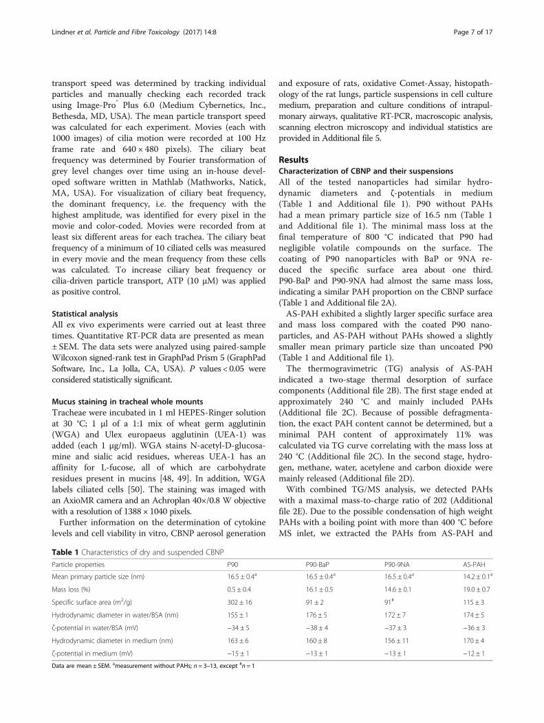
ResultsCharacterization of CBNP and their suspensionsAll of the tested nanoparticles had similar hydro-dynamic diameters and ζ-potentials in medium(Table 1 and Additional file 1). P90 without PAHshad a mean primary particle size of 16.5 nm (Table 1and Additional file 1). The minimal mass loss at thefinal temperature of 800 °C indicated that P90 hadnegligible volatile compounds on the surface. Thecoating of P90 nanoparticles with BaP or 9NA re-duced the specific surface area about one third.P90-BaP and P90-9NA had almost the same mass loss,indicating a similar PAH proportion on the CBNP surface(Table 1 and Additional file 2A).AS-PAH exhibited a slightly larger specific surface area
and mass loss compared with the coated P90 nano-particles, and AS-PAH without PAHs showed a slightlysmaller mean primary particle size than uncoated P90(Table 1 and Additional file 1).The thermogravimetric (TG) analysis of AS-PAH
indicated a two-stage thermal desorption of surfacecomponents (Additional file 2B). The first stage ended atapproximately 240 °C and mainly included PAHs(Additional file 2C). Because of possible defragmenta-tion, the exact PAH content cannot be determined, but aminimal PAH content of approximately 11% wascalculated via TG curve correlating with the mass loss at240 °C (Additional file 2C). In the second stage, hydro-gen, methane, water, acetylene and carbon dioxide weremainly released (Additional file 2D).With combined TG/MS analysis, we detected PAHs
with a maximal mass-to-charge ratio of 202 (Additionalfile 2E). Due to the possible condensation of high weightPAHs with a boiling point with more than 400 °C beforeMS inlet, we extracted the PAHs from AS-PAH and
Table 1 Characteristics of dry and suspended CBNP
Particle properties P90 P90-BaP P90-9NA AS-PAH
Mean primary particle size (nm) 16.5 ± 0.4a 16.5 ± 0.4a 16.5 ± 0.4a 14.2 ± 0.1a
Mass loss (%) 0.5 ± 0.4 16.1 ± 0.5 14.6 ± 0.1 19.0 ± 0.7
Specific surface area (m2/g) 302 ± 16 91 ± 2 91# 115 ± 3
Hydrodynamic diameter in water/BSA (nm) 155 ± 1 176 ± 5 172 ± 7 174 ± 5
ζ-potential in water/BSA (mV) −34 ± 5 −38 ± 4 −37 ± 3 −36 ± 3
Hydrodynamic diameter in medium (nm) 163 ± 6 160 ± 8 156 ± 11 170 ± 4
ζ-potential in medium (mV) −15 ± 1 −13 ± 1 −13 ± 1 −12 ± 1
Data are mean ± SEM. ameasurement without PAHs; n = 3–13, except #n = 1
Lindner et al. Particle and Fibre Toxicology (2017) 14:8 Page 7 of 17
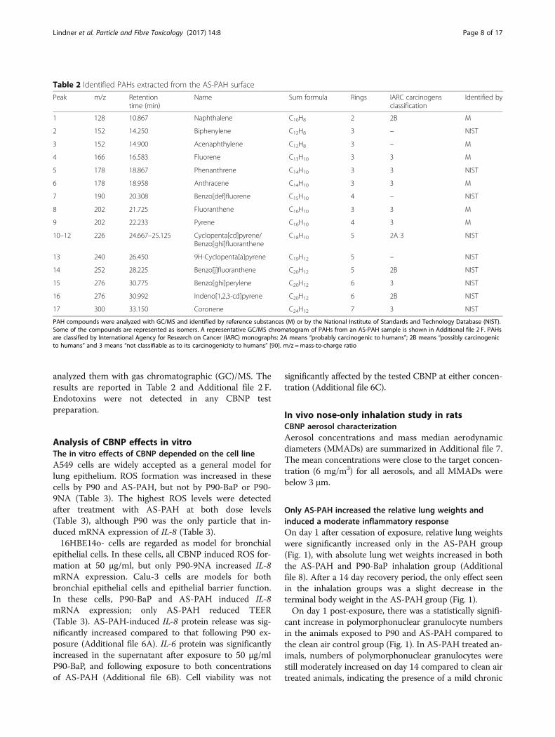
analyzed them with gas chromatographic (GC)/MS. Theresults are reported in Table 2 and Additional file 2 F.Endotoxins were not detected in any CBNP testpreparation.
Analysis of CBNP effects in vitroThe in vitro effects of CBNP depended on the cell lineA549 cells are widely accepted as a general model forlung epithelium. ROS formation was increased in thesecells by P90 and AS-PAH, but not by P90-BaP or P90-9NA (Table 3). The highest ROS levels were detectedafter treatment with AS-PAH at both dose levels(Table 3), although P90 was the only particle that in-duced mRNA expression of IL-8 (Table 3).16HBE14o- cells are regarded as model for bronchial
epithelial cells. In these cells, all CBNP induced ROS for-mation at 50 μg/ml, but only P90-9NA increased IL-8mRNA expression. Calu-3 cells are models for bothbronchial epithelial cells and epithelial barrier function.In these cells, P90-BaP and AS-PAH induced IL-8mRNA expression; only AS-PAH reduced TEER(Table 3). AS-PAH-induced IL-8 protein release was sig-nificantly increased compared to that following P90 ex-posure (Additional file 6A). IL-6 protein was significantlyincreased in the supernatant after exposure to 50 μg/mlP90-BaP, and following exposure to both concentrationsof AS-PAH (Additional file 6B). Cell viability was not
significantly affected by the tested CBNP at either concen-tration (Additional file 6C).
In vivo nose-only inhalation study in ratsCBNP aerosol characterizationAerosol concentrations and mass median aerodynamicdiameters (MMADs) are summarized in Additional file 7.The mean concentrations were close to the target concen-tration (6 mg/m3) for all aerosols, and all MMADs werebelow 3 μm.
Only AS-PAH increased the relative lung weights andinduced a moderate inflammatory responseOn day 1 after cessation of exposure, relative lung weightswere significantly increased only in the AS-PAH group(Fig. 1), with absolute lung wet weights increased in boththe AS-PAH and P90-BaP inhalation group (Additionalfile 8). After a 14 day recovery period, the only effect seenin the inhalation groups was a slight decrease in theterminal body weight in the AS-PAH group (Fig. 1).On day 1 post-exposure, there was a statistically signifi-
cant increase in polymorphonuclear granulocyte numbersin the animals exposed to P90 and AS-PAH compared tothe clean air control group (Fig. 1). In AS-PAH treated an-imals, numbers of polymorphonuclear granulocytes werestill moderately increased on day 14 compared to clean airtreated animals, indicating the presence of a mild chronic
Table 2 Identified PAHs extracted from the AS-PAH surface
Peak m/z Retentiontime (min)
Name Sum formula Rings IARC carcinogensclassification
Identified by
1 128 10.867 Naphthalene C10H8 2 2B M
2 152 14.250 Biphenylene C12H8 3 – NIST
3 152 14.900 Acenaphthylene C12H8 3 – M
4 166 16.583 Fluorene C13H10 3 3 M
5 178 18.867 Phenanthrene C14H10 3 3 NIST
6 178 18.958 Anthracene C14H10 3 3 M
7 190 20.308 Benzo[def]fluorene C15H10 4 – NIST
8 202 21.725 Fluoranthene C16H10 3 3 M
9 202 22.233 Pyrene C16H10 4 3 M
10–12 226 24.667–25.125 Cyclopenta[cd]pyrene/Benzo[ghi]fluoranthene
C18H10 5 2A 3 NIST
13 240 26.450 9H-Cyclopenta[a]pyrene C19H12 5 – NIST
14 252 28.225 Benzo[j]fluoranthene C20H12 5 2B NIST
15 276 30.775 Benzo[ghi]perylene C20H12 6 3 NIST
16 276 30.992 Indeno[1,2,3-cd]pyrene C20H12 6 2B NIST
17 300 33.150 Coronene C24H12 7 3 NIST
PAH compounds were analyzed with GC/MS and identified by reference substances (M) or by the National Institute of Standards and Technology Database (NIST).Some of the compounds are represented as isomers. A representative GC/MS chromatogram of PAHs from an AS-PAH sample is shown in Additional file 2 F. PAHsare classified by International Agency for Research on Cancer (IARC) monographs: 2A means “probably carcinogenic to humans”; 2B means “possibly carcinogenicto humans” and 3 means “not classifiable as to its carcinogenicity to humans” [90]. m/z =mass-to-charge ratio
Lindner et al. Particle and Fibre Toxicology (2017) 14:8 Page 8 of 17
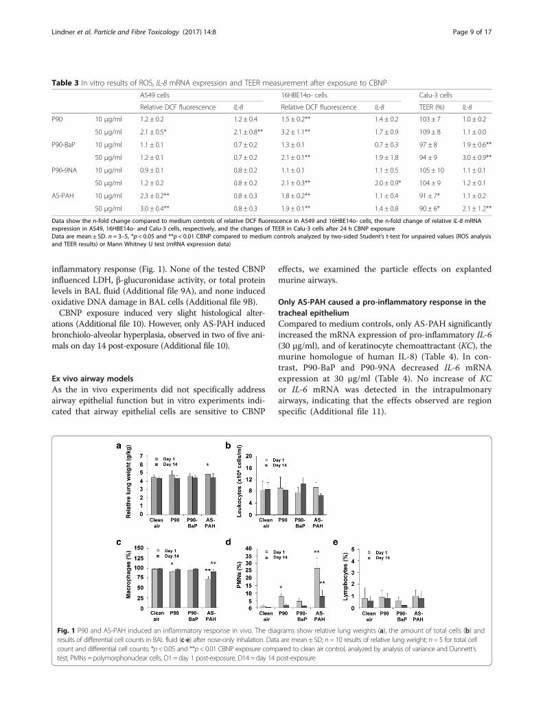
inflammatory response (Fig. 1). None of the tested CBNPinfluenced LDH, β-glucuronidase activity, or total proteinlevels in BAL fluid (Additional file 9A), and none inducedoxidative DNA damage in BAL cells (Additional file 9B).CBNP exposure induced very slight histological alter-
ations (Additional file 10). However, only AS-PAH inducedbronchiolo-alveolar hyperplasia, observed in two of five ani-mals on day 14 post-exposure (Additional file 10).
Ex vivo airway modelsAs the in vivo experiments did not specifically addressairway epithelial function but in vitro experiments indi-cated that airway epithelial cells are sensitive to CBNP
effects, we examined the particle effects on explantedmurine airways.
Only AS-PAH caused a pro-inflammatory response in thetracheal epitheliumCompared to medium controls, only AS-PAH significantlyincreased the mRNA expression of pro-inflammatory IL-6(30 μg/ml), and of keratinocyte chemoattractant (KC), themurine homologue of human IL-8) (Table 4). In con-trast, P90-BaP and P90-9NA decreased IL-6 mRNAexpression at 30 μg/ml (Table 4). No increase of KCor IL-6 mRNA was detected in the intrapulmonaryairways, indicating that the effects observed are regionspecific (Additional file 11).
Table 3 In vitro results of ROS, IL-8 mRNA expression and TEER measurement after exposure to CBNP
A549 cells 16HBE14o- cells Calu-3 cells
Relative DCF fluorescence IL-8 Relative DCF fluorescence IL-8 TEER (%) IL-8
P90 10 μg/ml 1.2 ± 0.2 1.2 ± 0.4 1.5 ± 0.2** 1.4 ± 0.2 103 ± 7 1.0 ± 0.2
50 μg/ml 2.1 ± 0.5* 2.1 ± 0.8** 3.2 ± 1.1** 1.7 ± 0.9 109 ± 8 1.1 ± 0.0
P90-BaP 10 μg/ml 1.1 ± 0.1 0.7 ± 0.2 1.3 ± 0.1 0.7 ± 0.3 97 ± 8 1.9 ± 0.6**
50 μg/ml 1.2 ± 0.1 0.7 ± 0.2 2.1 ± 0.1** 1.9 ± 1.8 94 ± 9 3.0 ± 0.9**
P90-9NA 10 μg/ml 0.9 ± 0.1 0.8 ± 0.2 1.1 ± 0.1 1.1 ± 0.5 105 ± 10 1.1 ± 0.1
50 μg/ml 1.2 ± 0.2 0.8 ± 0.2 2.1 ± 0.3** 2.0 ± 0.9* 104 ± 9 1.2 ± 0.1
AS-PAH 10 μg/ml 2.3 ± 0.2** 0.8 ± 0.3 1.8 ± 0.2** 1.1 ± 0.4 91 ± 7* 1.1 ± 0.2
50 μg/ml 3.0 ± 0.4** 0.8 ± 0.3 1.9 ± 0.1** 1.4 ± 0.8 90 ± 6* 2.1 ± 1.2**
Data show the n-fold change compared to medium controls of relative DCF fluorescence in A549 and 16HBE14o- cells, the n-fold change of relative IL-8 mRNAexpression in A549, 16HBE14o- and Calu-3 cells, respectively, and the changes of TEER in Calu-3 cells after 24 h CBNP exposureData are mean ± SD. n = 3–5, *p < 0.05 and **p < 0.01 CBNP compared to medium controls analyzed by two-sided Student’s t-test for unpaired values (ROS analysisand TEER results) or Mann Whitney U test (mRNA expression data)
Fig. 1 P90 and AS-PAH induced an inflammatory response in vivo. The diagrams show relative lung weights (a), the amount of total cells (b) andresults of differential cell counts in BAL fluid (c-e) after nose-only inhalation. Data are mean ± SD; n= 10 results of relative lung weight; n= 5 for total cellcount and differential cell counts; *p< 0.05 and **p< 0.01 CBNP exposure compared to clean air control, analyzed by analysis of variance and Dunnett’stest; PMNs = polymorphonuclear cells, D1 = day 1 post-exposure, D14 = day 14 post-exposure
Lindner et al. Particle and Fibre Toxicology (2017) 14:8 Page 9 of 17
All PAH containing CBNP induced Cyp-expression but noneof the CBNP induced mRNA for indicators of oxidativestress in tracheal epithelial cellsWe analyzed the mRNA expression of the cytochromeP450 enzymes Cyp1A1 and Cyp1B1 as indicators ofPAH metabolism, and of Gpx3 and Gr as indicators ofoxidative stress.All PAH-containing CBNP induced Cyp1a1 and
Cyp1b1 mRNA in the tracheal epithelium (Table 4).The most potent CBNP was AS-PAH, followed byP90-BaP. P90-9NA only induced Cyp1a1 and Cyp1b1mRNA expression at 30 μg/ml. In contrast, the un-coated P90 nanoparticles did not induce Cyp1a1mRNA expression, and significantly decreased Cyp1b1mRNA expression compared to medium controls intracheal epithelial cells (Table 4). None of the CBNPincreased the mRNA expression of Gpx3 or Gr. Inintrapulmonary airways PAH-containing CBNP in-duced a similar expression pattern of Cyp1a1 mRNAcompared to the tracheal epithelium; no increase ofmRNAs for anti-oxidative enzymes was detected(Additional file 11).Since the mRNA data of Cyp-expression indicated that
the CBNP affect airway epithelial cells and to detectchanges in epithelial function, we assessed their impacton cilia-driven transport.
Only P90 forms microscopically visible agglomerates;these bind to mucus and interact with ciliated cellsOn microscopic examination of tracheae, visible agglom-erates were observed on the airway epithelium after 24 hof incubation with P90 at 10 μg/ml and 30 μg/ml(Additional file 12A). These agglomerates were bound ei-ther to mucus on the epithelium or directly to cilia of cili-ated cells (Fig. 2a and b and Additional files 13 and 14).None of the PAH-containing CBNP showed microscopic-ally visible agglomerates at either tested concentration(Additional file 12A).
At 10 μg/ml, P90 agglomerates increased ciliary beatfrequency but mucus reduced particle transport speedCompared to medium control, P90 at 10 μg/ml in-creased mean ciliary beat frequency (Fig. 2c), although itdid not increase the mean of cilia-driven particle trans-port speed (Fig. 2d). Cells with bound agglomerates ex-hibited a higher ciliary beat frequency compared to cellswithout visible agglomerates (Additional file 12B and C).Furthermore, particle transport speed was reduced inareas with mucus and increased in areas without mucuscompared to medium control (Additional file 12D). In-cubation with P90 at a concentration of 1 μg/ml did notresult in formation of agglomerates, and therefore didnot increase ciliary beat frequency (Additional files 12Aand 15A). However, this concentration was sufficient toincrease mucus (Additional files 15B and C), and conse-quently to reduce particle transport speed (Additionalfile 15D).
At 10 μg/ml, PAH-coated P90 did not increase ciliary beatfrequency but differently influenced mucus release andparticle transport speedAs expected from the inability to form agglomerates,neither P90-BaP nor P90-9NA increased ciliary beat fre-quency (Fig. 2e and f). Incubation with P90-BaP in-creased amounts of mucus on the epithelial cells(Additional files 16 and 17), with a consequent reducedparticle transport speed compared to control (Fig. 2gand h). In contrast to P90 at 10 μg/ml, we observed deadcells on the epithelium during the experiments withboth coated particles (Additional files 16, 17 and 18).
At 10 μg/ml, AS-PAH increased ciliary beat frequency andmucus release but reduced particle transport speedAlthough AS-PAH did not form agglomerates, it in-creased ciliary beat frequency and resulted in mucus re-lease (Fig. 2i, and Additional files 12A and 19). Comparedto control, AS-PAH reduced particle transport speed
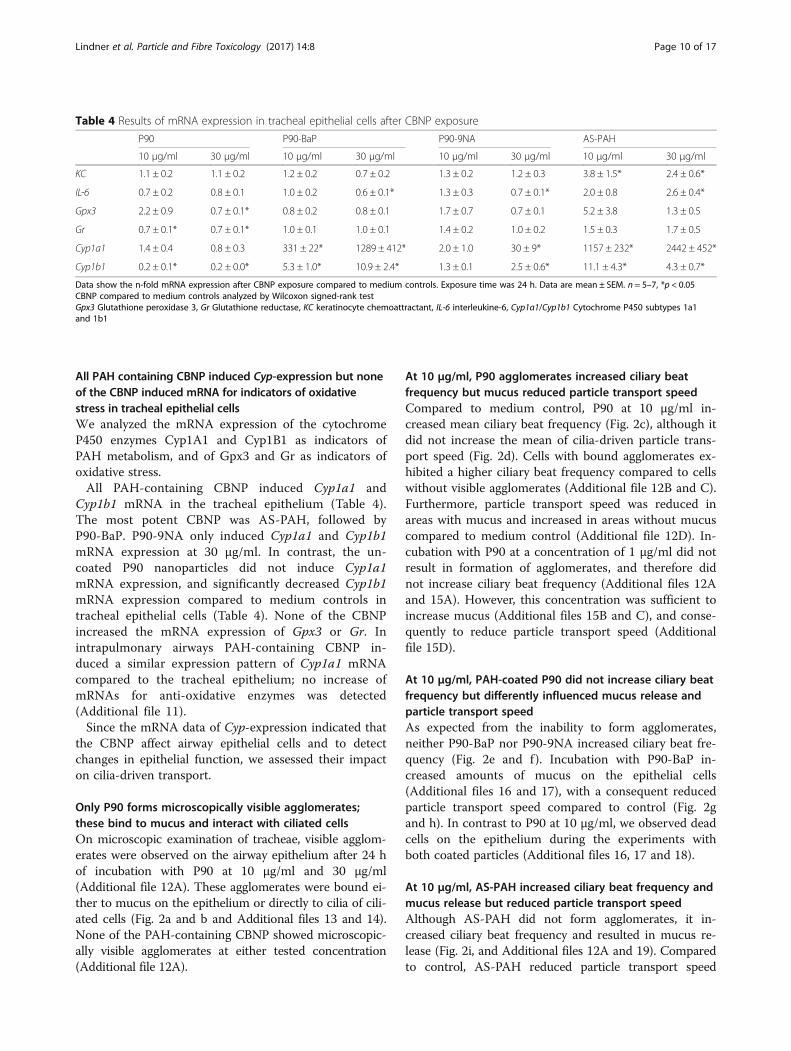
Table 4 Results of mRNA expression in tracheal epithelial cells after CBNP exposure
P90 P90-BaP P90-9NA AS-PAH
10 μg/ml 30 μg/ml 10 μg/ml 30 μg/ml 10 μg/ml 30 μg/ml 10 μg/ml 30 μg/ml
KC 1.1 ± 0.2 1.1 ± 0.2 1.2 ± 0.2 0.7 ± 0.2 1.3 ± 0.2 1.2 ± 0.3 3.8 ± 1.5* 2.4 ± 0.6*
IL-6 0.7 ± 0.2 0.8 ± 0.1 1.0 ± 0.2 0.6 ± 0.1* 1.3 ± 0.3 0.7 ± 0.1* 2.0 ± 0.8 2.6 ± 0.4*
Gpx3 2.2 ± 0.9 0.7 ± 0.1* 0.8 ± 0.2 0.8 ± 0.1 1.7 ± 0.7 0.7 ± 0.1 5.2 ± 3.8 1.3 ± 0.5
Gr 0.7 ± 0.1* 0.7 ± 0.1* 1.0 ± 0.1 1.0 ± 0.1 1.4 ± 0.2 1.0 ± 0.2 1.5 ± 0.3 1.7 ± 0.5
Cyp1a1 1.4 ± 0.4 0.8 ± 0.3 331 ± 22* 1289 ± 412* 2.0 ± 1.0 30 ± 9* 1157 ± 232* 2442 ± 452*
Cyp1b1 0.2 ± 0.1* 0.2 ± 0.0* 5.3 ± 1.0* 10.9 ± 2.4* 1.3 ± 0.1 2.5 ± 0.6* 11.1 ± 4.3* 4.3 ± 0.7*
Data show the n-fold mRNA expression after CBNP exposure compared to medium controls. Exposure time was 24 h. Data are mean ± SEM. n = 5–7, *p < 0.05CBNP compared to medium controls analyzed by Wilcoxon signed-rank testGpx3 Glutathione peroxidase 3, Gr Glutathione reductase, KC keratinocyte chemoattractant, IL-6 interleukine-6, Cyp1a1/Cyp1b1 Cytochrome P450 subtypes 1a1and 1b1
Lindner et al. Particle and Fibre Toxicology (2017) 14:8 Page 10 of 17
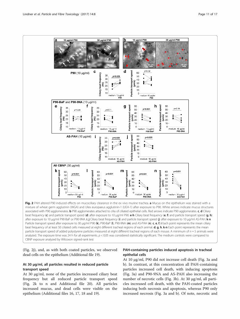
(Fig. 2j), and, as with both coated particles, we observeddead cells on the epithelium (Additional file 19).
At 30 μg/ml, all particles resulted in reduced particletransport speedAt 30 μg/ml, none of the particles increased ciliary beatfrequency but all reduced particle transport speed(Fig. 2k to n and Additional file 20). All particlesincreased mucus, and dead cells were visible on theepithelium (Additional files 16, 17, 18 and 19).
PAH-containing particles induced apoptosis in trachealepithelial cellsAt 10 μg/ml, P90 did not increase cell death (Fig. 3a andb). In contrast, at this concentration all PAH-containingparticles increased cell death, with inducing apoptosis(Fig. 3a) and P90-9NA and AS-PAH also increasing thenumber of necrotic cells (Fig. 3b). At 30 μg/ml, all parti-cles increased cell death, with the PAH-coated particlesinducing both necrosis and apoptosis, whereas P90 onlyincreased necrosis (Fig. 3a and b). Of note, necrotic and
Fig. 2 PAH altered P90 induced effects on mucociliary clearance in the ex vivo murine trachea. a Mucus on the epithelium was stained with amixture of wheat germ agglutinin (WGA) and Ulex europaeus agglutinin-1 (UEA-1) after exposure to P90. White arrows indicate mucus structuresassociated with P90 agglomerates. b P90 agglomerates attached to cilia of ciliated epithelial cells. Red arrows indicate P90 agglomerates. c, d Ciliarybeat frequency (c) and particle transport speed (d) after exposure to 10 μg/ml P90. e-h Ciliary beat frequency (e, f) and particle transport speed (g, h)after exposure to 10 μg/ml P90-BaP or P90-9NA. i, j Ciliary beat frequency (i) and particle transport speed (j) after exposure to 10 μg/ml AS-PAH. k-nParticle transport speed after exposure to 30 μg/ml P90 (k), P90-BaP (l), P90-9NA (m) and AS-PAH (n). c, e, f, i Each point represents the mean ciliarybeat frequency of at least 50 ciliated cells measured at eight different tracheal regions of each animal. d, g, h, k-n Each point represents the meanparticle transport speed of added polystyrene particles measured at eight different tracheal regions of each mouse. A minimum of n = 5 animals wereanalyzed. The exposure time was 24 h for all experiments. p < 0.05 was considered statistically significant. The medium controls were compared toCBNP exposure analyzed by Wilcoxon signed-rank test
Lindner et al. Particle and Fibre Toxicology (2017) 14:8 Page 11 of 17
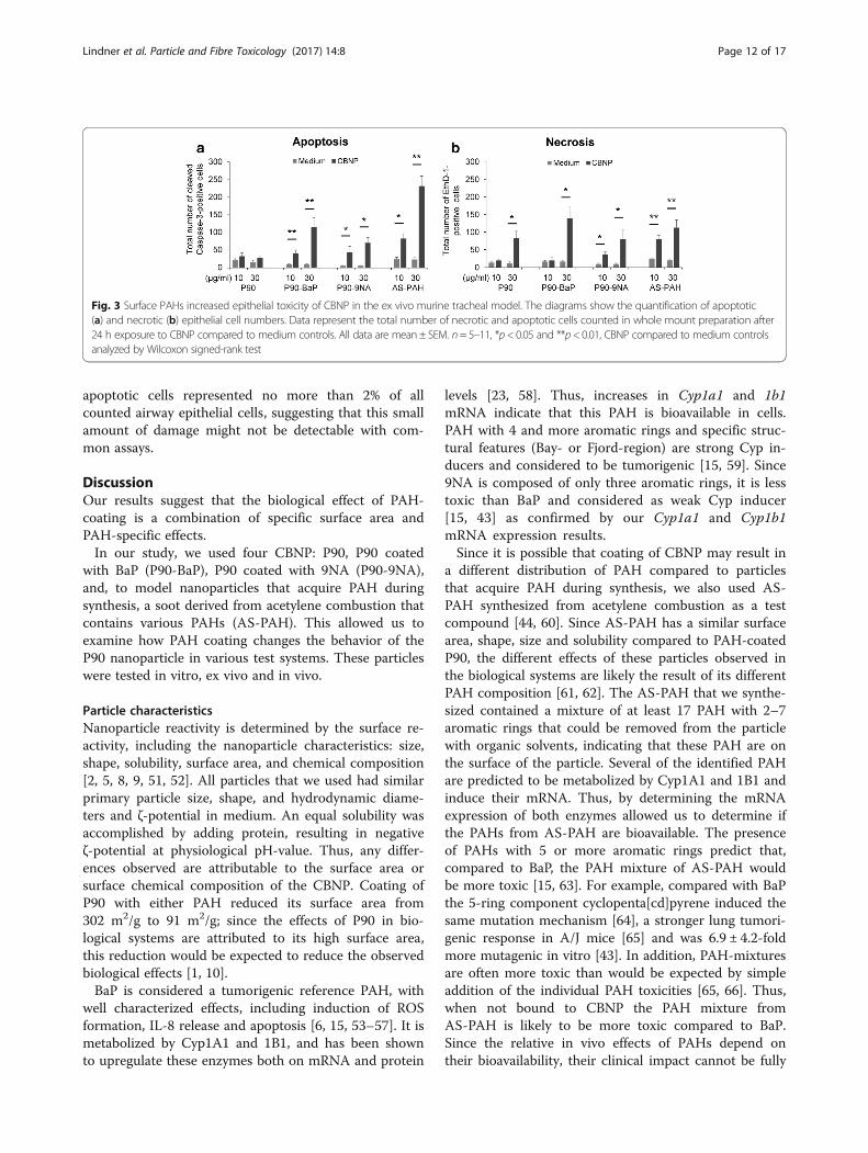
apoptotic cells represented no more than 2% of allcounted airway epithelial cells, suggesting that this smallamount of damage might not be detectable with com-mon assays.
DiscussionOur results suggest that the biological effect of PAH-coating is a combination of specific surface area andPAH-specific effects.In our study, we used four CBNP: P90, P90 coated
with BaP (P90-BaP), P90 coated with 9NA (P90-9NA),and, to model nanoparticles that acquire PAH duringsynthesis, a soot derived from acetylene combustion thatcontains various PAHs (AS-PAH). This allowed us toexamine how PAH coating changes the behavior of theP90 nanoparticle in various test systems. These particleswere tested in vitro, ex vivo and in vivo.
Particle characteristicsNanoparticle reactivity is determined by the surface re-activity, including the nanoparticle characteristics: size,shape, solubility, surface area, and chemical composition[2, 5, 8, 9, 51, 52]. All particles that we used had similarprimary particle size, shape, and hydrodynamic diame-ters and ζ-potential in medium. An equal solubility wasaccomplished by adding protein, resulting in negativeζ-potential at physiological pH-value. Thus, any differ-ences observed are attributable to the surface area orsurface chemical composition of the CBNP. Coating ofP90 with either PAH reduced its surface area from302 m2/g to 91 m2/g; since the effects of P90 in bio-logical systems are attributed to its high surface area,this reduction would be expected to reduce the observedbiological effects [1, 10].BaP is considered a tumorigenic reference PAH, with
well characterized effects, including induction of ROSformation, IL-8 release and apoptosis [6, 15, 53–57]. It ismetabolized by Cyp1A1 and 1B1, and has been shownto upregulate these enzymes both on mRNA and protein
levels [23, 58]. Thus, increases in Cyp1a1 and 1b1mRNA indicate that this PAH is bioavailable in cells.PAH with 4 and more aromatic rings and specific struc-tural features (Bay- or Fjord-region) are strong Cyp in-ducers and considered to be tumorigenic [15, 59]. Since9NA is composed of only three aromatic rings, it is lesstoxic than BaP and considered as weak Cyp inducer[15, 43] as confirmed by our Cyp1a1 and Cyp1b1mRNA expression results.Since it is possible that coating of CBNP may result in
a different distribution of PAH compared to particlesthat acquire PAH during synthesis, we also used AS-PAH synthesized from acetylene combustion as a testcompound [44, 60]. Since AS-PAH has a similar surfacearea, shape, size and solubility compared to PAH-coatedP90, the different effects of these particles observed inthe biological systems are likely the result of its differentPAH composition [61, 62]. The AS-PAH that we synthe-sized contained a mixture of at least 17 PAH with 2–7aromatic rings that could be removed from the particlewith organic solvents, indicating that these PAH are onthe surface of the particle. Several of the identified PAHare predicted to be metabolized by Cyp1A1 and 1B1 andinduce their mRNA. Thus, by determining the mRNAexpression of both enzymes allowed us to determine ifthe PAHs from AS-PAH are bioavailable. The presenceof PAHs with 5 or more aromatic rings predict that,compared to BaP, the PAH mixture of AS-PAH wouldbe more toxic [15, 63]. For example, compared with BaPthe 5-ring component cyclopenta[cd]pyrene induced thesame mutation mechanism [64], a stronger lung tumori-genic response in A/J mice [65] and was 6.9 ± 4.2-foldmore mutagenic in vitro [43]. In addition, PAH-mixturesare often more toxic than would be expected by simpleaddition of the individual PAH toxicities [65, 66]. Thus,when not bound to CBNP the PAH mixture fromAS-PAH is likely to be more toxic compared to BaP.Since the relative in vivo effects of PAHs depend ontheir bioavailability, their clinical impact cannot be fully
Fig. 3 Surface PAHs increased epithelial toxicity of CBNP in the ex vivo murine tracheal model. The diagrams show the quantification of apoptotic(a) and necrotic (b) epithelial cell numbers. Data represent the total number of necrotic and apoptotic cells counted in whole mount preparation after24 h exposure to CBNP compared to medium controls. All data are mean ± SEM. n= 5–11, *p< 0.05 and **p< 0.01, CBNP compared to medium controlsanalyzed by Wilcoxon signed-rank test
Lindner et al. Particle and Fibre Toxicology (2017) 14:8 Page 12 of 17
predicted without appropriately designed and conductedstudies, such as those we report here.When predicting the behavior of PAH-coated versus
uncoated nanoparticles in biological systems, three dif-ferent scenarios are possible. First, the reduction of thesurface area could reduce the biological effects of CBNP,and differences in the PAHs would therefore not have animpact. Second, the PAH content could increase the bio-logical effects of CBNP due to the toxicity of PAH – andso irrespective of the particle’s surface area. A third pos-sibility is that toxicity could be decreased in some celltypes due to the reduced surface area, but increased inother cell types due to PAH toxicity.
Biological effects of uncoated and PAH-coated CBNPSupporting the hypothesis that a reduced surface areareduces the biological effects of CBNP [1, 10, 67], thecoating of P90 with BaP prevented the P90-induced in-crease of the neutrophil attracting cytokine IL8 mRNAin A549 cells. As the high surface area of P90 is stronglyassociated with induction of oxidative stress, the reducedbiological effect can be explained by the reduced ROSrelease that we observed with P90-BaP and P90-9NA inA549 cells compared to uncoated P90. This reducedbiological effect is also supported by our observationthat P90, and not P90-BaP, led to a transient increase ingranulocyte influx in the in vivo experiment. This transi-ent increase in granulocytes without changes of total cellnumber has previously been observed [68, 69]. Themechanism for the reduced number of macrophages isnot known and would be an interesting area for futureresearch. However, these results clearly demonstrate thatsurface area can be an important factor of CBNP effectsin biological systems.However, other results support the theory that PAH
can increase the effects of CBNP [29]. Firstly, we foundthat P90-BaP, P90-9NA and AS-PAH induced apoptosisin tracheal epithelial cells ex vivo, whereas P90 did not.This is consistent with previous reports describing apop-tosis induction by PAH [54, 56, 57, 70]. Secondly, PAH-coated P90 and AS-PAH strongly induced Cyp mRNAexpression. Cyp1a1 and Cyp1b1 mRNA are known to beinduced by PAH in a subgroup of airway epithelial cells[71], and so their induction in both the trachea andintrapulmonary airways demonstrate that the PAH arebiologically active. Interestingly, the assumed differencesbetween P90-9NA, P90-BaP and AS-PAH are alsoreflected in the level of induction of Cyp1a1 and Cyp1b1mRNA, in that P90-9NA resulted in the least induction,followed by P90-BaP, with the strongest induction byAS-PAH. Finally, AS-PAH was the only CBNP to reduceTEER in vitro, to increase BAL granulocyte content14 days after the last application, and induce bothhistological changes in vivo and inflammatory mediator
mRNA expression (KC and IL-6) ex vivo. Furthermore,AS-PAH had the highest apoptosis rate in trachealepithelium ex vivo.Our data therefore suggest that the overall biological
impact of a PAH depends on the relative contribution ofsurface area reduction and PAH-induced toxicity, that is,in turn, influenced by the biological system. This is ex-emplified by cilia-driven particle transport speed, whichis influenced by ciliary beat frequency, mucus releaseand epithelial cell death.In the ex vivo trachea, P90 was the only particle to
form aggregates. These aggregates bound to ciliated cellsand increased ciliary beat frequency. Furthermore,microscopically visible nanoparticle agglomerates havebeen shown to increase ciliary beat frequency in themurine trachea [72]. A similar effect has been observedin the bovine trachea where polystyrene particles>200 nm in diameter that attach to cilia increase ciliarybeat frequency due to mechanical stimulation of axo-nema possibly involving second messengers such ascAMP or cGMP [73], both of which are known to in-crease ciliary beat frequency [74]. In addition, mechan-ical stimulation of cilia triggers Ca2+ influx, with aconsequent increase in ciliary beat frequency [75]. Coat-ing of P90 with PAHs prevented aggregate formationand consequently neither P90-BaP nor P90-9NA impactciliary beat frequency. Thus, this shows that PAH coat-ing of P90 reduced its effects on a biological system bychanging its surface properties and not by a specificPAH toxicity. However, we detected an increase in epi-thelial cell apoptosis after application of P90-BaP andP90-9NA compared to P90. This increase in apoptosistogether with the induction of Cyp enzymes can directlybe ascribed to PAH toxicity [70].AS-PAH increased ciliary beat frequency without
forming microscopically visible agglomerates. Interest-ingly, fluoranthene, a PAH that we detected on the sur-face of AS-PAH, has previously been shown to induce apersistent increase in Ca2+ in Calu-3 cells, whereas BaPand anthracene did not [76]. Since an increase in intra-cellular Ca2+ is known to increase ciliary beat frequency[74], it is possible that fluoranthene is directly respon-sible for the increased ciliary beat frequency. Despiteincreasing ciliary beat frequency there was a markedreduction in particle transport speed, possibly due toincreased apoptosis and mucus release.Previous research has shown that despite being bound
to the surface of CBNP, PAHs can be bioavailable [77].Our data confirm that PAH bound to CBNP exertsbiological effects in epithelial cells, as demonstrated byCyp1a1 mRNA induction. Cyp1a1 mRNA expression isa very sensitive parameter for detecting biologicallyactive PAHs [78]. The induction of Cyp1a1 mRNA istriggered by binding of PAH to the aryl hydrocarbon
Lindner et al. Particle and Fibre Toxicology (2017) 14:8 Page 13 of 17
receptor (AhR) that then translocates into the cell nu-cleus, and interacts with AhR nuclear translocator [79].This complex then induces Cyp1a1 mRNA expressionby binding to xenobiotic response elements [79]. In lowconcentrations, AhR activation suppresses the activity ofthe transcription factor p65 [80], which is known to in-duce the mRNA expression of IL-6 and IL-8 [81, 82].We also observed a small but significant decrease inIL-6 mRNA expression in the epithelium of explantedtracheae, possibly due to AhR activation. At higher con-centrations, PAHs can lead to IL-6 and IL-8 mRNAexpression [80] – as we observed with AS-PAH. In gen-eral, AS-PAH had more effects than the PAH-coatedCBNP, probably due to the composition of PAH presenton the particle’s surface. As discussed above, the PAHfluoranthene induces prolonged increase in intracellularCa2+ in Calu-3, whereas BaP and anthracene do not[76]. Yamaguchi et al. also observed an increase of intra-cellular Ca2+ that was independent from AhR [83], indi-cating that PAH have additional activities apart from theclassical AhR pathway. Identification of the specific PAHand/or PAH mixture that is responsible for the increasedeffects of AS-PAH would require additional research,although fluoranthene and compounds such as cyclo-penta[cd]pyrene are possible candidates.We have shown that AS-PAH exerts more toxic effects
than P90 despite its reduced surface area. We attributethis to the composition of the surface PAHs. The relativetoxicity of P90-9NA and P90-BaP, and the resultant bio-logical effect, depended on the test system used.
Choice of test systemsIn general, cell lines can give valuable information if thetest substance is interfering with basal cell functionspresent in all cells. However, if cell lines lack specificpathways that are important for toxic effects, their valuefor predicting in vivo toxicity is limited [84].The prediction of an in vivo effect based on results ob-
tained in a single cell line is made more challenging,since both changes in surface area and direct PAHtoxicity impact the overall effect of particles. This mayexplain why we had different results in terms of IL-8mRNA production in different cell lines. The reason forthese differences was most likely that some cell lineswere more sensitive to the toxic effects of the PAHs,whereas others were more sensitive to changes in sur-face area. An in vivo experiment in which all target celltypes are present is therefore in principle better suitedto detect effects [84]. However, in a single in vivo experi-ment, only a limited number of parameters can be deter-mined. The readouts predetermined in the OECDexperiment focus on inflammation and cell death. Theydo not address effects on mucociliary clearance. Itshould be noted, however that sustained impairment of
mucociliary clearance can have substantial health effects[85–88] that are missed in animal models by focusingsolely on inflammation and cell death. In principle, invivo experiments can be designed to also include theseparameters. However, this would be time consuming andcost intensive. The strength of short-term organ culturemodels is that they can be adapted to screen for specificorgan functions that are not routinely examined in stan-dardized in vivo experiments. This was demonstrated byusing our ex vivo trachea model that allowed examiningthe complex interplay between mucus production, ciliarybeating and cell death [72, 89].
ConclusionsUsing a range of different test systems our results dem-onstrate that the biological effect of CBNP is determinedby a combination of specific surface area and the com-position of the surface-bound PAH, and varies in differ-ent target cells. Specifically, AS-PAH exerted more toxiceffects than P90 despite its reduced surface area. We at-tribute this to the composition of the surface PAHs.However, the relative toxicity of P90-9NA and P90-BaPdepended on the test system used.
Additional files
Additional file 1: Electron microscopic images of P90 and AS-PAH.(PDF 428 kb)
Additional file 2: AS-PAH exhibited a less PAH content and a PAH mixcompared to the coated P90 nanoparticles. (PDF 308 kb)
Additional file 3: Primer pairs and probes used by quantitative real timeRT-PCR. (PDF 75 kb)
Additional file 4: Staining of necrotic and apoptotic cells in the epitheliallayer. (PDF 295 kb)
Additional file 5: Supplemental material and methods. (DOCX 31 kb)
Additional file 6: CBNP did not affect the viability of epithelial cells, butP90-BaP and AS-PAH induced cytokine release in vitro. (PDF 91 kb)
Additional file 7: CBNP aerosol concentration and results of Marple impactormeasurements used by nose-only inhalation experiments. (PDF 56 kb)
Additional file 8: P90-BaP and AS-PAH increased the lung wet weightsafter nose-only inhalation. (PDF 67 kb)
Additional file 9: CBNP did not increase enzyme nor total protein levelsin BAL and did not induce oxidative DNA-damage in BAL cells after nose-only inhalation. (PDF 80 kb)
Additional file 10: Only AS-PAH induced exposure-related histologicalalteration in lung tissue after nose-only inhalation. (PDF 72 kb)
Additional file 11: CBNP did not induce oxidative stress or a pro-inflammatory response in intrapulmonary airways. (PDF 106 kb)
Additional file 12: P90 agglomerates increased ciliary beat frequencyand released mucus impaired particle transport speed. (PDF 573 kb)
Additional file 13: P90 agglomerates attached to cilia of ciliated cellsafter incubation with 10 μg/mlP90. The movie shows cilia movement andattached P90 agglomerates on cilia of tracheal epithelium after 24 hexposure to 10 μg/ml P90. The movie was recorded with 100 Hz and aresolution of 640 × 480 pixels. The image sequence contains 1000images. (WMV 11853 kb)
Additional file 14: P90 agglomerates attached to cilia of ciliated cellsafter incubation with 30 μg/ml P90. The movie shows the cilia
Lindner et al. Particle and Fibre Toxicology (2017) 14:8 Page 14 of 17
movement and bound P90 agglomerates on the cilia of trachealepithelium after 24 h exposure to 30 μg/ml P90. The movie was recordedwith 100 Hz, a resolution of 640 × 480 pixels and contains 1000 images.(WMV 11658 kb)
Additional file 15: P90 did not cause agglomerates at 1 μg/ml, but releasedmucus impaired particle transport. (PDF 1144 kb)
Additional file 16: P90-BaP induced cell death. (PDF 707 kb)
Additional file 17: P90-9NA induced cell death at the higher concentration.(PDF 641 kb)
Additional file 18: P90 induced mucus release. (PDF 1021 kb)
Additional file 19: AS-PAH induced mucus release and cell death.(PDF 639 kb)
Additional file 20: No CBNP increased ciliary beat frequency at 30 μg/ml.(PDF 5594 kb)
AcknowledgementsThe authors thank Sabine Bartel and Susanne Krauss-Etschmann (Borstel) forproviding the 16HBE14o- cells for IL-8 mRNA expression experiments. Additionally,they thank all the participating patients and Dieter C. Gruenert for establishing the16HBE14o- cell line, and Juliane Artelt (Borstel) and Gudrun Knebel (Lübeck) forexpert technical assistance.David Young, a professional medical writer from Young Medical Communicationsand Consulting Limited, critically reviewed and language-edited the manuscriptprior to submission. This support was funded by the Borstel Research Center andthe Universität zu Lübeck.
FundingThis work was funded by the Bundesministerium für Forschung und Bildung(03X0093).
Availability of data and materialsAll data that are necessary to understand the theme of the study are includedin this published article and its supplementary information files. Additionaldatasets generated or analyzed during the current study are available fromcorresponding author on reasonable request.
Authors’ contributionsHF, PK and TH designed the study. MS and HB modified P90, produced theAS-PAH and characterized the CBNP. PG measured and analyzed the aerosolconcentration and MMAD. EH performed and analyzed the histologicalanalysis. CZ performed and analyzed the genotoxicological experiments. OCdesigned and supervised the rat inhalation study. GP was responsible for thegeneration and characterization of the exposure atmosphere. TT was the vet-erinarian responsible for the rat inhalation study. KL performed and analyzedthe ex vivo studies on tracheal epithelial cells. CB, PK and GH designed thesoftware for the ciliary beat frequency analysis. SS performed and analyzedthe ex vivo studies on intrapulmonary bronchioles. TH and JK carried outand analyzed the in vitro experiments (ROS, TEER). IL-8 mRNA expression of16HBE14o- and Calu-3 cells was analyzed by SW and AJ. AJ determined theIL-6 and IL-8 protein release from Calu-3 cells. JK characterized the CBNP inwater and media. KL and PK drafted the manuscript. All authors contributedto the intellectual content of the manuscript. All authors read and approvedthe final manuscript.
Competing interestsThe authors declare that they have no competing interests.
Consent for publicationNot applicable
Ethics approvalThe study was performed in accordance with the German animal protectionlaw and approved by the appropriate governmental authorities.The OECD inhalation study on rats was approved by the NiedersächsischesLandesamt für Verbraucherschutz und Lebensmittelsicherheit. The ex vivostudies on mouse trachea and intrapulmonary bronchioles were approvedby the Ministerium für Landwirtschaft, Umwelt und ländliche Räume desLandes Schleswig-Holstein.
Publisher’s NoteSpringer Nature remains neutral with regard to jurisdictional claims inpublished maps and institutional affiliations.
Author details1Institut für Anatomie, Zentrum für medizinische Struktur- und Zellbiologie,Universität zu Lübeck (UzL), Airway Research Center North (ARCN), GermanCenter for Lung Research (DZL), Ratzeburger Allee 160, 23562 Lübeck,Germany. 2Karlsruher Institut für Technologie, Engler-Bunte-Institut, BereichVerbrennungstechnik, Karlsruhe, Germany. 3Forschungszentrum Borstel,Leibniz-Zentrum für Medizin und Biowissenschaften, ExperimentellePneumologie, Borstel, Airway Research Center North (ARCN), German Centerfor Lung Research (DZL), Borstel, Germany. 4Forschungszentrum Borstel,Leibniz-Zentrum für Medizin und Biowissenschaften, Angeborene Immunität,Borstel, Airway Research Center North (ARCN), German Center for LungResearch (DZL), Borstel, Germany. 5Fraunhofer Institut für Toxikologie undExperimentelle Medizin ITEM, Hannover, Biomedical Research in Endstageand Obstructive Lung Disease Hannover (BREATH), German Center for LungResearch (DZL), Hannover, Germany. 6Institut für Biomedizinische Optik,Universität zu Lübeck (UzL), Lübeck, Airway Research Center North (ARCN),German Center for Lung Research (DZL), Lübeck, Germany.
Received: 15 November 2016 Accepted: 7 March 2017
References1. Stoeger T, Reinhard C, Takenaka S, Schroeppel A, Karg E, Ritter B, et al.
Instillation of six different ultrafine carbon particles indicates a surface areathreshold dose for acute lung inflammation in mice. Environ Health Perspect.2006;114:328–33.
2. Sahu D, Kannan GM, Vijayaraghavan R. Carbon black particle exhibits sizedependent toxicity in human monocytes. Int J Inflam. 2014;2014:1–10.
3. Marzaioli V, Aguilar-Pimentel JA, Weichenmeier I, Luxenhofer G, WiemannM, Landsiedel R, et al. Surface modifications of silica nanoparticles are crucialfor their inert versus proinflammatory and immunomodulatory properties.Int J Nanomedicine. 2014;9:2815–32.
4. Cheng WJ, Rong Y, Shi TM, Zhou T, Liu YW, Chen WH. Size-dependentbiological effects on vascular endothelial cells induced by different particulatematters. J Huazhong Univ Sci Technolog Med Sci. 2014;34:314–21.
5. Braakhuis HM, Park MV, Gosens I, De Jong WH, Cassee FR. Physicochemicalcharacteristics of nanomaterials that affect pulmonary inflammation. Part Fibre Toxicol.2014;11:18.
6. Ma JYC, Ma JKH. The dual effect of the particulate and organic componentsof diesel exhaust particles on the alteration of pulmonary immune/inflammatory responses and metabolic enzymes. J Environ Sci Health C.2002;20:117–47.
7. Gatoo MA, Naseem S, Arfat MY, Dar AM, Qasim K, Zubair S. Physicochemicalproperties of nanomaterials: implication in associated toxic manifestations.Biomed Res Int. 2014;2014:498420.
8. Shin SW, Song IH, Um SH. Role of physicochemical properties in nanoparticletoxicity. Nanomaterials. 2015;5:1351–65.
9. Stoeger T, Schmid O, Takenaka S, Schulz H. Inflammatory response to TiO2and carbonaceous particles scales best with BET surface area. Environ HealthPerspect. 2007;115:A290–1.
10. Schmid O, Stoeger T. Surface area is the biologically most effective dosemetric for acute nanoparticle toxicity in the lung. J Aerosol Sci. 2016;99:133–43.
11. Hussain S, Boland S, Baeza-Squiban A, Hamel R, Thomassen LC, Martens JA,et al. Oxidative stress and proinflammatory effects of carbon black andtitanium dioxide nanoparticles: role of particle surface area and internalizedamount. Toxicology. 2009;260:142–9.
12. Vesterdal LK, Mikkelsen L, Folkmann JK, Sheykhzade M, Cao Y, Roursgaard M,et al. Carbon black nanoparticles and vascular dysfunction in culturedendothelial cells and artery segments. Toxicol Lett. 2012;214:19–26.
13. Ma-Hock L, Strauss V, Treumann S, Kuttler K, Wohlleben W, Hofmann T, et al.Comparative inhalation toxicity of multi-wall carbon nanotubes, graphene,graphite nanoplatelets and low surface carbon black. Part Fibre Toxicol.2013;10:23.
14. Zhang R, Dai Y, Zhang X, Niu Y, Meng T, Li Y, et al. Reduced pulmonaryfunction and increased pro-inflammatory cytokines in nanoscale carbonblack-exposed workers. Part Fibre Toxicol. 2014;11:73.
Lindner et al. Particle and Fibre Toxicology (2017) 14:8 Page 15 of 17
15. Boström CE, Gerde P, Hanberg A, Jernstrom B, Johansson C, Kyrklund T, etal. Cancer risk assessment, indicators, and guidelines for polycyclic aromatichydrocarbons in the ambient air. Environ Health Perspect. 2002;110:451–88.
16. Kong HT, Xia K, Pan L, Zhang JC, Luo Y, Zhang Y, et al. Autophagy andlysosomal dysfunction: a new insight into mechanism of synergistic pulmonarytoxicity of carbon black-metal ions co-exposure. Carbon. 2017;111:322–33.
17. Valavanidis A, Fiotakis K, Vlachogianni T. Airborne particulate matter andhuman health: toxicological assessment and importance of size and compositionof particles for oxidative damage and carcinogenic mechanisms. J Environ SciHealth C Environ Carcinog Ecotoxicol Rev. 2008;26:339–62.
18. Lighty JS, Veranth JM, Sarofim AF. Combustion aerosols: factors governingtheir size and composition and implications to human health. J Air WasteManag Assoc. 2000;50:1565–618.
19. Oberdörster G, Oberdörster E, Oberdörster J. Nanotoxicology: an emergingdiscipline evolving from studies of ultrafine particles. Environ HealthPerspect. 2005;113:823–39.
20. Bonvallot V, Baeza-Squiban A, Baulig A, Brulant S, Boland S, Muzeau F, et al.Organic compounds from diesel exhaust particles elicit a proinflammatoryresponse in human airway epithelial cells and induce cytochrome p450 1A1expression. Am J Respir Cell Mol Biol. 2001;25:515–21.
21. World Health Organization. Air quality guidelines for Europe. WHO Reg PublEur Ser. 2000;V-X:1–273.
22. Shimada T, Fujii-Kuriyama Y. Metabolic activation of polycyclic aromatichydrocarbons to carcinogens by cytochromes P450 1A1 and 1B1. CancerSci. 2004;95:1–6.
23. Baird WM, Hooven LA, Mahadevan B. Carcinogenic polycyclic aromatichydrocarbon-DNA adducts and mechanism of action. Environ Mol Mutagen.2005;45:106–14.
24. Dasenbrock C, Peters L, Creutzenberg O, Heinrich U. The carcinogenic potency ofcarbon particles with and without PAH after repeated intratracheal administrationin the rat. Toxicol Lett. 1996;88:15–21.
25. Goulaouic S, Foucaud L, Bennasroune A, Laval-Gilly P, Falla J. Effect of polycyclicaromatic hydrocarbons and carbon black particles on pro-inflammatory cytokinesecretion: impact of PAH coating onto particles. J Immunotoxicol. 2008;5:337–45.
26. Mroz RM, Schins RP, Li H, Drost EM, Macnee W, Donaldson K. Nanoparticlecarbon black driven DNA damage induces growth arrest and AP-1 andNFkappaB DNA binding in lung epithelial A549 cell line. J Physiol Pharmacol.2007;58 Suppl 5:461–70.
27. Mroz RM, Schins RP, Li H, Jimenez LA, Drost EM, Holownia A, et al. Nanoparticle-driven DNA damage mimics irradiation-related carcinogenesis pathways. EurRespir J. 2008;31:241–51.
28. Borm PJA, Cakmak G, Jermann E, Weishaupt C, Kempers P, van Schooten FJ,et al. Formation of PAH-DNA adducts after in vivo and vitro exposure of ratsand lung cells to different commercial carbon blacks. Toxicol Appl Pharmacol.2005;205:157–67.
29. Chin BY, Choi ME, Burdick MD, Strieter RM, Risby TH, Choi AMK. Induction ofapoptosis by particulate matter: role of TNF-alpha and MAPK. Am J PhysiolLung Cell Mol Physiol. 1998;275:L942–9.
30. Delfino RJ. Epidemiologic evidence for asthma and exposure to air toxics:linkages between occupational, indoor, and community air pollution research.Environ Health Perspect. 2002;110 Suppl 4:573–89.
31. Balmes JR. How does diesel exhaust impact asthma? Thorax. 2011;66:4–6.32. OECD. Guideline for Testing of Chemicals, Test Guideline 412. Repeated Dose
Inhalation Toxicity: 28-day or 14-day Study. 1981; http://www.nikkakyo.org/ontai/merumaga/Challenge/OECD/TG_412.pdf. Accessed on 21th December 2016.
33. Sager TM, Castranova V. Surface area of particle administered versus mass indetermining the pulmonary toxicity of ultrafine and fine carbon black:comparison to ultrafine titanium dioxide. Part Fibre Toxicol. 2009;6:15.
34. Landsiedel R, Ma-Hock L, Kroll A, Hahn D, Schnekenburger J, Wiench K, et al.Testing metal-oxide nanomaterials for human safety. Adv Mater. 2010;22:2601–27.
35. Verma N, Pink M, Rettenmeier AW, Schmitz-Spanke S. Review on proteomicanalyses of benzo[a]pyrene toxicity. Proteomics. 2012;12:1731–55.
36. Boysen G, Hecht SS. Analysis of DNA and protein adducts of benzo[a]pyrene inhuman tissues using structure-specific methods. Mutat Res. 2003;543:17–30.
37. Shen YM, Troxel AB, Vedantam S, Penning TM, Field J. Comparison of p53mutations induced by PAH o-quinones with those caused by anti-benzo[a]pyrene diol epoxide in vitro: role of reactive oxygen and biologicalselection. Chem Res Toxicol. 2006;19:1441–50.
38. Zhang LJ, Bao Y, Li J. Nuclear respiratory factor-1 is involved in mitochondrialdysfunction induced by benzo(a)pyrene in human bronchial epithelial cells.Basic Clin Pharmacol Toxicol. 2011;109:115–22.
39. Ovrevik J, Arlt VM, Oya E, Nagy E, Mollerup S, Phillips DH, et al. Differentialeffects of nitro-PAHs and amino-PAHs on cytokine and chemokine responsesin human bronchial epithelial BEAS-2B cells. Toxicol Appl Pharmacol. 2010;242:270–80.
40. Harrigan JA, McGarrigle BP, Sutter TR, Olson JR. Tissue specific induction ofcytochrome P450 (CYP) 1A1 and 1B1 in rat liver and lung following in vitro(tissue slice) and in vivo exposure to benzo(a)pyrene. Toxicol In Vitro. 2006;20:426–38.
41. Fu PP, Von Tungeln LS, Chou MW. Metabolism of 9-nitroanthracene by ratliver microsomes: identification and mutagenicity of metabolites. Carcinogenesis.1985;6:753–7.
42. Fu PP, Heflich RH, Von Tungeln LS, Yang DT, Fifer EK, Beland FA. Effect of thenitro group conformation on the rat liver microsomal metabolism and bacterialmutagenicity of 2- and 9-nitroanthracene. Carcinogenesis. 1986;7:1819–27.
43. Durant JL, Busby Jr WF, Lafleur AL, Penman BW, Crespi CL. Human cellmutagenicity of oxygenated, nitrated and unsubstituted polycyclic aromatichydrocarbons associated with urban aerosols. Mutat Res. 1996;371:123–57.
44. Stroebele MR, Bockhorn H. Effects of synthesis conditions on the content ofpolycyclic aromatic hydrocarbons on the surface of carbon black nanoparticles.Proceedings of the European Combustion Meeting. 2013:Paper P5-52.
45. Cozens AL, Yezzi MJ, Kunzelmann K, Ohrui T, Chin L, Eng K, et al. CFTRexpression and chloride secretion in polarized immortal human bronchialepithelial cells. Am J Respir Cell Mol Biol. 1994;10:38–47.
46. Andersen CL, Jensen JL, Orntoft TF. Normalization of real-time quantitativereverse transcription-PCR data: a model-based variance estimation approachto identify genes suited for normalization, applied to bladder and coloncancer data sets. Cancer Res. 2004;64:5245–50.
47. Zehethofer N, Bermbach S, Hagner S, Garn H, Muller J, Goldmann T, et al.Lipid analysis of airway epithelial cells for studying respiratory diseases.Chromatographia. 2015;78:403–13.
48. Crouzier T, Beckwitt CH, Ribbeck K. Mucin multilayers assembled throughsugar-lectin interactions. Biomacromolecules. 2012;13:3401–8.
49. Kawano K, Uehara F, Sameshima M, Ohba N. Application of lectins fordetection of goblet cell carbohydrates of the human conjunctiva. Exp EyeRes. 1984;38:439–47.
50. Chazotte B. Labeling membrane glycoproteins or glycolipids with fluorescentwheat germ agglutinin. Cold Spring Harb Protoc. 2011;2011:pdb prot5623.
51. Knaapen AM, Borm PJ, Albrecht C, Schins RP. Inhaled particles and lungcancer. Part A: mechanisms. Int J Cancer. 2004;109:799–809.
52. Yang H, Liu C, Yang D, Zhang H, Xi Z. Comparative study of cytotoxicity,oxidative stress and genotoxicity induced by four typical nanomaterials: therole of particle size, shape and composition. J Appl Toxicol. 2009;29:69–78.
53. Podechard N, Lecureur V, Le Ferrec E, Guenon I, Sparfel L, Gilot D, et al.Interleukin-8 induction by the environmental contaminant benzo(a)pyreneis aryl hydrocarbon receptor-dependent and leads to lung inflammation.Toxicol Lett. 2008;177:130–7.
54. Jyonouchi H. Airway epithelium and apoptosis. Apoptosis. 1999;4:407–17.55. Kim KB, Lee BM. Oxidative stress to DNA, protein, and antioxidant enzymes
(superoxide dismutase and catalase) in rats treated with benzo(a)pyrene.Cancer Lett. 1997;113:205–12.
56. Tesfaigzi Y. Roles of apoptosis in airway epithelia. Am J Respir Cell Mol Biol.2006;34:537–47.
57. Revel A, Raanani H, Younglai E, Xu J, Rogers I, Han R, et al. Resveratrol, anatural aryl hydrocarbon receptor antagonist, protects lung from DNA damageand apoptosis caused by benzo[a]pyrene. J Appl Toxicol. 2003;23:255–61.
58. Tsuji PA, Walle T. Inhibition of benzo[a]pyrene-activating enzymes and DNAbinding in human bronchial epithelial BEAS-2B cells by methoxylated flavonoids.Carcinogenesis. 2006;27:1579–85.
59. Elovaara E, Mikkola J, Stockmann-Juvala H, Luukkanen L, Keski-Hynnila H,Kostiainen R, et al. Polycyclic aromatic hydrocarbon (PAH) metabolizingenzyme activities in human lung, and their inducibility by exposure tonaphthalene, phenanthrene, pyrene, chrysene, and benzo(a)pyrene as shownin the rat lung and liver. Arch Toxicol. 2007;81:169–82.
60. Donnet J-B, Bansal RC, Wang M-J. Carbon black - science and technology.Secondth ed. New York - Basel: Marcel Dekker, Inc; 1993.
61. Schreiber N, Strobele M, Kopf J, Hochscheid R, Kotte E, Weber P, et al. Lungalterations following single or multiple low-dose carbon black nanoparticleaspirations in mice. J Toxicol Environ Health A. 2013;76:1317–32.
62. Schreiber N, Strobele M, Hochscheid R, Kotte E, Weber P, Bockhorn H, et al.Modifications of carbon black nanoparticle surfaces modulate type IIpneumocyte homoeostasis. J Toxicol Environ Health A. 2016;79:153–64.
Lindner et al. Particle and Fibre Toxicology (2017) 14:8 Page 16 of 17
63. Moorthy B, Chu C, Carlin DJ. Polycyclic aromatic hydrocarbons: from metabolismto lung cancer. Toxicol Sci. 2015;145:5–15.
64. Keohavong P, Melacrinos A, Shukla R. In vitro mutational spectrum ofcyclopenta[cd]pyrene in the human HPRT gene. Carcinogenesis. 1995;16:855–60.
65. Nesnow S, Mass MJ, Ross JA, Galati AJ, Lambert GR, Gennings C, et al. Lungtumorigenic interactions in strain A/J mice of five environmental polycyclicaromatic hydrocarbons. Environ Health Perspect. 1998;106 Suppl 6:1337–46.
66. Gibb HJ, Chen CW. Multistage model interpretation of additive and multiplicativecarcinogenic effects. Risk Anal. 1986;6:167–70.
67. Monteiller C, Tran L, MacNee W, Faux S, Jones A, Miller B, et al. The pro-inflammatory effects of low-toxicity low-solubility particles, nanoparticlesand fine particles, on epithelial cells in vitro: the role of surface area.Occup Environ Med. 2007;64:609–15.
68. Renwick LC, Brown D, Clouter A, Donaldson K. Increased inflammation andaltered macrophage chemotactic responses caused by two ultrafine particletypes. Occup Environ Med. 2004;61:442–7.
69. Kyjovska ZO, Jacobsen NR, Saber AT, Bengtson S, Jackson P, Wallin H, et al.DNA damage following pulmonary exposure by instillation to low doses ofcarbon black (Printex 90) nanoparticles in mice. Environ Mol Mutagen.2015;56:41–9.
70. Solhaug A, Refsnes M, Lag M, Schwarze PE, Husoy T, Holme JA. Polycyclicaromatic hydrocarbons induce both apoptotic and anti-apoptotic signals inHepa1c1c7 cells. Carcinogenesis. 2004;25:809–19.
71. Chang H, Chang LW, Cheng YH, Tsai WT, Tsai MX, Lin PP. Preferential inductionof CYP1A1 and CYP1B1 in CCSP-positive cells. Toxicol Sci. 2006;89:205–13.
72. Bermbach S, Weinhold K, Roeder T, Petersen F, Kugler C, Goldmann T, et al.Mechanisms of cilia-driven transport in the airways in the absence of mucus.Am J Respir Cell Mol Biol. 2014;51:56–67.
73. Navarrette CR, Sisson JH, Nance E, Allen-Gipson D, Hanes J, Wyatt TA. Particulatematter in cigarette smoke increases ciliary axoneme beating through mechanicalstimulation. J Aerosol Med Pulm Drug Deliv. 2012;25:159–68.
74. Salathe M. Regulation of mammalian ciliary beating. Annu Rev Physiol.2007;69:401–22.
75. Li WE, Chen W, Ma YF, Tuo QR, Luo XJ, Zhang T, et al. Methods to measureand analyze ciliary beat activity: Ca2+ influx-mediated cilia mechanosensitivity.Pflugers Arch. 2012;464:671–80.
76. Ito Y, Son M, Sato S, Ohashi T, Kondo M, Shimokata K, et al. Effects offluoranthene, a polycyclic aromatic hydrocarbon, on cAMP-dependent anionsecretion in human airway epithelia. J Pharmacol Exp Ther. 2004;308:651–7.
77. Gerde P, Muggenburg BA, Lundborg M, Dahl AR. The rapid alveolar absorption ofdiesel soot-adsorbed benzo[a]pyrene: bioavailability, metabolism and dosimetry ofan inhaled particle-borne carcinogen. Carcinogenesis. 2001;22:741–9.
78. Totlandsdal AI, Cassee FR, Schwarze P, Refsnes M, Lag M. Diesel exhaustparticles induce CYP1A1 and pro-inflammatory responses via differentialpathways in human bronchial epithelial cells. Part Fibre Toxicol. 2010;7:41.
79. Rowlands JC, Gustafsson JA. Aryl hydrocarbon receptor-mediated signaltransduction. Crit Rev Toxicol. 1997;27:109–34.
80. Ovrevik J, Lag M, Lecureur V, Gilot D, Lagadic-Gossmann D, Refsnes M, et al.AhR and Arnt differentially regulate NF-kappaB signaling and chemokineresponses in human bronchial epithelial cells. Cell Commun Signal. 2014;12:48.
81. Kunsch C, Rosen CA. NF-kappa B subunit-specific regulation of the interleukin-8 promoter. Mol Cell Biol. 1993;13:6137–46.
82. Libermann TA, Baltimore D. Activation of interleukin-6 gene-expressionthrough the NF-kappa B transcription factor. Mol Cell Biol. 1990;10:2327–34.
83. Yamaguchi K, Near R, Shneider A, Cui HL, Ju ST, Sherr DH. Fluoranthene-induced apoptosis in murine T cell hybridomas is independent of the aromatichydrocarbon receptor. Toxicol Appl Pharmacol. 1996;139:144–52.
84. Saeidnia S, Manayi A, Abdollahi M. From in vitro experiments to in vivo andclinical studies; pros and cons. Curr Drug Discov Technol. 2015;12:218–24.
85. Munkholm M, Mortensen J. Mucociliary clearance: pathophysiological aspects.Clin Physiol Funct Imaging. 2014;34:171–7.
86. Hoegger MJ, Fischer AJ, McMenimen JD, Ostedgaard LS, Tucker AJ, Awadalla MA,et al. Impaired mucus detachment disrupts mucociliary transport in a pigletmodel of cystic fibrosis. Science. 2014;345:818–22.
87. Livraghi A, Randell SH. Cystic fibrosis and other respiratory diseases of impairedmucus clearance. Toxicol Pathol. 2007;35:116–29.
88. D'Amato G. Environmental urban factors (air pollution and allergens) andthe rising trends in allergic respiratory diseases. Allergy. 2002;57 Suppl 72:30–3.
89. König P, Krain B, Krasteva G, Kummer W. Serotonin increases cilia-drivenparticle transport via an acetylcholine-independent pathway in the mousetrachea. Plos One. 2009;4:e4938.
90. International Agency for Research on Cancer. IARC Monographs on theEvaluation of Carcinogenic Risks to Humans. http://monographs.iarc.fr/ENG/Classification/. Accessed on 16th November 2016.
• We accept pre-submission inquiries
• Our selector tool helps you to find the most relevant journal
• We provide round the clock customer support
• Convenient online submission
• Thorough peer review
• Inclusion in PubMed and all major indexing services
• Maximum visibility for your research
Submit your manuscript atwww.biomedcentral.com/submit
Submit your next manuscript to BioMed Central and we will help you at every step:
Lindner et al. Particle and Fibre Toxicology (2017) 14:8 Page 17 of 17


![[Toxicology] toxicology introduction](https://img.pdfslide.net/doc/110x75/55c46616bb61ebb3478b4643/toxicology-toxicology-introduction.jpg)