Embed Size (px)
Citation preview

PATHOLOGY AND ENTOMOLOGY OF TREES AND SHRUBS Chapter 2 - Leaf Diseases and Defoliation I. Relevant anatomy. The deciduous tree leaf starts out as a small protrusion of differentiated cells on the apical meristem. Depending the tree species considered, the leaf primodia are fully formed within the bud up to 9 months before they actually emerge as leaves. The significance of this bit of information to tree pathologists is that some injuries sustained while the leaf is in the bud manifest themselves much later in the life of the leaf. Thus, a minute hole or lesion incurred while the leaf is tucked within the bud scales may involve a significant portion of photosynthetic tissue when the leaf is fully expanded in the following growing season. From Foster, A.S. 1932. Amer. J. Bot. 19: 75-99. The mature leaf, as viewed in cross section is a relatively simple structure bounded on the upper and lower surfaces by layers of epidermal cells which are coated with a waxy substance - the cuticle. The role of the cuticle in protecting leaves is of interest for two reasons: 1. The cuticle itself protects the leaf from direct penetration by many potential disease causing agents and … 2. The cuticle affects how easily or effectively the leaf surface can be wetted. Many pathogens as well as pesticides are carried in water, and if the cuticle effectively prevents the surface of the leaf from being wetted, it could also reduce deposition of inoculum and/or chemicals intended to protect the plant from pathogens. Dicot leaf X-section from Kramer and Kozlowski ( 1979)

Recall that epidermal cells may have one or more modifications to become trichomes (leaf hairs)... or nectaries... or to be part of the stomatal apparatus. These modified cells are sometimes called "idioblasts" - a term used to denote cells with marked morphological and physiological differences from surrounding cells in any part of a plant. In most of the common dicots of north temperate climates, there are many more stomata on the undersides of leaves than on tops, but the relative percentage of top to bottom varies with species. The location of stomata has some bearing in the study of leaf diseases because many pathogens depend on stomata as infection courts and because it is best to have fungicides deposited where infection courts are. Also, in most woody dicots, leaves contain two types of chlorenchyma ( parenchyma with chlorophyll): * Palisade cells - elongate, almost cylindrical cells on the upper (adaxial) leaf surface sometimes confused with fungal structures when view microscopically * Spongy mesophyll - irregularly shaped cells on the lower (abaxial) leaf surface. Both palisade and spongy mesophyll cells only have primary cell walls. Each cell is surrounded with a somewhat rigid primary cell wall comprised of a loosely arranged network of cellulose, hemicellulose, and pectin. Leaf cells only have a relatively small amount of the phenolic polymer, lignin, largely associated with the veins. This is significant because the complex chemistry of lignin imparts considerable protection from microbial degradation. Leaves just don't have much and panels of parenchyma bordered by veins are especially vulnerable to attack by pests and pathogens. In fact, there are more diseases (and insect pests?) of foliage than of any other living part of a tree or shrub. The deciduous tree leaf grows bigger via activity of marginal meristems such that the youngest cells are at the margin of the leaf and the oldest ones are nearest the center. Conifer foliage has a simpler structure. The cuticle is usually much thicker and therefore more difficult to wet - a particular problem when one attempts to apply fungicides. The epidermis is comprised of a single layer of cells with a simpler stomatal apparatus and there is usually just one type of chlorenchyma, and a singular vascular bundle.
Conifer needle X-section from Kramer and Kozlowski ( 1979)

One oft-overlooked feature of conifer needle growth is that the meristem is at the base, near the site of needle attachment. Thus, the youngest tissue is at the base, the oldest is at the tip.
In all cases, the primary function of leaves is to capture the sun’s energy in the manufacture of carbohydrates via photosynthesis. We'll return to this issue later as products of photosynthesis allow us to measure impact of defoliation. Predictably, the amount of photosynthesis occurring in leaves in various parts of the crown of a mature tree varies considerably. Consider graphs below. The first shows that, at least in the case of Douglas-fir in this study, foliage on branches that are about 8 years old contributes the largest percentage of photosynthate to the tree. This seems logical inasmuch as 8-year-old healthy branches should have 5-6 years worth of needles and at that height in the crown, most are probably exposed to maximum sunlight. Younger branches would have fewer years' worth of needles, older ones might be shaded from above and might also have reached a threshold where loss of old needles exceeds
CO2 + H2O + + C-C-C-C-C 2 (c-c-c)
FRUCTOSEGLUCOSE
OSUCROSE
CELLULOSE
STARCH
GWHudler , Cornell Univ .
Adapted from Siminovitch et al.(1953)

From Woodman ( 1971)
From Clark ( 1956) Bi-Monthly Progress Report Vol 12, No. 5, Can. Dept. Agric.

In both deciduous and coniferous foliage, there is another facet of "normal" growth and development that was long overlooked by botanists, plant pathologists, and other concerned with plant health. Within the last 15 years, however, it has become a hot topic. The subject is endophytic fungi; fungi that live within foliage or buds or bark or wood of trees causing no discernable injury to their hosts and, perhaps, conferring some defense against attack by real pathogens. For a long time, pathologists bent on culturing known pathogens from diseased tissues dismissed the intruders as contaminants - survivors of careless surface cleansing. But with each passing year, it seems that more of these hitchhikers were found to actually be inside host tissue rather than outside, and there was considerable interest in trying to understand what role, if any, they play in the host plant's survival (Carroll, 1995; Saikkonen et al, 2003; . Many species are closely related to pathogenic species (e.g. Douglas-fir needle endophyte = Rhabdocline parkeri, D-f needle pathogens = R. weirii, R. pseudotsugae: Discula umbrinella in Fagus sylvatica foliage) and it is commonly assumed that such present-day endophytes were, themselves, once pathogens. But there is no real proof to support that assumption. Unfortunately, there are only a few instances of host-endophyte-pest/pathogen interaction that have been subjected to the kind of experimental scrutiny that would allow for reliable measures of cause and effect. Most recently (and perhaps most rigorously), A.E. Arnold and colleagues demonstrated that fungal endophytes in cacao leaves protect them from a challenge inoculation with Phytophthora sp. The protection was limited to areas known to be colonized by the endophyte and was not a consequence of a systemic acquired resistance reaction. (For more information on systemic acquired resistance, see Section ), this chapter. See: Carroll, G. 1995. Forest endophytes: pattern and process. Can. J. Bot. 73(Suppl. 1): S1316-1324. Faeth, S.H, and Hammon, K.E. 1997. Fungal endophytes in oak trees: experimental analyses of interactions with leafminers. Ecology 78: 820-827. Saikkonen, K., Faeth, S.H. Helander, M.L. & Sullivan T.J. 1998. Fungal endophytes: a continuum of interactions with host plants. Annual Review of Ecology and Systematics 29: 319-343. Arnold, A.E., Mejia, L.C., Kyllo, D, Rojas, E.I., Maynad, A, Robbins, N., and Herre, E.A. 2003. Fungal endophytes limit pathogen damage in a tropical tree. PNAS 100(26): 15649-15654. Petrini, O. 1994. Colonization of beech leaves (Fagus sylvatica) by the endophyte Discula umbrinella (teleomorph Apiognomonia errabunda). Mycological Res. 98(4): 423-432 Nash, B.L., Stanosz, G.R., and Taylor, G. 1994. Discula cambestris infection of sugar maple leaves associated with pear thrips injury. Plant Disease 78(3): 285-289. Wilson, D. and Carroll, G.C. 1994. Infection studies of Discula quercina, an endophyte of Quercus garryana. Mycologia 86(5): 635-647.

II. Diagnosis: For purposes of the rest of this discussion, we define defoliation as the loss of functional leaf tissue from the tree in any way, shape, or form. Thus tissue lost as a result of an insect chewing all or part of the leaf, tissue killed from frost, tissue killed or spotted by a pathogenic fungus on all or part of a leaf classifies as defoliation. If an insect eats half of the leaves on a tree completely, we would consider that to be 50% defoliation. However, if the insect eats half of each leaf on a tree, that is also 50% defoliation. It is not our intent here to illustrate all of the different types of leaf diseases. That is an exercise better left to the laboratory and field where fresh specimens can be properly observed and examined. However, it is important for you to realize that almost anything that goes wrong with a tree or shrub is reflected in the foliage. To be sure, there are leaf diseases that cause such things as spots, blotches, shot holes, generalized chlorosis, etc. But such agents as air pollutants, herbicides and other chemicals, drought, and other abiotic agents can also induce injuries in the foliage that are expressed as spots, blotches, wilting, discoloration, and other symptoms. Insects may also cause direct injury to the foliage in such a way that the injury resembles symptoms of a disease. To further complicate matters, many pathogens and animals, including insects, have primary sites of attack at places other than the leaves which may result in leaf symptoms first. In an effort to simplify our study, we will begin by considering the subject of leaf diseases as a distinct entity. You will not be challenged to separate leaf symptoms caused by things other than leaf diseases but as other kinds of pest problems come to the fore, you should continually ask yourself how you might differentiate leaf symptoms caused by direct parasitism from those caused by other means. III. Organisms that cause leaf diseases:
Some bacteria cause leaf diseases and so do a few viruses, but by far and away, most leaf diseases of woody plants are caused by Fungi: A. Ascomycetes and Deuteromycetes B. Basidiomycetes - Exobasidium, Insolibasidium Rusts
And there are a few leaf diseases caused by Oomycota, microbes that once were considered to be fungi but that have since been placed in the kingdom Stramenopila because of unique morphological and chemical features. The downy mildews (Plasmopara) and a few species of Phytophthora are in this group. A few leafspot diseases are also caused by algae and nematodes. IV. How do fungi that cause leaf disease enter plants?
Routes of ingress, in order of frequency: A. Spores land on leaf surface, germinate, and penetrate directly through cuticle and epidermis via formation of an appressorium followed by a haustorium or undifferentiated hyphae that move from cell to cell with or without successive formation of appressoria at each cell wall.

B. Penetrate via germ tube growth directly through stomata. C. Through leaf hairs D. Through wounds
V. How do fungi on or in leaves actually cause disease? Several ways: A. Direct parasitism --> Defoliation. Recall that some leaf pathogens are biotrophs (= obligate parasites) (powdery mildews),
Others are facultative biotrophs (Pestalotia, Botrytis), And still others are facultative saprophytes (Lophodermium, Gloeosporium). In any case, it is apparent that damage to the photosynthetic activity of a leaf from direct parasitism may exceed that associated with tissue that is obviously diseased based on symptoms. Most recently, Pinkard and Mohammed (2006) reported that Eucalyptus leaves infected with Mycosphaerella spp. showed significantly less light-saturated CO2 uptake than would have been expected based on percent foliage visibly damaged and concluded that colonization of part of a leaf may reduce carbon assimilation in asymptomatic tissue as well. They postulated that hyphae may cross host barriers in sufficient numbers to affect metabolism without causing visible symptoms, but it seems reasonable to think that diffusion of a “mild” toxin might also have the same effect. B. Toxin production. It is suspected that in many cases, fungal pathogens of leaves produce toxins that are responsible for sometimes sizeable chlorotic halos that surround tissue actually colonized by the pathogen. However, for tree pathogens, there are few concrete examples. Perhaps the best (and most dramatic?) example in the literature is that of Dothistroma needle blight (caused by Mycosphaerella septospora) and its associated toxin, dothistromin. The toxin is thought to be the cause of a reddish purple color at the margins of needle spots surrounding cultures on agar media. What is especially unnerving for loggers and others who are involved in harvesting timber from Dothistroma-ravaged lodgepole pine stands in the Pacific Northwest region of the U.S. is that the dothistromin is similar in structure to aflatoxin, a secondary metabolite of Aspergillus flavus with extreme potency as a human carcinogen. Fears that dothistromin-laced dust may be in the air around diseased trees - especially as they are disturbed by harvesting activities – have workers thinking twice about wearing respirators as they work and showering immediately after a shift to minimize contamination. C. Production of growth hormones. Sometimes, leaf disease fungi induce symptoms that are particularly characteristic. Fungi in the genera Exobasidium and Taphrina are noted for their ability to cause hypertrophy and hyperplasia - presumably because they either produce plant growth hormones or they stimulate their hosts to produce them in an excess amount. D. Induction of premature leaf shedding. Several tree species shed diseased leaves long before the leaves seem to have sustained enough visible damage to cause physical deterioration of the petiole. In the northeast U.S., Malus, Ulmus (especially species of European origin),Salix, and Populus are noteworthy in this regard. Addicott (1984) cites published reports of leaf pathogen-
induced premature defoliation of almond and Ribes, in addition to Malus but acknowledges that the physiological mechanisms that lead to such are poorly understood. Increased production of ethylene by microbe-infected plants is a common occurrence and, owing to the important role of ethylene in abscission layer formation, it alone may be the cause. However, it is entirely possible that other plant and/or pathogen generated chemicals also play a role. Premature leaf abscission in coffee is due to high levels of fungus-produced IAA-oxidase which presumably lowers IAA concentrations below favourable thresholds (Sequeira and Steeves, 1954). E. Predisposition to other pests (insects or diseases). It seems that for as long as people have been writing about trees and their problems, the role of insect defoliation in predisposing plants to “secondary” arthropods and diseases has been well documented. Armillaria spp. as killers of gyspy moth defoliated (thus, weakened) deciduous trees in the eastern U.S. have achieved almost legendary status (Wargo 1977, 1981 a,b)
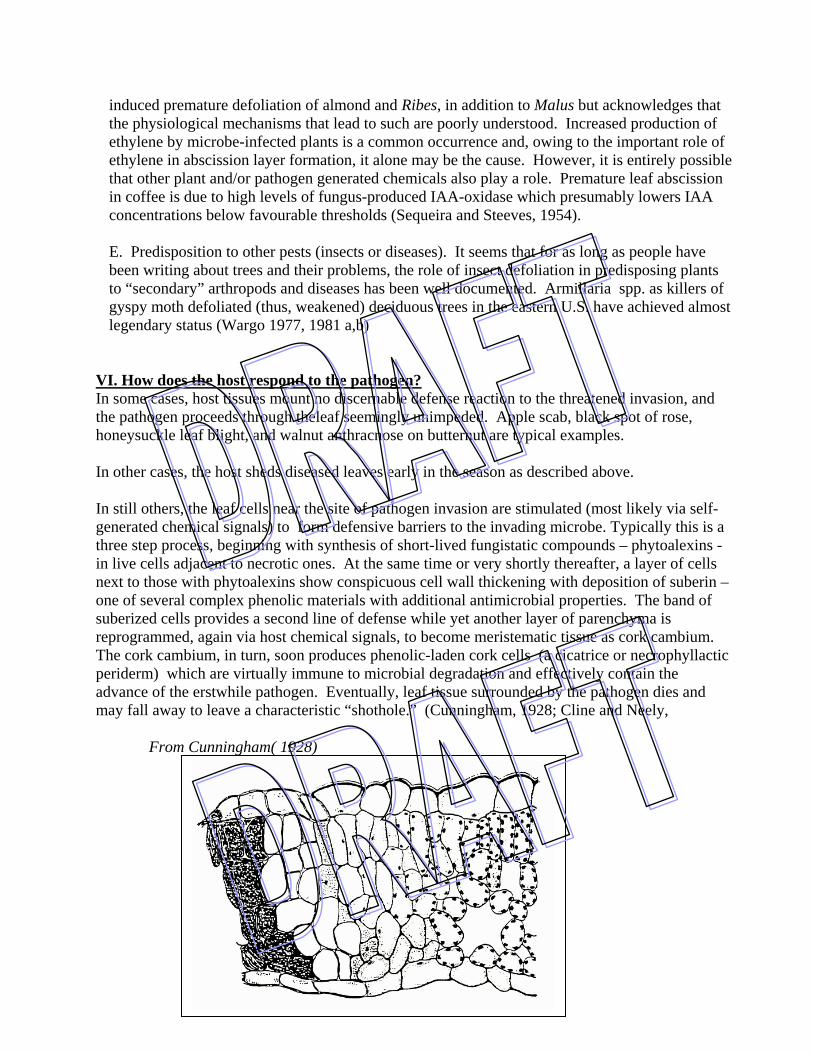
VI. How does the host respond to the pathogen? In some cases, host tissues mount no discernable defense reaction to the threatened invasion, and the pathogen proceeds through theleaf seemingly unimpeded. Apple scab, black spot of rose, honeysuckle leaf blight, and walnut anthracnose on butternut are typical examples. In other cases, the host sheds diseased leaves early in the season as described above. In still others, the leaf cells near the site of pathogen invasion are stimulated (most likely via self-generated chemical signals) to form defensive barriers to the invading microbe. Typically this is a three step process, beginning with synthesis of short-lived fungistatic compounds – phytoalexins - in live cells adjacent to necrotic ones. At the same time or very shortly thereafter, a layer of cells next to those with phytoalexins show conspicuous cell wall thickening with deposition of suberin – one of several complex phenolic materials with additional antimicrobial properties. The band of suberized cells provides a second line of defense while yet another layer of parenchyma is reprogrammed, again via host chemical signals, to become meristematic tissue as cork cambium. The cork cambium, in turn, soon produces phenolic-laden cork cells (a cicatrice or necrophyllactic periderm) which are virtually immune to microbial degradation and effectively contain the advance of the erstwhile pathogen. Eventually, leaf tissue surrounded by the pathogen dies and may fall away to leave a characteristic “shothole.” (Cunningham, 1928; Cline and Neely,
From Cunningham( 1928)

Some cells in the periderm are permeated with a group of chemicals known as phenolic compounds, which are toxic to many microorganisms and check further growth of the pathogen. Because phenolic compounds are going to come up periodically during the course, take just a moment to learn a bit more about them. First, what are they? Any compound with this structure is considered to be a phenolic.
However, those that are important in plant defense look like this
-OH
-OH
OH

The next logical question is where do they come from?
With the formation of tyrosine, then, you have the building block upon which other chemical reactions can occur to yield a variety of fungitoxic phenols.
What makes phenolics toxic is that upon oxidation, they are converted to quinones. And in that conversion, they pass through a stage known as a free radical. These are highly reactive, and among other things will "lock on" to reactive sites on enzymes.
One of the most common phenolic compounds that is implicated in plant resistance to infectious pathogens is chlorogenic acid
OH
OH OHO
O* O

In addition to the nonspecific and very obvious deposition of phenolic compounds in cell walls near sites of mechanical wounding or invasion by pathogens, there is a large array of other chemical reactions are triggered by pathogens. For instance, we know from exhaustive experimental evidence that soon after a challenge inoculation by a non-pathogenic microbe, plant cells, in response to the challenge, will produce low molecular weight antibiotics collectively called phytoalexins. They vary enormously in chemical structure among plant species and their mode of action is poorly understood, but the association of their rapid synthesis with a first line of defense against plant pathogens is unequivocal.
Unlike phytoalexins, which by definition are synthesized near the sites of inoculation and do not migrate far beyond those sites, there are other chemicals for which synthesis is apparently induced by a primary inoculation with a pathogen, and then those chemicals migrate through the plant to trigger syntheis of secondary compounds that actually confer resistance to secondary inoculations. The nature of the chemical signals remains a challenge to science, but one well known product of their action is synthesized as salicylic acid, a simple phenolic compound.
Unfortunately, little work on this signal-mediated systemic resistance (a.k.a. "systemic acquired resistance” or “systemic induced resistance" has been done on leaf diseases of trees. But its common occurrence in the plant kingdom suggests that it is, indeed, a significant phenomenon and one that should eventually attract the attention of tree pathologists seeking to understand resistance reactions. For more information on systemic acquired resistance in plants, but not trees, see: Hammerschmidt, R. and Nicholson, R.L. 1999. A survey of plant defense responses to pathogens. Pp. 55-72 in A.A. Agarawal, S.Tunzun, and Bent, E. eds. Induced Plant Defenses Against Pathogens and Herbivores. APS Press. St. Paul Hammerschmidt, R. and Smith-Becker, J. 1999. The role of salicylic acid in disease resistance Pp. 37-54 in A.A. Agarawal, S.Tunzun, and Bent, E. eds. Induced Plant Defenses Against Pathogens and Herbivores. APS Press. St. Paul
COOH
OH
VII. Ecological considerations:
There are literally hundreds of leaf diseases on trees and shrubs in the northeast U.S. and as you move south, you find even more. Learning how to identify the diseases is a formidable task in

and of itself, but to require that one also know all of the details of life cycles and other germane facts about the natural history of these pathogens borders on cruel and unusual punishment. Fortunately, most leaf pathogens can be neatly fit into one of three categories, and knowing which of the three fits a particular situation can be very important in making management decisions later on. If you can just learn about a few in some detail, you can safely draw some rather broad conclusions about many others. AN ARTIFICIAL BUT FUNCTIONAL CLASSIFICATION OF FOLIAGE DISEASES Primary inoculum only Spring Tar spots of maple, willow, holly, etc. Rhabdocline, Ploioderma needlecasts Azalea petal blight Primary inoculum only Mid-summer Lophodermium needlecast Primary & secondary Spring Many anthracnoses inoculum then Apple scab season-long Powdery mildews Many other leaf diseases
Now, let us carry this study of leaf pathogens a bit further by considering a group of diseases known as anthracnose diseases. ANTHRACNOSE is a collective term used to refer to all of those diseases of deciduous plants caused by fungi that produce conidia in acervuli. If the fungi produce ascospores (and most do), those ascospores will be produced in perithecia. In some crops (and even some trees and shrubs), anthracnose diseases attack twigs and branches as well as leaves, but for purposes of developing concepts here, only anthracnoses of leaves will be considered.

SOME COMMON ANTHRACNOSE DISEASES IN NEW YORK Ash, white ..........................………… Gnomoniella fraxini (Discula fraxinea) Birch: Paper/ Eur. white …………... Discula betulina, Cryptocline betularum Dogwood........…………………… Sphaceloma corni (Elsinoe corni), Discula destructiva Hickory..............................……....... Gnomonia caryae (Asteroma caryae) Hornbeam / Hophornbeam ..……… Apiosporiopsis carpinea (Monostichella robergei) Maple........................................... Kabatiella apocrypta, Didymosporina aceris, Discula campestris Populus spp.......................... …… Drepanopeziza populi-albae (Marssonina castagnei)
Oak (esp. white)..................…….. Apiognomonia quercina (Discula quercina) Plane tree ..........................……..... Apiognomonia veneta Privet...................................…..…... Glomerella cingulata Snowberry ........................…….... Sphaceloma symphoricarpi Walnut...................................….... Gnomonia leptostyla (Marssoniella juglandis) The preceding list gives some indication of the diverse array of fungi that can be included under the category of "anthracnose" diseases, but despite the large number, all are relatively similar in the way they grow, reproduce, and cause disease. By focusing on one of these and learning its intricacies, you should be in a position to make some assumptions about others in the same group. The one anthracnose disease which has received the most attention in recent times is walnut anthracnose caused by Gnomonia leptostyla (Marssoniella juglandis) The pathogen overwinters (OW) as immature perithecia in leaves infected the previous year, but it may also OW in leaf rachises which are attached to the tree. With the onset of warm weather, perithecia mature and ascospores are liberated. They are then carried to newly unfolding leaflets where they germinate and cause new infections. Under favorable conditions of temperature and moisture, spots develop on the leaves 12-24 days later, and acervuli are produced. Conidia produced in the acervuli are rainsplashed to new infection sites and the cycle continues throughout the summer. The reproductive potential for acervuli is tremendous. In some measurements for a study on black spot of rose (not an anthracnose, but much like one), a 1 square inch lesion produced approximately 3600 acervuli on the upper leaf surface and 2400 on the lower leaf surface. Each acervulus produces about 4000 conidia per day and continues to produce conidia for about 10 days. Thus, a 1 square inch lesion produces approx. 2.4 X 10⁸ conidia in its lifetime. Effects of various environmental parameters on leaf disease incidence Weather is crucial for determining whether or not spores are produced, whether or not they are disseminated, whether or not they infect leaves, and - if all of that happens, whether or not

they cause symptoms. One disease that received considerable attention in this regard (albeit 25 years ago!) is walnut anthracnose caused by Gnomonia leptostylla. G. leptostylla is typical of most anthracnose pathogens. The fungus overwinters on leaf debris on the ground OR in rachises that may remain suspended in the crown. In spring, the pathogen produces ascospores that are shot into the air and carried by wind currents to new leaves. If infection is successful, the spores will begin to decay the leaf tissue and within as little as 10 days, new fruit bodies - acervuli - will appear. Each acervulus will produce conidia, and each conidium is capable of causing more infection. With such a phenomenal reproductive capacity, the disease quickly escalates IF weather for all of these events is favorable. So…what is that? William Black and Dan Neely [1978, Phytopathology 68 (7):1054-1056] of the University of Illinois wanted to find out so they conducted a series of experiments, inoculating walnut seedlings with conidia produced in culture. First, they sprayed the seedlings with a spore suspension of spores and then suspended a plastic bag over each plant to keep humidity high. Then, they put some plants in each of several chambers to observe symptom development over a 2 week period. As you can see from the graphs below, after two weeks at 10C, there were little or no symptoms. At 20C, 80 per cent of leaves receiving approximately the same number of spores showed symptoms, and at 30C, there were virtually no symptoms. So, it's probably safe to conclude that the optimum temperature for infection and symptom expression is somewhere around 20C. By the way, the dotted line indicates the amount of symptom expression 2 more weeks later after the seedlings that had been held at 10C were moved to a 20C chamber. Obviously, the cooler temperature didn't kill the spores but it did delay some part of the process that eventually leads to symptom expression. From Black and Neely(1978)

In a second set of experiments, they considered both temperature and moisture. Leaves on seedlings were inoculated as before and the seedlings were covered with plastic bags and moved to chambers of differing temperatures. The difference in these experiments was that at various time intervals thereafter, the bags were removed and the leaf surface dried a bit. From the results, it is clear that at the lowest temperature, maximum infection was achieved when leaves were kept wet for at least 48 hr. At 20C however, 24 hr of leaf wetness was sufficient and at 30C, the spores were apparently killed.
In yet a third set of experiments, Black and Neely wondered how long after leaves were inoculated, the spores could tolerate drying before rewetting would trigger germination and infection. So, leaves were inoculated as before and moved to 20C chambers where they dried. At periodic intervals thereafter, some leaves were misted with water and bagged. Clearly, drying had an adverse effect on disease incidence, but what is especially impressive is that even after 12 days, enough spores remained alive to cause some visible symptoms.

Finally, Black and Neely inoculated seedlings as before and while doing their best to maintain constant temperatures - placed the seedlings in various light regimes. The results weren't surprising, but they did lend added insight into the lives of anthracnose pathogen. Most have relatively thin-walled spores that readily succumb to the toxic effects of UV light. EFFECT OF LIGHT INTENSITY AFTER INOCULATION:
PLANTS HELD IN SHADE -- 80-90% INFECTED UNDER GLASS -- 13% INFECTED FULL SUN -- 8% INFECTED In summary, results of these experiments as well as results of experiments with related foliar pathogens provide good evidence that temperature, moisture, and light intensity are the key factors that determine whether or not you will have a leaf disease epidemic. What do each of these factors contribute to the epidemic?
Moisture: Needed for production and dispersal of spores. Needed internally by spores for fundamental metabolic processes. If the external environment is dry, then spores will dry out and be unable to grow. Water is a medium for transport of leachates (nutrients) from leaf surface. Some spores apparently use host plant exudates to nourish their growth and search for a place of penetration.
One might think that since water seems to be so crucial to success of pathogenic fungi, you can never have too much. But, in fact, that's not the case. Many powdery mildew spores are killed by too much free water on the leaf surface, and years ago one of my grad students had trouble keeping spores alive under water for just a few hours as he travelled from Ithaca to the Adirondacks to conduct some experiments. Temperature: Cool temperature slows down leaf growth and keeps leaves in a young, succulent, easily-invaded (?) stage longer. Cooler temps also retard evaporation, thus prolonging leaf wetness. Light Intensity: In the real world, as light goes up, temperatures usually do too. And increased temperatures speed evaporation. But the other consideration is that UV light is toxic to many spores; especially those that are thin walled and colorless. And most leaf disease pathogens are just that way.

Every once in a while, things come together just right and then - WOW! WAVE YEAR OF INFECTION - modified data of Hitchcock and Cole, 1980 (N.Z.J. of Science Vol. 23: 69-72) on sycamore anthracnose.
Note also that the year after a wave year, even if weather conditions are not especially favorable, leaf disease incidence may be unusually high. That's because even if the window for infection is short, when it is open there is usually ample inoculum there to take advantage of it.
Virtually all leaf disease fungi have these same basic requirements for high moisture, cool temps, and low light, but one group is an exception and those are the powdery mildews. Those fungi can thrive when moisture is low and temperatures are high because a gelatinous covering that is 99.9% water surrounds the conidia. When a powdery mildew spore lands on a leaf, it can utilize its own water supply to germinate and grow until it penetrates the leaf. Thus, powdery mildews thrive in the hottest and driest parts of the year when other pathogens are inactive. From Delp ( 1954)


VIII. Management of leaf diseases: A. Use fungicides Spray Conventional things like: copper hydroxide (Kocide, Phyton 27) chlorothalonil (Bravo, Daconil 2787, Twosome) mancozeb (Dithane M-45, Manzate 200) triadimefon (Bayleton, Strike) thiophanate methyl (Topsin M, Cleary's 3336) propiconazole (Banner MAXX) azoxystrobin (Heritage, Quadris) Inject: triadimefon, Fungisol New developments: Antagonistic phylloplane bacteria collected from abandoned orchards Baking soda (sodium or potassium bicarbonate) Horticultural oil Antitranspirants *Special note: As distasteful as use of fungicides may be to some of you, the fact of the matter is that they do prevent disease when the right material is applied at the right time. Furthermore, in the case of needlecast diseases in the Cornell Plantations, we have found that one fungicide application every 3-4 years seems to be sufficient to keep disease incidence at a tolerable level. B. Modify climate / Avoid high hazard areas 1. Weed control in nurseries & xmas trees 2. Increase spacing 3. In nurseries - limit overhead irrigation 4. Avoid planting in high hazard areas - ie. courtyards, N sides of houses Instead, select gentle slopes or flats where good air drainage is promoted C. Limit primary inoculum - an especially effective control measure for a disease like tar spot that only produces primary inoculum
Unfortunately, this approach is less effective in controlling diseases with primary and secondary inoculum because slight reductions in initial inoculum will be overcome with secondary inoculum cycles. In any case, if you want to try reducing primary inoculum, here are possible strategies:
1. Rake the leaves
2. Accelerate decomposition of leaves on the site. Destroy residual leaves by increasing decay via introduction of saprophytes or promotion of their
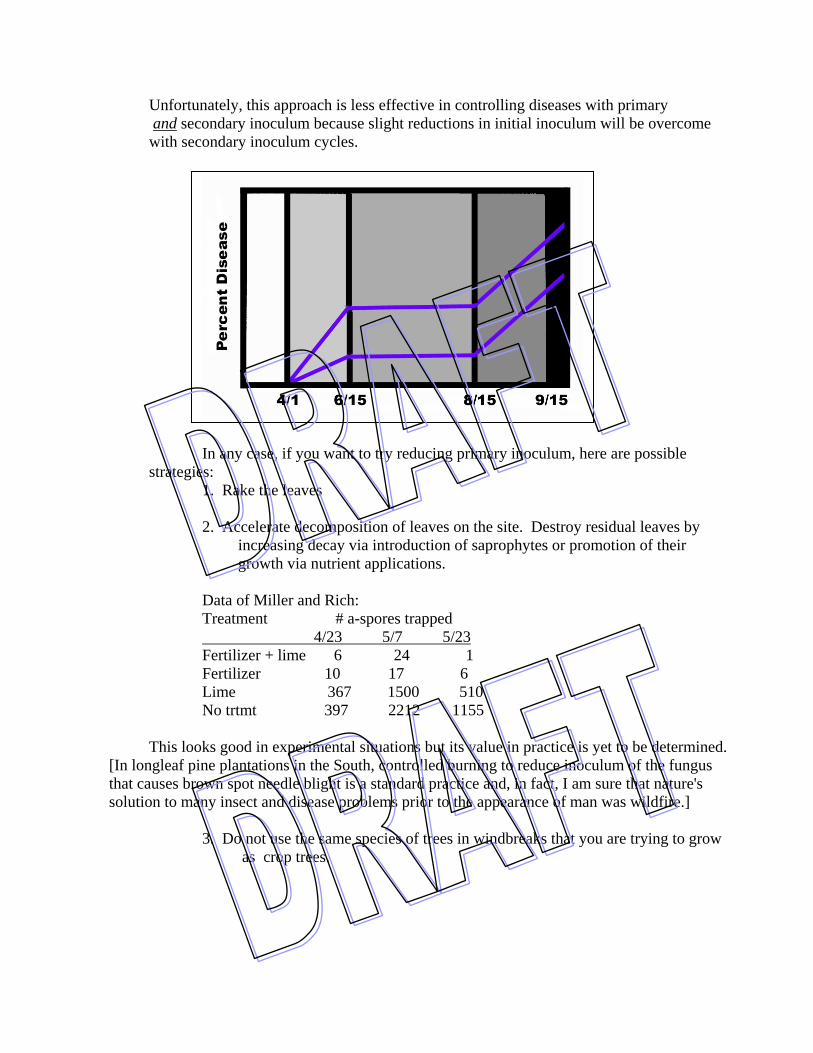
growth via nutrient applications. Data of Miller and Rich: Treatment # a-spores trapped
4/23 5/7 5/23 Fertilizer + lime 6 24 1 Fertilizer 10 17 6 Lime 367 1500 510 No trtmt 397 2212 1155 This looks good in experimental situations but its value in practice is yet to be determined. [In longleaf pine plantations in the South, controlled burning to reduce inoculum of the fungus that causes brown spot needle blight is a standard practice and, in fact, I am sure that nature's solution to many insect and disease problems prior to the appearance of man was wildfire.] 3. Do not use the same species of trees in windbreaks that you are trying to grow as crop trees.

D. Use resistant varieties: • leaf blight resistant hawthorns - English (crataegus oxycantha) are very susceptible to blight • disease resistant flowering crabapples rust, powdery mildew, scab, fireblight • London plane: American sycamore (susceptible) x Turkish plane (resistant) =
London plane - looks like a sycamore but anthracnose resistant
E. Don't do anything? What are the consequences if trees lose some leaves every year or every once in awhile?
And how many leaves do they have to lose before it makes any difference to them? First, look first at how a normal deciduous tree functions, and begin by considering this simplified version of a graph of starch content in a deciduous tree adapted from data collected by Parker and Houston ( 1971 ) and by Siminovitch et al (1953) When leaves first start to grow in the spring,, virtually all of the resources they need to get bigger come from stored reserves; they might legitimately be considered parasites because they give little or nothing back. However, once they reach full size, they become suppliers. Furthermore, we know from earlier discussions that leaves in various parts of the crown of a tree may differ significantly in the amount of photosynthate they contribute to the overall resources of the tree. Some may do so little for the overall health of the tree that their loss might have no demonstrable effect.

So it's logical to expect that some leaves in the crown of a tree may be more expendable than others. But how can we predict what the impact of various levels of defoliation and times of defoliation might be? It seems that there are three possible approaches:
1. Look at data from non-defoliated trees, such as that above, and make inferences about effects of defoliation.
2. Actually go out and pick leaves off of trees and then return later to measure the impact of that action. 3. Sit around and wait until some insect or disease comes along and does the job for you.
All of these have been used and all have advantages and disadvantages, but when results from several efforts to study effects of defoliation yield similar results, we can gain some measure of confidence that the method of data collection and the conclusions are valid. In the case of deciduous trees, some of the best information on this subject comes from studies where people actually go out and pick the leaves off of trees. A classic series of experiments were carried out by Heichel and Turner who were interested in effects of defoliation of red oak and red maple at the Harvard Forest. Their methods weren't rocket science but their conclusions have been the foundation for much of the work on defoliation of deciduous trees that has followed. Essentially, they manually defoliated the study trees just after maximum leaf expansion. On some trees, they took half the leaves, on others 75 percent and on still others 100 percent In an effort to simulate insect injury, they took only the leaf blades and not the petioles. They recorded a wide array of responses including whether or not new leaves were produced and, if so, how many and how large. It turns out that with their trees, up to 50% of the leaves could be removed from either species with little or no apparent effect on the tree. No new leaves were produced and growth in the subsequent year was apparently also unaffected. If defoliation exceeded 50 percent, however, the trees responded by forming new leaves that were smaller and were fewer in number than the amount removed. Overall, photosynthetic area was reduced. Purposeful defoliation above 50% to encourage replacement with smaller leaves has advantages inasmuch it is routinely used in bonsai culture.

Mid- to late-season (July 14 was latest) defoliation that results in sufficient loss to leaves to trigger refoliation also delays hardening off of twigs, making them susceptible to early winter injury. The buds swell in preparation for refoliation but because it is late in the season, the leaves may not actually emerge and the tender, swollen bud is extremely susceptible to freezing injury.

Consider also the data of Parker and Houston (197 )who measured starch stored in roots of trees that had been defoliated just as leaves had reached maximum expansion. They took measurements before budbreak, just after budbreak, just after defoliation, and just after budbreak the following year.
.
SUMMARIZE: CRUCIAL FACTORS TO CONSIDER IN PREDICTING THE FATE OF DEFOLIATED TREES.
1. SEVERITY of defoliation (40 - 50% tolerable) 2. FREQUENCY of defoliation (one year complete = OK)
Spring, 2002
Summer, 2001(after defoliation & refoliation )
Spring, 2001 after new leaf growth
Fall, 2000
3. TIMING of defoliation
4. HOST VIGOR . Predictions from each item above are predicated on notion that tree is otherwise in good condition.
GWHudler , Cornell Univ .
1
32

So far, the discussion has centered on the fate of deciduous trees faced with the prospects of defoliation. But what about conifers - the evergreen kind? Their needles are intended to serve a useful function for more than just one year and one might expect that losing them could have a more profound effect. In fact, that does seem to be the case most of the time. All you have to do is go to an abandoned Christmas tree plantation where needlecast diseases or needle feeding insects have been allowed to progress uncontrolled and you will find that many of the trees are either dead or close to it. But predicting the the responses of conifers to loss of various age classes can involve far more than simple subtraction of the contribution of that age class to the overall photosynthate budget. For instance, consider the case with balsam fir and two insects that feed on its foliage. One, the balsam fir sawfly, only eats needles that are one year old and older. The other, the spruce budworm, only eats current year's needles unless the population is extremely high. Knowing that the current year's foliage contributes the largest percentage of photosynthate to the overall budget might lead one to expect that removal of that age class of needles would be cause more damage than loss of older needles. But it doesn't . In fact, spruce budworm defoliation of current years needles causes older needles to increase photosynthetic activity and the defoliation also triggers elongation of epicormic buds with some amount of replacement foliage. The responses and explanations therefore are detailed in: Vanderklein, D.W. and Reich, P.B. 1999. The effect of defoliation history on photosynthesis, growth and carbon reserves of two conifers with contrasting leaf lifespans and growth habits. New Phytol. 144: 121-132. Little, C.H.B, Lavigne,M.B. and Ostaff, D.P. 2003. Impact of old foliage removal, simulating defoliation byh the balsam fir sawfly, on balsam fir tree growth and photosynthesi of current-year shoots. Forest Ecology and Management 186: 261-269. In contrast, data of O'Neil(1962) clearly shows that in the case of jack pine, loss of current years needles is far more damaging than loss of older needles. Thus age class as well as defoliation intensity are variables to consider..
.
Mean Height Growth in 1956 and 1957 and Mean Diameter Growth in 1957 for the Control and Six Groups of Jack Pine Artificially Defoliated in
August 1956
Mean Height Growth (inches) Age Class of Needles
Removed
Number of Trees 1956 1957
Mean Diameter Growth (in) -
1957 Current Year 1 yr old only 2 yr old only 3 yr old only 2 & 3 yr old 1, 2, 3 yr old
None
10 8 10 8 7 10 10
15.30 16.75 15.25 13.75 15.10 14.75 14.80
1.6* 8.55 8.65 8.90 8.
6.55 9.90
0.15 0.20 0.23 0.23 0.20 0.12 0.24
* Six of these trees produced no leaders in 1957 as a result of the defoliation From O'Neill ( 1962)

LITERATURE CITED: Black, W. and Neely, D. 1978. Effects of temperature, free moisture, and relative humidity on the occurrence of walnut anthracnose. Phytopathology 68 (7):1054-1056. Bradshaw, R.E., Bhatnagar, D., Ganley, R.J., and Gillman, C.J. 2002. Dothistroma pini, a forest pathogen, contains homologs of aflatoxin biosynthetic pathway genes. Applied and Environmental Microbiology 68: 2885-2892. Clark, J. 1956. Photosynthesis of white spruce and balsam fir. Bi-Monthly Progress Rep., Can. Dept. of Agriculture 12(5): 1-2. Cunningham, H.S. 1928. A study of the histologic changes induced in leaves by certain leaf-spotting fungi. Phytopathology 18: 717-751. Heichel, G.H., N.C. Turner, and G.S. Walton. 1972. Anthracnose causes dieback of regrwoth on defoliated oak and maple. Plant Dis. Rep. 56: 1046-1047. Heichel, G.H. and N.C. Turner. 1976. Phenolong and leaf growth of defoliated hardwood trees. Pp. 31-40 in J.Anderson and H. Kaya, eds. Perspectives in Forest Entomology. Academic Press. New York. Miller and Rich. Dormant season nitrogen and apple scab incidence. Parker, J. 1974. Effecs of defoliation, girdling, and severing of sugar maple trees on root starch and sugar levels. USDA For. Serv. Res. Paper NE306. Parker, J. and Houston, D.R. 1975. Effects of repeated defoliatioin on root and root collar extractives of sugar mpale trees. For. Sci. 17: 91-95. Siminovitch, D., Wilson,C.M., and Briggs, D.R. 1953. Studies on the biochemistry of the living bark of the black locust in relation to its frost hardiness. V. Seasonal transformations and variations in the carbohydrates: starch-sucrose interconversions. Plant Physiol. 28: 383-400, Wargo, P.M. , Parker, J., and Houston, D. 1972. Starch content in roots of defoliated sugar maple. For. Sci. 18: 203-204. Wargo, P.M. 1978. Insects have defoliated my tree - Now what's going to happen? J. Arboriculture 4: 169-175. Wargo, P.M. 1975. Estimating starch content in roots of deciduous trees: a visual technique. USDA For. Serv. Res. Pap. NE-313. Wargo, P.M. 1972. Defoliation-induced chemical changes in sugar mpale roots stimulate growth of Armillaria mellea. Phytopathology 62: 1278-1283.

Webb, W. L. and Karchesy, J.J. 1977. Starch content of Douglas-fir defoliated by the tussock moth. Can. J. For. Res. 7: 186-188. Woodman, J.N. 1971. Variation of net photosynthesis within the crown of a large forest-grown conifer. Photosynthetica 5: 50-54.