Embed Size (px)
Citation preview
SYSTEMIC PATHOLOGY II
VPM 222
PATHOLOGY OF LIVER &
PANCREAS
Lab 2
Enrique Aburto Winter 2015
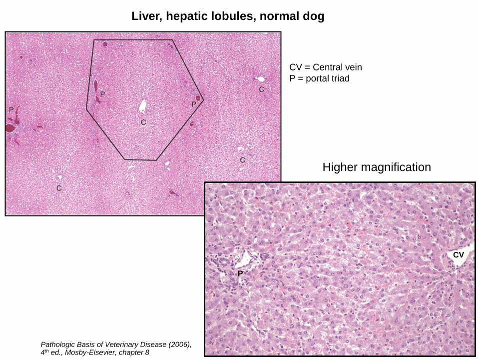
Liver, hepatic lobules, normal dog
Higher magnification
Pathologic Basis of Veterinary Disease (2006), 4th ed., Mosby-Elsevier, chapter 8
CV
P
CV = Central vein
P = portal triad
Regenerative
nodule
F
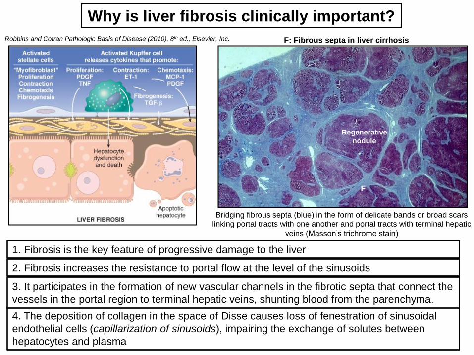
F: Fibrous septa in liver cirrhosis
Why is liver fibrosis clinically important?
2. Fibrosis increases the resistance to portal flow at the level of the sinusoids
Bridging fibrous septa (blue) in the form of delicate bands or broad scars
linking portal tracts with one another and portal tracts with terminal hepatic
veins (Masson’s trichrome stain)
4. The deposition of collagen in the space of Disse causes loss of fenestration of sinusoidal
endothelial cells (capillarization of sinusoids), impairing the exchange of solutes between
hepatocytes and plasma
3. It participates in the formation of new vascular channels in the fibrotic septa that connect the
vessels in the portal region to terminal hepatic veins, shunting blood from the parenchyma.
1. Fibrosis is the key feature of progressive damage to the liver
Robbins and Cotran Pathologic Basis of Disease (2010), 8th ed., Elsevier, Inc.
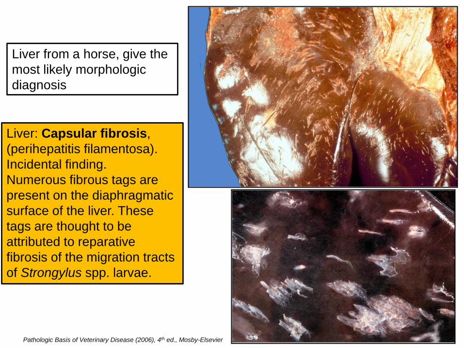
Liver from a horse, give the
most likely morphologic
diagnosis
Liver: Capsular fibrosis,
(perihepatitis filamentosa).
Incidental finding.
Numerous fibrous tags are
present on the diaphragmatic
surface of the liver. These
tags are thought to be
attributed to reparative
fibrosis of the migration tracts
of Strongylus spp. larvae.
Pathologic Basis of Veterinary Disease (2006), 4th ed., Mosby-Elsevier
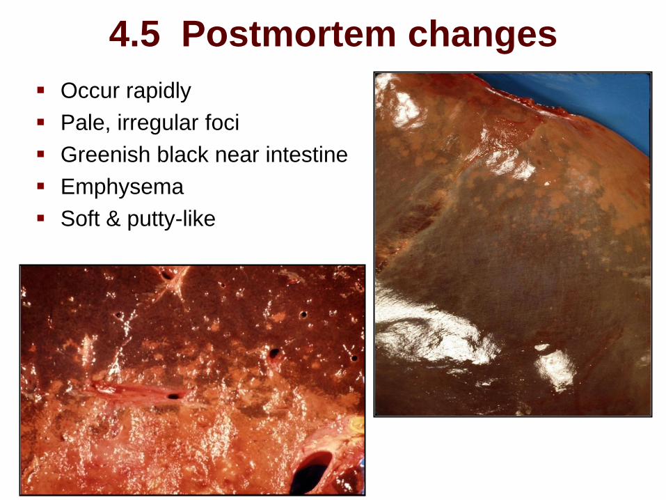
4.5 Postmortem changes
Occur rapidly
Pale, irregular foci
Greenish black near intestine
Emphysema
Soft & putty-like
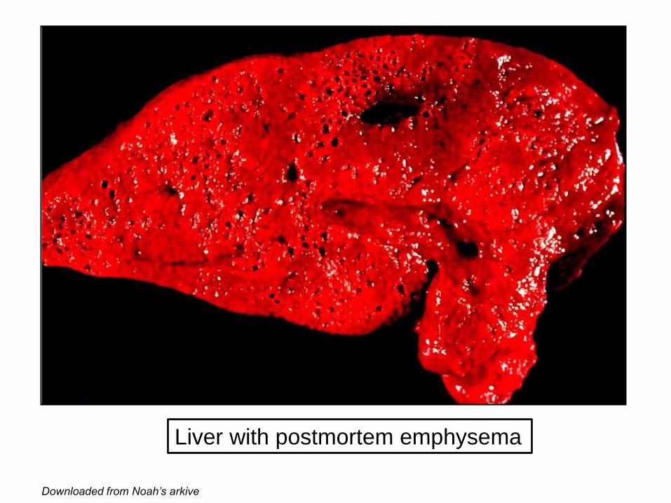
Liver with postmortem emphysema
Downloaded from Noah’s arkive
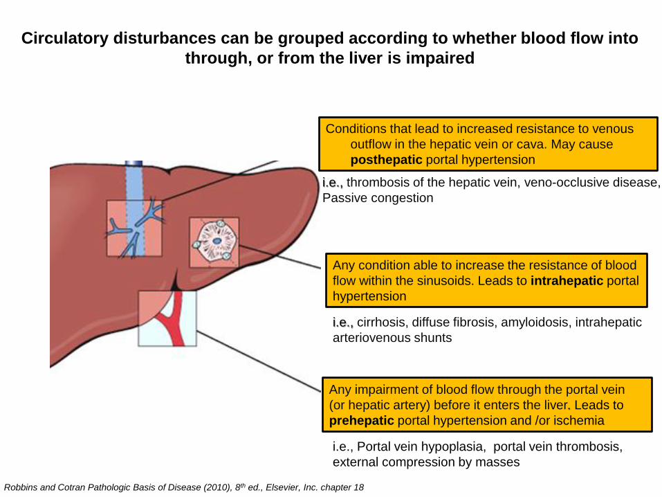
Circulatory disturbances can be grouped according to whether blood flow into
through, or from the liver is impaired
Any impairment of blood flow through the portal vein
(or hepatic artery) before it enters the liver. Leads to
prehepatic portal hypertension and /or ischemia
Any condition able to increase the resistance of blood
flow within the sinusoids. Leads to intrahepatic portal
hypertension
Conditions that lead to increased resistance to venous
outflow in the hepatic vein or cava. May cause
posthepatic portal hypertension
i.e., Portal vein hypoplasia, portal vein thrombosis,
external compression by masses
i.e., cirrhosis, diffuse fibrosis, amyloidosis, intrahepatic
arteriovenous shunts
i.e., thrombosis of the hepatic vein, veno-occlusive disease,
Passive congestion
Robbins and Cotran Pathologic Basis of Disease (2010), 8th ed., Elsevier, Inc. chapter 18
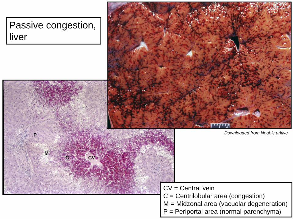
Passive congestion,
liver
C
P
M CV
CV = Central vein
C = Centrilobular area (congestion)
M = Midzonal area (vacuolar degeneration)
P = Periportal area (normal parenchyma)
Downloaded from Noah’s arkive
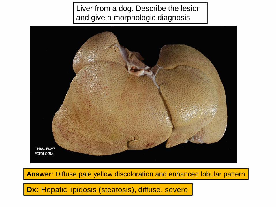
Dx: Hepatic lipidosis (steatosis), diffuse, severe
Liver from a dog. Describe the lesion
and give a morphologic diagnosis
Answer: Diffuse pale yellow discoloration and enhanced lobular pattern
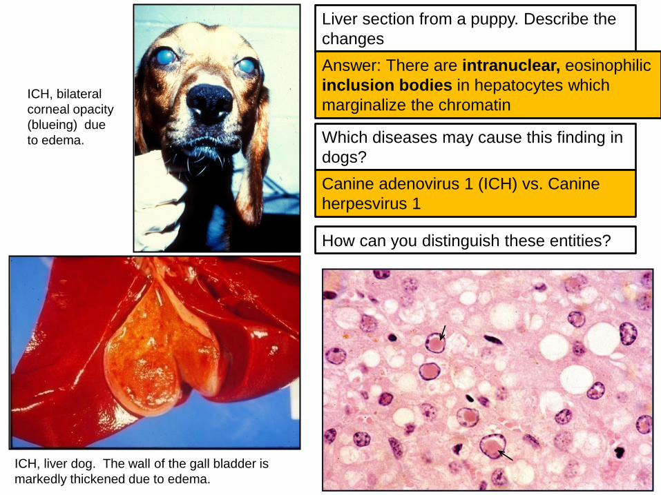
Liver section from a puppy. Describe the
changes
Answer: There are intranuclear, eosinophilic
inclusion bodies in hepatocytes which
marginalize the chromatin
Which diseases may cause this finding in
dogs?
Canine adenovirus 1 (ICH) vs. Canine
herpesvirus 1
How can you distinguish these entities?
ICH, bilateral
corneal opacity
(blueing) due
to edema.
ICH, liver dog. The wall of the gall bladder is
markedly thickened due to edema.

Livers from cows, give a
morphologic diagnosis
for each case
Dx: Hepatic abscesses,
multifocal
Dx: Hepatic granulomas, multifocal
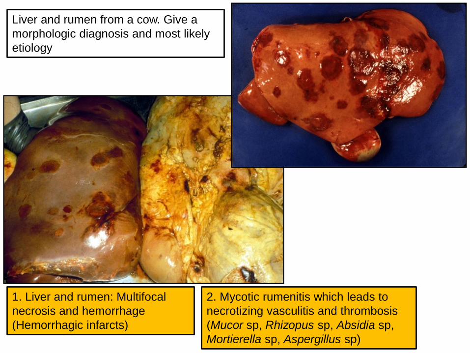
Liver and rumen from a cow. Give a
morphologic diagnosis and most likely
etiology
1. Liver and rumen: Multifocal
necrosis and hemorrhage
(Hemorrhagic infarcts)
2. Mycotic rumenitis which leads to
necrotizing vasculitis and thrombosis
(Mucor sp, Rhizopus sp, Absidia sp,
Mortierella sp, Aspergillus sp)
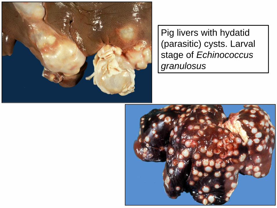
Pig livers with hydatid
(parasitic) cysts. Larval
stage of Echinococcus
granulosus
Downloaded from Noah’s arkive
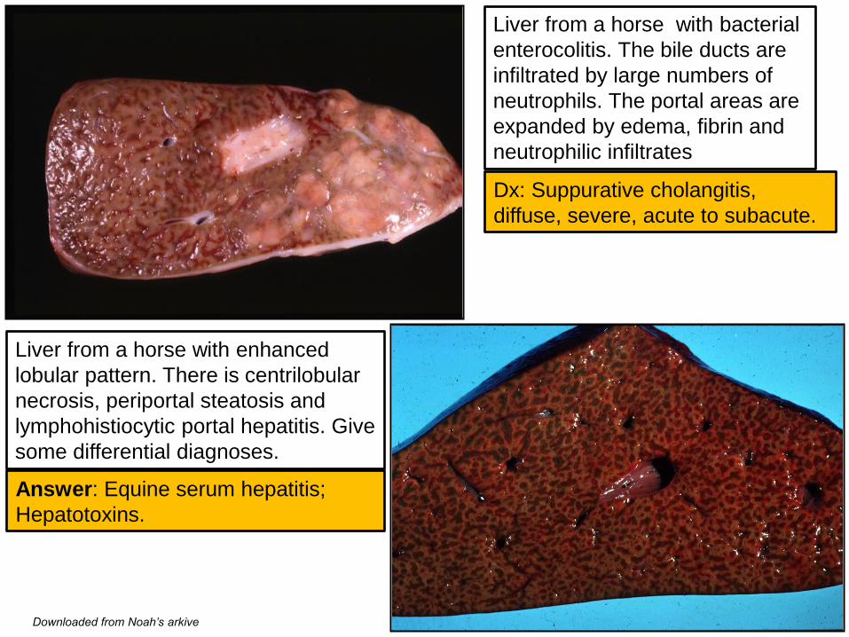
Liver from a horse with bacterial
enterocolitis. The bile ducts are
infiltrated by large numbers of
neutrophils. The portal areas are
expanded by edema, fibrin and
neutrophilic infiltrates
Liver from a horse with enhanced
lobular pattern. There is centrilobular
necrosis, periportal steatosis and
lymphohistiocytic portal hepatitis. Give
some differential diagnoses.
Answer: Equine serum hepatitis;
Hepatotoxins.
Dx: Suppurative cholangitis,
diffuse, severe, acute to subacute.
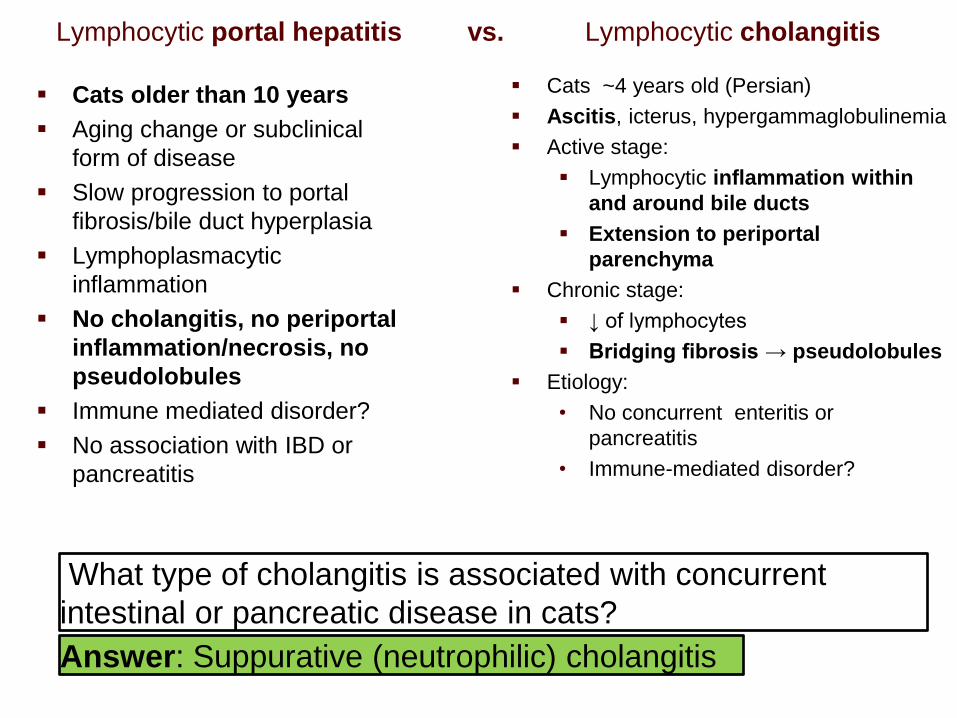
Lymphocytic portal hepatitis vs. Lymphocytic cholangitis
Cats ~4 years old (Persian)
Ascitis, icterus, hypergammaglobulinemia
Active stage:
Lymphocytic inflammation within
and around bile ducts
Extension to periportal
parenchyma
Chronic stage:
↓ of lymphocytes
Bridging fibrosis → pseudolobules
Etiology:
• No concurrent enteritis or
pancreatitis
• Immune-mediated disorder?
What type of cholangitis is associated with concurrent
intestinal or pancreatic disease in cats?
Answer: Suppurative (neutrophilic) cholangitis
Cats older than 10 years
Aging change or subclinical
form of disease
Slow progression to portal
fibrosis/bile duct hyperplasia
Lymphoplasmacytic
inflammation
No cholangitis, no periportal
inflammation/necrosis, no
pseudolobules
Immune mediated disorder?
No association with IBD or
pancreatitis
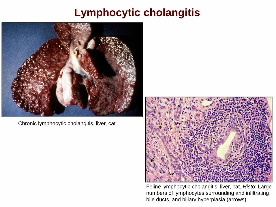
Lymphocytic cholangitis
Chronic lymphocytic cholangitis, liver, cat
Feline lymphocytic cholangitis, liver, cat. Histo: Large
numbers of lymphocytes surrounding and infiltrating
bile ducts, and biliary hyperplasia (arrows).
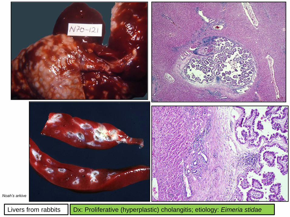
Livers from rabbits
Noah’s arkive
Dx: Proliferative (hyperplastic) cholangitis; etiology: Eimeria stidae

BEST WISHES IN THE FINAL