Embed Size (px)
Citation preview

Pathology of the Hematopoietic
System
Lecture 2:
Myeloproliferative diseases
Lymph nodes
Paul Hanna,
Fall 2014

Hematopoietic Neoplasia
Lymphoproliferative Disease
Lymphoma
Lymphoid leukemia
Plasma cell tumours
Myeloproliferative
Disease
Histiocytic Neoplasia
Myeloid leukemia
Myelodysplastic Syndrome
Mast cell tumour?
Primary Hematopoietic Neoplasia
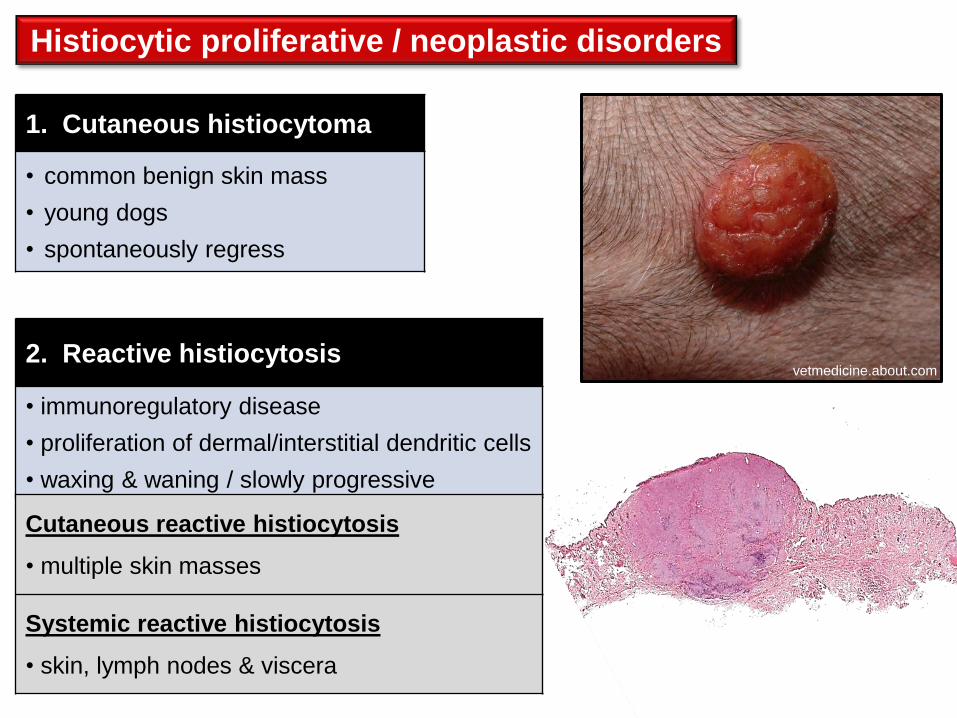
Histiocytic proliferative / neoplastic disorders
1. Cutaneous histiocytoma
• common benign skin mass
• young dogs
• spontaneously regress
2. Reactive histiocytosis
• immunoregulatory disease
• proliferation of dermal/interstitial dendritic cells
• waxing & waning / slowly progressive
vetmedicine.about.com
Cutaneous reactive histiocytosis
• multiple skin masses
Systemic reactive histiocytosis
• skin, lymph nodes & viscera

Histiocytic proliferative / neoplastic disorders
33. Histiocytic Sarcoma
• malignant neoplasia of histiocytes
(macrophages or dendritic cells)
• breed predispositions
Bernese Mountain dog,
Rottweiler, Flat-coated retriever
• solitary lesions
periarticular, skin / subcutis,
lymph nodes, spleen or liver
• multisystemic lesions
disseminated histiocytic sarcoma
= malignant histiocytosis
Dr E Aburto

• aggressive multisystemic disease
tumor masses in many organs, eg spleen,
liver, lung, BM, LN’s, CNS, (rarely skin)
Disseminated histiocytic sarcoma (= malignant histiocytosis)
Histiocytic sarcoma, hilar
& mesenteric lymph nodes
Courtesy of Dr A Lopez, AVC
Histiocytic proliferative / neoplastic disorders
Dr E Aburto
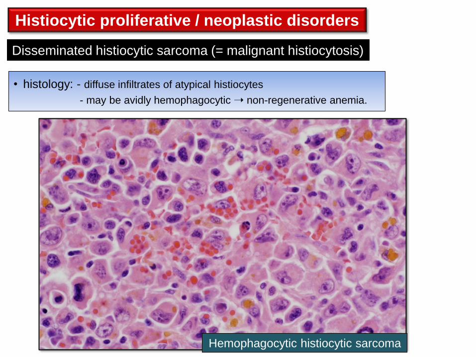
• histology: - diffuse infiltrates of atypical histiocytes
- may be avidly hemophagocytic non-regenerative anemia.
Disseminated histiocytic sarcoma (= malignant histiocytosis)
Hemophagocytic histiocytic sarcoma
Histiocytic proliferative / neoplastic disorders

Mast cell neoplasia
• common skin tumors of dogs
Cutaneous mast cell tumours
• rare, mostly cats.
• two main forms: splenic and intestinal
Systemic mastocytosis*
Mast cells are widely distributed in connective tissues - but originate in BM
For information only
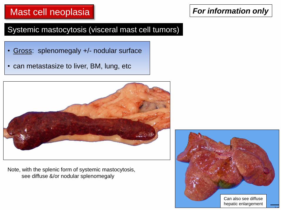
• Gross: splenomegaly +/- nodular surface
• can metastasize to liver, BM, lung, etc
Systemic mastocytosis (visceral mast cell tumors)
Can also see diffuse
hepatic enlargement
Mast cell neoplasia
Note, with the splenic form of systemic mastocytosis,
see diffuse &/or nodular splenomegaly
For information only
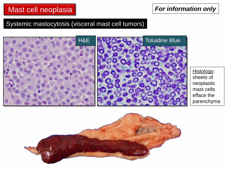
Histology:
sheets of
neoplastic
mast cells
efface the
parenchyma
Mast cell neoplasia
Systemic mastocytosis (visceral mast cell tumors)
Toluidine Blue H&E
For information only
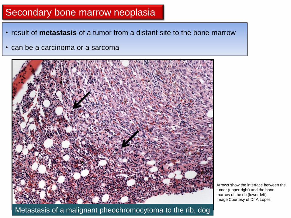
Secondary bone marrow neoplasia
• result of metastasis of a tumor from a distant site to the bone marrow
• can be a carcinoma or a sarcoma
Metastasis of a malignant pheochromocytoma to the rib, dog
Arrows show the interface between the
tumor (upper right) and the bone
marrow of the rib (lower left)
Image Courtesy of Dr A Lopez
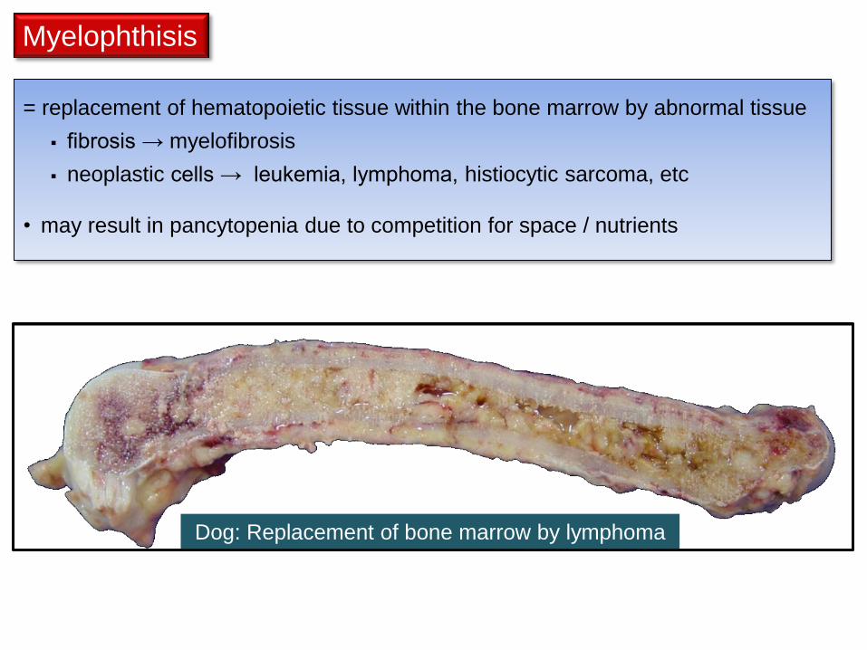
Myelophthisis
= replacement of hematopoietic tissue within the bone marrow by abnormal tissue
fibrosis → myelofibrosis
neoplastic cells → leukemia, lymphoma, histiocytic sarcoma, etc
• may result in pancytopenia due to competition for space / nutrients
Dog: Replacement of bone marrow by lymphoma
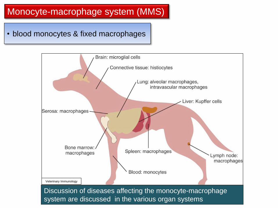
Monocyte-macrophage system (MMS)
• blood monocytes & fixed macrophages
Discussion of diseases affecting the monocyte-macrophage
system are discussed in the various organ systems
Veterinary Immunology
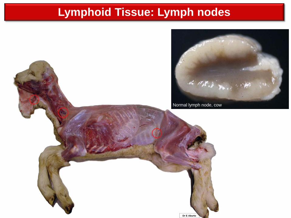
Lymphoid Tissue: Lymph nodes
Normal lymph node, cow
Dr E Aburto
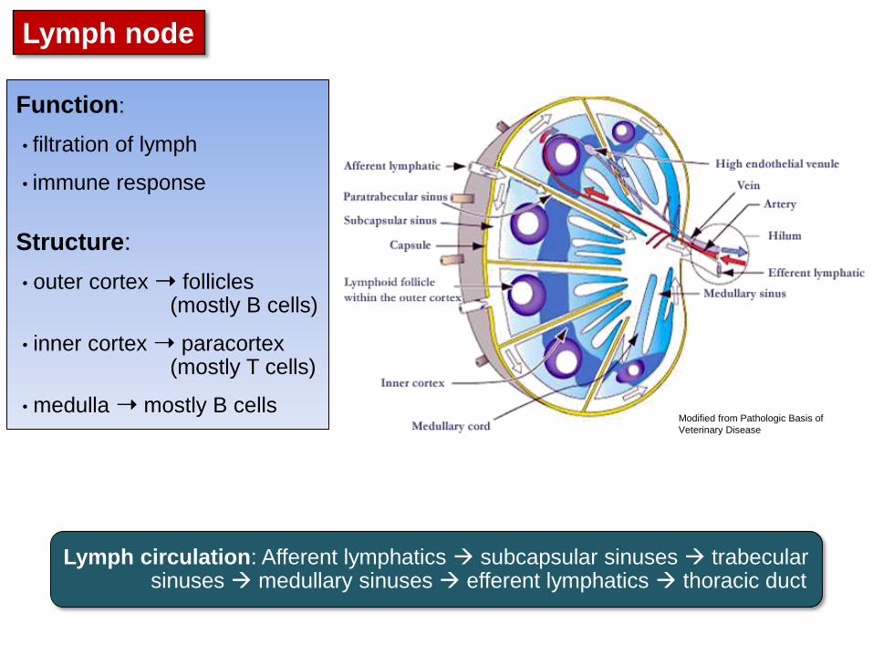
Lymph node
Function:
• filtration of lymph
• immune response
Structure:
• outer cortex ➝ follicles (mostly B cells)
• inner cortex ➝ paracortex (mostly T cells)
• medulla ➝ mostly B cells
Lymph circulation: Afferent lymphatics subcapsular sinuses trabecular sinuses medullary sinuses efferent lymphatics thoracic duct
Modified from Pathologic Basis of
Veterinary Disease
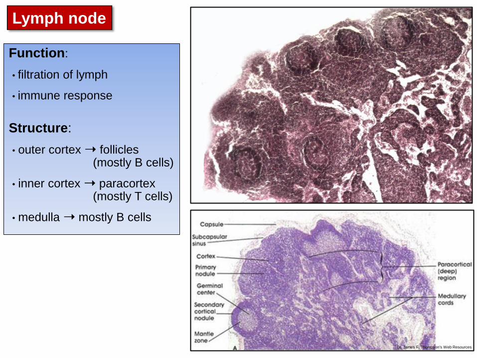
Lymph node
Noah’s Arkive
Normal lymph node, cow Function:
• filtration of lymph
• immune response
Structure:
• outer cortex ➝ follicles (mostly B cells)
• inner cortex ➝ paracortex (mostly T cells)
• medulla ➝ mostly B cells
Dr. James F. Thompson's Web Resources

Lymph node: Anthracosis
Lymph node: Miscellaneous changes
• black discoloration due to carbon within phagocytes
Lymph node: Hemorrhage
• can originate within the lymph node or in the tissues drained by the node
• red discolouration of the lymph nodes
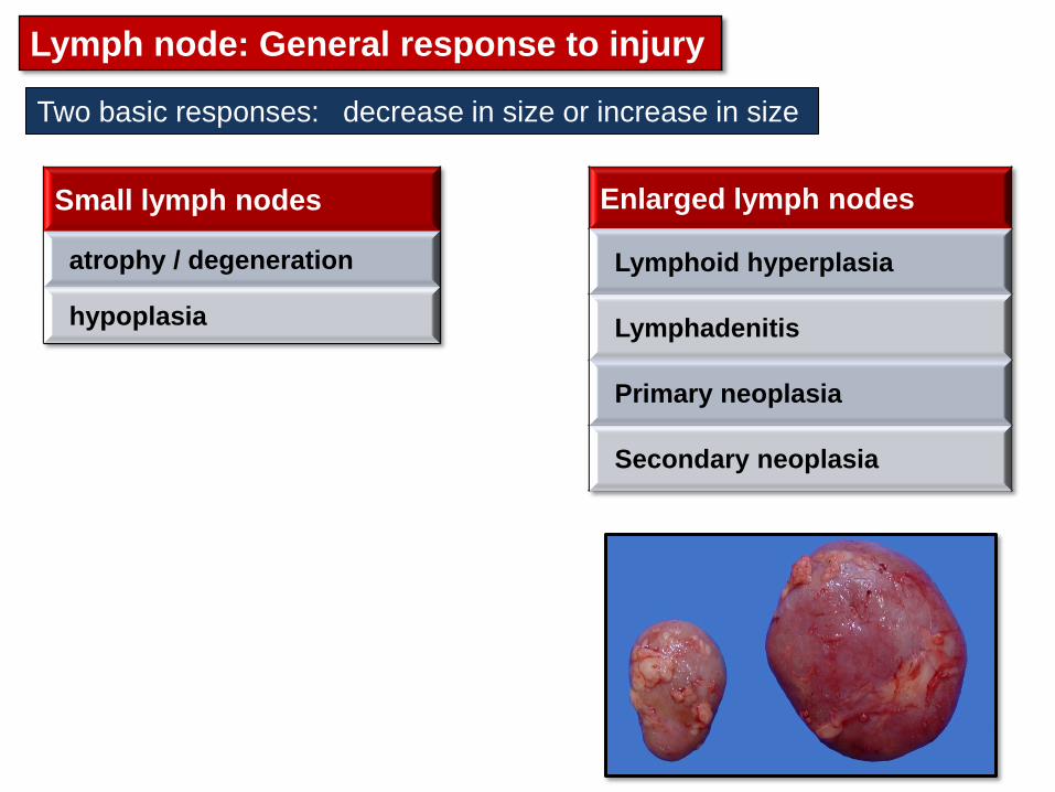
Lymph node: General response to injury
Enlarged lymph nodes
Lymphoid hyperplasia
Lymphadenitis
Primary neoplasia
Secondary neoplasia
Small lymph nodes
atrophy / degeneration
hypoplasia
Two basic responses: decrease in size or increase in size

Lymph node atrophy / degeneration
Senile atrophy (aging)
Cachexia / malnutrition
Viral infection
• BVD
• FIV
• CDV
• FPV / CPV
Toxins / Drugs / Irradiation
Lesions:
• Gross: small lymph nodes
• Histo: lymphoid depletion /
degeneration
Small lymph nodes
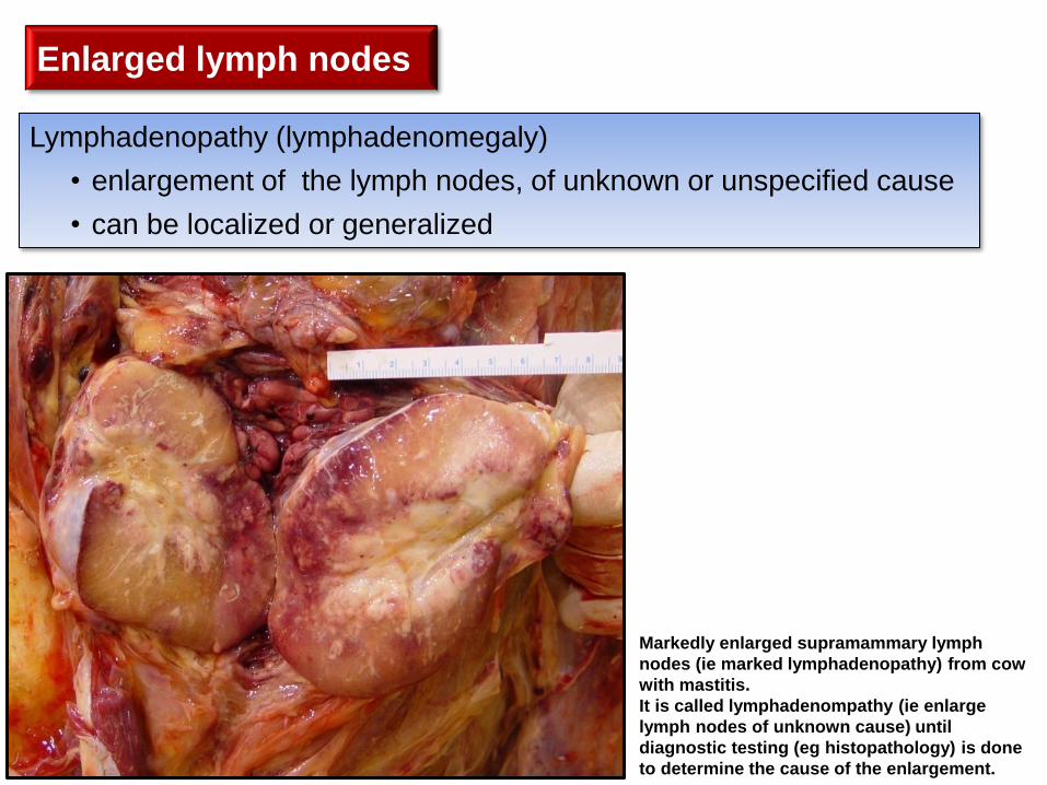
Lymphadenopathy (lymphadenomegaly)
• enlargement of the lymph nodes, of unknown or unspecified cause
• can be localized or generalized
Enlarged lymph nodes
Markedly enlarged supramammary lymph
nodes (ie marked lymphadenopathy) from cow
with mastitis.
It is called lymphadenompathy (ie enlarge
lymph nodes of unknown cause) until
diagnostic testing (eg histopathology) is done
to determine the cause of the enlargement.
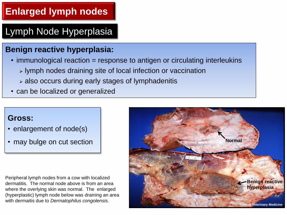
Benign reactive hyperplasia:
• immunological reaction = response to antigen or circulating interleukins
lymph nodes draining site of local infection or vaccination
also occurs during early stages of lymphadenitis
• can be localized or generalized
Lymph Node Hyperplasia
Gross:
• enlargement of node(s)
• may bulge on cut section
Cornell Veterinary Medicine
Normal
Benign reactive
hyperplasia
Peripheral lymph nodes from a cow with localized
dermatitis. The normal node above is from an area
where the overlying skin was normal. The enlarged
(hyperplastic) lymph node below was draining an area
with dermaitis due to Dermatophilus congolensis.
Enlarged lymph nodes
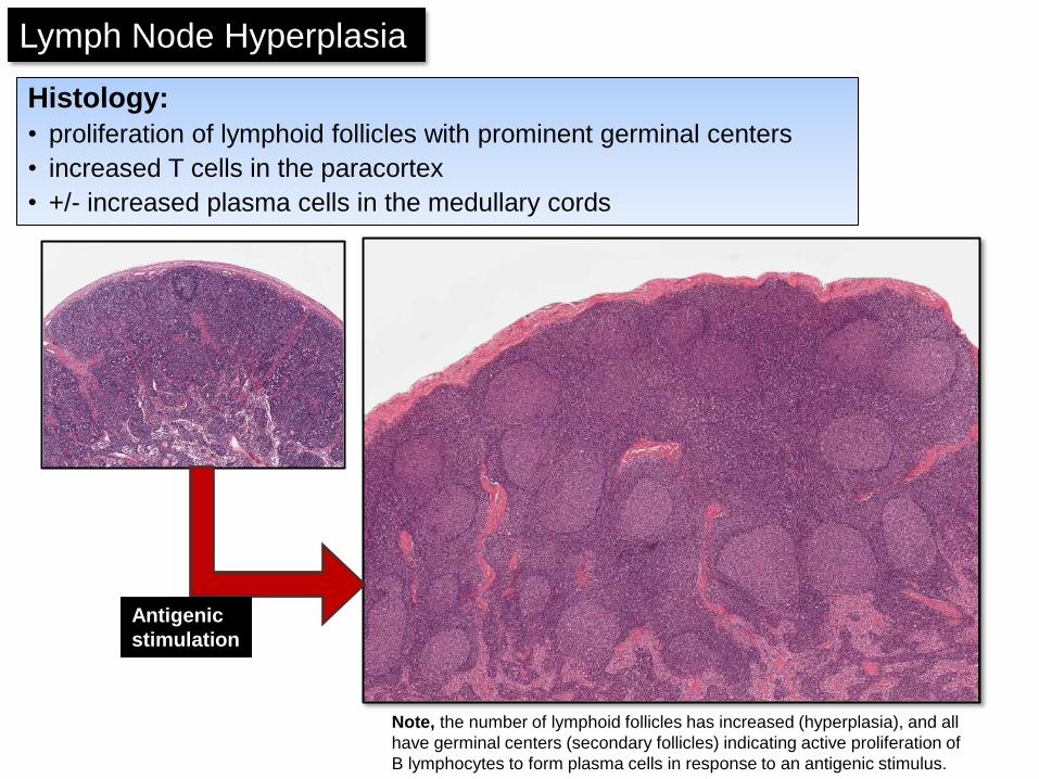
Histology:
• proliferation of lymphoid follicles with prominent germinal centers
• increased T cells in the paracortex
• +/- increased plasma cells in the medullary cords
Note, the number of lymphoid follicles has increased (hyperplasia), and all
have germinal centers (secondary follicles) indicating active proliferation of
B lymphocytes to form plasma cells in response to an antigenic stimulus.
Normal
Antigenic
stimulation
Lymph Node Hyperplasia
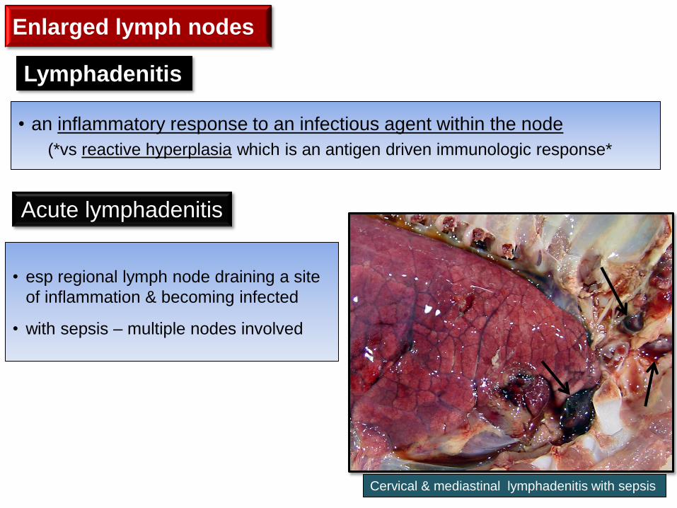
• an inflammatory response to an infectious agent within the node
(*vs reactive hyperplasia which is an antigen driven immunologic response*
Lymphadenitis
• esp regional lymph node draining a site
of inflammation & becoming infected
• with sepsis – multiple nodes involved
Cervical & mediastinal lymphadenitis with sepsis
Acute lymphadenitis
Enlarged lymph nodes

Gross lesions:
• enlarged, soft, wet, red lymph nodes
• often bulging & hyperemic on cut surface
• exudates are usually serous
Tracheobronchial
lymphadenitis
Figure 13-71 (Zachary) Acute lymphadenitis, tracheobronchial
lymph nodes, pig. The nodes are enlarged and reddened from
draining the pneumonic cranial lung lobes. Note the red
consolidation of the dorsal portion of the cranial lung lobes.
Acute lymphadenitis
Mesenteric lymphadenitis
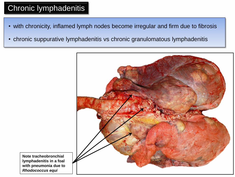
Chronic lymphadenitis
Note tracheobronchial
lymphadenitis in a foal
with pneumonia due to
Rhodococcus equi
• with chronicity, inflamed lymph nodes become irregular and firm due to fibrosis
• chronic suppurative lymphadenitis vs chronic granulomatous lymphadenitis

Chronic Suppurative Lymphadenitis
• swollen lymph node with pus-filled area surrounded by fibrous capsule
= lymph node abscess
• response to pyogenic bacteria
• can fistulate to the skin surface
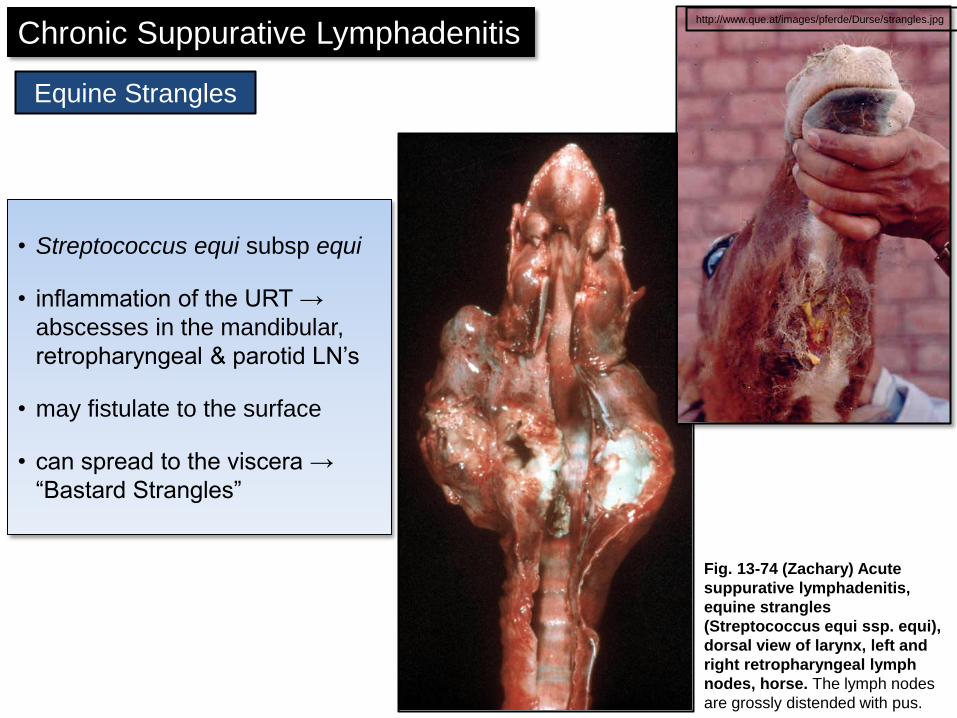
Chronic Suppurative Lymphadenitis http://www.que.at/images/pferde/Durse/strangles.jpg
• Streptococcus equi subsp equi
• inflammation of the URT →
abscesses in the mandibular,
retropharyngeal & parotid LN’s
• may fistulate to the surface
• can spread to the viscera →
“Bastard Strangles”
Equine Strangles
Fig. 13-74 (Zachary) Acute
suppurative lymphadenitis,
equine strangles
(Streptococcus equi ssp. equi),
dorsal view of larynx, left and
right retropharyngeal lymph
nodes, horse. The lymph nodes
are grossly distended with pus.
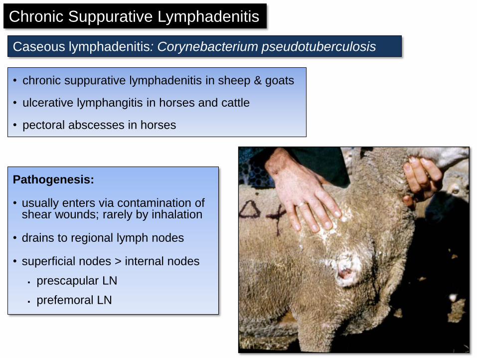
Pathogenesis:
• usually enters via contamination of shear wounds; rarely by inhalation
• drains to regional lymph nodes
• superficial nodes > internal nodes
prescapular LN
prefemoral LN
Caseous lymphadenitis: Corynebacterium pseudotuberculosis
• chronic suppurative lymphadenitis in sheep & goats
• ulcerative lymphangitis in horses and cattle
• pectoral abscesses in horses
Chronic Suppurative Lymphadenitis

• chronic suppurative inflammation and caseous necrosis
• as lesion progresses characteristic concentric laminations
Caseous lymphadenitis: Corynebacterium pseudotuberculosis
Fig. 13-71 (Zachary) Caseous lymphadenitis, Corynebacterium
pseudotuberculosis, lymph node, sheep. The whole lymph node
has been replaced by an abscess containing mostly semifluid
yellowish pus. This is an early stage of caseous lymphadenitis,
before the pus has become inspissated and caseous.
Chronic Suppurative Lymphadenitis
Note, the concentric laminations often seen in
the abscesses of “caseous lymphadentitis” are
considered characteristic of the disease.

• some cases have systemic involvement with abscesses in the internal organs
Caseous lymphadenitis: Corynebacterium pseudotuberculosis
Goat, caudal mediastinal lymph nodes: lymph
node abcesses
Chronic Suppurative Lymphadenitis
Dr E Aburto
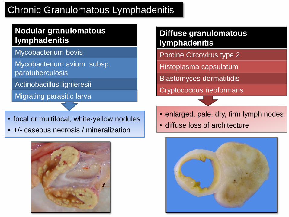
Nodular granulomatous
lymphadenitis
Mycobacterium bovis
Mycobacterium avium subsp.
paratuberculosis
Actinobacillus lignieresii
Migrating parasitic larva
Diffuse granulomatous
lymphadenitis
Porcine Circovirus type 2
Histoplasma capsulatum
Blastomyces dermatitidis
Cryptococcus neoformans
• focal or multifocal, white-yellow nodules
• +/- caseous necrosis / mineralization
• enlarged, pale, dry, firm lymph nodes
• diffuse loss of architecture
Chronic Granulomatous Lymphadenitis
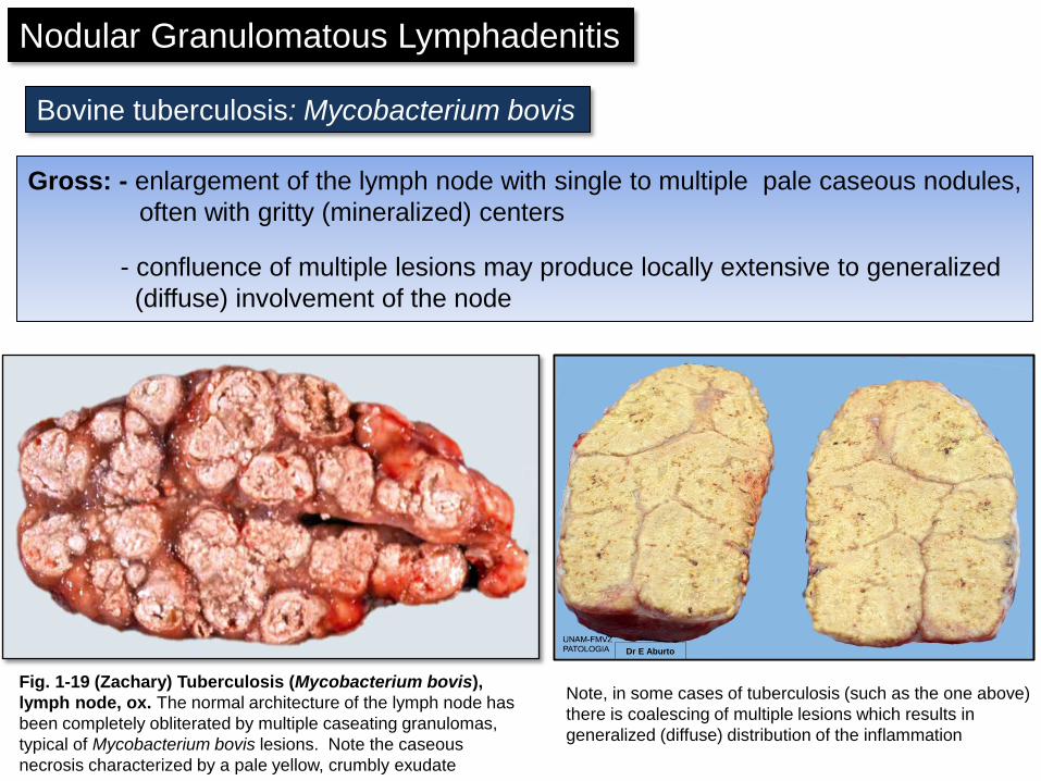
Gross: - enlargement of the lymph node with single to multiple pale caseous nodules,
often with gritty (mineralized) centers
- confluence of multiple lesions may produce locally extensive to generalized
(diffuse) involvement of the node
Bovine tuberculosis: Mycobacterium bovis
Fig. 1-19 (Zachary) Tuberculosis (Mycobacterium bovis),
lymph node, ox. The normal architecture of the lymph node has
been completely obliterated by multiple caseating granulomas,
typical of Mycobacterium bovis lesions. Note the caseous
necrosis characterized by a pale yellow, crumbly exudate
Note, in some cases of tuberculosis (such as the one above)
there is coalescing of multiple lesions which results in
generalized (diffuse) distribution of the inflammation
Nodular Granulomatous Lymphadenitis
Dr E Aburto

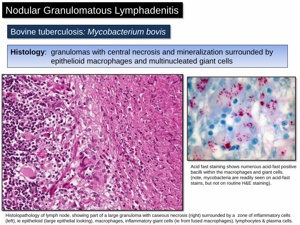
Histology: granulomas with central necrosis and mineralization surrounded by
epithelioid macrophages and multinucleated giant cells
Bovine tuberculosis: Mycobacterium bovis
Nodular Granulomatous Lymphadenitis
Bovine tuberculosis: Mycobacterium bovis
Acid fast staining shows numerous acid-fast positive
bacilli within the macrophages and giant cells.
(note, mycobacteria are readily seen on acid-fast
stains, but not on routine H&E staining).
Histolopathology of lymph node, showing part of a large granuloma with caseous necrosis (right) surrounded by a zone of inflammatory cells
(left), ie epithelioid (large epithelial looking), macrophages, inflammatory giant cells (ie from fused macrophages), lymphocytes & plasma cells.
Histology: granulomas with central necrosis and mineralization surrounded by
epithelioid macrophages and multinucleated giant cells
Nodular Granulomatous Lymphadenitis
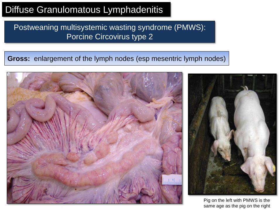
Diffuse Granulomatous Lymphadenitis
Postweaning multisystemic wasting syndrome (PMWS):
Porcine Circovirus type 2
Pig on the left with PMWS is the
same age as the pig on the right
Gross: enlargement of the lymph nodes (esp mesentric lymph nodes)
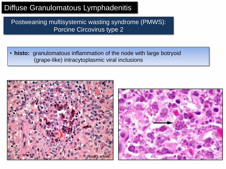
• histo: granulomatous inflammation of the node with large botryoid
(grape-like) intracytoplasmic viral inclusions
Noah’s arkive
Diffuse Granulomatous Lymphadenitis
Postweaning multisystemic wasting syndrome (PMWS):
Porcine Circovirus type 2
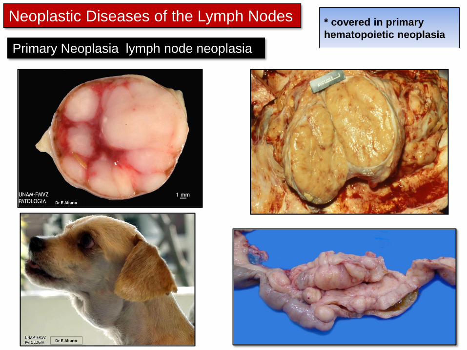
Neoplastic Diseases of the Lymph Nodes
Primary Neoplasia lymph node neoplasia
* covered in primary
hematopoietic neoplasia
Dr E Aburto
Dr E Aburto
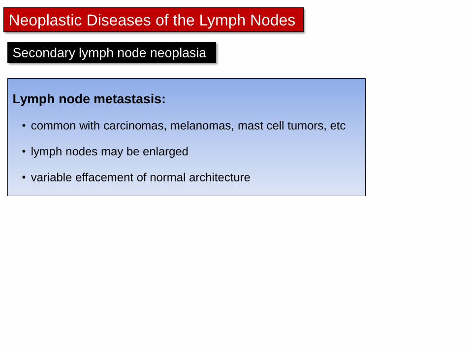
Neoplastic Diseases of the Lymph Nodes
Lymph node metastasis:
• common with carcinomas, melanomas, mast cell tumors, etc
• lymph nodes may be enlarged
• variable effacement of normal architecture
Secondary lymph node neoplasia
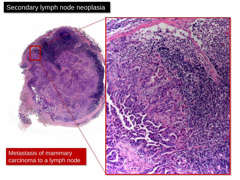
Metastasis of mammary
carcinoma to a lymph node
Secondary lymph node neoplasia