Embed Size (px)
Citation preview
Pathophysiology 8-26Cell injury and death
-etiology-cause of disease-pathogenesis-how disease comes about-morphologic changes-alterations at cellular and organ level-clinical result-what happens to the patient (Sx)
*diseases always start at the cellular level
Causes of cellular injury
1. Hypoxia (three mechanisms)ischemia (lack of blood flow)-main way cell gets deprived of O2.lung malfunctionanemia
2. Physical agentstemperature (+/-)pressureradiationelectric shock
3. chemicals/drugs4. infectious agents (germs)5. immune reactions6. genetic7. nutritional imbalances
How these things injure cells
Disrupt cell membraneInterfere with mitochondria (cut off respiration) therefore energy supplyInterfere with protein production (RNA/DNA synthesis)Damage nucleus (DNA inside)
Reversible vs irreversible injury
There is a point to which injury to a cell is reversible. After this point the damage is irreversible and the cell will die (cell cannot heal)
Apoptosis- Programmed cyclic death of cells (eg. Thymus, epithelium)Text definition- form of cell death designed to eliminate unwanted host
cells through activation of a coordinated, internally programmed series of events effected by a dedicated set of gene products.
1
Intercellular accumulations
-Nl substances can accumulate inside the cell—fatty liverhepatocytes stuffed with fat (DM/obese,etc); cholesterol (hormones),protein (in Alzheimer’s) in brain cells
-Abnl stuff-asbestos, etc.
Calcification—tissues can calcifyDystrophic (older people)-tissues accumulate Ca++ but serum Ca++ is nlMetastatic (younger)-tissues nl but serum Ca++ is high
*if see heart valves/bronchi on x-raycalcification-look pg.45
8/31 Cellular Growth and DifferentiationThree types1. Continuously dividing cells—never exit growth cycle (skin, all blood cells, bone marrow, mucosa)
Stem cells—give rise to other cells—precursor cell that other cells originate frompredifferentiatedcan differentiate to any cell (forebearers, like seeds—can turn into more specific cells2. Quiescent cells—normally don’t divide, but if stimulated properly, they could. Eg. Liver (basement membrane);blood vessels (cuts)3. Non-dividing cells—cannot divide/regenerate. Eg. Brain;neurons
if damagedreplaced by a scarnever regenerate. Eg. Cardiac muscle; peripheral nerves—can’t stim dead nerves.
Molecular events and cell growth Cells grow because of signalsgrowth factor (GF)made of polypeptide (protein) [cells take orders from proteins (GF)]. Hormones can induce cell to tell cell to grow
Oncogenes—genes involved with cancersthey look like GF’s but cause unregulated cell growth
1. GFinteract with receptorreceptor can be on cell surface or intracellular (eg steroids are
intracellular)2. Cell then grows
Growth inhibitor (GI)—suppresses growth.CA usually loss of inhibitionthen GI’s don’t work and GF’s are too
strong
2
Cellular Adaptation—Growth and Differentiation p.32 READ***Any organPhysiologic—response to a nl stimulus (eg. Puberty)Pathologic—response to an abnl stimulus (eg. Abcess)
Hyperplasia—increase in number of cells1. physiologic—breast tissue during puberty2. pathologic—unopposed estrogen—lead to hyperplasia of uterus (womb)—need to take estrogen and progesterone together to prevent overstimulation of the uterus
progesterone prevents pathologic hyperplasia of the uterus*postmenopausal women with uterus need estrogen and progesterone
without uterus—can have estrogen only.
Hypertrophy—increase in size of cells1. physiologic—skeletal muscle in response to exercise2. pathologic—cardia
Atrophy—decrease in size of cells1. lack of workload2. cut nerve (dennervation) –muscle cells need nerve supply to maintain integrity
Lou Gerhig’s Disease—cells in spinal cord deteriorate, therefore, muscles atrophy (dennervation atrophy)3. decreased blood supply (renal artery stenosis)4. poor nutrition5. loss of endocrine stimultion (uterus)after menopause—no hormones—uterus atrophies
PEARL—can’t feel uterus after menopause—if could—bad 6. aging
Metaplasia—reversible change whereby one cell type is replaced by another (eg. Squamous to columnar) (Squamous,columnar,glandular)
Bronchus has columnar cellsciggarette smokesinge ciliaafter timecells change to squamous (squamous are more tolerable to heat)
Chronic refluxcells in lower esophagus change to protect itself from acidthe new cells can become malignant
Cervix (draw papsmear)--outside is squamous cells--lining is columnar (high up)if these switch—then pathologic
zone where cells are changing from squamous to columnar is called the transition zonethis is where cervical ca arisestherefore need sample from transition zoneIf papsmear says metaplasia—then you did it right Dysplasia—bad –look up
3
The way to approach a patient—
Chief complaint—what brought them inHPI—history present illnessPMH—past medical historyMEDS—medicationsFH—family historySH—social history (smoke,oh,work,hobby)ROS—review of systems (rest of body)PE—physical exam(Differential)—all possible diagnosesLAB/XRAY—help sort out the listRX—treatment
9/2/99 Immunology
Immunoglobulins (antibody)—proteins of the immune system produced in rspns to ag’s.Multi-chained proteins that bind antigens (substance that induces formation of
antibodies)—antigens can be soluble (dissolved in H2O) or insoluble
Soluble antigens—eg. Toxin; foreign proteinsInsoluble antigens—eg. Pollen; bacteria; viruses; fungi; (all germs); tissues/cells (autoimmune rxns)
Where antibodies are made—B-cells—develop in bone marrowwhen maturesecreted into plasma
(serum)on antigenic stimulation they differentiate into plasma cells and secrete ab’s
B-cells—secrete ab’sHumoral immunity [SPECIFIC] (in the blood)—b-cellsplasma cells
(secrete)abTonsils; gut; lymph nodes; spleenstore and secrete ab’s.
T-cells participate in cellular immunity [NON-SPECIFIC]Ab’s bind ag’s
Sensitizitation—make a lot of ab’s after first contact with specific antigen
Draw picture of antibody
Top portion (the v part of the y shape) is Fab—it is the binding site—where ag’s bindEach ab has a specific binding site—this binding site is preprogrammed in the
DNA of the plasma cell. It is based on the arrangement of amino acids and therefore hoe they unfold.
Monoclonal Ab—one B-cell makes one kind of Ab—each plasma cell makes different Ab’s.
4
Cross reactivity—when one ab reacts with different (more than one) antigen. Eg. Pcn and rocephin—doesn’t have the same receptor, but in 5% of the people who are allergic to pcn, it is close enough.
An antibody can be present in the body without ever having the ag.
The bottom part of the antibody (the straight part of the Y) is called the Fc region and is the constant.
Agglutination—once ag and ab hook together, they clump and congeal. They group together and stick to eachother—reason—they need to be cleared—can’t be floating around the bloodstream.
5 Classes of Ab’sThe classes are based on the Fc portion of the antibody molecule. It varies slightly between classes.
I. IgG—most common comprising about 70% of all ab’s.G is for gamma—gammaglobulin—it has a clinical use because it has so many
ab’s. It also stays in the body for a long time so it can be used as a marker for past infections.
When IgG binds with ag, the Fc portion twists—called conformational change—this change in the molecule activates the compliment system—[liver made series of 9 proteins that are inactive until compliment system is turned on] the compliment system is the bridge between ag/ab binding and the actual clearing of the molecule (by phagocytosis)
In ab/ag unbound state—the compliment system is turned off
Cascade of rxns—pg. 203 fig.7-131. ab/ag binding2. conformational change3. compliment system activated4. clearing by phagocytosis
mom Rh-; dad Rh+--need rhogam (2nd pregnancy especially)
neutrophils—Fc portion of IgG ab’s binds to receptor on neutrophils—then clear entire molecule
IgG is very small and can pass through the placenta—therefore giving passive immunity
II. IgA—the “secretory” antibody**--found in saliva, resp tract, gi tract, mouth, stomach, tears, sweat, colostrum (breast milk)
5
Can have selective deficiency of IgA—sx are getting cold/flu very frequently—7-10x/year is typical in children, so it has to be a lot more than that!!
III. IgM—big—does not cross the placenta—first class of ab to respond to infection/immunization
Can order certain titers of certain ab’s—this is only acute acting
IV. IgD—don’t know what it does
V. IgE—synonym is reagin—mediates allergic reactions
Hypersensitivity rxnsFloats on mast cells—draw picture---
Fc is bound to mast; Fab is ready to bindMast cell is granular—each granule is filled with histamine—mast cells are found in skin, conjunctiva, nose, etc.
--when ag hits Fab of IgEmast bursts and lets the histamine escape
Histamine-potent vasodilator (ie. Red hives eyes, etc.)—more blood to periphery (no warm bath—only increases the vasodilitation)-increased vascular permeability (rest of immune system leaks out)—edema-bronchoconstrictor
9/7/99Topics—cells (continued); Hypersensitivity Rxns; Histocompatibility Ag’s; Rejection/Graft Tissues; Tumor Immunity
Chapter 7T-cells—mature in the thymus
Found in spleen; lymph nodes; blood(Cellular immunity)
The shape of the t-cell receptor (TCR) is based on charges of aa’s in DNA
CD4+ / T-helper cell—when ag interacts with the receptor of this helper cellT-helper produces and secretes CYTOKINES (induces movement of other cells to the area of the T-helper)—these cells include—T-cells/B-cells/macrophages
Macrophages—Big eaters—1. Engulf and eat (lys) (when at the site of infection) 2. Can also secrete cytokines. 3. Also present antigen to other cells.
Monocytes—a macrophage that is in the circulation (a macrophage is in tissue)
6
CD8 Cell (T8)—refers to what is on the membraneCYTOTOXIC celldirectly kills cellsputs hole in membrane; lyses the cell.
Hypersensitivity Rxns
I. Immediate--occurs within hoursAg/IgE reactionmast cell degranulationrelease histamine and leukotrienes
Leukotrienes are potent bronchoconstrictorscausing bronchospasm
*Scratch test—IgE in tissueAg/IgE—mast degranulation (histamine released)
VasodilationIncreased vascular permeability
Scratch test is qualitative—just tells you what, of the ag’s you were tested for, you are allergic to.
*Rast test—from bloodQuantitative result—tells you the extent of allergy to each allergen
Take bloodCentrifuge it downPut serum on pre-plated specific antigenAdd radiolabeled ab to Fc portionRadioactive compound is used to count the number of ab’s that attack the ag
Tx1. avoidance is primary2. med that keeps mast cells from degranulating
II. Involves Ab/Ag interaction when Ag is on the cell surface (ie. Blood cells)Eg. Transfusion reactiongave wrong blood typeA+ person got AB blood—
the body will destroy the new blood
1. Compliment Dependent—interaction between Ab/Ag activates the compliment systemthen a compliment molecule fragment lyses the cell.
Picture
Or—the compliment fragment can attach to the cell surface (opsonization) and macrophages will think it’s a cell that needs to be phagocytized
Picture
7
2. Antibody/antigen dependentlysisAb/ag—reactnonsensitized cells that have Fc receptors (including
monocytes, neutrophils, eosinophils, and NK cells)will lys the cell without phagocytosis—
This does not involve the compliment system
3. Cellular dysfunction without lysisAntigen on cellab interacts and the result is interference with cell
function (as a direct result of the attachment)Eg. Receptor on thyroidab bindsresult is overstimulation of the
thyroid (Graves)
III. Ag/Ab complex disorders
--these complexes (cellag/ab) are too big for the kidney to filter therefore they get stuck—they can also get deposited in b.v. walls, skin, etc.--this big molecule can cause tissue damage as a result of its capacity to activate the compliment system
Cechlor is a drug that is associated with this type of rxn.*damage is done by the complexinflammation/phagocytosis by the compliment system
IV. Delayed HyperS rxns (occurs within days)*cell mediatedeg. Poison ivy
Reason it takes so long is because it requires T-cells to recognize the agsynthesize proteins (cytokines such as interferon and interleuken)recruit other cellsmacrophages
Granulomanondegradeable ag’s (such as tubercle bacilli)the initial lymphocyte infiltration is replaced by macrophages(2-3weeks)these accumulated macrophages undergo morphologic transformation into epitheloid cells (resembling epithelium)these cells then become surrounded by lymphocytesthis is referred to as a granulomagranulomatous inflammation
Self/non selfHistocompatability Antigens
Markers on the cell surface of every cell in the bodyIt is an antigen that comes from chromosome 6
HLA—human leukocyte antigensthe ag’s that are expressed on the surface of WBC’s
--principal function is to bind peptide fragments of foreign proteins for presentation to appropriate antigen-specific T cells. (T cells in contrast to B cells can recognize only membrane bound ag’s therefore histocompatability ag’s are crucial to t cell immunity.
8

Class I—bind proteins made within cells to present to T-cells (t-cells recognize and attack (eg. Hepatitis agpresent that protein of cell surface)
Class II—present exogenous proteins (soluble proteins-bacteria, etc)
[Class III—code for compliment proteins]
Rejection
* HLA antigens do not match—most rejection is cellular (T-cells/macs/etc)Hyperacute rxnhumoralab already made to react with the ag.
Immunosuppressants—inhibit cytokinescan’t have cellular immune reaction without them
Graft vs. Host Tissues New organ has some immune T-cells from old host—they can react with the new host tissue
Tumor immunity—in this case tumor is ca
Technical definition of tumor—mass/growth
The immune system is connected with ca. Homosexual males epidemic of kaposis sarcomalesions
Immune surveillance—something about a ca cell gives a non-self signalt-cells kill itIf have ca—the immune system must have missed it, etc.
Tumor antigens—
Specific—ag expressed only in ca cells (specific to the tumor and the individual)Associated—ag’s expressed to a large degree on ca cells, but also on other cells
(other disorders)Eg. Prostate specific antigen (PSA)—it is an associated ag high level means
you may have prostate ca.you don’t definitely have it
Eg. Cea-25—ovarian ca.Also associated—can be found on ovarian ca and also nl cells—can also
be associated with colon ca
NK cells—float around ready to act—need no prior sensitization—have receptor
9
Fight CaTake out a ca tumor and get T-cells from it. The t-cells had the right receptor—have affinity for the camaybe ca only growing b/c don’t have enough of these t-cells
Shot of interferon—cytokine—to speed up cellular immunity
9/9/99 Inflammation
Complex rxn to vascularized connective tissueleading to the accumulation of fluid and leukocytes in extravascular space
4 signs of inflammationhotred (rubor)swellingpain
Pathophys behind inflVascular changesStructural changesEmmigration of leukocytes
Acut infl (minutes to days)
1. vascular changes (minutes to half hour after acute anfl)a. vasodilation—starts in arteriole—increases blood flowheat and rednessb. slowing of local circulation (at site of injury)—this is secondary to an
increased vascular permeabilitythe cause of increased vascular permeability is as follows: the junctional
gaps between endothelial cells in the tunica interna of blood vessel walls widenc. leukocytes marginate—they fall to the sides of b.v. (b/c of the slow circulation)
2. increased vascular permeability (some or all or one can happen)a. endothelial cell contraction—pulling apart eg. Histamineb. cytoskeletal reorganization—cells retract from one another—causing
endothelial gapsc. direct injury—shock, necrotizing fascitis, etcd. leukocyte mediated injury—release proteolytic enzymesendothelial
injury/detachmentincreased vasc permeabilitye. immature b.v.’s—eg. Burn—grow new b.v.’s (androgenesis)—don’t have tight
gaps
10
3. Emmigration of leukocytesa. marginate
bound to inside wall via adhesion molecules (attract WBC’s)—normally, adhesion molecules are not there—they are provided in rx to injury (endothelial cells make them after injury
b. collagenase—leukocyte puts hole through basement membrane—cell can now move out of the b.v.
c. once out—move by chemotaxis(cytokines, compliment proteins, etc—at infl site)
Recognition and attachment p. 62(bottom L)
Opsonins—coated microorganisms—Fc portion on it (ag with ab on it)—so that leukocytes can recognize it—1. Recognize 2. Engulf 3. Digest (wbc’s have degrading chemicals inside so they can digest)
Chemical Mediations p. 65
--Mediators originate from either plasma or from cellsCan float in plasma inactive and then b/c active (compliment pro’s, clotting
factors), or can be made by the cell at the time of need--Need receptor on target cell for mediator to work--Are cell specific, or interact with many cells--Not stable—they are short lived compounds—degraded quickly--can be harmful under certain circumstances
Chemical Mediators
Vasoactive amines—proteins that effect changes in b.v.’sEg. Histamine; serotonin (which is also a neurotransmitter)
ProteasesEg. Complement
Kinin bradykinin—proteinIncrease vascular permeabilityIncrease vasodilationHas correlation with pain
Clotting system
Arachidonic Acid (AA)—normally found on the surface of the cell membrane. In rx to injury—phospholipase is produced and cleaves AA from the cell membrane (this is where steroids work to decrease inflammation)AA is then degraded into two molecules as follows
AAcyclooxygenase (NSAIDS can work here to halt process)if notgoes to prostaglandins (associated with vasodilation and easy flow of platelets)
11
Thromboxane is then made (by plateles)—it squeezes down on b.v.’s and aggregates platelets.AAlipoxygenase (hemotatic for neutrophils) [asthma drugs work here]if notleukotrienes (severe bronchspasm)
The main cell in acute infl is neutrophils
Autocrine—chemical made by same cell that it effectsParacrine—cells near by are effectedEndocrine—target organ is a long way away
Outcomes of acute inflammation
1. Resolution—complete healingFluid/cells return through lymphatics
2. Fibrosis—heal by scarFibroblasts—cells that secrete collagen, therefore scar
3. Abcess—(walled off infection)—can’t heal so body localizes it by putting membrane around it4. Turns into chronic infl (weeks to months)
involves more lymphocytes and macrophagescauses tissue destruction
Settings for chronic infl1. persistent infection (tb)2. prolonged exposure to toxins
talcum; coal; bakers lung; atherosclerosis (mac’s filled with cholesterol)
3. SELF—antigen—AUTOIMMUNITYbody’s own cells are ag’s
Morphologic Patterns of Infl--serous—thin watery fluid (lines cavities, etc)--fibrinous—serous and protein—sticky yellow fluid--supparative/purulent—PUS--ulcer—local defect of an organ
sloughing of necrotic tissue
Systemic effects of infl--fever--disturbed sleep--increased white count--sleepy--no appetite
Wound healing
12
Primary intervention—stitch (only stitch if <6 hours old)Secondary intervention—leave it open. Eg. Abcess—let it drain or else new infectionFactors that inhibit wound healing--poor nutrition
need to build protein, collagen, etc--steroids
eg. Prednisoneinhibit infl, therefore can’t healeg. Rhematoid arthritis
--infection--foreign bodies—splinter, etc--pressure on wound (eg. Obese)
dehiscence—when wounds open back up from surgery--poor vascular supply—DM, smoking
9/14/99
Diseases of white blood cells
“ontogeny recupitulates phylogeny”—as we go through evolution, we retrace our evolutionary past
yolk sac—where stem cells are first formed (from hypoblast—forms part of U.C.—fetal membrane)
Pluripotent (mother cell)—ability to develop into any cell type (p. 603)—as the steps progress, the cells become more specific and can differentiate into less cell types:
Pluripotentmyeloid stem trackend products include: eosinophil, monocyte, neutrophil, platelet, RBC
Pluripotentlymphoid stem trackplasma cell, NK cell, T-cell
Once the cell is myeloid or lymphoid—can only become the above listed.
Stem cells start at the yolk sac and then migrate first to the liverInfants—differentiate blood cells in liver and spleenChildren—blood cells made in all bonesAdults—axial skeleton—red marrow makes blood cells
Blood cells develop by cytokines that induce cell to develop into more specific cell typesEg. Infectionsignal (cytokine)make more macrophages
13
Disorders of WBC’s
Two types—benign and malignantI. Benign white cell disorders (leukopenia/leukocytosis)
A. Leukopenia—increased susceptibility to infectionTwo pathologic mechanisms
1. decreased production2. increased destruction
--Most commonly, stem cells are being inhibited, therefore bone marrow is suppressed (drugs often produce this effect [acutane for acne], along with chemo)
--vitamin deficiency of B12 and folate—this alters DNA synthesis—can’t make blood cells
--neutrophil—important in fighting bacteria--granulocytes (inside neutrophils)—digest particles after they are
engulfed--Immune related destruction—leukocytes are ag’s--splenic sequestration—wbc’s stuck in spleen--chronic infection—eg. Abcess—all wbc’s go out of circulation to tissue
Clinical presentations of leukopeniaSee mostly in oncology (secondary to chemo)Agranulocytosis (referring to neutrophils)—drastic leukopenia
Fever/overwhelming infection Reverse isolation—isolate us from them—can’t fight infection
Distinguish b/t increased destruction and decreased production
--look at bone marrowif destruction (cells destroyed by immune rxn)—marrow will be
hypercellular b/c it is trying to compensateif decreased production—hypocellular, therefore the problem is in the
bone marrowB. leukocytosis (nl wbc count is 4000-10000)
Termed reactive when it is physiologicInfection, burns, trauma, MI, allergic rxns, autoimmune
diseases, chronic infections (tb, etc)PEARL—differentiate between swollen lymph node (just sick) and infected
lymph node (abcess in node)—Swollen/tender—swollenRed/exquisitely tender—infected (bacterial)
14
II. malignant (central [in bone marrow] and peripheral)A. leukemias (central)
Neoplastic transformation of stem cells in the marrow. Starts with one ca stem cell (monoclonal)subsequent divisions = more ca
Etiology—radiation, chemicals, viruses—these change DNA therefore more prone to malignant transformation
Four types—Acute
LymphocyticMyeloid
ChronicLymphocyticMyeloid
Acute—abrupt onset—days-weeksStem cells stop differentiatingimmature cells called blaststhe normal cell
precursors get pushed out by leukemic cellsMay present as acute infection—bleeding (nose/gums), fatigue (secondary to
decreased erythrocyte production)PEARL—unilateral bone pain—consider worst
ALL (Acute Lymphocytic Leukemia)—can distinguish between types by surface markers on cell—used to determine tx and prognosis
--most common childhood ca--common to 4-5y old--prognosis very good—90%remission/2/3 cured
AML (Acute Myelogenous Leukemia)--Myeloid line—Neutrophil / eosinophil,etc precursor--Adult leukemia--not as easily cured—30%/5y survival
Chronic (over years)CML (chronic myelogenous leukemia)
--adult onset--blast crisis—division takes off--granulocyte precursor is most common
CLL (chronic lymphocytic leukemia)--adult onset—men more common--50-60-70,000 white count—doctors won’t treat until around 100,000blood
becomes too viscous reason—risk benefit ratio—tx is harmful
15
DiagnosesCBC with peripheral smear
If it shows blastsit’s a possibilityThen need bone marrow biopsy from hip
Polycythemi Vera—leukemia of red cellsUnregulated growth of red cell precursorsHb to 20g (nl-16)Viscous blood
FatigueCirculatory disturbances
Tx—blood letting
Lymphoma—ca arising in lymph tissues peripherally (mainly lymph nodes—GI included)
2 types—hodgkins and non-hodgkinsclasses based on what cells and lymph nodes look like
Hodgkins—Arises in peripheral lymph nodesSpreads continuously (ie. Cervicalclavicularaxillaryetc)Reed-Sternberg cellCommon in young adults—curableDon’t know cell of originRarely in GIRare extra nodal (brain, spleen, etc)
Non-Hodgkins Skipping of areas when spreading is more common
Commonly found in GIFrequently extra nodalMost cell lineage from B-cells/sometimes T-cells
Staging—relates to prognosis—same for hodg and non-hodgI—1 lymph node regionII—2 lymph node regions on same side of diaphragmIII—both sides of diaphragmIV—both sides of diaphragm including extra nodal organ
A—no symptomsB—symptoms (fever, night sweats, weight loss)
Painless lymph node enlargement is the chief presentation
Pea/lima bean—nlWalnut/peach—abnl
16
If abnl node—follow it to resolution—recheck 2-4 weeks until sure node is at nl size
Multiple Myeloma--ca of plasma cell—clonal expansion—one plasma cell that turns cancerous--mature plasma cell released into circulationsecretes ab’s
DiagnosesSerum protein electrophoresis
Electrical charge to serumseparate out different proteinsget pattern with a spike of one abnl immunoglobulin (most commonly IgG)
Can make full or partial immunoglobulins—can measure eitherElectrophoresis of blood is for fullElectrophoresis of urine is for partial
The ca cells secrete cytokines (interleuken)stimulates osteoclastsbreakdown boneelevated serum Ca++causes fatigue/calcification in kidneyrenal failure common
Prone to infection b/c the normal cells are pushed outTypical survival—5y
9/16/99
Diseases of immunity
Autoimmunity—immune responses to self antigensThree characteristics of true autoimmune disorder:1. immune rxn needed2. primary autoimmune—pathogenesis from disorder of immune system/lack of
self tolerance—not secondary to an exogenous antigeneg. Rheumatic fever is not primary autoimmune b/c strep firstthen
destroy heart valves3. no other known cause
Tolerance—lack of immune reaction to antigenAutoimmune disease suggests lack of self tolerance
Immunologic tolerance
1. clonal deletion in thymis—rich in displaying self antigens T-cells, as released from bone marrow sponge in thymus. The autoreactive T-cells then undergo apoptosis. (the same happens with B-cells but in the bone marrow.2. Peripheral supression by T-cells—some autoreactive T-cells escape from the thymuscertain T-cells in the periphery will then suppress (down-regulate) the activity of these autoreactive T-cells
17
Mechanisms of Autoimmune Diseases
1. Self antigens combine with other moleculesthe new molecule is different and induces an immune response (change in self antigens)
eg. Drug induced hemolysis2. molecular mymicry—antigen(infection/germ/protein) is fought by immune systemmolecule of self resembles the antigen, therefore the body cross reacts to the foreign ag and self
eg. Rhematic fever—strep—also attcks heart valvesMS—myelin looks like viral protein
3. imbalance between supressor and helper cells—usually loss of supressor cells4. direct infections—stimulates a lot of T-cellsduring the course, may recruit some autoreactive T-cells as well
Isolated IgA deficiency1 in 600commonly completely asymptomaticprone to develop infections because IgA (the secretory ab) is normally acts as a mucosal barrier against
foreign proteins and ag’s, it can be speculated that unregulated absorption of these substances may trigger abnl immune responses. Ie. SLA, rhematoid arthritis
other clinical significance—if transfused with blood containing nl IgA, they can develop severe, possibly fatal anyphylactic reactions.
SLE—systemic lupus erythematosis
Hallmark autoimmune diseaseSystemic (anywhere in body) disease of immunity (not organ specific)Failure of self toleranceEtiology—some evidence of genetics; not sure about environmental component;
certain people by genetic make-up are more prone based on environmental fators
Blood disordersType II hypersens—direct Ab/Ag inteactionTypeIII—immune complex mediated—ab/ag activate compliment (set off
inflammation process)eg. kidney damage; joint deterioration
ANA—antinuclear antibody—gold standard blood test for lupus—+ANA does not mean lupus is present. However people with lupus almost always have a +ANA
ANAantibodies to nucleus of cells—damage and destroy cells
1 in 2500 people get lupus; much more common in women—autoimmune diseases in general favor women
SLE—illustrates a chronic illness with flare-ups and remisses
18
Diagnoses—clinical diagnoses—there is no one test that can confirm itList of eleven criteriameet 4probably have Lupus
Increase number from four—more chance of having
**1. malor rash—red cheeks and across bridge of nose2. discoid rash—red, raised, scaly (need skin biopsy to determine what type of
rash)3. photosensitivity (also in migrains)4. oral ulcers5. arthritis—from synovial rxns (ab/ag complex in jointactivate
complimentinfl)6. serositis—inflammed serosa—pleuritis (pleuracy), pericarditis, etc**7. Kidney involvement—constant infl rxn in kidneyresult in renal failure8. neurologic sx—destruction in brain (psychosis)9. hematologic blood disorders (anemia)10. false + RPR (syphillus)11. +ANA—antinuclear ab—sensitive but not specific—+ANA by itself does not
diagnose SLE—need at least 4 of theseprocess of ANA test
dilute serum and check for antibodiesmore dilut, ie. 1:160 as opposed to 1:4, means more ab
12. frequent miscarriages (normal is 1 in 3 automatically miscarriage)
Tx—antiinflammatoryChronic steroids—very anti-immunePlasmaphoresis—take blood out and “clean” and put back inSupport—treat sx
Sjogrens Syndrome
Two body systems—lacrimal and salivary gland are destroyed by autoimmune rxnTherefore—dry eyes, dry mouth“sicca”lymphocyteskill salivary glandfibrosisno blood test, no specific etiologyTx—drink H2O
19
Scleroderma/Systemic Sclerosis
Systemic scarring (fibrosis)Very pronounced in the skin (hands)Etiology—unknownMiddle aged womenOther organ involvement—
Esophagus—dysphagiaJoints—scar—arthrofibrosis
Pulmonary fibrosisKidney scarring—especially blood vessels to kidneyrenal failure
Mixed connective tissue disease (MCTD)
Some sx of lupus; some sx of sclerodermaMany patients in this category suffer joint pain—not as severe as rhemetoid arthritis
Acquired Immunodeficiency Syndrome
HIV—epidemiology—Blood-borne HomosexualityIV drug useHemophiliacs—many transfusions for new clotting factorsBlood transfusions (decreased chance now with screening)Maternal-fetal can be prevented if know ahead of time)Heterosexual contacts with infected people
Etiology of AIDSHIVretrovirus—RNA virus gets into cells and copies itself to DNAthen
makes more RNA virus
PathogenesisBinds to ag on CD4+ cellsmacrophages display CD4+ also(it binds to them
too)taken into cytoplasmin DNAcan remain latentthen change RNA-DNA-RNACD4+diescytokines gone, therefor the rest of t-cell immunity can’t work
20
SX1. acute infection—flu-like (fever, aches, etc)2. Latency3. Crisis—cells actively replicatevast #’simmune system ruinedinfection/tumor
When HIV becomes AIDS1. CD4 below 200 microliters2. HIV + opportunistic infection—one that an intact immune system would have no problem with
EXAM I TO HERE
21
9/28/99
Chapter 5—Hemodynamic disorders and Thrombosis
--major portion of ER patients will have one of these disorders
Hemodynamic Disorders--disorders of blood flow and fluid balanceEdema—non-inflammatory(abnormal—not from an inflammatory rxn—this is pathological edema)
Edema—abnl accumulation of fluid in the body cavity or within cells
Types of edema—1. ascites—in abdominal cavity2. pulmonary edema—in lung3. cerebral edema—in brain4. anasarca—generalized edema (nutrient deficiency—rare in US)
An imbalance of two forces causes edema—Hydrostatic pressure—force/pressure through a vessel. Eg. Heart beatpressure
on vasculatureOncotic pressure—force exerted by solutes—sugar, salt, protein—fluid moves
TOWARDS solutes—therefore it is the tendency of H2O to move towards solutes
Heart contractscreates pressure at arterial end of vessel (hydrostatic force)fluid seeps out at arterial end of capillary bed Venous end—hydrostatic pressure is much lower because of velocity decrease through capillary bedfluid does not seep out.
The hydrostatic pressure is higher on the arterial end of the vessel.
Normally, an increased osmotic pressure on the venous end brings fluid back in to the vessel—lymph gets the rest of the lost fluid and dumps it back into the circulation (via left and right subclavian vein) to create a virtually constant volume of blood in vasculature.
22
Disruptions between hydrostatic pressure and oncotic pressure
1. increases in hydrostatic pressure a. localized increase in hydrostatic pressure—push too much fluid out—
can’t get all of the fluid back ineg. Blood clot in the leg—localized swelling because of a clot in the
venous endfluid proximal to the clot causes an increased hydrostatic pressure in the venous end, however oncotic pressure remains constantedema
b. generalized increase in hydrostatic pressure—eg. Pulmonary edema—the heart is not working well (decreased cardiac output)pulmonary veins get an increased osmotic pressure b/c of fluid accumulationfluid seeps out of pulmonary capillariesswelling in belly; increased pressure in the legs (dependent edema)
Tx—improve heart function
Renin-angiotensin-aldosterone—kidney chapter
2. Reduced oncotic pressureHeart beat pushes fluid out but there is not enough oncotic pressure on
venous end to draw fluid backEg. Low protein states (decreased synthesis) will reduce oncotic pressureKidney failure will cause this from the loss of proteinMalnutrition of protein
3. Lymphatic obstructionimpaired lymphatic drainage secondary to tumors that press vessels
closed, filariasis (in inguinal region)—lead to elephantiasis; also cancer surgery for breast cancer—can remove/damage axillary nodes
4. Sodium retentioncauses non-inflammatory edema by increasing the plasma volume
(secondary to the increased osmotic pressure)—therefore it causes an increased hydrostatic pressure
--if heart failure—no salt
PEARL—edema in ankles b/c of gravityIf bed ridden—look sacral area
23
Pitting edema—finger pressure over substantially edematous subq tissue displaces the interstitial fluid (at the local level) and leaves a finger shaped depression
Hyperemia—redness due to arteriolar dilitation
Congestion—a backflow of blood--congestion of chest in pulmonary edema--congestion of liverincrease force in vena cavapush blood back to
liver
Hemorrhage—rupture of a blood vessel—bleeding
Hematoma—collection of blood within the tissues1. subdural hematoma—blood below dura in the brain2. petechiae—punpate(very small) hemorrhages in the skin (purple spots)3. purpura—larger than #2—purple4. eccymosis—sub epithelial hematoma
HemostasisFlowing and clotting of blood
Pathologic Thrombus—blood clot on a non-traumatized vessel—eg. not from a cutThis is the basis for an MI, stroke, blood clot to leg, retinal thrombi
Formation of a clotEpithelial cells sit on basement membrane—structural proteins are beneath the
basement membrane p. 118—fig. 5.5
1. cut blood vessel—vaso constriction from chemicals and verve reflex2. blood now exposed to collagen (one of the structural proteins)—platelets stick
to the exposed collagen—this become the primary plug and stops bleedingthe end point of the clotting system is the enzyme thrombin which
converts figrinogen (inactive) to fibrin (active)fibrin forms the actual scab
Factors that prevent clotting1. insulation of platelets from collagen2. endothelial cells produce antithrombinsinhibit clotting3. endothelial cells can secrete plasminogenbreaks down fibrin
--TPA (tissue plasminogen activator)—Tx Mi
Factors that promote clotting1. endothelial cells secrete von Willebrand factor—helps platelets stickplatelets drawn to it2. endothelial cells produce tissue factor to begin the clotting cascade3. endothelial cells produce plasminogen inhibitors
24
*be generally familiar with the clotting system and its components
Clotting cascade1. clotting proteins made in the liver (in liver disease—bleed)2. made in inactive forms—need stimulus to be activated3. endpoint—thrombin converts fibrinogen (soluble) to fibrin (insoluble)--the clotting cascade is essentially a series of conversions of proenzymes to activated enzymes--two pathways—extrinsic and intrinsic--Intrinsic—initiated by Hageman Factor (XII) / tissue factor (VIIa)--extrinsic—initiated by tissue factor (from the site of tissue injury)
Pathogenesis of Thrombi1. endothelial injury—increased tendency to form clots—smoking damages endothelial cellsthrombiheart disease2. alterations in nl blood flow—something about the way blood flows passed endothelial cells
--increased BP—form plaques in arteries—the high pressure must alter/damage endothelial cells
--stagnant blood flow—pooling of bloodplatelets fall out of solution and form clotsclinically from A. fibblood clots in atria3. Hypercoaguable states—predisposed to clots
a. deficiencies of some anti-clotting (from endothelial cells) proteins—genetic—rare
b. immobility—such as post-op (stockings, etc)c. estrogen—increases clotting factors (liver—makes more clotting factors)d. cae. smoking
Arterial thrombi—blocks arterial vessel (especially associated with atherosclerosis—cholesterol blockage)
3 p’s—pale, pain, pulseless
Venous thrombi—much more commonMost show up in lower extremity
Outcomes of venous thrombi—1. clot can propogate—grows bigger2. embolization—clot dislodges and travels3. reabsorb—clot disintegrates 4. become organized—scar—can develop channels through it—it underwent
organization b/c it was subjected to inflammation
25
—Disseminated intravascular coagulation (DIC)the sudden or insidious onset of widespread fibrin thrombi in the microcircuation.[Systemic clotting]—all of the coagulation proteins become depleted because of
thisNot a primary disease but a potential complication of any condition associated
with widespread activation of thrombin.Effects—brain, lungs, heart, kidneysNot visible on gross inspection
Embolism—any dislodged solid, liquid, or gas1. pulmonary embolism—PE—almost always comes from lower extremity
occlusion of pulmonary a.—inhibit oxygenation of that segment of the lung
Presentation is sudden onsetPleuritic chest pain (hurts upon inspiration)DyspneaCough up blood (usually means lung tissue death)
Same risk factors as hypercoaguable statesEverone with chest pain has PE until proven otherwise!
Tx—reverse cause—thinners (coumadin, etc), etc.Most commonly the chest X-ray is nl with people that have PE
2. Systemic emboli—SE—clots that originannaly form in the heart (arterial emboli)
most common (75%) embolize to lower extremity—painful, pulseless, pale leg
10% to brain15% other—kidney, spleen, mesentery, etc.
Causes of SE—Post MIA. FibSEstroke
3. Amniotic fluid embolism—amniotic fluid enters mothers circulation through a tear in the placental membrane and rupture of uterine veinscoagulation cascadepulmonary edema, diffuse alveolar damage, as well as systemic fibrin thrombi indicative of DICOnset—severe dyspnea, cyanosis, hypotensive shock, followed by seizures and coma 1 in 50,000 deliveries
4. Air embolism—gas/air bubbles cause shock/death
26
diver come up to fast—nitrogen bubbles outkills themneed 100cc’s of air to kill someone
5. Fat embolism—fracture long bonemicroscopic fat globule in circulationmechanical obstruction and biochemical injury
Microemboli occlude pulmonary or cereral microvasculature1-3 d after injury--Tachypnea, dyspnea, tachycardia
Infarction—an area of ischemic necrosis—dead cells within a tissue or organ which is secondary to a vascular occlusion
Vast majority—arterial occlusionsPinched hernia/twisted bowel—can cause occlusionRed vs. white infarctions
Red—occurs…1. with venous occlusions2. in loose tissues (such as lung) which allow blood to collect in
the infarcted zone3. in tissues with dual circulations (lung; small intestine)
permitting flow from the unobstructed vessel into the necrotic zone4. in tissues that were previously congested b/c of sluggish venous
outflow5. when flow is reestablished to an area of previous arterial
occlusion and necrosis
White—occurs…In arterial occlusions or in solid organs (heart, spleen, kidney)
Septic infarctions—something that occludes blood flow with germs in it (especially bacteria—eg. Endocarditis
Bland infarct—no bacteria
Clinical significance (consequences) of infarctionCollateral circulations—alternate route of blood flow
--patient can do fine if good collateral circulation--two routes (liver—hepatic and portal) [good collateral circ]--acue block can be much worse because the collateral circulation did not
have as much time to developeg. 75% blockage with good collateral circulation is better than 40% blockage
with bad/no collateral circulation
Shock
-systemic hypoperfusion owing to reduction either in CO or in the effective circulating blood volume—results in hypotension followed by impaired tissue perfusion and cellular hypoxia
27
3 types of shock1. cardiogenic shock—low perfusion state due to bad pump—eg. Massive MI, etc
tx—fix the heart2. hypovolemic shock—trauma, bleeding
tx—fluids/stop bleeding3. septic shock—bacteria—especially gram negative organisms
will go over in micrbio section
The hypovolemia is so severe that the microvasculature becomes necrotic
Shock is most susceptible in two organsheart and kidney and brainIschemic encephalopathy—vegetable—15 minutes
If no intervention by the irreversible stagecomplete renal failure due to acute tubular necrosis
9/30/99
Types of infectious agents
1. Virus—ubiquetus (lots)—the most common infectious agent-obligate intracellular agents—cannot divide; spread without host cells-classified by the type of nucleic acid they have—this tells us which coat they
have and which cells they infect-DNA viruses-RNA viruses—HIV
can’t see virus with microscopeeg. of viruses—hanta, herpes, hepABC, etc
2. Bacteria—-not obligate intracellular -bigger than virus—can see with microscope-classification—based on staining characteristics of cell wall
-gram + --purple stain binds to cell wall—looks purple-gram - --purple stain washes off—looks pink
-gram + infections usually above the diaphragm—pneumonia, strept, earaches, sinusitis
-gram – infections usually below the diaphragm—abdominal, urinary
Commensal bacteria—good bacteria
28
-intestine, skin, mouth, vagina-prevent bad bacteria from invading
3. Plasmids—fragments of DNA that infect (get into the DNA) of bacteria-important with antibiotic resistance –(code for proteins that resist antibiotics)-code for proteins that make adhesions (germ stick to cellinfxn)-turn good bacteria into bad via these routes
4. Fungi—have cell walls-reproduce with spores (resistant to extreme environmental conditions)-topical—thrush, ringworm, vaginitis, jock itch—all localized-opportunistic fungi—ubiquetous and colonize human skin and GI without
causing illness -common cause of serious disease in three clinical scenarios (when opportunistic
fungi become systemic and life threatening infections1. immuno-compromised (suppressed) people 2. chemotherapy3. with antibiotics (good bacteria keep fungi awayantibiotics kill
commensal bacterianow fungi can take over
5. Protozoa-single celled living organisms—can live on their own-giardia—from beavers-malaria
6. Helminthes-worms-learn what worms are where you live
7. Ectoparasites-bugs-lyce, crabs, scabies, fleas, ticks
Host Defenses
1. Skin—first line of defense-outer layer karotinized (protein)—keeps germs out-moist skin is more permeable than dry skinstaph, impetigo-can get infetion from neddle pushing bacteria in (unclean site)
2. respiratory tract—nosehair, mucus, cilia3. urogenital—constant flushing of bladder
-urine is sterile-if don’t flush frequentlycause infxn-foley catheter—forms path for germs to get in sterile bladder from outside-womens’ urethra shortermore bladder infections
29
-harmonious bacteria—women prevent urogenital infxns with good flora4. GI
-ph in stomach-pancreatic enzymes can digest germs-antibodies secreted—IgA from lymph tissue-commensal bacteria
How to Detect Germs—two main ways1. take sample and do stain (gram, acid-fast (for tb)2. Culture—grow it
Inflammatory response to infxns—need to see under microscopep. 345
1. Supparative inflammation—caused by—-neutrophils (polymorphonucleocytes)/can also refer to them as granulocytes-pus is the result-common in bacterial infxn
2. Mononuclear inflammation—typical of lymphocytes (T and B cells and macrophages)-common in viral infections
3. Cytoproliferative/cytotoxic—virus-mediated damage to individual host cells in the absence of host inflammatory response—can also cause epithelial cells to proliferate and for unusual individual and aggregate morphologic lesions (venereal warts)
4. Necrotizing inflammation—not common—cell death is the dominant feature as a result of strong rapid toxins secreted by organisms—flesh eating bacteria
5. Chronic inflammation/scarring—-cirrhosis of liver-pelvic inflammatory diseasescarring—steril female
Spread of germs on host-Cell to cell-lymph tissue-blood stream—faster—more serious—bacteremia, viremia
Spread of germs from host to host-airborne—cough, sneeze-direct skin to skin contact-phomites—inanimate objects that transfer disease-sexually-shredding skin—spread genital warts (Hpv virus) through tanning beds-spores
30
How germs make us sick:1. cytotoxic—direct injury to cell2. toxins—certain bacteria release toxins (diphtheria—bateria—chemical
released from it injures nerve cells)3. enzymes released from bacteria to break down tissue (nec.fas.)4. immune response—although directed at the intruder, cause additional
damagesupparation, scarring, hypersens rxns—double edged sword—necessary to overcome infxn, but at the same time contribute to tissue damage
10/5/99
How viruses make us sick:
Tropism—before virus gets in cell and makes you sick, it must bind to a specific receptor(viruses are trophic for different organs)eg. Rhinovirus—cold virus binds receptor in noseeg. Hep virus—virus binds receptor in liver
--different organsisms have different affinities for different organs based on receptors
Virus life cycle(Pathogenesis of a viral infxn)1. Binding2. virus gets within cell3. virus uncoats (unwraps itself)exposes DNA/RNA4. integrates itself with host DNA (virus RNA/DNA binds with host DNA)5. replicate (divide)—hijacks cell machinery to make viral proteins6. repackage self7. leave cellgo to other cells and do the same thing
How viruses make us sickinterupt our nl call function1. While integrating in our DNA and hijacking machinery, our cell can’t funtion.2. virus damages host cell membrane3. after virus has replicatedbreaks outlyses cell membrane—irreversible injury4. viral proteins (Pr) exposed on cell surfacemark cell for immune destruction5. weaken our cellssusceptible to other infxns (our immune system fighting virusmore susc. To other infxns)6. ruin one cellkill other areas/cells also
eg. Polio trophic for nerve tissuemuscles served by that nerve lose stimatrophy of those muscles7. induce cellular transformationcauses cell to undergo unregulated growthca
eg. Human Papilloma viruscervical ca
31
Bacteria
Adhesions—molecules produced by bacteria to stick to host cells-bacteria don’t have to make them all the time—only when conditons are right for
bacteria to thrive.Eg. Bladder infxncran juiceprevent adhesion molecules
Gram (-) bacteria—Endotoxin—within cell wall of gram (-) [pink]Endotoxin is a lipolysaccharideEndotoxin has many biological properties
-in small amounts—stimulate immune system (reactive)-in large amounts—suppress heart; cause widespread
vasodilitationmajor mediator of septic shockendotoxin levels raise secondary to killing germspatients get worse before they
get better.Urinary tract infxn is the most common scenarioif you go into shock from a
urinary tract infxnit is from an endotoxin
Supparative infxn—BAND (immature neutrophils); increased white
Exotoxins—gram (-)/(+)—chemical that bacteria produces and exportsDifferent bacteria make different exotoxinsEg. BotulismEg. Tetanusdamage nervesEg. E.coli—commensal—no exotoxins
Other strains—secrete toxins
How germs try to beat us:1. Hide—eg. In lumen of gut—not in contact with bloodno WBC’saren’t destroyed2. form cysts—barrier to immune system3. cleave antibiotics (certain bacteria can bkdn antibiotics)—get properties from plasmids4. resist bkdn by hiding in a capsule that they secrete. Coating around them (made of CHO)5. can change antigens (through mutations)—body doesn’t recognize it
eg. HIV6. cause immunosuppression—HIV is hallmark of this
32
Specific Infections:
Respiratory System Infections—major cause of people coming inCOLD, Bronchitis, pneumonia, laryngitis, sinusitis, influenza (flu)
VIRAL—1. Cold—#1 respiratory syndromeaverage kid—8 colds/y (no immunity to subtypes)almost never seriousclassic virus is rhinovirus- >100 subtypes/serotypesmay have T-immunity to one type—but still susceptible to the other types—keep
getting colds-binds in nasal mucosa thru molecule called ICAM (adhesion molecule)-Sx—runny nose, headache, fever, sore throat, lasts 7-10d-Rx—supportive care/Dr. Mom
do nothing—fluids,restdo not Rx cold (URI) with antibiotics—because colds are viral
risks of rx cold with antibiotics—--antibiotic resistance—#1 cause is overuse of antibiotics--#1 way antibiotics get overused is in colds
give them cough medicine with codeine -When cold is more than a coldeg. Sinus infection
-if more than 2 weeks—more than a cold—need antibiotics because immune system is lowsusceptible to other infections (baterial, etc)need antibiotics to protect
Bronchitis does not equal antibiotics-vast majority is viral (rarely can be from bacteria, but mostly not)-bacterial bronchitis—smoker, prolonged deep coughs (1.5-2weeks), brown
mucus
Colds/Bronchitis—can be serious1. predispose body to serious infxns (b/c of decreased immunity)—super infxn2. can trigger asthma (in dormant patients)3. in COPD patientssuper infection can kill them
*green nose means nothing
Phlegm—neutrophil granules and cell breakdown products, and cytokines
2. Laryngitis—acute-1 week—froggy voice--99.9% viral--no antibiotics
33
3. Influenza (flu)--RNA virus—comes from AsiaN. America--many same sx as cold--distinguishing characteristics:
-severity of sickness-higher fever—104 not uncommon-dry hacking cough-severe myalgia
--see it b/t November and March--cytotoxic T-cell kills influenza—takes longer1-2 weeks--not humoral
flu shot—the ag’s from viral coat of 3 most likely strains--ag doesn’t attachhave T-cell to kill it
*comes in epidemics—eg. Influenza doesn’t happen in July
--no antibiotics unless DM, elderly, nursing homeside effects of flumore susceptible to bacteriakill them
Bacterial—
1. Pneumonia—infxn in lung—can be viral but is usually bacterial--common pathogens
a. streptococcus pneumoniae (#1 cause)—gram (+) (purple)travel in pairs—if see gram (+) diplococci—pneumonia-there is a vaccine for this—get ag’s from capsulemake
ab’s for itb. H. flu—another bacterial cause of pneumonia
*see white consolidated area on lung X-ray (from pus)Sx1. look sick2. chills—rigor—teeth chatter3. pleuritic chest pain4. high fever5. sputum is disgusting
Walking pneumonia/atypical pneumonia—lung tissue infxn but not bed ridden-specific germ—mycoplasma=atypical pneumonia
Rx—E-mycin—kills usualls and atypical (use this!)TCN—kills usualls
Rales—Physical findings in pneumonia—sounds like hair crinkling—it is
from alveoli with pus in it
34
Complications/Outcomes of Pneumonia1. antibiotics work/immune system heals it2. form abscess—in lung—that area can’t exchange gas (looks like a bubble on x-ray)
infl so severe that that part of lung scarsthat part is DONE3. Abscess—causes scar4. empyema—lung infxn spread to (visceral)pleural surface5. Disseminated infxn—if pneumonia bacteria gets into bloodbacteremiavery very high fevermeningitis; endocarditis (secondary infxns)
*look at different pneumonias in book
2. Tb (Tuberculosis)—-micobacterium tuberculosis—an acid-fast bacteria—refers to how it is
detected-acid-fast = Tb-HIV increased the incidence of Tb (because of immunodeficiency)can
also spread it-passed by respiratory inhalation-aerobic bacteria—may migrate to lung (most common site)-may lie dormant and activate when sick (has better conditions)-tissue destruction—from cellular immunity to germcauses granuloma
in lung tissue—less available space for gas exchange-can be systemic (disseminated/widespread)then its called miliary
Pott’s Disease—TB in the vertebral spine (x-ray with destructive process in vertebra—can be from TB or Ca)
Rx—for inactive TbIf <35—antimicrobials kill TbIf >35—medicine ruins liver—risk of effects of Tb is less than taking the
medicineACTIVE Tbtreat no matter what!
PPD yearly—
Fungi—1. Histoplasmosis—fungi cause respiratory disease
-see granulomas in lung tissue, therefore (T) cellular mediated rxn to histoplasmosis
>1 month with antibiotics—maybe think histo-associate in bird and bat shit-spelunker—caverchicken shit too
35
10/7/99
*nomograms—summary sheet of germs grew and the antibiotics that they are sensitive to
GASTROINTESTINAL DISEASES
1. Viral Gastroenteritis (GE)—most common GI diseasecause—whole family of viruses—
a. Rotavirus—most commonb. Enterovirusc. Norwalk Agent
spread—fecal/oraldiarrhea—transmitted from stool to mouth
Sx—-V and D-usually happens in bunches of people at the same time-if vomiting, be suspicious of calling it GE-find out if other people around them are sickif so then probably GE
Main Pathways—-virusinvade intestinal lining mucosal cells in small and large
intestineprevent reabsorption of H2OH2O stays in lumendiarrhea-24h—less than a week-should NOT be longer than that
Complications—-primary—dehydration-10% dehydration (loss of 10%body weight)they need IV fluids-infants much more vulnerable to dehydration than oldr b/c they weigh less
Rx—Let bowel resetLet IgA workClear liquids—jello, popsicle, gingerale, chick broth, PEDIALYTE
2. Dysentery—
clinical syndrome—refers to a group of signs and sxDysentery describes diarrhea with abdominal cramping and tenesmus in which
loose stools contain blood, pus, and mucusMany etiological agents—diarrhea (+/-) blood, real bad cramping, fever,
“toxic”—ill appearing—sicker than GE/flu
Protozoa and bacteria can cause it
Transmition—fecal/oral, contaminated food
36
Bacterial causes of dysentery—CampylobacterHelicobacter*e. coli (the toxin producing strands); or e. coli with fimbriashigella*salmonellayersiniavibrio cholera—bacteria common in raw shellfish—secretory—produces toxins
that cause SECRETION of H2O from intestine mucosal cells—get dehydrated very fast
Protozoan causes of dysentery—Giardia—beaversEntamoeba histolytica
Different drugs kill bacteria/protozoa—fid out cause thru stool culture—look for ova and parasites
Gram (+)—pyogenic cocci—little purple circles
Two major species of this:
Staph. Aureus--Common skin pathogen (normal)--impetigo—skin infection—it got under skin--stye on eye--boil (skin abscess)--pneumonia—staph. Aureus—common cause in CF patients--toxic shock—colonization of s. aureussecrete toxinsshock
Strep.--classed by surface antigens--Class A—tonsilitis/Quinsy (peritonsular abscess)--Class B—carriers—in vagina—cause pre-term labor, new born illnessgroub B strep in the newborn (overwhelming infxn in newborn)test for it and tx mom b/f birth--Class D—enterococcus (lives in colon)—important in urinary tract infection--strept pneumoniae—most common bacterial cause of pneumonia—has vaccine from pieces of capsule--no spleeninrease chance to get strept pneumonia--sickle cell patints too
37
ANAEROBIC INFECTIONS--bacteria species live where no O2
Clostridium perfingins—rare gram (+) purple rod (gram (+) usually cocci)SporessoilDirty wound—gets deep in body and cause gangrenegerms produce degrading
enzymeseat tissue (important in trauma)
Clostridium tetanis—toxin paralyzes—severe skeletal muscle contraction (ie lock jaw)important in puncture wounds
Clostridium botulinum—toxincanned food
Clostridium difficile—prolonged/high power/broad spectrum antibioticsAntibiotics killing commensal bacteria—wiping them outdifficile takes
oversignificant diarrhea and pseudomembranous colitis--collect stool—look for toxinneed another antibiotic to kill it
OPPORTUNISTIC INFECTIONS—infxn that wouldn’t happen in intact (nl immune system) host
CMV—cytomegalovirus—--in healthy—mild flu-like sx--in HIV—cause encephalitis
Pseudomonas—bacteria-cause of disease in burns-CF patients-DM (get otitis externa OE)—swimmers ear-common in broad spectrum antibiotics (used frequently in ICU)-smells bad
Legionella—Philadelphia— gram (-) bacteria-thrives in moisture (A.C.)
Candida—fungus—-common cause of thrush/yeast infxn-opportunistic in immunocompromised patients
38
Cryptococcus—fungus-yeast infxn-INDIA INK—special stain from spinal tap—if positivefungus in brain-often found in bird crap
PCP Pneumonia (pneumocystic coriniai pneumonia)—fungus—common in HIV+ patients
-one of 1st clues of AIDS’ first manifestationcommon with T4 <200
Crytosporidia—protozoan—lives in H2O-significant diarrhea in immunocompromised host
Toxoplasmosis—protozoan—cause serious illness in HIV-also to fetuses—pregnant stay away from cat litter box
Lyme’s Disease—-caused by spirochete Borrelia burgdorferi-transmitted from rodents to people by ticks-multiple organ systems involved—3 stages
-Stage I-acute illness-weeks--spirochetes multiply and spread within dermis—causing an
expanding area of redness with a pale center. Fever, lymphadenopathy.—then rash disappears in a few weeks-Stage II-early dissemination stage-weeks to months
--spread throughout body--joint and muscle pain--cardia arrhythmias--meningitis (cranial nerve involvement)--make ab’s to spirochete flagellar proteins--some escape and go intracellular CNS
-Stage III-late dissemination stage-2-3 years after initial bite--chronic arthritis/severe damage to large joints--encephalitis from mild to debilitating
39
CHILDHOOD ILLNESSES
Rubeola—cause of measles-not prevelant in US-spread by aerosol (respiration-coughing)--SX-URI sx—cold-like, pneumonia-GI sx—diarrhea-Encephalitis--Physical findings—-Koplik’s spot—ulcers on inside of cheek (buccal mucosa)-diffuse papular rash
Mumps—parotid swelling-mumps trophic for parotid-orchitis not common (leads to sterility)
Infectious Mononucleosis--etiology-epstein barr virus—-close physical contact—deep kissing
-Hallmark—-horrific pharyngitis-real bad sore throat-fatigue-posterior cervical lymphadenopathy-splenomegaly
-2 weeks-6 months
Polio—enterovirusSx—diarrhea--vaccine—1st 2 doses injections—non-living (to build immunity)
--2nd 2 doses oral—stronger--kids were getting polio
1/100—virus invads neurons in spinal cord—dennervation atrophy
40
Chicken Pox—Shingle (related)VariellaRespiratory spread
Sx—URI—2 weeks; later vescicular (blistering) rash
Whooping coughBacteria—bordetella pertusisViolent coughing fits followed by whooping inspirationToxin injures respiratory cilia and causes lots of secretionsVomit and spit up frothy white stuff
Rx—E-Mycin
10/12/99
Cancer/Oncology--2nd only to cardiovascular disease in US deaths--easier to prevent then to treat--Ca screening should be part of everyday treatment--LAWSUITS—don’t miss ca
Origins—--cancer/malignant disease —crab-like growth—--tumor = swelling
--can have benign tumor or malignant ca but no tumor (ie. Leukemia)
Cancer/malignant—--uncontrolled growth /autonomous growth--steals nutrients and O2 that would be used for rest of body--disrupt nl organ function by virtue of where it is growing (ie. can push things
out like in leukemia)
Benign tumors/swellings/masses—--generally won’t metastasize (a few exceptions-brain)
Ca Type Risk Factors Initial SxLung Smoking Cough/SOB (small tumor won’t do it)Breast Family history LumpColon Family history Blood in stool/change in bowel habitCervix Promiscuity (ie. genital warts);
can get from one encounterVaginal bleeding
Uterus Hormonal imbalance (endogenous/exogenous)
Vaginal bleeding (post menopausal bleeding is ca until proven otherwise)
Skin Sun exposure Skin lesionProstate Old age Bone pain from metastisis/trouble urinating
41
Safeguards against CaBreast (71y most common onset) Regular month self-exam; yearly check-upLung Stop smokingColon-rectal Increase fiber; low fatCervix Monogamy/abstinence/pap smearsSkin Out of sun-strongest 10-2/tanning bedsOral Stop chewing/smoking
Nomenclature RulesMalignant—-- carcinoma—epithelial cell-- sarcoma—mesenchymal (fat/muscle/cartilage)-- adenocarcinoma— glandular organ (ie. colon, pancreas)-- squamous cell carcinoma—sheets/stacks (lips, skin)-- mixed—cancer of different cell types—parotid
-- dysplasia—pre-cancer (ie. dysplastic mole)—something abnl about the cells
Benign—-- add oma to anything-- chondroma—cartilage; malignant—chondrosarcoma-- osteoma—bone; malignant—osteosarcoma-- lipoma—fat; malignant—liposarcoma -- myoma—muscle; malignant—myosarcoma -- fibroma—fibrous mass-- adenoma—benign glandular tumor
EXCEPTIONS TO OMA RULE--melanoma—a skin ca--lymphoma—peripheral cancer in lymph nodes (hodg/non)--glioma—brain ca
Distinction under microscopeCancerous tumors Benign tumorsPoorly differentiated Well differentiatedPolymorphic nuclei; increased nucleus:cytoplasm ratio
Homogenous/smaller nucleus:cytoplasm ratio
Increased number of mitosis Fewer mitosisFaster growing Slower growingInvasive to nl tissue Noninvasive—may push nl tissue aside; may have a
capsuleMetastasis No metastasis
42
Differentiation—the maturity/development of a cell—the degree to which it has attained its final function and has turned off the genes that are not part of it. Eg. Fully functioning RBC vs. blastMetastasis—1. Lymph—most common/critical
--swollen lymph node will be in the same region as the ca. Eg. Breast ca and axillary nodes—if the lymph node is (-) then ca hasn’t metastasized2. Seeding—cell to cell spread
--eg. Ovarian caperitoneum (typical place of spread)—direct contact--needle biopsy may cause seeding
3. Hematogenous—blood--mostly through veins—lower pressure; thin walled
Epidemiology of Ca--Changes in health habitschange in epidemiology
--women and breast ca—in the past there was less; now more b/c of smoking--cervical ca—decreased b/c of pap smear screening--more women die of lung cancer, but more women get breast cancer--prostate ca increasing—b/c men are living longer
Biology of Cancer1. Monoclonal—one cell line proliferates—homogenous
--can be heterogenous through mutations, but origin is always monoclonal2. Cell—one of three parts goes awry to cause monoclonal growth
--activation of growth promoting genes called oncogenes--inhibit growth suppressor genes--deactivate apoptosis genes
3. Carcinogenesis—evolution of Ca--process by which Ca is formed—starts with a change in the
cellgrowth/spread/etcca--doubling time—by the time a woman feels a lump she’s had ca for more than a
year
Oncogenes— --parts of DNA sequence that promote growth
--not necessarily a negative thing (protoongogenes are normal)--some sequences are identical to sequences found in some viral genes. Eg. Part of
HepB share genetic growth promoting sequenceliver cirrhosis--oncoproteins—this is what has the actual effect—acts as the growth signal—the
expression of the gene
Properties of Oncoproteins—1. abnormal growth factors2. code for growth receptors
43
3. signal transduction—GF binds to receptor—need communication (transduction) from the cell membrane——if increase transduction—amplify the effect therefore increase growth4. unconvolution/unwrap DNA—so it can divide
Certain proteins can turn oncogenes bad:1. mutations (TATATATC); change DNA2. chromosomal translocation—move pieces of chromosomes—lose normal
regulation3. loss of suppressor function
Doubling times are different for different tumors1 gram (1/454 lb) takes 30 doubling times (can’t feel/see on X-ray)more aggressive tumors—shorter doubling timeschemo works on dividing therefore cellscells that divide more rapidly are more
sensitive to chemo—slow growing tumors are resistant to chemogive us more time to find caless time when it is big
Tumors can’t grow without blood supply—--nutrients/O2/etc--angiogenesis—tumor creating its own blood supply--ca can secrete chemicals to stimulate growth of new blood vessels--thalidomide—inhibits angiogenesis—use for ca treatment
10/14/99
Ca—continued--carcinogenesis—something that induces malignant transformation
Classes of chemicals—--initiators of ca—changes the cell via DNA damage and makes it likely to become cancerous--promoters of ca—actually causes ca in an already initiated cell (must have initiator first)--a given chemical can be both initiator and promoter or be one or the other
--many chemicals are metabolized in the liver—-everyone is different-may ingest material that does not cause caone person may convert it to an
initiating chemical and vice versa
44
KNOWN CARCINOGENS
Chemical causes of ca—--Alkylating agents—drugs that damage DNA
-given by oncologists (chemo)—goaldisrupt DNA of dividing tumor cells. The problem is the association with secondary malignancies (can damage nl cells as well as cancer cells)
--Polycyclic hydrocarbons—found in cig smoke, smoked meats, seared/grilled/flame broiled meat
--Azo dyes—well associated with bladder ca--Aflatoxin—chemical secreted by fungus—in moldy peanuts--Radon--Nitrosamines—slim jim/hot dog
Ames test—used to detect if a chemical is carcinogenic-chemical plated on growth medium-subject salmonella to the chemical-look for mutations of the salmonella-if it causes mutationscan cause ca
Radiation causes of ca—--Ultraviolet radiation—sun—skin ca; TV--X-ray--nuclear power plants--therapeutic radiation—in 50’s for acnethyroid ca--radiation is cytotoxic
Viral causes of ca—--HPV—human papilloma viruscervical ca (HPV found in cervical ca, but not
in nl cervical cells)—association—may be initiator or promotor--epstein-bar virus—lymphoma, nose ca. May cause trivial illness in one and
devstating in another--Hep B—liver ca
-damages hepatocytes-liver tries to regenerate-may be cause by rapid growth of liver and therefore mutations-initiator
--HTLV-1—human t-cell leukemia virus type I—passed thru sex(HPV, Hep B, and HTLV-1 are all transmissible)
45
CLINICAL EFFECTS OF CA/TUMORS--How does ca make someone sick
1. Location—sx/problems are determined by where the ca iseg. Lung ca in periphery may not produce too many sx vs. a tumor the same size
on main stem bronchidisrupt air flowmuch more seriouseg. Brain vs belly—compress brain (even benign)—very serious. Belly may not
compress—more space
2. Functional Activity—malignant tumors can secrete hormones/chemicals that are biologically active
eg. Lung cahormone/chemicalact like nl physiologic hormonesecrete parathyroid hormoneautonomously mobilize calcium
ACTH can do it too--benign adenoma can secrete hormones also
eg. Pancreas—nan ca adenomasecrete insulin autonomously(ie. with no relation to the negative feedback system)
3. Bleeding/Infxeg. Kidney—tumor can erode into aortaeg. Lung ca—proliferate into arterybleedcough up bloodinfx eg.—most common infx is in lungpneumonia—everything below tumor is
susceptible to pneumonia b/c it closes off the bronchus and the lung tissue is cut off from air (Post Obstructive Pneumonia). X-ray—must repeat in six weeks (when pneumonia is cured)—can not see the tumor through the pus—
infx eg.—colon—b/c of germscolonic flora invade
4. Torsion/Infarction—kill tissue--twist artery—can’t flow blood eg.—testicular caeg.—ovarian catwistcut off own blood supplyinfarct of ovary
Cachexia—descriptive term of melting away—loss of appetite, loos of weight, weaknessgoing down hill
Causes—--tumor steals nutrients/O2--proteins secreted by the ca tumor (hormone-like)--immune response—cytokines—flu-like sxnever get better
46
Paraneoplastic Syndrome--something else besides a new growth--how ca makes you sick besides 1-4--effects of ca that aren’t related to the specific location of the ca
1. autonomous hormone secretion is an example 2. hypercouaguability—blood clots for no reason may be ca3. acanthosis nigricans—diffuse black skin rash—related to autonomous
hormone secretion (MSH)—subcategory of autonomous hormone secretion
--sometimes paraneolplastic syndrome, not the ca is what presents
Grading vs. Staging
Staging—macroscopic—TNM—size of tumor, lymph node involvement, metsStaging—direct relationship to tx and prognosis
Grading—microscopic—1-3--looks at differentiation--1 if looks like cell of origin; 3 if looks nothing like cell of origin--mitosis low—1--mitosis everywhere—3grading has some relationship to staging, but we are concerned with staging
Diagnosing Ca
Biopsy1. excision—take out piece or whole2. fine needle aspiration—needle in mass and suck cells out
--not appropriate for lymph nodes3. Cytologic exam—sample some cells
--pap smear--withdraw fluid (eg. Ascites), pleural tap, CSF, bronchoscope
**need to know what it is to treat it
47
10/19/99
Genetic Disorders
46 chromosomes—--44 are somatic non-sex chromosomes (autosomes)--2 are germ chromosomes (sex)
23 from mom23 from dad
-kid gets one of each number from each parent
Germ Cellsova and sperm--need two germ cells to make 46--all other cells referred to as somatic
Hereditary / Familial—--traits / illnesses passed down thru DNA
Congenital—born with it—it can be familial, but it doesn’t have to be (FAS is non-familial but congenital)Congenital familialDown’s, etc.
Mutation—permanent change in DNA causes change in RNA therefore a change in protein that was coded for by DNA
Classes of Mutations—--gene—part of a chromosome that codes (gives rise to) a specific protein. Each chromosome has several genes.
1. Gene mutation—end up with an abnl protein illness / disease
2. Chromosome mutation—mutation of more than one gene
3. Genome mutations—loss / gain of whole chromosomes--extra / not enough chromosomes—extra 21st is Down’s Syndrome--most genome mutations are spontaneously aborted—only a few live so we don’t
know about them all
48
Types of Gene Mutations—
1. Point mutation--nucleotide codes are the blueprints for aa’s. each triplet is a code for 1 aa.--20 aa’s--aa lines upthe line up determines what protein will be madea. change in one specific nucleotidewrong aa gets coded for and therefore the
protein is abnlwon’t function in the cell like it is supposed to.b. during transcription there are certain nucleotides that are stop signalspoint
mutation can happen here and protein is too shortshortened proteins*point mutation either causes abnormal proteins or short proteins
--FRAME SHIFT MUTATIONS2. Deletion—if one nucleotide is deleted, it changes the entire frame (bunch of wrong aa’s)3. Insertion—add a nucleotidechanges the entire frame (throws off transcription of subsequent aa’s)
Causes—chemicals, radiation, virus
Mendelian Genetics--simple, predictable patterns of inheritance--family tree—genetic counseling
1. Autosomal dominant—--not germ cells—--one copy of abnl gene will cause disease--heterozygous—1 nl, 1 abnl
*one parent w/ disease, one without50% chance of kid getting it, however kid doesn’t have to show it
--penetrence—the % of people with an abnormal gene that SHOW THE DISEASE—everyone doesn’t have to show sx
eg. Huntington’s
2. Autosomal Recessive--need both mutant genes (2 abnl copies) to express the disease--homozygous—two abnl genes--eg. CF--1/25 has abnl gene for CF--1/625 couples may have CF abnl copy--1/4 gets the disorder
--A1 a1 x A2 a2--1/625 x ¼ = 1/2500 caucasian live births have CF baby--1/1 x 1/25 = ½; 1/50 = chance of CF patient having CF child
49
3. Sex-linked (disorders of the x chromosome)--germ cells--x chromosome—act as if they are recessive--males just need one copy—female needs two abnl copies--xx=girl; xy=boy--Hemophelia
xx x*x xx*
--mom’s nl prevents disease from being expressed—but women can be carriers--almost never have muscular dystrophy or hemophelia --boys can use nl from mom—but because they have the y they have more chance
of getting the disorder--infected dad, nl mom can get nl boy or abnl--if mom carrier and dad infected, probably going to get it
Mutations of proteins—--receptor proteins--structural proteins (Marfan’s syndrome)--enzyme defects (PKU—metabolic disorder—can’t met certain proteins)
Result—dysfunctional protein
Receptor disorderFamilial hypercholesteremia--cause of severe premature cardiovascular disease--1 in 500 people
--genetic defect of LDL receptor in liverisn’t taken up by liver therefore gets deposited in b.v.’s
--young people in strokes, CAD, etc.--xanthoma—yellow faty deposit in skin (usually over eye)--autosomal DOMINANT
--heterozygous—will have some receptors--homozygous—no receptors
both are abnl
Multifactorial inheritance—
--Genetic inheritance and environmental factors--Multiple genes—groups of genes and environment--Not a single gene that causes it***most common situation that we seeCAD, DM, HTN-no prenatal test for DM b/c it is multifactorial--DM risk factor for CAD--environment is VERY important
50
Specific genetic disorders
Down’s Syndrome--Genome mutation—xtra 21st chromosome--trisomy 21--1/800 births (general pop)--teenage girl—1/1500; 45—1/25--older eggs—something about them is abnl--xxx 21--some degree o fmental handicap--cardiac anomalies--protruding tongue--folds in hands--slanting eyes--pre natal testingblood test
Turner’s Syndrome--45 XO--women don’t have 2nd x chromosome (need y for male)--short, webbed neck, no ovariesno periods (primary amenorrhea) and no
secondary sex characteristics--diagnose Turner’s—blood anylyzation for karyotype (set of genes)
51
FOR EXAM III
10/26/99
V ascularI nfectiousN eoplasticD rugsI atrogenic (side effect of treatment)C ongenitalA utoimmuneT raumaE ndocrine
Sudden onset—think vascular (torsion, embolus, stroke, MI)
DISEASES OF INFANCY AND CHILDHOOD--many diseases--infant—acclimatization of systems --adolescents—trauma--teens—psych
Newborns—--Critical factors to determine how kid will do:
-gestational age-birth weight
7 lbs is goodThree combinations of gestational age and birth weight
--SGA—small for gestational age (done with averages)--AGA—appropriate for gestational age--LGA—large for gestational age
-each category has its own set of problems-use these to anticipate certain problems
Term—37-43 weeks of gestation is considered<37 = premature
SGA—ICU for immature organs (mostly respiratory)AGA—good organsLGA—too big—problems being born (broken bones; trauma)
52
IUGR—intrauterine growth retardation--smaller than average infant--due to lack of growth rather than lack of time--not growing properly--can be SGA because of IUGR
Reasons—--mom—poor nutrients, drugs, smoking, alcohol--placental insufficiency—multiple pregnancies (not enough placenta to go
around); placental abruption (placenta rips off wall); aged placenta (infarct; death. Seen in post term)
--fetus—infx (from mom eg. Toxoplasmosis);chromosomal abnormalities, congenital problems (heart, etc)
measure moms belly to check growthDo ultrasound / amniocentesis (depending on what point in the pregnancy)—to find out how everything is
POTENTIAL PROBLEMS
Pre-term infants (<37 weeks)--organ maturity1. especially respiratory problems b/c of premature lungs
--lungs are one of the last organs to function because surfactant (decrease surface tension to keep prevent alveolar collapse) is formed late**must anticipate respiratory problems with pre-term labor
2. Liver—--liver is even immature in term infants (physiologic jaundice)--even worse in pre-term infantssignificant levels of bilirubin
(jaundiced)injure brain (kernicterus)3. Brain and Kidneys** can treat the respiratory and liver but not the brain
APGAR score—--no relationship with IQ--named after Dr. Virginia Apgar--tool used to assess how newborn is adjusting to aerobic life (from intra to extra
uterine)--measured at 1 min and 5 min and possibly more
5 categories—scored on 0-1-2 (best)1. reflex (startle/screaming vs. floppy)2. color (blue=0; pink=2, pale=1)3. respiratory rate (crying and screeming=2; nothing=0; weak/grunting=1)4. HR (none=0; >100=2; 0-100=1)5. Tone (muscle tone)—(floppy=0; vigorous movement=2; somewhat=1)
53
--HR is objective, but all of the others are subjective--can’t diagnose specifics on APGAR--can realize general trouble
BIRTH INJURIES--SGA infants—fragile skullsintracranial bleeding--LGA infants—most prevalent—
--trauma (fractures, traction injuries [brachial plexus], shoulder dystocia, injuries from forceps [injure nerves]
Bruises on the head--caput—a bruise that crosses suture lines--cephalohematoma—bruise doesn’t cross over suture lines
--can have enough blood at bruise to cause anemia and bleeding (RBC’s and platelets used up)
CONGENITAL MALFORMATIONS--born with abnl development of something
--Deformities—physical position; related to packaging problems in utero--Disruption—organ was forming ok then something happened (infx, etc)--Sequence—refers to where one problem leads to another—malformation b/c of
problem with another organ (heart defectsmall organs b/c of poor blood flow);kidney--Syndromes—groups of problems/findings that don’t have a single etiology
(many times they are chromosomal like in Down’s; others are FAS, congenital syphilis)
FLK—funny looking kidBook—House of God
10/28/99Pathophysiology behind perineal infxs
Two ways:1. Ascending route—vaginal canalcervixbaby (eg. Group B strep—gram + pyogenic cocci). This category also includes descending route (during birth)—child can swallow secretions (genital herpes)—blisters on perinium indicates C-section.
2. Hematogenous routemoms bloodplacentababy
Diseases infants get through birth canal—--group B strep--pneumonia--newborn sepsis / bacteremiameningitis
54
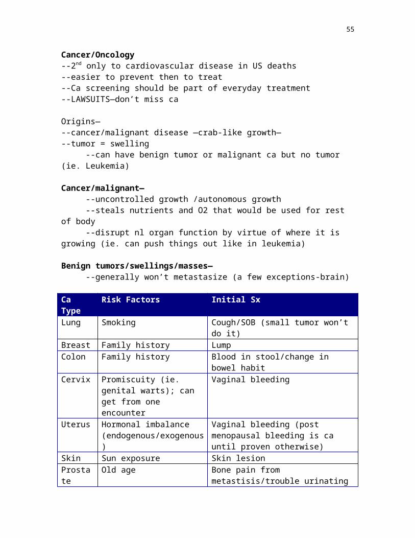
Diseases infants get through hematogenous route—--if this happens early on it usually has more potential to be life-threatening to the
baby--if later—the harm will probably be less severe--it also depends on the specific germ--TORCH titer—checks for ab’s of these germs--toxoplasmosis--other--rubella--cytomegalovirus--herpes (can get this from birth canal or blood)
Encephalitis is the worst infx a baby can get
**Know the differences between ascending infx and hematogenous infx
RESPIRATORY DISTRESS SYNDROME (RDS) / HYALIN MEMBRANE DISEASE(proteinacious material that fills the lung)
--many signs and sx--abnormality in the newborn—dyspnea--suspect this when see the kid having trouble breathing--Clinical sx
--retractions—see neck muscles contract; nasal flaring; intercostal retractions (can see all ribs)
--tachypnea--grunting (trying to open collapsed alveoli)--cyanotic
--Etiologies--pre-term delivery (immature lungs—lack of surfactant)--aspiration of fluid (obstruct airway)—amniotic fluid; blood; meconium
(worst)—first shit—must highly suspect RDS--narcotics / sedation—suppress respiratory drive of baby
--any brain damage is high potential for RDS--nuchal cord—can choke child--idiopathic--C-section—may be prone to respiratory distress (look up why)
TTN—transient tachypnea of the newborn—not RDS
PEARL—listen to the newborn nursery nurses (ER and ICU too)
Tx—O2 support and intubate (ginger use of O2—excess bad for eyes)
In kids that have previously had RDS—--asthma--need flu shots
55
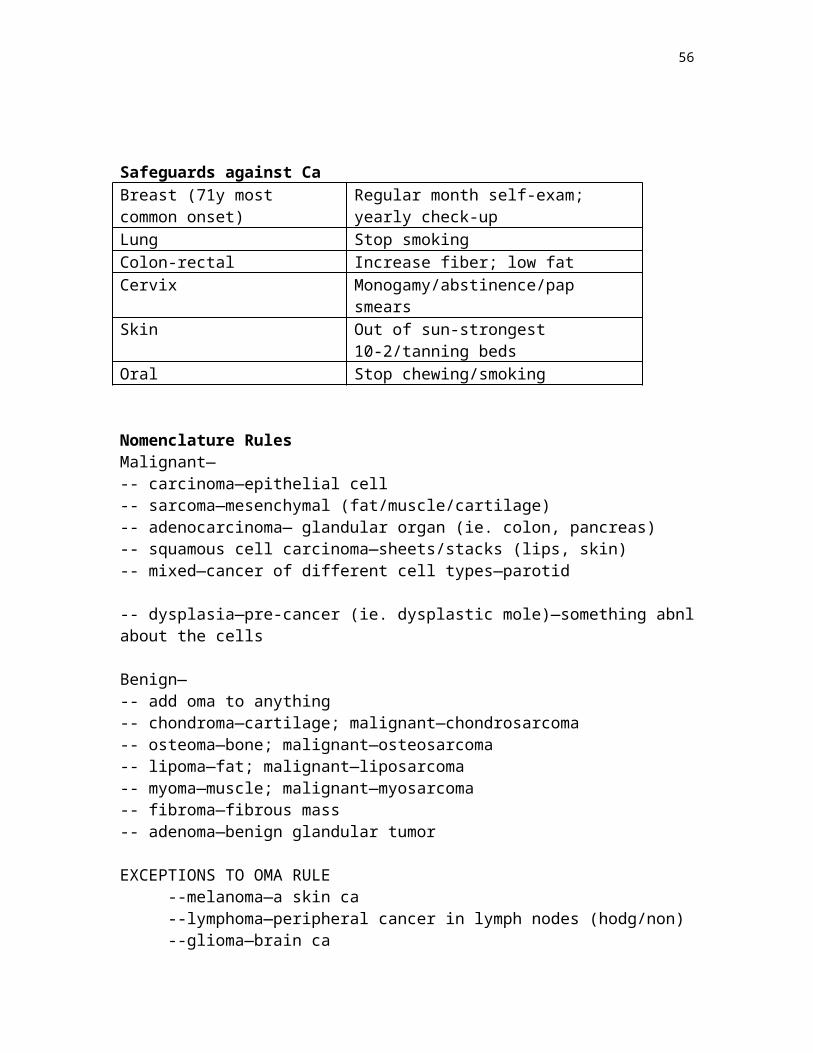
Hemolytic Diseases in the Newborn
(adult—artificial heart valve may lyse RBC’s)
Most common—erythroblastosis fetalis
Mom is blood type O (no a or b ag on RBC; have anti a and anti b abs)Dad—AB(O is universal donor; AB is universal recipient)(if don’t have ag, may have ab)
Presentation—--anemia--jaundice (severe)—RBC breakdown products in bilirubinkernicterous--hydrops fetalis—in utero destruction of child (picture in book)
--worse with subsequent pregnancies--worse if mom had prior blood transfusion
METABOLIC ERRORS (autosomal recessive)
Stick heel—screened for at birth
1. PKU—the amino acid phenylalanine can’t be metabolizedbuilds uptoxic to the brain
--diet of no phenylalanine--incidence—4 / year
2. Galactosemia—inability to break down galactose
Tx—dietary for both
No SIDS
3. Cystic Fibrosis (CF)--1 in 2500 Caucasian carriers--genetic defect—need to get right gene in lung and have it expressed--gene involved in Cl- ion transport therefore Cl- ion transport defectthick
secretions--hallmark—thick secretions in the lungs (tenacious mucus)--prone to infx—morbidity is lung--two bacterial pathogens
-pseudomonas-staphylococcus aureus (gram + pyogenic cocci)-need different antibiotics to get these—usual antibiotics won’t
--also thick secretions in pancreas
56
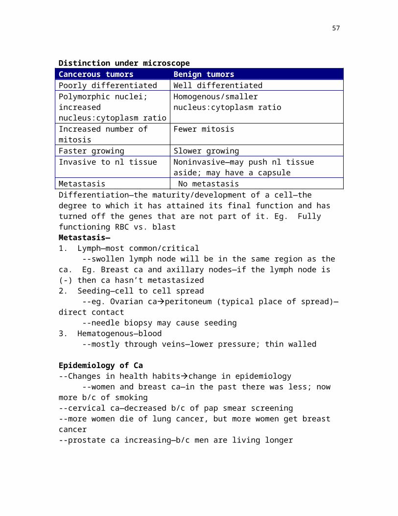
-can’t get pancreatic juice thru pancreatic duct to duodenumcan’t break down fooddon’t grow well b/c cant digest the food
-can take exogenous pancreatic enzymesEtiology—
-family history-slow growth-look at growth charts—clue for many diseases-sick all the time (frequent pulmonary infx)-wheeze at rest-meconium ileus—bowel obstruction—plug of meconium very thick
-approximately 10% of meconium ileus is CFDiagnosis—
-sweat tet—analyze sweat for Cl- content—determine abnormality of transport group
---need to be aggressive with flu shots—choose the right ab’s
Tumors—know neuroblastoma, retinoblastoma, Wilm’s tumor
Neuroblastoma—--most common childhood solid tumor--most common tumor in <1 year old—500 new cases each year--15% of all childhood ca deaths--25-35% arise in medulla of adrenal gland--the rest somewhere on symp chain, with the 2nd most common on the
paravertebral region of posterior mediastinum--presentation (under 2)—large abdominal mass, fever, possible weight loss--older children—no sx until metsbone pain, resp sx, GI, --age and stage are important determinants of outcome-- <1 year old—excellent
prognosis regardless of stage
Retinoblastoma—--very rare—1 in 20,000 infants and children--caused by a loss of suppressor function (suppressors regulate cell growth)--60% are sporadic--40% are familial (autosomal dominant)--familial—affected parent + mutation in one retinal cell (two hit theory)--sporadic—both mutations occur simultaneously on a single retinal cell
Wilm’s Tumor—--most common primary renal tumor of childhood--age 2-5--increased risk in 3 groups of congenital malformations
--WAGR syndrome (characterized by aniridia, genital anomalies, and mental retardation33% chance of Wilm’s)
--Denys-Drash Syndrome (characterized by gonadal dysgenesis and nephropathyrenal failure)
57
--Beckwith-Wiedemann Syndrome (characterized by enlargement of body organs)
11/2/99
Pulmonary Physiology
Lung Functions—--macrophages—phagocytes for germs--regulate BP--major function—gas exchange—O2 in; CO2 out--Lung tissue—exchanges gas at the level of the alveoli
Ventilation Apparatus—--medulla oblongata contains respiratory center—CO2 levels are the drive to
breathe--C3-C5—Phrenic nerve—motor to diaphragm--diaphragm / accessory muscles (neck; intercostals)--thoracic cage / sternum / scapula—protection
--a neuromuscular disorder can lead to pulmonary problems b/c can’t use ventilatory apparatus
O2 is carried on Hb—Hb binds O2 in the alveoli--3% O2 dissolved in plasma
Lung Failure—the pathophysiology is hypoxiaVentilatory Apparatus Failure—the pathophysiology is hypercapnia (high CO2)
Oxygenation—the exchange of gas in the alveoli with the blood cells in the pulmonary capillaries
--need gas in the alveoli and RBC’s in the pulmonary capillary--simple (passive) diffusion—
--PO2 higher in alveolus than RBCO2 moves into RBC--PCO2 higher in pulmonary capillary than airCO2 moves to air
Oxygenation is dependent on several factors—1. PO2 in alveolus—nl is 21% (most air is N; some H2O vapor; CO2)
--can increase this therapeutically2. Barriers (or lack of) to diffusion
--wall / interstitial space / alveolus—shouldn’t e anything else there--eg—baker’s lung—scar tissue—pulmonary fibrosis
3. Matching of ventilation to perfusion—need air and blood cells
*alveolar air = atmospheric air
58
PO2 of alveolar air is about 100mmHgPO2 in pulmonary capillary is about 40mmHg
--O2 moves air to bloodPCO2 in alveolar air is about 40mmHgPCO2 in capillary is about 46mmHg
--CO2 moves from blood to air
Ventilation—(causes of failure)1. excess CO2 production—associated with fever; seizure2. increase in dead space—parts of respiratory tree that are ventilated but not
perfused—physiological example is trachea (others may be a mismatch)3. decreased ventilation from neuromuscular causes—drug overdoses that cause
suppression of medulla; asthma attack—muscles fatigue
Lung Volumes—see in PFT’s
Vital Capacity (VC)—maximal inspiration followed by a maximal expiration –good estimate of pulmonary fnct
Tidal Volume (TV)—nl amount of air breathed with each breath--can estimate at 10cc/kg bw
Forced Expiratory Volume in 1s (FEV1)—peak flow—breathe out all the way as fast as you can—good assessment of asthmatics—charts to compare w/ age; wt; sex
Inspiratory Reserve Volume (IRV)—max amount of air that can be inspired after TV
Expiratory Reserve Volume (ERV)—max amount of expelled air after nl breath
Residual Volume (RV)—amount of air left in lungs after nl breath
--use these in diagnosis
Acid-Base Balance
ABG—arterial blood gas—typically take from radial a. ; sometimes femoral a.
--critical in various clinical settings—ICU, ER (shock, trauma), etc
Data—--pH—7.35-7.45--PO2—80-100 (this is peripheral O2)--PCO2—40 (this is peripheral)
<7.35 = acidosis>7.45 = alkalosis--also need to get some venous blood for a HCO3- level
59
**need ABG and venous stick to begin to interpret what is happening
H+ + HCO3- H2CO3H20 + CO2 (carbonic acid)
--acid base balance—lung and kidney--kidney controls H2O and HCO3---lung controls CO2
pH Primary disorder Compensatory mechanismRespiratory acidosis
Low Lung not removing CO2—increased CO2
Kidney will increase HCO3- to maintain nl pH (balance equation)
Respiratory alkalosis
High Low CO2 (below 30) Kidney will secrete HCO3- into urine—serum HCO3- will be low
Metabolic acidosis
Low Low HCO3- Lung will decrease CO2—hyperventilate
Metabolic alkalosis
High High HCO3- Lung will increase CO2—decrease respirations
--Sx of respiratory acidosis—fatigue, confusion
--Reasons for respiratory alkalosis—hyperventilation, anxiety attack, aspirin overdose
--Respiratory disturbances are primary from lung and compensatory from kidney--Metabolic disturbances are primary from kidney and compensatory from lung
--kidney and lung work together to keep CO2 and HCO3 equal
11/4/99
H+ + HCO3- H2CO3 H20 + CO2
--increased CO2 drives equation to the leftincrease of H+ ionsdecreased pH--decreased CO2 drives equation to the rightdecrease H+ ionsincreased pH
Cardiac Physiology—
Pump—--circulates O2, nutrients, and waste products, therefore a common cause of renal
failure is heart failure--endocrine function—ANF—atrial natriuretic factor—a protein to kidney—
regulates fluid balance—this is the natural connection b/t the heart and the kidney--myocardial cells—muscle cells of the heart
60
Electrical conduction—SA(RA)AV node (junction of a and v)bundle of His (R and L)purkinje
Cardiac Cycle—1. Atrial systole—30% of ventricular filling2. Ventricular systole—this is followed by repolarization3. Diastole—ventricular filling time (70% of total) / rest time
--perfusion of coronary vessels--RAtricuspidRVpulmonic valve--LAbicuspid (mitral)LVaortic valve
--ventricular systole—tricuspid and mitral valve closed; aortic and semilunar valves open--diastole—AV valves open; aortic and semilunar valves closed
BP—R heart—pulmonary circulation—25/10 mmHg--L heart—systemic—140/90 mmHg
--systolic—peak pressure during systole--diastole—least pressure during diastole
Heart Sounds—--AV valves closing—lubb (all valves closed at this point); ventricles have just
finished filling--pressure builds up in the ventriclessemilunar valves forced opensemilunar
valves closing is Dubb
Pulse—pressure wave from LV contraction
Murmur and bruit—sounds heard secondary to turbulent flowMurmur—heart—stenotic, blocked valve, open valve when it should be closedBruit—peripheral (eg. Carotid) (eg. Plaque); renal
Cardiac Output—--main indicator of cardiac status--5-6 L/min in nl adult
SV=EDV-ESVSV x HR = CO
Nl SV(EF) is about—60%
Ejection fraction—synonym for SVmeasure thru echocardiagram
--if hear heart murmurget echo
30% EF—fatigue on minimal exertion10% EF—live but SOB at rest
--high risk of strokepool bloodfall out of solution and clot—need coumadin
61
Factors that increase CO—--CO at sleep is about the same as at awake--exercise--anxietyincrease HRincrease SV somewhat; also increase symp tone--pregnancy
Factors that decrease CO—--sit and then stand up--heart disease—lowers SV therefore decrease CO--MI—less muscle massless pressuredecrease SVdecrease CO--rapid arrhythmias—150+--decrease fill time
CO=HR x SV
HR—chronotropic—time+chonotropes increase HR-chonotropes decrease HR
asthma puffer—beta-agonist—stim symp
SV—--contractility—the squeeze of the heart muscle
--the actin and myosin filaments slide across each other to shorten cell--ionotropic+ionotrope—increases contractility of the heart (this type used in most therapeutic
cases)-ionotrope—decreases contractility of the heart
--sympathetic and parasympathetic also have increase and decrease effects on contractility, respectively
Contractility—Starling’s Law—
62
ANF—low volumes—atria release hormone to tell kidneys to retain fluid
MI—filling same; less contractilitydecreased SV
Can manipulate preload and afterload (BP)—afterload specifically is a major part of what we do—possibility is there to move someone from too high afterload to too to low preload
Vasoconstriction—increases afterloadVasodilaton—decreases afterload
--can’t reduce afterload without reducing preload too(dilation—more blood in periphery and less in heart)
SV depends on—preload (blood in the heart); and afterload (BP)
Exercise—Relative state of increased sympathetic toneContractility and CO increasedHR and SV (contractility increaseincrease COIncrease venous returnincrease preloadPeripheral vasodilation for coolingdecreases afterload
Oxygen Consumption—of the heart--heart cells packed with mitochondria--LAD, RCA, circumflex—blood flow somewhat fixed thru these areas--if filled with cholesterol—less blood—less O2 to heart muscle therefore O2
delivery is limited by coronary a.’s –need supply to exceed demand
Factors that increase O2 Consumption—--increased peripheral resistance (afterload)—need to decrease BP--increased HR (exercise)—B-blockers (slow HR)--increase contractility—B-blockers (enderol; propanolol; etc)
63
11/9/99
Congestive Heart Failure—CHF
Hypertrophy—increase in the size of heart cells (LVH)Dilitation—the chamber walls thin and expand—increase chamber size
--cardiomegaly—big heart—can’t tell difference b/t LVH and dilitation on echo--myocardium—heart muscle--papillary muscles / chordae tendinae
CHF—heart cannot maintain its output to meet the metabolic demands of the body--pump failure--it is the endpoint of several other cardiac diseases--no one treatment b/c of the various causes
Clinical SX--SOB—hallmark (dyspnea)—orthopnia—made worse by laying down--fatigue--pallor--cough—from fluid in chest
Systolic heart failure—pump failure—fluid back-up—congestion failure due to primary pump (ventricle) failure. Eg—aortic stenosis—fluid back-up to lungs. **most common scenario
Diastolic heart failure—(dysfunction)—see on echos—filling defect—LV has trouble filling—can’t fill can’t pump—almost exclusively seen in hypertensive heart disease (increase BPhypertrophy heartcan’t fill)—balloon vs. punching bag
Etiologies / causes of CHFAcronym—heart
--Hypertension—hypertrophy. Can be acute hypertension b/c of increased afterload (systolic failure). Can by chronic hypertensionhypertrophy (diastolic failure)
--Embolism—especially pulmonaryright side failure. The embolus causes an obstructionheart has to pump against it
--Arrhythmia—decreased filling timedecrease preload (diastolic failure?)--Respiratory / Rheumatic—valvular causes of heart disease--Thyroid disease—uncommon—manifest thru arrhythmia. Thrombus—
manifest thru MI
64
Left sided heart failure—--more common--also referred to as forward failure--causes—
--failure of LV—most common scenario where LV will fail is MI / ischemia
--acute hypertension--valvular heart disease--myocarditis—primary diseases of the heart musclemost common is
viral ; ETOH--(myocarditis can cause)—hypertrophied heart—can ballon out and dilate
(down side of Starling’s)
Physical exam on forward failure—--frothy sputum--rales—alveoli filled with fluid
Organs that suffer—from left sided heart failure1. lungs—heart not pumping efficientlyincrease hydrostatic pressure in pulmonary capillariesfluid exudespulmonary edemacan’t exchange gas as good (extra barrier to alveoli)2. kidneys—can’t make urine b/c not being perfusedincreased BUN ; Cr3. Brain—not getting enough nutrients—hypoxic encephalopathy
Right sided heart failure—--most common cause is left sided heart failure--other causes—pulmonary embolusobstruction to outflow of RV--COPD—Cor Pulmonale—increases pressure of blood flow from the right
ventricle (right sided heart failure b/c of lungs)
Sx—--JVD—jugular venous distention—blood back-up into SVC—can see it in neck--IVC—liver congestion--peripheral edema—from increase in venous hydrostatic pressure--fluctuations in weight b/c of fluid gain and loss
Heart failure is usually chronic
Tx principles—acute or chronic--rest--O2--diuretics—major part—reduce volume--digoxin—increase contractility--ACE inhibitors—especially important in diastolic disordersdecrease afterload--nitrates—if dealing with an ischemic situation
65
CAD is the number one cause of deaths in the US—750,000 / y
Ischemia—imbalance b/t the demand and supply (in the heart)—need more blood than you’re getting. Rare cause is congenital—small coronary vessels. More common is a chronic build up of plaque in the coronary arteries
Blocked coronary vessels—ischemiaPathogenesis—atherosclerosis (build up of plaque in artery)
Presentation of CAD—--MI--angina--no sx
Epidemiology—--common as people age--risk factors—can use to decide how likely sx are due to CAD—help decide what
the chances are that CAD is present—however you can have CAD with 0 risk factors-smoking-family history-HTN-high cholesterol-DM
-minors—obesity; age; sedentary lifestyle
Pathogenesis of CAD—--long term build up of atheromas / cholesterol plaques on coronary arteries. Can
see the beginnings of the build up on a 2-year-old
--the plaque is a fixed occlusion / obstructionleads to angina (temporary mismatch of supply and demand)Angina—predictable amount of activity—fixed obstruction and demand excedes supply
Unstable angina—plaque cracks / bleadsexposes agplatelets aggregate and occlude all blood flowpossible MI
--the obstruction causes NO blood flow (in angina there is still some blood flow, although not enough)
--no heart muscle damage in angina / unstable angina until 60 minutes. (no cardiac enzymes)60 minutes—cardiac enzymes = heart muscle death
Vasospasm—Prinzmetal’s angina—sudden spasm of blood vessel--same problem for distal heart muscle—just a different cause--important with caths—give Ca++ channel blockers
66
asa—inhibits platelets sticking together—less chance of clotting
MI Sx—--chest heaviness--diaphoresis --SOB--radiation of heaviness--pale--nausea
MI—heart muscle death—two types1. transmural—across the thickness of the whole wall
--Q wave; ST elevation2. subendocardial / nonQ—inner layer of heart muscle
--ST depression
LCA (left main)—occlusion here is widow makerLAD—septum and anterior LVCX—lateral LVRCA—RV and some posterior walll of LV
--importance with the RCA is the SA node->arrhthymias --not important for the pump but for the conduction system
Complications / Consequences of MI—
(no complications—TPA—OK)1. death—50 % of deaths are first presentation2. Arrhythmia—especially ventricular—fib/tach—hypoxic heart cells b/c irritableectopic firing3. CHF—heart can’t pump as wellcardiogenic shock (a lot of heart muscle involved)4. Rupture papillary muscles—from ischemiamitral regurgitation (acute valvular disease)5. necrotic tissue doesn’t contractcause pooling of bloodclotsystemic emboli6. Ventricular aneurysm—scar on ventricular musclescar can balloon out (like blister on tire)—the risk is blood clotting—need long term blood thinners
Prognosis—Long term1. how much muscle was destroyed—more muscle = worse off2. condition of the rest of the coronary arteries (blocks)3. risk factor modification4. compliance with care—take medicine, etc
*every cardiac pt should be on asa and b-blockers (unless extreme circumstances)
67
11/11/99
Heart Failure—can have both left and right / systolic and diastolic
Hypertensive changes in the heart—long standing HTN is increased afterload
LVH—CONCENTRIC pattern (see on echo)—the ventricle grew consistently throughout. The cause is HTN (in the absence of valvular heart disease.
Hypertrophy decreases the size of the lumen (relative to the increased muscle mass) with time the heart muscle will undergo dilitation—
--pressure from the LV is transmitted back to the atria and causes left atrial dilitation—dilated atria is a risk for fibrillation and therefore clotting
--HALLMARK cause of atrial fibrillation is LVHbecause of the pressure being transmitted back to the atria and causing atrial dilitationirritable focia. fibclotting
--Right sided changesfrom pulmonary disease (cig smoke; coal dust)—wipes blood vessels out and forms scar tissuethis increases pressure in the lungRV has to work harder to pumpRVH
--LVH is most prevalentREMODELING—LVH can be reversed by decreasing BP—
--always suspect LVH in HTN cases--EKG shows LVH--echo proves LVH
Valvular Heart Disease—--Heart murmurs—congenital or acquired--Stenosis—inadequate opening of valve--Regurgitation—inadequate valve closure
--echo makes the diagnosis
68
Acquired Murmurs—1. Aortic stenosis—inadequate opening of aortic valve (obstruction to blood flow)
--most common etiology—AGE--dystrophic calcification—--two patient types
-congenital bicuspid aortic valve—calcify sooner (50-60) than the nl tricuspid aortic valve (70+)
-rheumatic fever / overwhelming strep—mitral is more common but its possible to attack aortic
-this is a secondary autoimmune rxn from strepown ab’s tag valve
Presentations of aortic stenosis—1. CHF—backflow to cause pulmonary edema2. Syncompe—result of poor perfusion to brain3. Angina—coronary arteries not perfused
Murmur in aortic stenosis—S1 (mitral valve closure)S2 (aortic valve closure)--a building and falling of the sound between S1 and S2 (creshendo decreshendo)
--there is no nl diastolic murmur--there are abnl and non-pathologic systolic murmurs
Tx of aortic stenosis—--replace valve--valvularplasty—balloon in it—but end up w/ valvular regurgitation (problems closing)
2. Aortic Sclerosis—--aortic valve is calcified but it is not obstructing the blood flow--no consequence
3. Mitral valve prolapse—(MVP)--mitral valve closes too hard and prolapses into the left atrium--hear a click during the heart cycle--mid SYSTOLIC click--more common in women--usually benign--occassionally associated with arrhythmias--if there is a click and murmur present (S1—click—murmur—S2)—need
endocarditis prophylaxis for dentist; colonoscopy; etc--B-blockers may be given to decrease contractility
69
4. Mitral stenosis—--most common etiology—rheumatic--hopefully won’t see anymore--LA will dilatethink fibrillationstroke--DIASTOLIC murmurif hear diastolic murmurthink mitral
stenosiscoumadin
--to uncover a diastolic mitral murmurs regurgitation / stenosis--lay down and roll on left sidebrings mitral valve close to chest wall—can hear
it better
Infective Endocarditis (IE)—
--infection of heart valves--typical etiology—strep / staph--vegetations—colonies of bacteria on heart valves--need transesophageal echo—swallow transducer and see it--endocarditis starts with bacteremia(dentist)if there is a heart murmur present—need antibiotics to protect; IV drug users—introduce bacteria into blood stream
Diagnosis—--vegetations can lead to obstructions--hear new heart murmur-- + blood culture
--we can prevent these valvular diseases with prophylaxisantibiotics b/f certain procedures (dentist; colonoscopy;etc)
Cardiomyopathy—general for heart muscle disease
Idiopathic Dilated Cardiomyopathy—big floppy heart doesn’t pump well—systolic problem / also risk of stroke—decreased ejection fraction
Sx—--fatigue--SOB—both because of decreased CO
--need heart transplant--it is idiopathic but there are some known causes—
--etoh--viral--amyloid—abnl protein in heart muscle cells--Fe overload in heart--iatrogenic—some chemo drugs
Myocarditis—--related to idiopathic dilated cardiomyopathy--viruses ; autoimmune diseases
70
IHSS—Idiopathic Hypertrophic Subaortic Stenosis / Septal Hypertrophy--nothing to do with the heart valve--thickened septal wall--ASSYMETRICAL hypertrophy (only septum is hypertrophied)--many sudden deaths in sports--important because it is associated with lethal arrhythmias
Diagnosis—--simple to diagnose with echo--questionnaire—any sudden unexplained deaths in the family--hear murmur during systole—the hypertrophied septum forms an obstruction
thru the aortic valve--two things in IHSS to make murmur louder
1—val salva maneuver—decrease blood return to heart (increase intraabdominal pressure)less blood in ventriclehole smaller thru aortic valve because the lower amount of blood allows the heart walls to be closer togetherlouder systolic murmur
2—have them squat down and stand upwhen they stand up the blood return to the heart is decreasedif the murmur is louder on there way upcould be IHSS
Sx—--exertional angina--exertional syncope (very serious complaint)
Tx—--surgery—remove the hypertrophied portion of the septum
Pericardial Effusion—--fluid in pericardial sacrestricts filling of the heart--may be a muffled heart sound--see in heart failure / autoimmune disorders--pericarditis (infl of pericardium) can also cause this (viral; lupus; renal failure)
--in pericarditis you hear sandpaper (friction rub)Tamponade—
--aspirate fluid in pericardial sac with needle--eg—post MI—ventricular aneurysm rupture—er situation
Post CABG—pericardium is healing—how do you tell the differenceDressler’s Syndrome—Post MI pericarditis
Heart Ca—--rare--atrial myxoma—mass in the atrium—hear a plop
71
Congenital heart disease—--Ventricular Septal Defect (VSD)—most common congenital defect--free communication b/t R and L ventricles--associated with other septal defects such as tetralogy—--only 30% are isolated anomalies--effects depend on size of hole
Tetralogy of Fallot—--VSD--obstruction to right ventricular outflow (subpulmonary stenosis)--RVH--the aorta overlies the VSD--most common cyanotic congenital heart disease--outcome depends on severity
Pulmonary Edema—--heart related--end stage of CHF--pathophysiology—increased hydrostatic pressure in pulmonary capillaries
(because heart not pumping efficiently)fluid exudespulmonary edema--the fluid is transudate—watery (filtered)--when the fluid seeps out gravity pulls it to the bottom of the lung--therefore hear it from base to apex--if rales descend—improvement
Etiology of Pulmonary Edema—1. HEART FAILURE—from any cause MI; valvular; etc2. Fluid overload—usually IV
--pneumonia in hospitalfluids--post-opoverload of fluid
Second Pathologic Mechanism—--decreased oncotic pressure—--liver failure—lack of albumin--renal failure—proteinurea--severe malnutrition
Another way to get fluid in the lung is injury to pulmonary capillaries—--cause loose gap junctions—fluid seeps out--causes—
--overwhelming infx--sepsis--anesthetics--drugs
72
Adult Respiratory Distress Syndrome—--caused by diffuse pulmonary capillary damage --pathogenesis—increased vascular permeabilityedema
--also called non-cardiogenic pulmonary edema—--very sick--not exchanging O2 b/c of H2O in lungs--put them on ventilator--diuretics won’t work blood vessels are leaking--put them on PEEP (positive end expiratory pressure)
11/16/99
Pulmonary Embolus—blood clot in legRight heartlodges in pulmonary artery (big in central / small in periphery)
Risk factors—--if at risk for clotsat risk for PE--hypercoaguable states—immobility (post-op), smoking, estrogen (BC pills), ca
Presentation—--Sudden Onset--Pleuritic chest pain--cough--dyspnea
*chest X-Ray will look nl—can’t diagnose PE on chest X-ray--need to do VQ—ventilation/perfusion—tracer isotope—PE is an obstruction to blood but not air—if see air but no bloodsuggests PE (pneumonia—no air or blood)
Preventative therapy—--move post-op--heparin--stockings--not in car too long
Recurrent emboli—basket in vena cave (thru femoral v)catch clots b/f get to lung
RESTRICTIVE VS OBSTRUCTIVE PULMNARY DISEASE
Restrictive—pulmonary disease characterized by decreased TLC—restricted to deep inspiration (restricts the filling of the lung)
--polio--spinal cord injury (can’t ventilate)--severe kyphosis--severe pectis (caved in chest)--neuromuscular disorder--obesity—big body cavity—hard to move chest—sleep apnea--mesothelioma—lung pleura tumor—encases the lung—can’t expand as well
73
Obstructive Lung Disease—obstruction to air flow (can’t get air out)--very prevalent—just behind CHF--COPD—chronic obstructive pulmonary disease--major risk factor—smoking--chronic bronchitis--asthma--emphysema
regular coldin COPDvery sick
--Chronic Bronchitis—cough and mucus production for 3 months at a time for two consecutive years
--in reality—always coughing--major risk factor—smoking--pathogenesis of cough and mucus—chronic irritation of the respiratory tree
(cilia; goblet cells—secrete mucus to try to protect lower lung tissue)--the mucus is the obstruction to air flow--very susceptible to infx—cold / flu. B/c of mucus they are also very susceptible
to bacterial infxs--put COPD on antibiotics—prevent super-infx
Clinical SX—--cough--sputum production--dyspnea (mucus impeding air flow)
--can lead to heart problems—b/c of the COPD (Cor Pulmonale)
--Asthma--pathogenesis—hypersensitive airways (bronchioles)—hyper-responsive irritable
bronchioles with reversible bronchoconstriction (spasm) and airway infl (mucus in smaller bronchioles)
--hypersensitive airway--reversible spasm--airway infl (mucus)
Tx—--must address spasm and airway obstruction (oral/inhaled steroids)
--no antibiotics
Asthma triggers—--COLDS--allergens—dust mytes; cats; pollen; smoke (cig/wood); spores--cold air--exercise--pollution (cities)
--if asthma is caused by an allergen—type I hypersensitivity rxn (immediate)—IgE—mast—histamine--may need desensitization tx / specific allerge tx
74
Sx of asthma—--cough--dyspnea (wheezing)--trouble exercising--family history is important--association with excema (dry infl hypersensitive skin)
--Emphysema—pink-puffers--obstruction to airflow more esoteric --almost never in a non-smoker--can rarely have genetic factor--the cig smoke destroys the walls of the alveoli
--within normal walls there is a protein that gives elastic recoil (muscle)—so upon exhalation, some force comes from the alveoli relaxing—like letting air out of a balloon—emphysema degrades these walls so that the lung has trouble decreasing its dimensions and getting air out
--it is obstructive b/c you can breathe it in but can’t get it out
Sx—--hallmark—dyspnea (trapped air in end of bronchiole)--not cough b/c no mucus
--smoke serves as the irritantattracts neutrophils and macrophageswalls get destroyed as the product of the infl rxn
BRONCHIECTASIS—refers to a chronic almost permanent infection in the lung in the setting of an obstruction
Eg—physical condition like in lung ca--think it in COPD w/infx that won’t clear--very hard to treat—chronic antibiotics-scrape it
FINAL—12/14/99—6-9p
--no pulmonary infxs for this exam but will be on final—Tb; bacterial pneumonia; etc
75
SARCOIDOSIS—stereotype is african american women but he’s never seen it with them--common causes—hylar lymphadenopathy--bilateral hylar lymphadenopathy—either ca or sarcoidosis--biopsy the node—histologic hallmark featurenon-caseating granulomas (Tb
has caseating granulomas)--when severe it also infiltrates the lunglung disease
clinical sx—--cough--SOB--small % get infiltrating lung disease—very sick--majority get better on own
Tx—--steroids—suggests it is an immune response --most people just get better on there own
Etiology—can breath stuff in (genetic predisposition)granulomas
--other causes of lung damage in general—radiation (lung ca); some heart meds; look book
LUNG CANCER
Epidemiology--#1 cause of cancer deaths in US
Cell of origin is bronchogenic—epithelial cells lining the airways (not the alveoli)
Typical mets—--adrenals (if see this think lung ca)--liver--brain--bone—these three are usualls of other cancers
Etiology—--smoking and 2nd hand smoke--asbestos / industrial chemicals --radon--genetic clusters--radiation
76
Biopsy report on lung nodules—--1 of 4 histologic types—all ca—impacts tx and prognosis1. squamous cell—
--high link to smoking--found more central / large bronchi
2. adenocarcinoma—--more peripheral (towards pleura)--common in non-smokers--could be radon, radiation, etc
3. oat cell / small cell—--most aggressive--almost all nucleus--paraneoplastic syndrome—ectopic hormones--high association with smoking--very radiosensitive—more treatable but not curable
4. large cell—--poorly differentiated version of #1 or #2
--practically—say oat cell or not oat cell—that’s where the tx varies
Staging of lung ca—--info needed
-size of tumor-where is the primary place--where it spread (which nodes ; which distant organs)
--staging relates to prognosis--stage I can be cured with surgery--stage IV—mets, etc—not curable
on diagnosis of lung CA—10% will survive 5 years
History----smoker--hemoptysis—cough up blood = chest x-ray--change in regular cough--bone pain from mets--neck swelling—(lymphadenopathy; SVC obstruction)--headache / seizures—from brain mets--dysphagia--horseness—involvement of laryngeal n.
Diagnosis—1. biopsy—best way
--if tumor is central—close to hylum—use bronchoscope--if peripheral—CT guided aspiration
2. sputum cytology—look for lung ca cells—good way to start
77
Tx—Surgical tx reserved for early—stage I maybe stage II--if III / IV—surgery will do nothing
Complications—of lung ca--Pulmonary complications1. post obstructive adalectasis—airways distal to tumor collapse b/c no air flow2. pop—post obstructive pneumonia—get follow up film3. mets to pleura and or pericardium—get malignant effusions (gluid collections
b/t the lung and chest wall (pleural space)—can’t expand lung—dyspnea--need to draw the fluid out
4. SVC obstruction—swelling in neck/face/arm
PLEURA—
--lining of the lungs--diseases specific to the pleura
Primary diseases of the pleura—1. pleuricy—viral infx to pleura
--pleuritic chest pain--give advil--hard to distinguish from PE
2. mesothelioma—Secondary diseases—
1. pleural effusions—fluid in pleural space--CHF—from increased hydrostatic pressure--increased vascular permeability (ARDS, infectious)--decreased oncotic pressure--pressure phenomena—popped air sacs (needle, etc)--malignancies that cause seepage of fluid--trauma—blood in the space
2. PNEUMOTHORAX—air in pleural space—shouldn’t be there--can’t expand the lung
Causes—--trauma—--spontaneous pneumothorax—common in teens
--alveoli pop for no reason and air rushes into pleural space--can resolve on own or suck air out and re-expand the lung
END OF TEST III
78
FOR FINAL
11/30/99
BLOOD VESSELS—--four categories of things that can go wrong1. narrowed—atherosclerosisischemia to distal organs2. thrombosis—complete occlusion (in the setting of narrowing)3. weakened blood vessel—rupture (aneurysm) 4. infl of blood vessels
Three layers of arteries (and veins)—--intima—inner layer—blood touches here
--lined by endothelial cells--gap junctions--vascular permeability
--media—muscular middle layer--adventitia—outer layer
Atherosclerosis—--carotidstroke--coronary arteryMI--can happen anywhere
--atheroma—mixture of cholesterol / fat; smooth muscle (from hyperplasia); blood monocytesmacrophaes when leave tissueengulf fatfoamy macrophages
--atheroma is the technical word for plaque
--small artery—the plaque itself can acclude—eg—brain--larger arteries—atheroma is the blockage but the real problem doesn’t start until thrombosis on top of the atheromaischemic distal tissue
--also—pieces of the atheroma can embolize—important in caths / stents—--risk of procedure to clean carotid a.
atherosclerosis—MULTIFACTORIAL--genetics and environment (smokin, diet, geographical switchesnew lifestyle,
etc)
RISK FACTORS OF ATHEROSCLEROSIS--HTN--DM--high cholesterol (especially LDL)--family history—relatives under 50-60y--smoking--obesity / sedentary lifestyle
79
LDL—brings cholesterol to the tissues (what the macrophages engulf)
HTN—the increased pressure injures the endotheliumdisrupts endothelium and sets it up for plaque formation
DM—must tightly control blood sugarit helps
SMOKING—STOP—5-10y of stoppedcardiovascular risk is that of non-smokers
Fig—12.12
These risks are synergistic—+1 risk factor increases risk geometrically rather than additively
HTN—140/90 or less in nl young adult--as older—70-80—160/90 is still nl
the association between HTN and cardiovascular disease is diastolic BP--mild90-104 diastolic--moderate105-115--severe>115 mmHg
Framinghamm study—increased blood pressure is associated with MI, CVA, aneurysms (ballooning out of BV’s), and dissections (under intima but does not blow thru externa), and renal failure
--Essential HTN / Primary HTN— >95% of all HTN—means it was not caused by any other disease—idiopathic—usually no sx
--Approximately 4% of HTN is renovascular—renal arteries are atherosclerotickidney therefore senses the decreased blood volume entering it and secretes reninit therefore perfuses itself by increasing volumeincreased pressure
--1% of HTN is secondary to other diseases—mostly endocrine
White coat syndrome—just nervous—use BP machine to verify
Malignant HTN—sudden severe elevations in BP (diastolic up to 120-140)--SX—neurologic
--bad headache--blurred vision
80
HTN—MULTIFACTORIAL—--genetics--environment
1—blood volume—media is sensitive to blood borne / nerve impulses—diuretics can lower BVBP
2—diameter of the vessel—constriction or relaxation3—CO—if increaseincrease pressure4—kidneys—must assess kidneys when see HTN (BUN / Cr)—kidney
play a huge role in BV--kidney and BP—Blood volume; renin-angiotenses system
--salt is important in older people (60-80)—kidneys are not as efficient--salt is the biggest enemy of CHF
SPECIFIC DISEASES OF BLOOD VESSELS
A. Inflammatory Diseases1. Temporal Arteritis—inflammatory condition in the temporal artery
--disease of the elderly--important no to miss—if not treatedblindness in that eye
Sx—--bad headache where temporal artery is--tender and inflamed scalp--hurts when you touch it (other things don’t do that)
Diagnosis—--sedimentation rate—general measure of infl
Rx—--short course of prednisone to end headache and prevent monocular
blindness
2. Kawasakis Disease—--also called monocutaneous lymph node syndrome--inflammatory vascular disease—idiopathic --diagnosis—
--all clinical--constellation of syndromes and signs—a group of things that
make you think of it--won’t be considered on the first visit--disease mainly of children
81
--SX—--fever for +1 week--conjunctivitis--strawberry tongue--edema of hands and feet--red hands and feet (esp. palms and soles)--peeling skin--cervical adenopathy (nodes)
--may get better on its own but don’t missassociated with coronary aneurysms--Rx—aspirin (anti-inflammatory)—will fix it
--also give gamma globulin--don’t give asa in to kids in general with fever
Raynaud Disease—red white and blue--paroxysmal pallor or cyanosis of the digits of the hands or feet and infrequently
the tips of the nose and ears--cause—intense vasospasm of local small arteries / arterioles --young otherwise healthy women--fingers change to whitebluered--intimal proliferation can appear late in the disease--exaggerated central and local vasomotor response to cold / emotion--usually benign--in chroniccan have atrophy of the skin, subq tissue, and muscle--ulceration and ischemic gangrene are rare
Raynaud Phenomenon is contrasted to this—--arterial insufficiency of the extremities secondary to the arterial narrowing
induced by various conditions including systemic lupus erythematosus, atherosclerosis, or Buerger disease
--Raynaud phenomenon may be the first manifestation of any of these
Aneurysms and dissections—don’t memorize the classifications
--Aneurysm—abnl dilitation of any BV (mostly arteries)--berry aneurysm—small ones in head--bifurcations are common places for aneurisms--aortic aneurysms—associated with atherosclerosis (“wide mediatinum”)--pulsation in belly—could mean aneurysm--pulsatile mass = aneurysmRISKS OF ANEURYSM—
--rupture--embolize (piece of plaque)
Tx—--surgery in healthy --in elderly w/ many problems—leave alone
Dissections—50s-70s, HTN patients--blood under intima layercourses thru media layer
82
--RISK—eventual rupture of the artery--common site-AORTA--SX—
--sudden onset--tearing, searing, horrible pain (in belly)--radiate to back
--need CAT scan with contrast—see the abnormal channel--TX—IV B-blockers
Varicose veins and hemorrhoids—--dilated veins secondary to high pressure--risk—obesity--problem—stagnant blood flowulcerations--stockings; feet up
Superficial phlebitis—inflammation of superficial vein--red skin; can see it--tx—antiinfl and leg elevation and time--these will NOT embolize
Thrombosis in a deep vein—--blood clots here CAN embolize--can’t see it
Mangioma—congenital lesions--look like red birth mark
Lymphangitis—red streaksinfx--cut / leg ulcer / etcit is spreading to lymph vesselsneed antibiotics--painful--usually group A B-hemolytic streptococci--obstructs the lymph channel—obstructive lymphedema
Tx of Vascular Disease—Therapies1. risk factor modification—risk is 0 to the patient2. thrombolytics—clot buster—major risk is hemorrhage3. stent—conduit (metal / plastic)
--risk—can clot off (foreign body)--need to be on blood thinners--endothelial cells will migrate over it--risk of putting it in
83
12/2/99
Red Cell Physiology
p. 603—tableMyeloid line
Erythropoesis—process of stem cell (pluripotent)mature RBC (hematopoesis—general term for blood cell formation)
--adults—bone marrow of axial skeleton--fetus—other bones, liver, spleen
Erythropoeitin (EPO)—hormone from kidneystimulate RBC production
Extramedullary erythropoesis—red cell production outside of the marrow
Anemia--low RBC’sless O2 transportThree pathologic mechanisms—
1. decreased production2. blood loss
--acute—trauma--chronic—GI losses (bleeding ulcers; long term asa / ibuprofinbleed in
stomach; colon cairon deficiancy anemia), kidney tumor / bladder tumor—urine.3. increased destruction
PEARL—assume colon ca in elderly with anemia
Diagnosing Anemia—--CBC—Hb ; hematocrit (the % of the volume of whole blood contributed by
cells or RBC mass to total volume)--either low Hb or low crit
Clinical findings / Sx of Anemia
--fatigue / weakness--pallor--syncope--light-headed / dizzy--PICA—crave paint chips / ice chips--pale conjunctiva--angina / SOB (person with coronary artery blockages)--tachycardia (with acute blood loss)
84
How to differentiate between increased destruction and decreased production—--get a reticulocyte count—normal is 1%
--reticulocyte—the RBC just before full maturity
--low reticulocyte count = decreased production--high reticulocyte count = increased destruction
INCREASED DESTRUCTION OF RBC’S--main mechanism is hemolysis—cleave RBC’s
--clues—--increased bilirubin (metabolic biproduct of heme)jaundiced
--if anemia and jaundicedthink hemolysis--the bone marrow will also be hypercellular
--Causes of hemolysis—--mechanical—artificial heart valvephysical trauma--immune mediated—natural antigenserythroblastosis fetalis
--can also be foreign antigens—from drugs like methyl-dopa—these will bind with the RBC and mark it for destruction
--lupus--congenital—sphenocytosis—abnormal shape RBC—more susceptible to rupture
DECREASED PRODUCTION OF RBC’S--two categories1. decrease in substrate (building blocks)
--Fe deficiency—microcytic cell anemia--B12 / Folate—macrocytic cell anemia (coenzymes in DNA synthesis—without
them the cell can’t grow)--<1 kids fed cows milkinduce gastritisbleedFe deficiency
2. generalized marrow failure—inadequate bone marrow--reasons
--drugs (methotrexate) suppress bone marrowaplastic anemia—decrease in all types of blood cell production
--iatrogenic from chemo—suppresses marrow--tumor infiltration into bone marrow—push out stem cells (leukemia);
(prostate ca because of its common mets to bone)
nl Hb counts—--men—14-18 g/dL--women—12-16 g/dLinfants—10-11 g/dL
Anemia of Chronic Disease—--if can’t find the causecall it this
85
Polycythemia—increased RBC concentration--crit / Hb up
Relative Polycythemia—--looks increased on blood count but it’s just more concentrated
--eg—GEv & ddehydration--give fluids
Absolute Polycythemia—--actual increase in RBC’s
Primary Polycythemia Vera—(P. Vera)—bone marrow disease—red cell leukemia—stem cells in marrow proliferateincreased RBC’s
Tx—blood letting--risk—strokethick blood can’t get circulated in brain
Secondary Polycythemia—--increase in EPOincrease Hb because of RBC’s--smokers / altitude
BLEEDING DISORDERS—--Categories—
--clotting abnormalities--almost always related to liver failure--other—genetic / familial disorders
--hemophilia—no Factor VIII--Von Willebrands deficiency—cofactor of Factor VIII (link
between proteins and platelets)—like glue
Thrombocytopenia—decrease in number of platelets--see in chemo--some drigs can cause because of antigenic tagsdestroy platelets--Idiopathic Thrombocytopenic Purpurapurple hemorrhages under skin--the thinking is some immune rxn to some ag on the platelets--nl platelet—150,000-400,000mm2
--20,000—bleed with trauma of nl life--Tx—steroids—stop immune destruction
--platelet transfusion--remove spleen—can sponge and hide platelets
Hemolytic Uremic Syndrome—--e. coli sepsis--potential serious complication of food poisoning
86
DIC—--1-50,000 pregnancies--infx--trauma--surgery
--bleed everywhere because all clotting factors used up in clots--organs become ischemic because of the clots and decreased flow--Tx—clotting factors—stop bleeding
--heparin—stop clotting
87