Embed Size (px)
Citation preview

Pathophysiology ofPathophysiology of Asthma
PulmonaryPulmonary Function Testing
Wayne Kradjan, Pharm. D.

2007: Expert Panel 3 Asthma Guidelines
• Expert Panel 3 (EP3):Guidelines for the Diagnosis
d M t f A thand Management of Asthma• Summary report available
online:online: http://www.nhlbi.nih.gov/guidelines/asthma/asthsumm.gHtm
• (Original in 1997, updated in 2002 and 2007)

Definition of Asthma
• A chronic inflammatory ydisorder of the airways…
• In susceptible individuals, this i fl i i d finflammation causes episodes of wheezing, breathlessness, chest tightness and coughing, particularly g g g, p yat night or early morning.
• Usually associated with widespread but variable airflow obstructionbut variable airflow obstruction (bronchospasm) that is often reversible, either spontaneously or
ith t t twith treatment.• Inflammation also causes an increase
in bronchial hyperresponsiveness to yp pa variety of stimuli (triggers)

Key Indicators for Diagnosis of Asthma
• Wheezing– (absence does not exclude asthma)
• History of any of following:– Cough, worse particularly at night
R h– Recurrent wheeze– Recurrent difficulty in breathing
Chest tightness– Chest tightness• Symptoms occur or worsen at
night, awakening the patientnight, awakening the patient

Key Indicators (continued)
• Symptoms occur or worsen by:– Exercise– Viral infection– Animals with fur
House dust mites (mattresses– House dust mites (mattresses, pillows, carpets, upholstery)
– Mold– Smoke (wood, cigarettes)– Pollen– Weather changes– Airborne chemicals, dust
M i (l h )– Menses, emotions (laugh, cry)

Bronchial Hyperresponsiveness
More easily induced• More easily induced bronchospastic response to a variety of stimuli that may not otherwise cause a response in the general population.
Allergens– Allergens– Chemicals, irritants– Exercise
• Response may also be more intense and prolongedN h i d l• Non-asthma patients may develop a transient BHR after viral upper respiratory infection.esp ato y ect o .

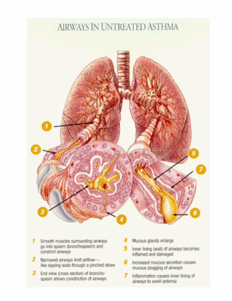
Inflammatory airway changes (“remodeling”)
I d i ll thi k• Increased airway wall thickness– Hypertrophy and hyperplasia of
airway smooth muscley– Thickened basement membrane
(under lumen epithelial lining)E d ti i fl t• Exudative inflammatory reaction with epithelial cell lining desquamation (denuding) g q ( g)and edema
• Mucous gland (goblet cell) h t h dhypertrophy and mucus hypersecretion.
• Eosinophilic infiltrationEosinophilic infiltration


Asthma Triggers
• Allergens (seasonal/ perennial)– Grass, weeds, pollen, mold, mildew– Animal dander, saliva, dust mites
Ch i l i it t d f• Chemical irritants and fumes– Cigarette smoke, pollution, perfume– Household cleaners, occupationalHousehold cleaners, occupational
• Viral infections, rhinitis, sinusitis, (“post nasal drip”)
• Gastroesophageal reflux (GERD)• Exercise; cold, dry air• Extreme emotions• Drugs (aspirin, beta blockers)

Epidemiology• 5% of US population• 5% of US population• 5,000 deaths per year in US
Hi h i id i i i• Higher incidence in inner city, especially African Americans and Hispanic populationsand Hispanic populations.– Racial vs. socioeconomic?

Childhood onset• Most common chronic disease ofMost common chronic disease of
children (6.9% of population)– More likely to be allergic basis– Atopy: genetic predisposition to
IgE mediated response to aeroallergensaeroallergens
– Common: child with positive family history of asthma and ll t t d llallergy to tree and grass pollen,
house dust mites, household pets and molds.
– 30-70% markedly improve or symptom free as adult

Adult onset• May be allergic or non-allergic• May be allergic or non-allergic• Often negative family history and
negative skin tests to common gallergens
• Often history of nasal polyps, aspirin sensiti it and chronicaspirin sensitivity and chronic sinusitis
• Environmental exposure: woodEnvironmental exposure: wood dust, chemicals, pollutants at workplace or in air
• Chemical sensitizers: viral infection, tobacco smoke, diet, perfumeperfume

Genetic Factorsili l d i b i b• Strong familial and genetic basis, but
no single genetic abnormality known.• Correlations with:
– Genetic alterations on chromosomes 5 and 11
– IgE polymorphisms causing high IgElevelslevels.
– 9 polymorphic forms of β2 adrenergic receptors (ADRB2 gene).
» Arg/Arg variant at position 16 (15% whites; 30% bl k d A i ) h 3% l k30% blacks and Asians) have 3% lower peak flow compared to Gly/Gly homozygotes. Susceptible to receptor down-regulation?
» Possible enhanced response with regular beta agonist use in Gly/Gly genotype at locus 16agonist use in Gly/Gly genotype at locus 16 and detrimental effects in Arg/Arg genotype.(may explain possible ADRs with salmeterol)
– 5 lipooxygenase promoter gene

Environmental Factors
• Increased time spent indoors– Indoor allergens (molds, mites,
cockroaches)cockroaches) • Tobacco smoke exposure
– maternal smoking risk for childmaternal smoking risk for child• Increased childhood infections
associated with lower risk– Having older or multiple
siblings or day care center attendance may lower riskattendance may lower risk (more childhood infection)
– Hygienic hypothesis (next lid )slide)

Hygienic Hypothesis of Asthma
• Imbalance of T- helper cells(Th2 and Th1 type lymphocytes)Early exposure to childhood• Early exposure to childhood infections more likely to activate Th1 responses (protective immunity)
• If immune response is predominately from Th2 cellspredominately from Th2 cells, there is a greater production of cytokines that mediate allergic inflammation.
• Details later in lecture

A iAssessing PulmonaryPulmonary Function


Pulmonary function:Bronchodilator tone
P k i t fl t (PEFR) i• Peak expiratory flow rate (PEFR) in liters/ minute
• Forced expiratory volume in one p ysecond (FEV1) in liters
• Normal values vary according to h i htsex, age, height
– Reported as absolute values or– Percentage of normal or of g
personal best
• Establish patient zonesGreen = 80 100% of normal– Green = 80-100% of normal
– Yellow = 50-79% of normal– Red = <50% of normal

Peak Expiratory Flow Rate
• First blast of air exhaled by the patient reaches this flow rate l t i di t lalmost immediately.
• The flow rate quickly slows as more air is exhaledmore air is exhaled.– Less elastic recoil by lung
• Indirect measure of lumen size• Indirect measure of lumen size of large airways and strength of expiratory muscles during p y gmaximal effort.

True Zone Peak Flow MeterTrue Zone Peak Flow Meter

Peak Flow MeterPeak Flow Meter

Directions for use of Peak Flow Meter
• “zero the pointer”– Move indicator to bottom of
numbered scale on meternumbered scale on meter.• Stand upright• Breathe in as deeply and p y
completely as possible• Close lips around mouthpiece to
f i h lform tight seal– Do not put tongue in opening
• Quickly blow out as hard and• Quickly blow out as hard and fast as you can.
• Note reading; repeat 3 timesg; p

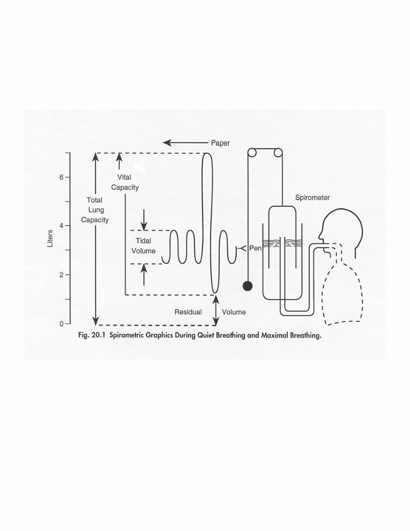
Spirometry
• Forced Vital Capacity (FVC)– Total volume of air exhaled with
maximal effort and continuedmaximal effort and continued exhalation until cannot move any more air.May take 3 5 seconds– May take 3-5 seconds
• Forced Expiratory Volume in one second (FEV1)( 1)– Amount of air exhaled in first
second during FVC measurementT picall 80% of FVC– Typically 80% of FVC
– May measure pre and post bronchodilator


FEV F d i l i 1 t dFEV1 = Forced expiratory volume in 1st second

Pulmonary functionbronchodilation
Bronchodilation: aim for 15• Bronchodilation: aim for 15-20% improvement
• Consider: easier to get higherConsider: easier to get higherpercent change with low baseline
• Look at absolute change(> 0.2 L FEV1)(> 20 L/min PEFR)( 20 L/min PEFR)
• Does the patient feel better?• Does the patient have side p
effects?• single dose vs 1-4 weeks

Case studyDay 1. Before treatment
• 32 year old female;5 .5 foot tall (66 inches)
• Predicted normal peak flow:520 L/ min
• Observed peak flow rate:255 L/ min
• Calculate her percentage of• Calculate her percentage of predicted normal.

Case studyDay 1. Percent of predicted
• 32 year old female;5 .5 foot tall (66 inches)
• Predicted normal peak flow:520 L/ minOb d k fl t• Observed peak flow rate:255 L/ min
• 255/ 520 = 49% of predicted l ( ll / d )normal (yellow/red zone)

Case studyDay 1. After albuterol
• 32 year old female;5 .5 foot tall (66 inches)
• Predicted normal peak flow:• Predicted normal peak flow:520 L/ min
• Observed peak flow rate:255 L/ min
• 255/ 520 = 49% of predicted normal
• 15 minutes after 2 puffs albuterol: 340 L/ min (65%); f l l h f b hfeels less short of breath
• Calculate a percent of change.

Case studyDay 1. Percent change
• Predicted normal peak flow:Predicted normal peak flow:520 L/ min
• Observed peak flow rate:255 L/ i255 L/ min
• 255/ 520 = 49% of predicted normal (yellow/red zone)(yellow/red zone)
• 15 minutes after 2 puffs albuterol: 340 L/ min (65%); feels less short of breath
• % Change = (340-255)/255:33 % i33 % improvement

Pulmonary Function:Hyperresponsiveness
• Measures sensitivity of airways.How easy it is to make them
t i t (b h )constrict (bronchospasm)• Use controlled dose of test
marker: methacholinemarker: methacholine, histamine, cold air
• PC20: “Provocative• PC20: Provocative Concentration 20%”.Largest dose of marker patient tolerates before provoking a 20% worsening of FEV1 or PEFRPEFR

Interpreting PC 20
• Lower PC 20 = more hyperresponsiveness
• Normal: no decline in function with doses < 8 -16 mg/mLA th 20% d li i• Asthma: 20% decline in function with doses as small as 0 125-4 mg/mL0.125 4 mg/mL
• Improved: 1-3 “doublings” of PC20 dosePC20 dose

Obstructive Airways Disease: S f ESequence of Events
Inflammation nerve exposureInflammation, nerve exposure
HyperresponsivenessHyperresponsiveness(↓ PC20, histamine, methacholine)
“Trigger”: allergen or irritant exposure(cold air, exercise)( , )
Bronchospasm (↓ FEV1, peak flow)p ( , p )mucous, edema, cough
OBSTRUCTION

Early and Late PhaseResponses
Chronic asthma characterized by cyclic “late phase responses”by cyclic late phase responses .

Pharmacologic/Physiologic Basis of AsthmaPharmacologic/Physiologic Basis of Asthma
Mast cell contentsHi iAllergen
+
IgE
Ca++ inflow(↓ cyclic AMP, ↑ cyclic GMP*)
- Histamine- Recruit mediators (IL’s,lymphokines, eosinophils)
Phospholipase A2
M t ll b tIgE Mast cell membrane rupture(phospholipid release)
Arachidonic acidArachidonic acid
Leukotrienes Prostaglandins
Lipooxygenase CyclooxygenaseZileuton(Zyflo)
inhibit
- LTD4- LTE4
“Receptors”
- PGE- prostacyclin- thromboxaneZafirlukast (Accolate)
Montelukast (Singulair)Blockreceptor
*Beta agonists increase cyclic AMPAnticholinergics decrease cyclic GMP,Corticosteroids block phospholipase A2

Mast Cells
• 3-5 fold increase in numbers found in walls of respiratory tract of patients with allergic component.
• Degranulation: Release of pre-formed g pcontents– Histamine– Eosinophil and neutrophil chemotacticEosinophil and neutrophil chemotactic
factors (recruitment)– Proteases, interleukins
• Cell wall membrane (phospholipid)• Cell wall membrane (phospholipid) rupture– Leukotrienes C4, D4, E4
P t l di– Prostaglandins– Platelet activating factor
• Early phase bronchoconstriction
Histamine
• Induces smooth muscle contraction and bronchospasm
(early phase response)– (early phase response)• Induces mucosal edema and
mucus secretion• Sensitizes airways to other
stimuli including irritants and ld d icold, dry air
• Antihistamines have little bronchodilating benefit,bronchodilating benefit, indicating other mediators play a greater role

Leukotrienes
• LC4, LD4, LE4 constitute what was formerly called “slow releasing substance ofreleasing substance of anaphylaxis.”
• LD4 and LE4 share a common receptor which when stimulated produces bronchospasm, mucous secretion microvascularsecretion, microvascular permeability and airway edema.
• Contribute to early and late phase responses
• Receptor antagonists: Zafirlukast and montelukastand montelukast

Inflammatory Cellsh d h i• Late phase response and chronic
sustaining mediators• Eosinophils
– Contain inflammatory enzymes, generate leukotrienes, express pro-inflammatory cytokines.
– Contain major basic protein thanContain major basic protein than contributes to airway epithelial damage and hyperresponsiveness.
• Macrophages– Bacterial and foreign matter
scavengers.– Mediator release to initiate and amplify
inflammatory responsesinflammatory responses.• Neutrophils. Increased numbers, but
role unclear.

T-Lymphocytes• Shift toward predominance of Th2
cytokine profile, either as overexpression of Th2 or punderexpression of Th1
• T helper 2 (Th2) subtypeProduces and releases pro– Produces and releases pro-inflammatory cytokines (IL-4, IL-5, IL6, IL9, IL-13) with recruitment of eosinophils, IgE (allergy sensitization)p , g ( gy )
• T helper 1 (Th1) subtype– Produce and release IL-2, interferon gamma
which enhance cellular defense mechanisms i t i f ti ( t ti i it )in response to infection (protective immunity)
• Both release IL-3, interferon alpha, and granulocyte-macrophage colony stimulating factor.

TH2 Lymphocytes• Primary cell for chronicityPrimary cell for chronicity
or memory in asthma• Orchestrate allergicOrchestrate allergic
inflammatory processes– Required for IgE production by
B llB cells.– IL-4 and IL-5 release play key
role by y» up-regulation of adhesion
molecules» Stimulating activation and» Stimulating activation and
enhanced survival of eosinophils

Nitric Oxide (NO)
• Generated by cells in respiratory tract from the amino acid L-arginine by the enzyme NO synthase.
• NO synthase induced by proinflammatory cytokines in airway epithelial cells, vascularairway epithelial cells, vascular endothelial cells, macrophages, and eosinophils
• High exhaled NO levels may be an• High exhaled NO levels may be an indicator of ongoing inflammation and responses to therapy.P d th l l ti• Produces smooth muscle relaxation (vasodilation and bronchodilation), but also amplifies inflammatory responses

Obstructive Airways Disease: S f ESequence of Events
Inflammation nerve exposureInflammation, nerve exposure
HyperresponsivenessHyperresponsiveness(↓ PC20, histamine, methacholine)
“Trigger”: allergen or irritant exposure(cold air, exercise)( , )
Bronchospasm (↓ FEV1, peak flow)p ( , p )mucous, edema, cough
OBSTRUCTION

Asthma Severity Classification(Not currently on long term control drugs)
Three age groupsee age g oups– Children, age 0-4 years– Children, age 5-11 years– Youth, > 12 years of age and adults, y g
• Stages of Severity– Intermittent (old = mild intermittent)– Mild Persistent– Moderate Persistent– Severe Persistent
• Components of severity (impairment)p y ( p )– Symptoms (episodes/ week or month)– Nighttime awakenings (per week or month)– Frequency of short acting beta agonist useq y g g– Interference with normal activity – FEV1 (age 5 or above, only)
• Exacerbation Risk (frequency & severity)( q y y)

Asthma Severity Classification(Example – see figures 11 from NIH ( p gguidelines for greater detail)
• Intermittent• Intermittent– Symptoms, beta agonist use ≤ 2 days/ week– Night time awakenings < 2x/ month
• Mild Persistent– Symptoms >2 days/ week, not daily– Night time awakenings 3-4x/ month– Night time awakenings 3-4x/ month
• Moderate Persistent– Symptoms daily– Night time awakenings >1 day/week
• Severe PersistentSymptoms throughout the day– Symptoms throughout the day
– Night time awakenings >4 days/week

Classification of Asthma Control (See figures 12 and 15)
h i• Same three age groupings as for classification of severity
• Classification of control– Well controlled– Not well controlled– Very poorly controlled
• Impairment markers– Symptoms– Nighttime awakenings– Interference with normal activity– Beta agonist use– FEV1 (over age 5 only)
Q lit f lif ti i ( d lt )– Quality of life questionnaires (adults)– Episodes of exacerbation– Treatment related adverse effects

Staging:Further Considerations
• Seasonality• Nocturnal symptomsy p• Exercise induced• Peak flow monitoring• Peak flow monitoring• Daily fluctuations
C h i• Cough variant• “Wheezy bronchitis” in
children

Assessing AsthmaAssessing Asthma Control: The Use of the A th C t l T t™Asthma Control Test™(ACT)
See separate slide set
Asthma Control Test is a trademark of
QualityMetric Incorporated.

What Does the Score Mean?• A score of 19 or less may mean
your patient’s asthma may not be controlled as well as it could becontrolled as well as it could be
• A score of 20 or more may mean your patient’s asthma is underyour patient s asthma is under control; however, there are other factors you may consider in assessing asthma control

Taking an Asthma History
• When did you first learn you had asthma? Describe.
• What symptoms of your asthma bother you most? DescribeH ft d th• How often does your asthma affect your sleep?Do you have coughing• Do you have coughing associated with you asthma?
• What things make your asthma• What things make your asthma worse? Probe for triggers, exercise induced. Describe

Asthma History (2)
• What medications are you taking for your asthma?
Have them explain name how– Have them explain name, how they take it (demonstrate MDI), understanding of “rescue” vs “controller” perceived benefitcontroller , perceived benefit, side effects experienced.
• Do you use a peak flow meter?– Demonstrate technique, personal
best, results recent and past• Overall assessment of control• Overall assessment of control• Concerns, goals• Do they have an asthma plan?Do they have an asthma plan?