Embed Size (px)
Citation preview
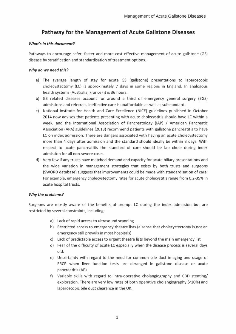
Management of Acute Gallstone Diseases
1
Pathway for the Management of Acute Gallstone Diseases
What’s in this document?
Pathways to encourage safer, faster and more cost effective management of acute gallstone (GS)
disease by stratification and standardisation of treatment options.
Why do we need this?
a) The average length of stay for acute GS (gallstone) presentations to laparoscopic
cholecystectomy (LC) is approximately 7 days in some regions in England. In analogous
health systems (Australia, France) it is 36 hours.
b) GS related diseases account for around a third of emergency general surgery (EGS)
admissions and referrals. Ineffective care is unaffordable as well as substandard.
c) National Institute for Health and Care Excellence (NICE) guidelines published in October
2014 now advises that patients presenting with acute cholecystitis should have LC within a
week, and the International Association of Pancreatology (IAP) / American Pancreatic
Association (APA) guidelines (2013) recommend patients with gallstone pancreatitis to have
LC on index admission. There are dangers associated with having an acute cholecystectomy
more than 4 days after admission and the standard should ideally be within 3 days. With
respect to acute pancreatitis the standard of care should be lap chole during index
admission for all non-severe cases.
d) Very few if any trusts have matched demand and capacity for acute biliary presentations and
the wide variation in management strategies that exists by both trusts and surgeons
(SWORD database) suggests that improvements could be made with standardisation of care.
For example, emergency cholecystectomy rates for acute cholecystitis range from 0.2-35% in
acute hospital trusts.
Why the problems?
Surgeons are mostly aware of the benefits of prompt LC during the index admission but are
restricted by several constraints, including;
a) Lack of rapid access to ultrasound scanning
b) Restricted access to emergency theatre lists (a sense that cholecystectomy is not an
emergency still prevails in most hospitals)
c) Lack of predictable access to urgent theatre lists beyond the main emergency list
d) Fear of the difficulty of acute LC especially when the disease process is several days
old.
e) Uncertainty with regard to the need for common bile duct imaging and usage of
ERCP when liver function tests are deranged in gallstone disease or acute
pancreatitis (AP)
f) Variable skills with regard to intra-operative cholangiography and CBD stenting/
exploration. There are very low rates of both operative cholangiography (<10%) and
laparoscopic bile duct clearance in the UK.
Management of Acute Gallstone Diseases
2
Why have a joint document?
Biliary disease is managed by most surgeons on the acute surgical take and LC is undertaken by
many of them. The majority of surgeons providing the EGS (Emergency General Surgery) service are
neither HPB surgeons nor have access to HPB colleagues on site. This document seeks to provide a
concise management plan for the majority of cases of gallstone disease presenting to the EGS in any
hospital rather than attempting to provide an exhaustive guide to every scenario. Facilities and skills
vary between units and between individual surgeons on emergency duty. At times this will influence
the best management and expert referral and transfer will be needed to deal with complex
problems in a specialist HPB unit. In principle it should be an institutional responsibility to provide
best practice care to all acute biliary disease patients presenting as acute admissions to a general
surgical service
Initial assessment
In the management of acute biliary disease, a number of factors will dictate the optimum
treatment pathway for any individual patient, including:
1. Severity and persistence of the biliary pain
2. Ultrasound findings
3. Results of blood tests
4. Overall fitness for surgery
History and examination
It is essential to establish the nature, severity and frequency of symptoms. Previous episodes of
biliary pain should be identified to permit optimum management planning. An overall assessment
of the patients’ fitness and desire for surgical intervention should be assessed.
Ultrasound
For a patient with first presentation of biliary pain (i.e. pain in the epigastrium or RUQ, usually
radiating round or through to the back, lasting for at least 20-30 minutes (often several hours or
more)) ultrasound (US) is essential to establish the diagnosis of gallstone disease and exclude other
diagnosis in the right upper quadrant. There is no correlation between the presence of, or severity
of biliary pain, with the number nor the size of the gallstones, nor the thickness of the gallbladder
wall. Patients who are already known to have gallstones do not routinely require further imaging.
The presence of dilated bile ducts with abnormal LFTs raises the possibility of CBD stones. Patients
with dilated ducts alone (ie with normal LFTs) do not require further routine preoperative
investigation of their bile ducts as stones in this group are no more common than in the non-dilated
GS population (ie <5%).
Blood Tests
A blood lipase (or amylase) will identify patients with acute pancreatitis. Elevated WCC and/or
inflammatory markers may indicate patients with severe inflammation or infection of the
gallbladder. Liver function tests may be elevated due to inflammation in the gallbladder or
pancreas or may be due to CBD stones.
Management of Acute Gallstone Diseases
3
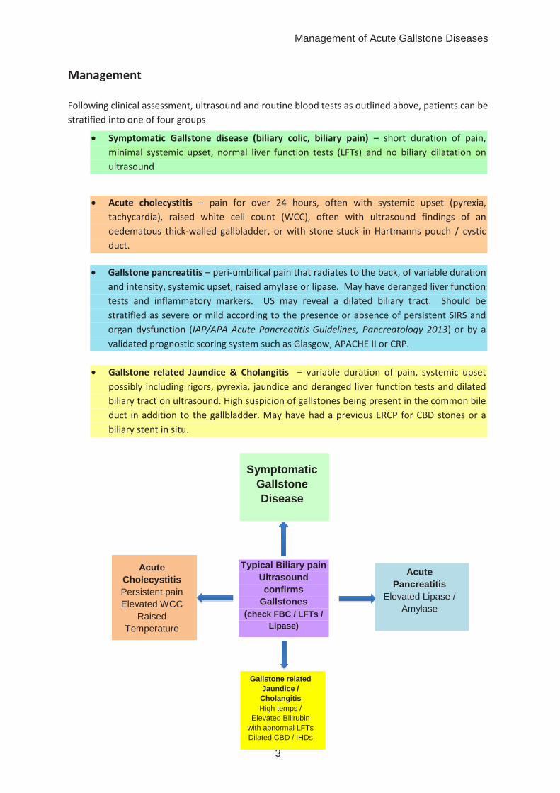
Management
Following clinical assessment, ultrasound and routine blood tests as outlined above, patients can be
stratified into one of four groups
Gallstone related Jaundice & Cholangitis – variable duration of pain, systemic upset
possibly including rigors, pyrexia, jaundice and deranged liver function tests and dilated
biliary tract on ultrasound. High suspicion of gallstones being present in the common bile
duct in addition to the gallbladder. May have had a previous ERCP for CBD stones or a
biliary stent in situ.
Gallstone pancreatitis – peri-umbilical pain that radiates to the back, of variable duration
and intensity, systemic upset, raised amylase or lipase. May have deranged liver function
tests and inflammatory markers. US may reveal a dilated biliary tract. Should be
stratified as severe or mild according to the presence or absence of persistent SIRS and
organ dysfunction (IAP/APA Acute Pancreatitis Guidelines, Pancreatology 2013) or by a
validated prognostic scoring system such as Glasgow, APACHE II or CRP.
Symptomatic Gallstone disease (biliary colic, biliary pain) – short duration of pain,
minimal systemic upset, normal liver function tests (LFTs) and no biliary dilatation on
ultrasound
Acute cholecystitis – pain for over 24 hours, often with systemic upset (pyrexia,
tachycardia), raised white cell count (WCC), often with ultrasound findings of an
oedematous thick-walled gallbladder, or with stone stuck in Hartmanns pouch / cystic
duct.
Symptomatic
Gallstone
Disease
Gallstone related
Jaundice /
Cholangitis
High temps /
Elevated Bilirubin
with abnormal LFTs
Dilated CBD / IHDs
Acute
Pancreatitis
Elevated Lipase /
Amylase
Acute
Cholecystitis
Persistent pain
Elevated WCC
Raised
Temperature
Typical Biliary pain
Ultrasound
confirms
Gallstones
(check FBC / LFTs /
Lipase)
Management of Acute Gallstone Diseases
4
Symptomatic Gallstone disease (Biliary colic, Biliary pain)
Most patients with biliary pain are suitable for early treatment in the ambulatory care setting or
by early inpatient cholecystectomy. Once the pain has been treated or resolved, management
options include:
a) Ideally confirm the clinical diagnosis with ultrasound identification of gallstones during the
index presentation. The patient should be offered a date for cholecystectomy, or reviewed in
a surgical clinic pending a patient or surgical decision to operate. If surgery is declined or not
appropriate (due to co-morbidities etc.) refer back to the GP with advice on a low fat diet
which may reduce attacks of pain in a proportion of patients.
b) All surgical triage units should develop the ability to offer diagnostic ultrasound scans within
working hours. If ultrasound is not available within a few hours, the patient should be
discharged from the surgical triage unit to have an early outpatient ultrasound with follow up
in either a “hot” biliary or acute general surgical clinic. Most patients who are medically fit will
be offered an elective laparoscopic cholecystectomy (within 6 weeks ideally) after one severe
attack of biliary colic, as the likelihood of symptomatic recurrence is high.
c) If pain is not controlled, or recurrent attacks occur at the index presentation, manage as acute
cholecystitis and proceed to acute inpatient cholecystectomy.
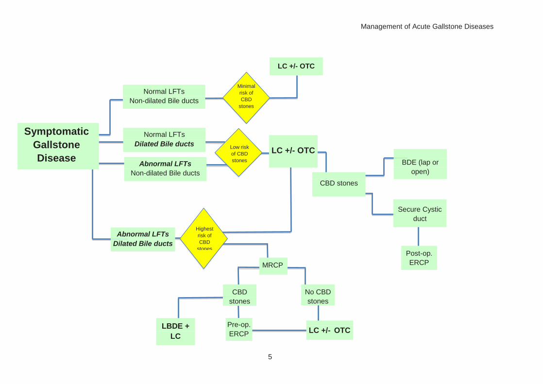
Management of abnormal LFTs
Patients with abnormal LFTs but normal bilirubin and a non-dilated biliary tract on ultrasound
may proceed to directly to LC. Although some surgeons may prefer to have pre-operative
identification of CBD stones (by MRCP or EUS) and duct clearance by ERCP prior to LC, further
investigation introduces unnecessary delay in management, increases costs and impacts upon
MRI / EUS resources. The presence of CBD stones is around 10% in this group of patients and
the status of the CBD should where possible be clarified by intra-operative cholangiogrphy (IOC)
or Laparoscopic Ultrasound. Where IOC has not been performed if LFTs remain abnormal post-
operatively they should be investigated by MRCP or EUS.
If CBD stones are found at LC the surgeon has several options. Ideally patients will have duct
clearance in a single staged procedure by LBDE (laparoscopic bile duct exploration). This may
not always be possible and securing of the cystic duct with subsequent post-operative ERCP
either during the same admission or within 1-2 weeks post LC is an alternative approach in non-
obstructed biliary ducts. Intra-operative ERCP, antegrade stenting of the CBD (with subsequent
post- operative ERCP) and Open BDE are viable alternatives.
Patients with a CBD >10mm and abnormal LFTs with an elevated bilirubin represent those at
highest risk of CBD stones and pre-operative (MRCP or EUS) assessment of the CBD will yield a
CBD stone rate of around 30% permitting CBD clearance pre-operatively. However, the most
cost effective algorithm for treatment in centres with the appropriate skill mix remains to
proceed to LC & OTC without pre-op investigation, as the majority of patients will not have CBD
stones, and the treatment options for managing CBD stones listed above may still be applied.
There should be a locally agreed patient pathway for managing patients in these various
scenarios dependent upon local skills and facilities.
Management of Acute Gallstone Diseases
5
Abnormal LFTs
Non-dilated Bile ducts
Pre-op.
ERCP
LC +/- OTC
CBD stones
BDE (lap or
open)
Secure Cystic
duct
Post-op.
ERCP
MRCP
Normal LFTs
Non-dilated Bile ducts
Abnormal LFTs
Dilated Bile ducts
Normal LFTs
Dilated Bile ducts
Symptomatic
Gallstone
Disease
LC +/- OTC
No CBD
stones
LC +/- OTC
Low risk
of CBD
stones
Minimal
risk of
CBD
stones
Highest
risk of
CBD
stones
LBDE +
LC
CBD
stones

Management of Acute Gallstone Diseases
6
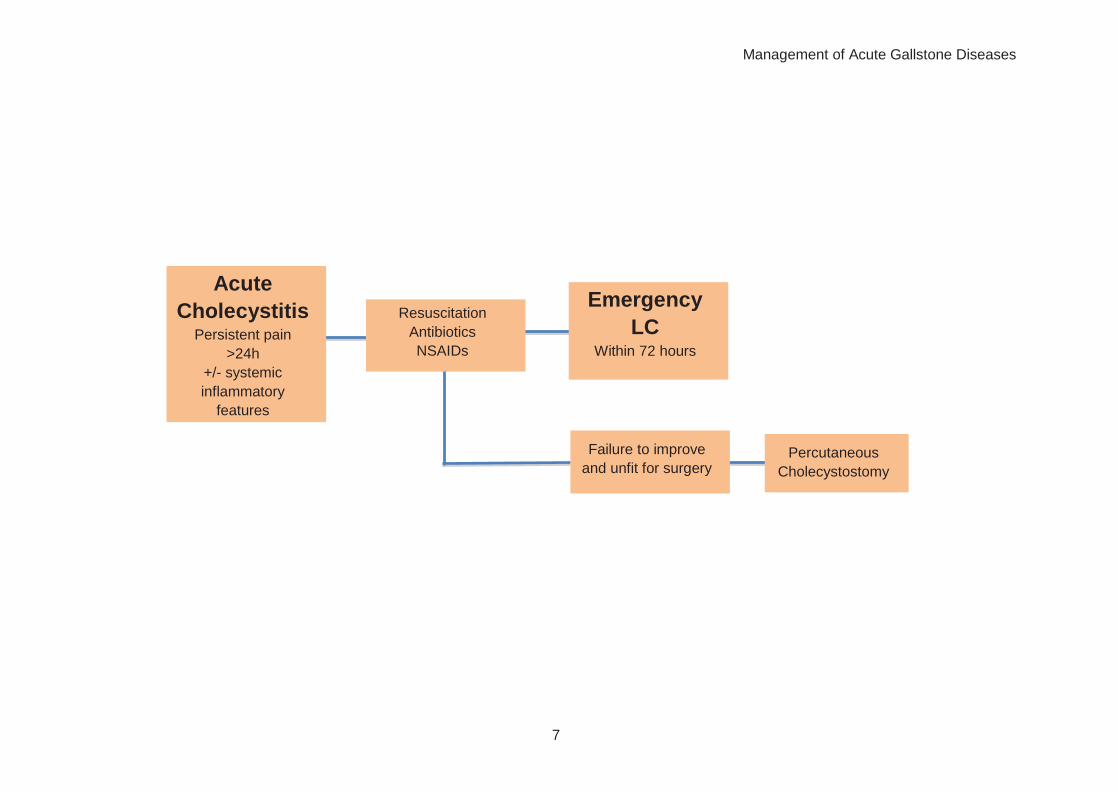
Acute Cholecystitis
Patients with acute cholecystitis should be admitted to hospital to have fluid resuscitation, antibiotics and analgesia. The diagnosis of acute
cholecystitis is a clinical one with a consistent history of ongoing pain and tendeness in the RUQ, usually accompanied by a raised temperature and
inflammatory markers. Ultrasound features may underestimate disease severity (a thickwalled gallbladder and pericholecystic fluid when present are
suggestive diagnosisic indicators) and overall the patient’s systemic symptoms and inflammatory markers, together with response to therapy are a
better guide to severity.
Patients diagnosed with acute cholecystitis should have their LC on the same admission within 72 hours (NICE guidelines, Oct 2014 state 1 week but 72
hours is preferable). Surgery for this sub-group of patients may be very challenging and is associated with a higher incidence of complications
(particularly beyond 96 hours) and a higher conversion rate. These patients should be operated on by surgeons with experience of operating on
patients with acute cholecystitis or if not available locally should be transferred to a specialist unit. This is preferable to leaving them for a delayed
procedure.
Patients with persistent and / or increasing biliary pain, but without systemic inflammatory response, should be managed in the same way as patients
with AC i.e. by early LC.
Patients with signs and symptoms of AC who are unfit for general anaesthesia and surgery who do not improve with antibiotics, may be treated by
percutaneous cholecystostomy. This treatment however is not recommended for patients who are fit for surgery since it may not be therapeutic in
patients with necrotic gallbladders causing delay in definitive management.
Management of Acute Gallstone Diseases
7
Acute
Cholecystitis Persistent pain
>24h
+/- systemic
inflammatory
features
Resuscitation
Antibiotics
NSAIDs
Failure to improve
and unfit for surgery
Emergency
LC Within 72 hours
Percutaneous
Cholecystostomy
Management of Acute Gallstone Diseases
8
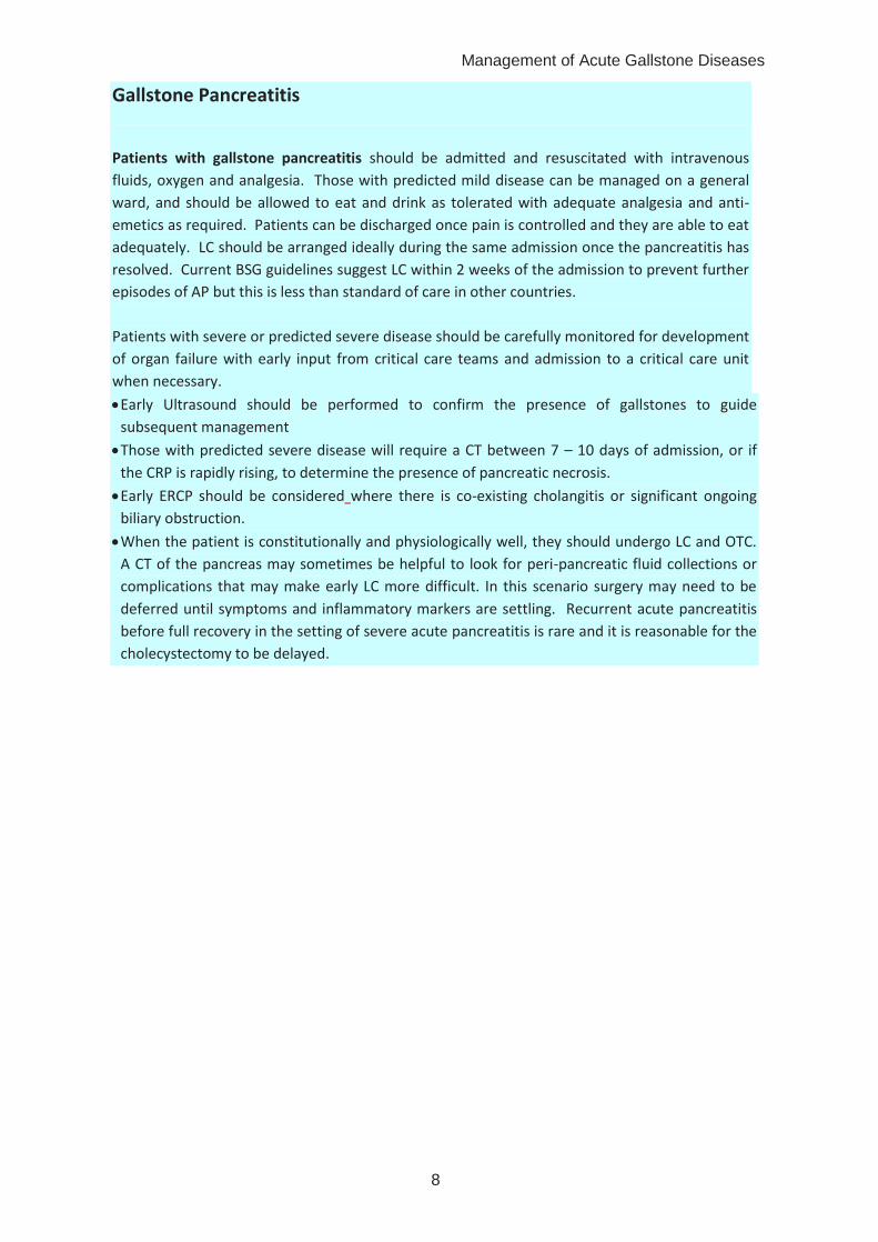
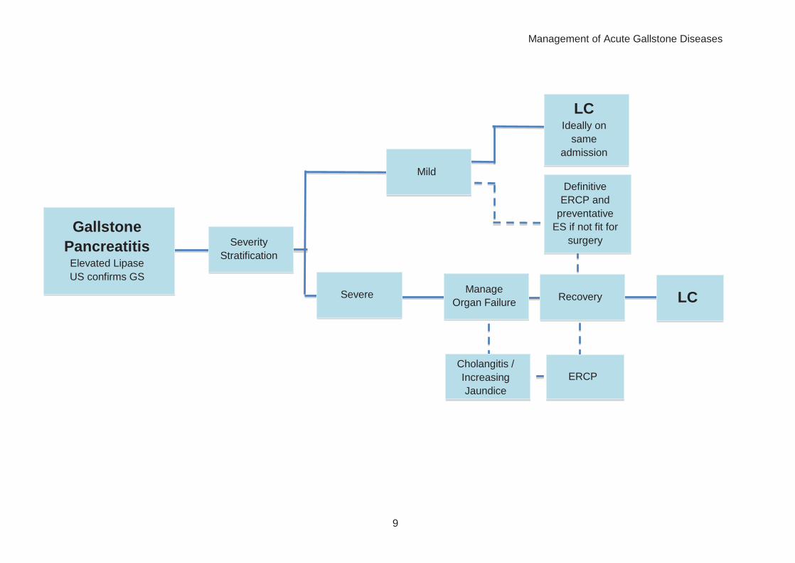
Gallstone Pancreatitis
Patients with gallstone pancreatitis should be admitted and resuscitated with intravenous
fluids, oxygen and analgesia. Those with predicted mild disease can be managed on a general
ward, and should be allowed to eat and drink as tolerated with adequate analgesia and anti-
emetics as required. Patients can be discharged once pain is controlled and they are able to eat
adequately. LC should be arranged ideally during the same admission once the pancreatitis has
resolved. Current BSG guidelines suggest LC within 2 weeks of the admission to prevent further
episodes of AP but this is less than standard of care in other countries.
Patients with severe or predicted severe disease should be carefully monitored for development
of organ failure with early input from critical care teams and admission to a critical care unit
when necessary.
Early Ultrasound should be performed to confirm the presence of gallstones to guide
subsequent management
Those with predicted severe disease will require a CT between 7 – 10 days of admission, or if
the CRP is rapidly rising, to determine the presence of pancreatic necrosis.
Early ERCP should be considered where there is co-existing cholangitis or significant ongoing
biliary obstruction.
When the patient is constitutionally and physiologically well, they should undergo LC and OTC.
A CT of the pancreas may sometimes be helpful to look for peri-pancreatic fluid collections or
complications that may make early LC more difficult. In this scenario surgery may need to be
deferred until symptoms and inflammatory markers are settling. Recurrent acute pancreatitis
before full recovery in the setting of severe acute pancreatitis is rare and it is reasonable for the
cholecystectomy to be delayed.
Management of Acute Gallstone Diseases
9
Manage
Organ Failure
Cholangitis /
Increasing
Jaundice
ERCP
LC
Severity
Stratification
Severe Recovery
Gallstone
Pancreatitis Elevated Lipase
US confirms GS
Mild
LC Ideally on
same
admission
Definitive
ERCP and
preventative
ES if not fit for
surgery
Management of Acute Gallstone Diseases
10
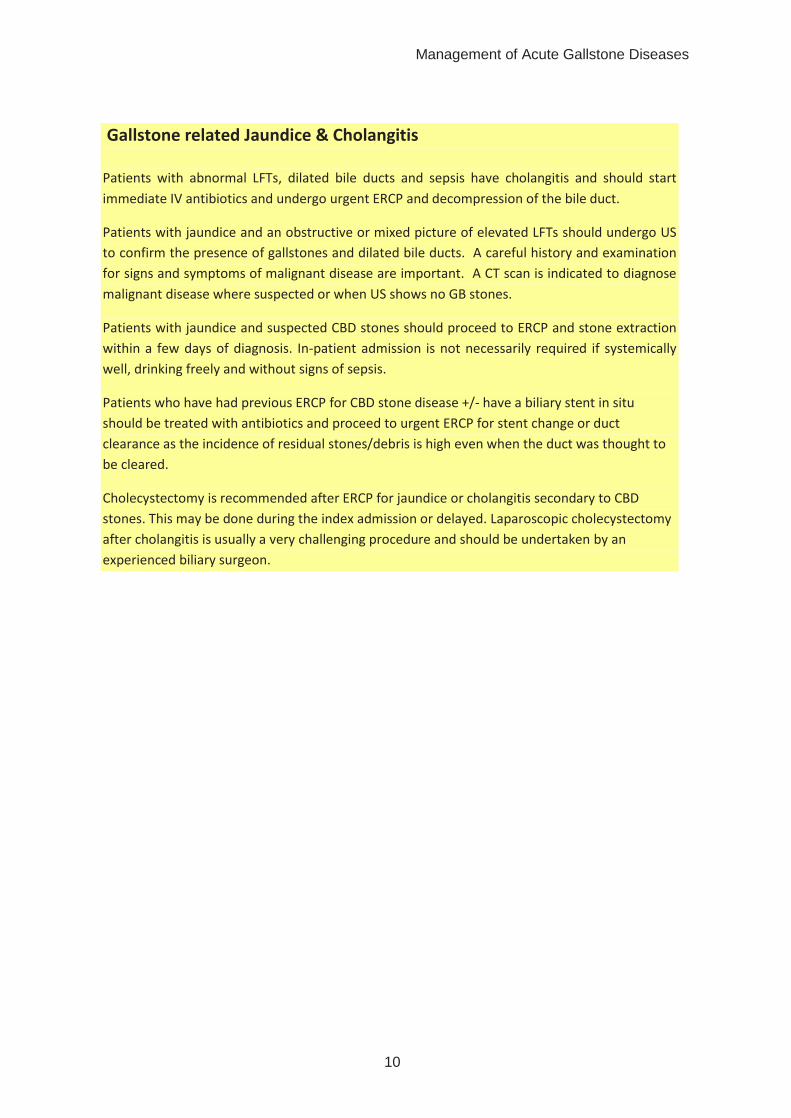
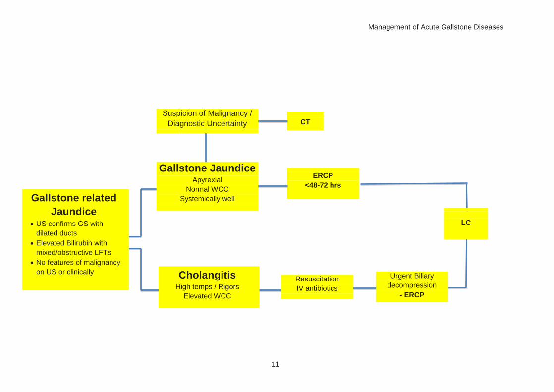
Gallstone related Jaundice & Cholangitis
Patients with abnormal LFTs, dilated bile ducts and sepsis have cholangitis and should start
immediate IV antibiotics and undergo urgent ERCP and decompression of the bile duct.
Patients with jaundice and an obstructive or mixed picture of elevated LFTs should undergo US
to confirm the presence of gallstones and dilated bile ducts. A careful history and examination
for signs and symptoms of malignant disease are important. A CT scan is indicated to diagnose
malignant disease where suspected or when US shows no GB stones.
Patients with jaundice and suspected CBD stones should proceed to ERCP and stone extraction
within a few days of diagnosis. In-patient admission is not necessarily required if systemically
well, drinking freely and without signs of sepsis.
Patients who have had previous ERCP for CBD stone disease +/- have a biliary stent in situ
should be treated with antibiotics and proceed to urgent ERCP for stent change or duct
clearance as the incidence of residual stones/debris is high even when the duct was thought to
be cleared.
Cholecystectomy is recommended after ERCP for jaundice or cholangitis secondary to CBD
stones. This may be done during the index admission or delayed. Laparoscopic cholecystectomy
after cholangitis is usually a very challenging procedure and should be undertaken by an
experienced biliary surgeon.
Management of Acute Gallstone Diseases
11
ERCP
<48-72 hrs
CT
Cholangitis High temps / Rigors
Elevated WCC
Resuscitation
IV antibiotics
Suspicion of Malignancy /
Diagnostic Uncertainty
Gallstone Jaundice Apyrexial
Normal WCC
Systemically well
Gallstone related
Jaundice US confirms GS with
dilated ducts
Elevated Bilirubin with
mixed/obstructive LFTs
No features of malignancy
on US or clinically
LC
Urgent Biliary
decompression
- ERCP
Management of Acute Gallstone Diseases
12
Summary
This document provides a guide to pathways for the management of Acute Gallstone related
diseases.
It is recognised that there are other alternative management algorithms that may be more
applicable in individual centres dependent upon local resources and available skills.
The algorithms presented here seek to set a standardised approach which is considered acceptable
amongst a reasonable body of General Surgeons practising within the UK. It also serves as a
template to which future research and audit studies can refine and improve upon and allow the
development of performance related outcome measures (PROMs) to be established.