Embed Size (px)
Citation preview
Physiology & Behavior 69 (2000) 187–201
0031-9384/00/$ – see front matter © 2000 Elsevier Science Inc. All rights reserved.PII: S0031-9384(00)00201-8
Patterns in the brain: Neuronal population coding in the somatosensory system
Gernot S. Doetsch*
Department of Surgery (Section of Neurosurgery) and Department of Physiology and Endocrinology, Medical College of Georgia, Augusta, GA 30912, USA
Received 5 June 1999; received in revised form 1 November 1999; accepted 8 November 1999
Abstract
The aim of this article is to review some basic principles of neural coding, with an emphasis on mechanisms of stimulus representationin ensembles of neurons. The theory of “across-neuron response patterns” (ANRPs), first suggested by Thomas Young (1802) and fullydeveloped by Robert Erickson (1963–2000), is summarized and applied to the problem of coding in primary afferent fibers and corticalneurons of the somatosensory system. The basic premise of the theory is that precise information about stimulus features cannot be en-coded by single neurons, but is encoded by patterns of activity across populations of neurons. Different stimuli produce uniquely differentpatterns of ensemble activity (ANRPs)—discrimination between two stimuli is based on the absolute difference in total amount of activ-ity (neural mass difference) of the ANRPs for those stimuli. Review of the literature shows that ANRPs and related population codes canaccurately represent and differentiate among various stimulus parameters that cannot be distinguished by single neurons alone. Finally,the behavior of neuronal ensembles can be used to account for the sensory-perceptual changes associated with plasticity of thalamocorti-cal circuits following selective sensorimotor deprivation or experience. © 2000 Elsevier Science Inc. All rights reserved.
Keywords:
Sensory coding; Population responses; Across-neuron response patterns; Somatosensory system; Primary afferent fibers; Cerebral cortex;
Plasticity; Review
1. Introduction
1.1. Purpose of the review
The sense of touch constitutes the interface between theexternal world of objects and our internal world of self. It isboth an active sense for exploring and manipulating the en-vironment, and a passive sense for receiving touch that canstir the emotions as well as inform. Unlike other senses,touch is simultaneously given and received by simple mu-tual contact of the skin. It is a young girl caressing hersilken, gray cat, a carpenter feeling the grain of his newlycarved table, a physician palpating a sensitive abdomen, alover’s touch that leads to a kiss, a blind man fingering apattern of raised dots in order to “see.”
How does this sense of touch come about? What are theneural mechanisms responsible for the rich variety of soma-tosensory experiences? As G. E. Smith [107] observed “. . .all the kaleidoscopic manifestations of mental activity aredependent upon, and determined by, physiological pro-cesses taking place in the nervous system.” But what is the
nature of those processes, especially as they apply to somes-thesis? The aim of this review is to address those questions,based primarily on the author’s own research in sensoryprocessing and on related neural coding studies by others.The review concentrates on stimulus coding by populations(ensembles) of neurons and is limited mainly to discussionof the somatosensory system.
1.2. Brief history of neural coding
The history of neural coding is marked by a pervasivetension between two seemingly contradictory ideas—thatdifferent sensory functions are mediated by activation ofdistinctly different neurons with highly specific stimulussensitivities, or that different functions are represented byunique patterns of activity in populations of neurons withrelatively broad stimulus sensitivities. The “specificity” the-ory had its origins in Johannes Müller’s doctrine of “spe-cific nerve energies” [79], which emphasized the uniquefunctional characteristics of the nerves mediating differentsense modalities. That doctrine was subsequently extendedby von Helmholtz [48] and von Frey [39] to become theprinciple of “specific fiber energies,” which held that eachstimulus submodality or quality within a sense modality isencoded by excitation of different specific nerve fibers or
* Corresponding author. Tel.: 706-721-7120; Fax: 706-721-8084
E-mail address
188
G.S. Doetsch / Physiology & Behavior 69 (2000) 187–201
neurons. The modern version of this theory is often referredto as the “labeled-line” code. Support for this view wasdriven largely by new electrophysiological techniques thatmade it possible to determine the physiological properties,response characteristics, and receptive fields of individualneurons. The predominance of the specificity idea is exem-plified by the ubiquitous phrase, “place- and modality-spe-cific neurons,” in the neuroscience literature. This notion isalso reflected in various proposals for different classes ofcells signaling “touch, vibration, pain, cold, position, move-ment, etc.” in somesthesis, “red, green, edge, orientation, di-rection of movement, etc.” in vision, and “salty, sweet, sour,etc.” in gustation. With its emphasis on individual cells, thelabeled-line notion has been explicitly or implicitly advocatedby many investigators [4,19,38,52,64,66,76,78,90,93–95].
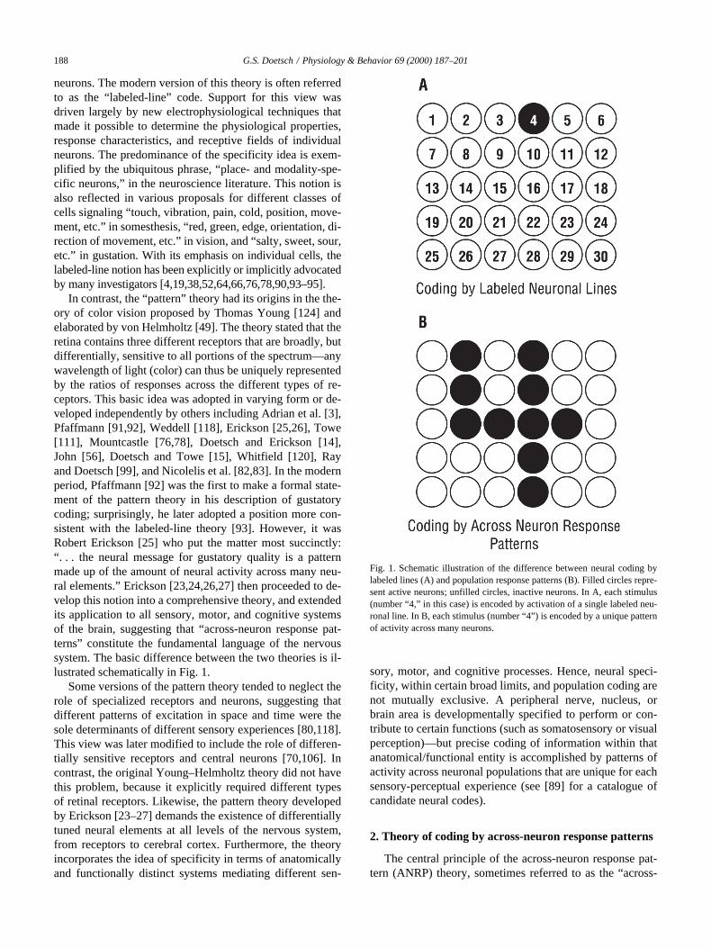
In contrast, the “pattern” theory had its origins in the the-ory of color vision proposed by Thomas Young [124] andelaborated by von Helmholtz [49]. The theory stated that theretina contains three different receptors that are broadly, butdifferentially, sensitive to all portions of the spectrum—anywavelength of light (color) can thus be uniquely representedby the ratios of responses across the different types of re-ceptors. This basic idea was adopted in varying form or de-veloped independently by others including Adrian et al. [3],Pfaffmann [91,92], Weddell [118], Erickson [25,26], Towe[111], Mountcastle [76,78], Doetsch and Erickson [14],John [56], Doetsch and Towe [15], Whitfield [120], Rayand Doetsch [99], and Nicolelis et al. [82,83]. In the modernperiod, Pfaffmann [92] was the first to make a formal state-ment of the pattern theory in his description of gustatorycoding; surprisingly, he later adopted a position more con-sistent with the labeled-line theory [93]. However, it wasRobert Erickson [25] who put the matter most succinctly:“. . . the neural message for gustatory quality is a patternmade up of the amount of neural activity across many neu-ral elements.” Erickson [23,24,26,27] then proceeded to de-velop this notion into a comprehensive theory, and extendedits application to all sensory, motor, and cognitive systemsof the brain, suggesting that “across-neuron response pat-terns” constitute the fundamental language of the nervoussystem. The basic difference between the two theories is il-lustrated schematically in Fig. 1.
Some versions of the pattern theory tended to neglect therole of specialized receptors and neurons, suggesting thatdifferent patterns of excitation in space and time were thesole determinants of different sensory experiences [80,118].This view was later modified to include the role of differen-tially sensitive receptors and central neurons [70,106]. Incontrast, the original Young–Helmholtz theory did not havethis problem, because it explicitly required different typesof retinal receptors. Likewise, the pattern theory developedby Erickson [23–27] demands the existence of differentiallytuned neural elements at all levels of the nervous system,from receptors to cerebral cortex. Furthermore, the theoryincorporates the idea of specificity in terms of anatomicallyand functionally distinct systems mediating different sen-
sory, motor, and cognitive processes. Hence, neural speci-ficity, within certain broad limits, and population coding arenot mutually exclusive. A peripheral nerve, nucleus, orbrain area is developmentally specified to perform or con-tribute to certain functions (such as somatosensory or visualperception)—but precise coding of information within thatanatomical/functional entity is accomplished by patterns ofactivity across neuronal populations that are unique for eachsensory-perceptual experience (see [89] for a catalogue ofcandidate neural codes).
2. Theory of coding by across-neuron response patterns
The central principle of the across-neuron response pat-tern (ANRP) theory, sometimes referred to as the “across-
Fig. 1. Schematic illustration of the difference between neural coding bylabeled lines (A) and population response patterns (B). Filled circles repre-sent active neurons; unfilled circles, inactive neurons. In A, each stimulus(number “4,” in this case) is encoded by activation of a single labeled neu-ronal line. In B, each stimulus (number “4”) is encoded by a unique patternof activity across many neurons.
G.S. Doetsch / Physiology & Behavior 69 (2000) 187–201
189
fiber pattern” theory [23–27], is that information is encodedby neuronal populations and not by individual neurons.More specifically, precise information is represented in thenervous system by spatiotemporal patterns of activity andamounts of activity in ensembles of nerve fibers and centralneurons. The basis for this proposition is that the sensitivityfunctions or neural response functions (NRFs) of singlecells are typically broad compared with the ability to dis-criminate between stimuli [26,31]. Young [124] recognizedthis when he first proposed his trichromatic theory of colorvision. Furthermore, most cells are sensitive to more thanone stimulus dimension. For example, somatosensory neu-rons may respond to variations in stimulus quality (submo-dality), location, intensity, orientation, direction of move-ment across the skin, etc. Consequently, there are certainvalues of each stimulus dimension that elicit identical firingrates in a given cell. It follows that an individual neuroncannot differentiate among those multidimensional stimulusvariables, and cannot precisely encode information as astrictly labeled line.
The general solution to this problem is that neurons withbroad, overlapping NRFs can accurately encode a specific
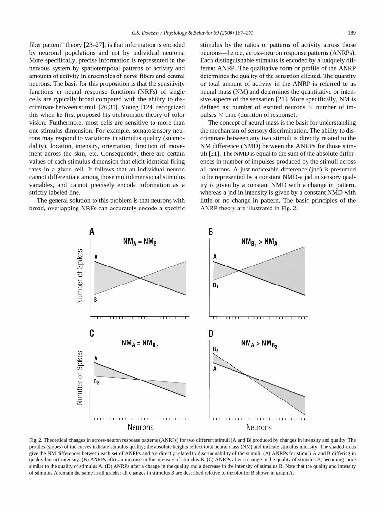
stimulus by the ratios or patterns of activity across thoseneurons—hence, across-neuron response patterns (ANRPs).Each distinguishable stimulus is encoded by a uniquely dif-ferent ANRP. The qualitative form or profile of the ANRPdetermines the quality of the sensation elicited. The quantityor total amount of activity in the ANRP is referred to asneural mass (NM) and determines the quantitative or inten-sive aspects of the sensation [21]. More specifically, NM isdefined as: number of excited neurons
3
number of im-pulses
3
time (duration of response).The concept of neural mass is the basis for understanding
the mechanism of sensory discrimination. The ability to dis-criminate between any two stimuli is directly related to theNM difference (NMD) between the ANRPs for those stim-uli [21]. The NMD is equal to the sum of the absolute differ-ences in number of impulses produced by the stimuli acrossall neurons. A just noticeable difference (jnd) is presumedto be represented by a constant NMD-a jnd in sensory qual-ity is given by a constant NMD with a change in pattern,whereas a jnd in intensity is given by a constant NMD withlittle or no change in pattern. The basic principles of theANRP theory are illustrated in Fig. 2.
Fig. 2. Theoretical changes in across-neuron response patterns (ANRPs) for two different stimuli (A and B) produced by changes in intensity and quality. Theprofiles (slopes) of the curves indicate stimulus quality; the absolute heights reflect total neural mass (NM) and indicate stimulus intensity. The shaded areasgive the NM differences between each set of ANRPs and are directly related to discriminability of the stimuli. (A) ANRPs for stimuli A and B differing inquality but not intensity. (B) ANRPs after an increase in the intensity of stimulus B. (C) ANRPs after a change in the quality of stimulus B, becoming moresimilar to the quality of stimulus A. (D) ANRPs after a change in the quality and a decrease in the intensity of stimulus B. Note that the quality and intensityof stimulus A remain the same in all graphs; all changes in stimulus B are described relative to the plot for B shown in graph A.
190
G.S. Doetsch / Physiology & Behavior 69 (2000) 187–201
In short, the ANRP theory holds that sensory-perceptualdistinctions are based on differences in neuronal populationresponses, differences in either the profile or amount of ac-tivity in the ANRP, or a combination thereof. All distin-guishable features of a stimulus—quality, location, inten-sity, orientation, direction of movement, etc.—can beencoded simultaneously by an ensemble of neurons, withoutrequiring a different coding mechanism for each stimulusfeature. As demonstrated by Erickson [24], each discrim-inable stimulus property corresponds to a different ANRP inthe same (or overlapping) neuronal ensembles.
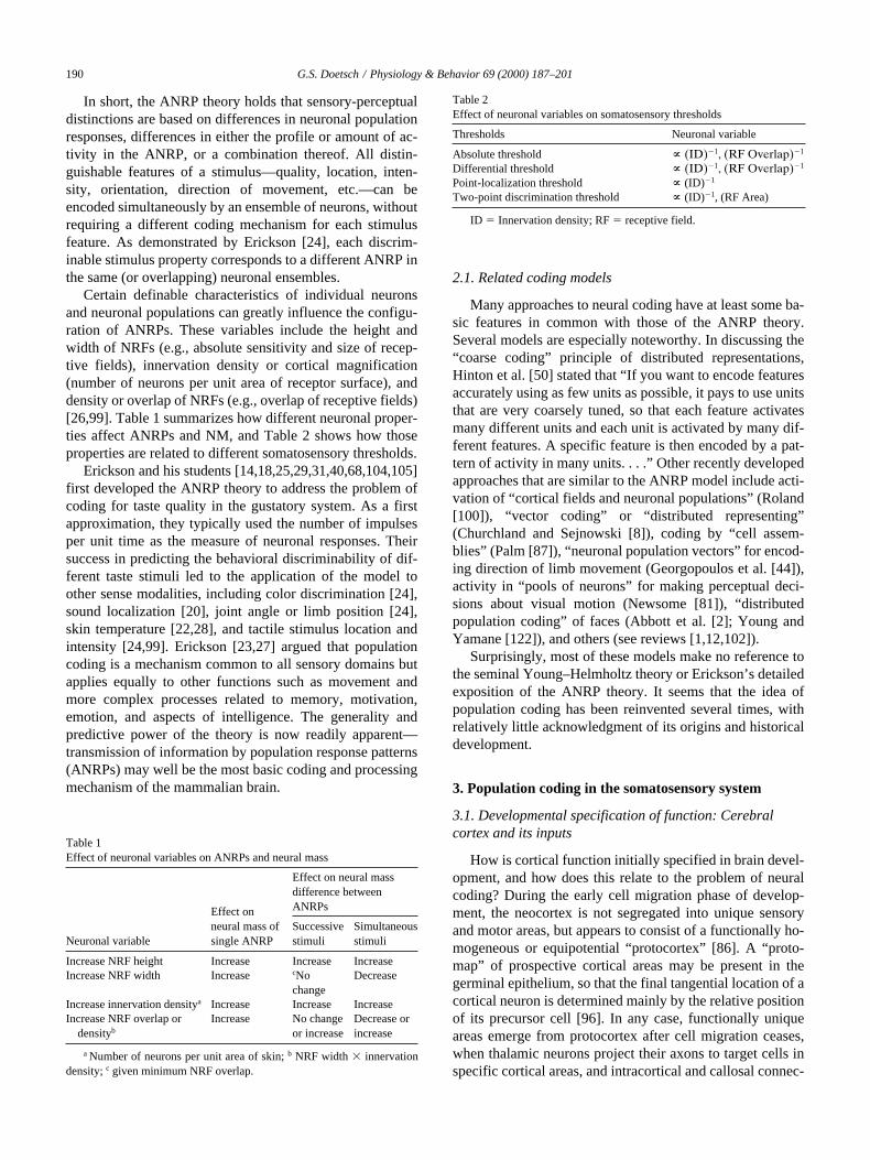
Certain definable characteristics of individual neuronsand neuronal populations can greatly influence the configu-ration of ANRPs. These variables include the height andwidth of NRFs (e.g., absolute sensitivity and size of recep-tive fields), innervation density or cortical magnification(number of neurons per unit area of receptor surface), anddensity or overlap of NRFs (e.g., overlap of receptive fields)[26,99]. Table 1 summarizes how different neuronal proper-ties affect ANRPs and NM, and Table 2 shows how thoseproperties are related to different somatosensory thresholds.
Erickson and his students [14,18,25,29,31,40,68,104,105]first developed the ANRP theory to address the problem ofcoding for taste quality in the gustatory system. As a firstapproximation, they typically used the number of impulsesper unit time as the measure of neuronal responses. Theirsuccess in predicting the behavioral discriminability of dif-ferent taste stimuli led to the application of the model toother sense modalities, including color discrimination [24],sound localization [20], joint angle or limb position [24],skin temperature [22,28], and tactile stimulus location andintensity [24,99]. Erickson [23,27] argued that populationcoding is a mechanism common to all sensory domains butapplies equally to other functions such as movement andmore complex processes related to memory, motivation,emotion, and aspects of intelligence. The generality andpredictive power of the theory is now readily apparent—transmission of information by population response patterns(ANRPs) may well be the most basic coding and processingmechanism of the mammalian brain.
2.1. Related coding models
Many approaches to neural coding have at least some ba-sic features in common with those of the ANRP theory.Several models are especially noteworthy. In discussing the“coarse coding” principle of distributed representations,Hinton et al. [50] stated that “If you want to encode featuresaccurately using as few units as possible, it pays to use unitsthat are very coarsely tuned, so that each feature activatesmany different units and each unit is activated by many dif-ferent features. A specific feature is then encoded by a pat-tern of activity in many units. . . .” Other recently developedapproaches that are similar to the ANRP model include acti-vation of “cortical fields and neuronal populations” (Roland[100]), “vector coding” or “distributed representing”(Churchland and Sejnowski [8]), coding by “cell assem-blies” (Palm [87]), “neuronal population vectors” for encod-ing direction of limb movement (Georgopoulos et al. [44]),activity in “pools of neurons” for making perceptual deci-sions about visual motion (Newsome [81]), “distributedpopulation coding” of faces (Abbott et al. [2]; Young andYamane [122]), and others (see reviews [1,12,102]).
Surprisingly, most of these models make no reference tothe seminal Young–Helmholtz theory or Erickson’s detailedexposition of the ANRP theory. It seems that the idea ofpopulation coding has been reinvented several times, withrelatively little acknowledgment of its origins and historicaldevelopment.
3. Population coding in the somatosensory system
3.1. Developmental specification of function: Cerebral cortex and its inputs
How is cortical function initially specified in brain devel-opment, and how does this relate to the problem of neuralcoding? During the early cell migration phase of develop-ment, the neocortex is not segregated into unique sensoryand motor areas, but appears to consist of a functionally ho-mogeneous or equipotential “protocortex” [86]. A “proto-map” of prospective cortical areas may be present in thegerminal epithelium, so that the final tangential location of acortical neuron is determined mainly by the relative positionof its precursor cell [96]. In any case, functionally uniqueareas emerge from protocortex after cell migration ceases,when thalamic neurons project their axons to target cells inspecific cortical areas, and intracortical and callosal connec-
Table 1Effect of neuronal variables on ANRPs and neural mass
Neuronal variable
Effect on neural mass of single ANRP
Effect on neural mass difference between ANRPs
Successivestimuli
Simultaneousstimuli
Increase NRF height Increase Increase IncreaseIncrease NRF width Increase
c
No change
Decrease
Increase innervation density
a
Increase Increase IncreaseIncrease NRF overlap or
density
b
Increase No changeor increase
Decrease or increase
a
Number of neurons per unit area of skin;
b
NRF width
3
innervationdensity;
c
given minimum NRF overlap.
Table 2Effect of neuronal variables on somatosensory thresholds
Thresholds Neuronal variable
Absolute threshold
~
(ID)
2
1
, (RF Overlap)
2
1
Differential threshold
~
(ID)
2
1
, (RF Overlap)
2
1
Point-localization threshold
~
(ID)
2
1
Two-point discrimination threshold
~
(ID)
2
1
, (RF Area)
ID
5
Innervation density; RF
5
receptive field.
G.S. Doetsch / Physiology & Behavior 69 (2000) 187–201
191
tions are made [53,69,73]. This presumably constitutes themajor stage of functional specification. After the basic iden-tity of each cortical area is established, the functional andanatomical microstructure of those areas can be fine-tunedby neuronal activity related to unique sensory experiences,motor activities, and learning. In the adult brain, majorfunctional respecification of a cortical area is very unlikely,although internal organizational changes may occur.
Thus, the parcellation of the cortex into somatosensory,visual, and auditory areas, etc., appears to be due primarilyto the specific developmental wiring of subcortical and cor-tical circuits. These circuits are driven by spinal or cranialnerves that innervate specialized receptors in the end organssubserving a particular modality. Somatosensory cortex be-comes “somatosensory” mainly because it receives inputfrom the somatosensory thalamus, which in turn, receivesafferent input from pathways that transmit sensory informa-tion from peripheral somatic structures. Whereas the modal-ity specificity of different sensory cortical areas is definedby their selective interconnections, the submodality speci-ficity of individual neurons within those areas may be quite“fuzzy,” given the broad NRFs and multidimensional sensi-tivity of those neurons. This leads directly to the idea thatprecise stimulus information within a particular modality isgiven, not by single neurons, but by neuronal ensembles lo-cated in the cortical areas devoted to that modality.
3.2. Neuronal typology and taxonomy: Basic issues
Can neuronal typology and classification help identifybasic principles of neural coding? Historically, most studiesof sensory coding focused on the specific response charac-teristics of individual neurons or nerve fibers. The physio-logical findings were often used to create a neuronal taxon-omy based on a single criterion, such as maximal sensitivityto a specific stimulus feature (submodality), receptive fieldorganization, rate of response adaptation, or conduction ve-locity. A common problem with such typologies is their es-sentialistic nature, using one or very few salient features toclassify neurons and ignoring less prominent characteristics.Compounding this problem, the class to which a neuron isassigned has often been reified to indicate the specific func-tion of that neuron. Hence, a category label such as “posi-tion, orientation, or edge detector” may implicitly be trans-formed into a labeled-line code for that discrete “function.”
When polythetic (multiple) taxonomic criteria are em-ployed, the results more closely reflect the functional rela-tionships among neurons, but the categories frequently be-come “fuzzy” or may even disappear [30,32,101,115]. Insome cases, polythetic taxonomy yields relatively consistentsubsets of neurons with differential characteristics that canbe used to help determine synaptic interconnectivity, neu-ronal circuitry, and various functional properties of neuralsystems. These data are valuable as criteria for decidingwhether a particular cell is a member of the coding popula-tion at a given synaptic level of the system. For example, itis important to differentiate between presynaptic and
postsynaptic neurons at all levels, and to know the selectiveprojections of those neurons to different targets that may beinvolved in sensory perception or some other function. Tounderstand the flow of information in the cortex from oneneuronal population to another requires knowledge of thelaminar position and synaptic connectivity of those popula-tions. Of course, the major criterion for determiningwhether a subset of neurons participates in stimulus codingis the degree to which inclusion of that subset enhances theability of the neuronal population as a whole to represent(encode) specific stimulus features (for a list of criteria forcandidate neural codes, see [88,89]).
3.3. Neuronal typology and taxonomy: Somatosensory nerve fibers
Can taxonomy of peripheral somatosensory fibers helpsolve the coding problem? Of all the sense modalities, thesomatosensory system may appear most amenable to taxo-nomic analysis, with putative neuronal classes for touch,pressure, flutter-vibration, temperature, pain, joint position,etc., and their subcategories [9,76,78]. For instance, fourmain types of mechanosensitive nerve fibers innervating theglabrous skin of the human hand are now generally recog-nized—RA (QA), Pc, SAI, and SAII fibers [9,55,58]. Eachclass is usually assigned a functionally different role, andthe information it carries is thought to be analyzed sepa-rately at higher levels. Consistent with this notion, Dykes[19] developed a comprehensive model of somatosensoryprocessing by anatomically segregated sets of parallel neu-ral channels made up of different submodality-specific cellpopulations extending from peripheral receptors to cerebralcortex. The specific sets of neurons included, but were notlimited to, the four types of glabrous skin mechanoreceptiveafferents. Dykes [19] proposed that information in the dif-ferent neuronal populations is processed independently, butnoted that a “serious deficiency” of the model is its failureto provide a mechanism for the integration of informationcarried by different afferent channels.
At first glance, data obtained by microstimulation of sin-gle human peripheral nerve fibers seemed to provide strongsupport for the notion that different groups of fibers mediatediscrete submodalities of sensation, especially near thresh-old [85,103,116,117]. However, as discussed by Ray andDoetsch [99], stimulation at different intensities and fre-quencies was found to produce considerable variations inthe sensory qualities evoked—RA fibers were especiallyversatile, yielding sensations of touch, pressure, vibration,tickle, or combinations thereof. In contrast, no sensationcould be elicited by stimulation of SAII fibers alone.Schady et al. [103] made the interesting comment thatchanging stimulus frequency from 5 to 30 Hz evoked 24“previously unrecognized sensations.” Similar variabilitywas encountered in attempting to spatially match the pro-jected sensory fields with the receptive fields of single fi-bers [85,103,116,117]. Finally, no simple relationship wasfound between the firing frequency of any one fiber and the
192
G.S. Doetsch / Physiology & Behavior 69 (2000) 187–201
perceived intensity of the corresponding sensation. In short,individual nerve fibers seem to be limited in their ability totransmit precise information about stimulus submodality,location, intensity, etc. A population code is apparently re-quired, even at the level of primary afferent fibers. The raresituation (near threshold) in which a single fiber or a smallnumber of fibers signals precise information may representa limiting case of the ANRP code—activation of the fiber(s)simply constitutes a small ensemble pattern that is distin-guishable from other patterns.
It is now clear that many somatosensory neurons as-signed to a given category (such as RA, SA, etc.) have com-plex profiles of sensitivity, and respond to more than onestimulus dimension. Gibson et al. [45] anticipated this situa-tion when they used an extensive array of stimuli and differ-ent measures of neuronal responsiveness to study the “cod-ing profiles” of single mechanosensitive nerve fibers ofkittens. They concluded that the response properties of thefibers were highly variable and were “continuously andbroadly distributed.” Given the diversity of coding profiles,the authors “were unable to find any justifiable basis fordesignation of discrete categories of S-R profiles.” The useof many different stimuli (and response measures) typicallyshows that single neurons have more complex, multidimen-sional sensitivity functions (NRFs) than is revealed by theuse of only one or a few stimuli. Furthermore, the findingsof such studies tend to break down taxonomic categoriesbased on one or a few characteristics. Hence, strict “placeand modality specificity” tends to become “fuzzy” indeed.
Consistent with that view, Ray and Doetsch [98] foundthat the size and organization of receptive fields, “on” and“off” responses, and even the rate of adaptation of mecha-nosensitive nerve fibers of the raccoon varied considerablywith stimulus location and intensity. They concluded that fi-ber classification based on adaptation rate was unreliable,and that single fibers could not be assigned a specific cod-ing function. Furthermore, the response properties of indi-vidual fibers had little psychophysical predictive power, andcould not account for variations in tactile acuity across theskin. In this case at least, neuronal taxonomy was found tobe of limited value in studying coding.
3.4. Receptive fields and brain maps
Can neuronal receptive fields (RFs) and somatotopicmaps contribute directly to neural coding mechanisms?Specifically, do they convey useful information to thebrain? These questions are especially appropriate becauseneurophysiologists have long been preoccupied with map-ping the RFs of single neurons and mapping sensory/motorrepresentations in the brain, with the hope of gaining insightinto neural coding. It is clear that RFs and maps are bothempirical constructs defined by the investigator in terms ofan arbitrary level of neuronal firing in response to stimula-tion of specific skin locations. At any given moment, RFsand sensory maps are static and highly simplified represen-tations of the skin by individual neurons (RFs) and popula-
tions of neurons (maps), respectively. In fact, the RFs ofcentral neurons can vary greatly in size and organization,depending on various subcortical and cortical modulatinginfluences; likewise, the topography of brain maps canchange considerably with localized sensory deprivation andexperience (see reviews [62,63,71,72]).
Thus, the momentary properties of a RF reflect not onlythe dominant input connectivity of a neuron, but also thecurrent level of excitability of the cell. Similarly, the basicorganization of a sensory map is established by develop-mental processes that provide for easy lateral cell interac-tions; short-term changes in the map reflect modifications inthe balance of excitatory and inhibitory inputs reaching cer-tain neurons. One might argue that the maximum size andlocation of neuronal RFs, and the maximum variability inmap organization, provide markers that define the spatiallimits on the skin to which sensation produced by neuronalexcitation is projected. But, in fact, afferent input from theperiphery finds its way to the appropriate set of central neu-rons without separate information about RFs or maps.
In short, neither RFs nor maps provide direct informationabout profiles or levels of neuronal activity produced by dif-ferent stimuli. Fields and maps can be interpreted or “read”only by an external observer; they cannot be utilized inter-nally by the brain, which “knows” only neuronal activity orthe lack thereof. This leads to the conclusion that informa-tion about different stimuli and their functional significanceis conveyed not by RFs or maps, but by afferent and centralpatterns of activity in ensembles of neural elements.
3.5. Population response patterns of primary somatosensory nerve fibers
Can activity in neuronal populations solve the codingproblem? In 1931, Adrian et al. [3] commented on the over-lapping distribution of the RFs of mechanosensitive nervefibers: “. . . stimulation of any point on the skin will causeimpulse discharges in several fibres, and the particular com-bination of fibres in action, together with the relative inten-sity of the discharge in each, would supply all the dataneeded for localization.” Hence, the notion of neuronal pop-ulation coding in somesthesis is not new. However, moststudies of ensemble activity have been based on extrapola-tions made from the responses of a “representative” singlenerve fiber to stimulation of different points within the RFof that fiber. Moreover, population behavior was usually ex-amined separately for each class of fibers [9,58].
In contrast, Ray and Doetsch [99] used a direct approachto determine the ANRPs of median (forepaw) and tibial(hindpaw) nerve fibers of the raccoon. They recorded the re-sponses of single fibers to punctate stimulation at standardtest locations (using a series of intensities) on the glabrousskin of the forepaw and hindpaw, regardless of the preciselocation of the stimulus within any one RF. Furthermore,they pooled the responses of the fibers from each nervewithout making assumptions about the possible classifica-tion or function of the fibers. The ANRPs of the median and
G.S. Doetsch / Physiology & Behavior 69 (2000) 187–201
193
tibial fiber samples were then reconstructed for each stimu-lus location and intensity, and the sample ANRPs were ad-justed for differences in innervation density measuredacross each paw and between the two paws.
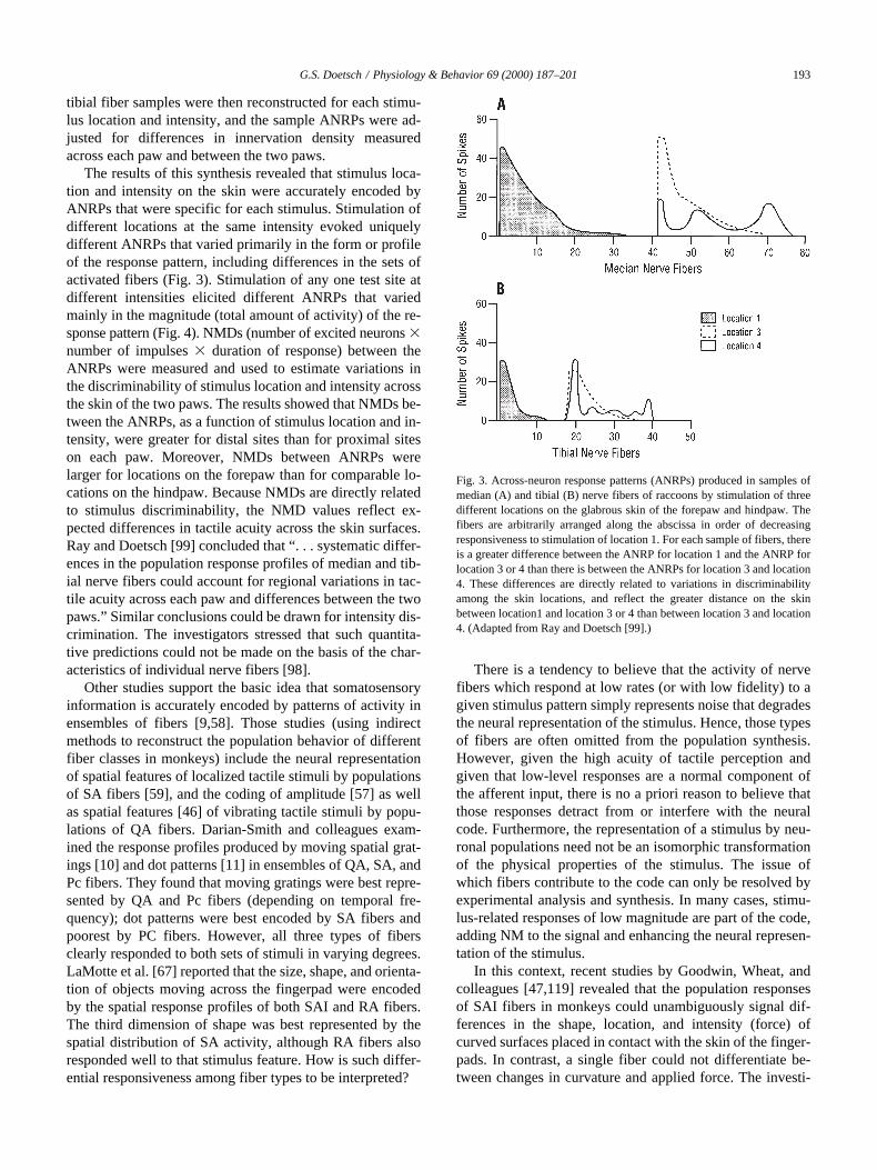
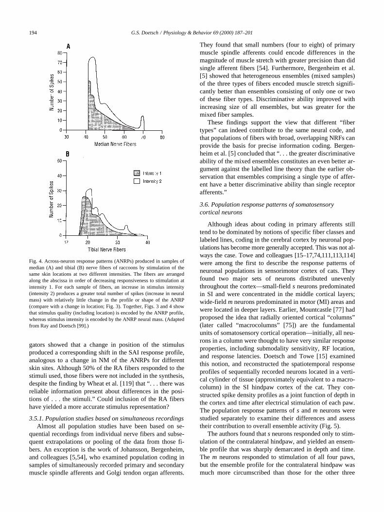
The results of this synthesis revealed that stimulus loca-tion and intensity on the skin were accurately encoded byANRPs that were specific for each stimulus. Stimulation ofdifferent locations at the same intensity evoked uniquelydifferent ANRPs that varied primarily in the form or profileof the response pattern, including differences in the sets ofactivated fibers (Fig. 3). Stimulation of any one test site atdifferent intensities elicited different ANRPs that variedmainly in the magnitude (total amount of activity) of the re-sponse pattern (Fig. 4). NMDs (number of excited neurons
3
number of impulses
3
duration of response) between theANRPs were measured and used to estimate variations inthe discriminability of stimulus location and intensity acrossthe skin of the two paws. The results showed that NMDs be-tween the ANRPs, as a function of stimulus location and in-tensity, were greater for distal sites than for proximal siteson each paw. Moreover, NMDs between ANRPs werelarger for locations on the forepaw than for comparable lo-cations on the hindpaw. Because NMDs are directly relatedto stimulus discriminability, the NMD values reflect ex-pected differences in tactile acuity across the skin surfaces.Ray and Doetsch [99] concluded that “. . . systematic differ-ences in the population response profiles of median and tib-ial nerve fibers could account for regional variations in tac-tile acuity across each paw and differences between the twopaws.” Similar conclusions could be drawn for intensity dis-crimination. The investigators stressed that such quantita-tive predictions could not be made on the basis of the char-acteristics of individual nerve fibers [98].
Other studies support the basic idea that somatosensoryinformation is accurately encoded by patterns of activity inensembles of fibers [9,58]. Those studies (using indirectmethods to reconstruct the population behavior of differentfiber classes in monkeys) include the neural representationof spatial features of localized tactile stimuli by populationsof SA fibers [59], and the coding of amplitude [57] as wellas spatial features [46] of vibrating tactile stimuli by popu-lations of QA fibers. Darian-Smith and colleagues exam-ined the response profiles produced by moving spatial grat-ings [10] and dot patterns [11] in ensembles of QA, SA, andPc fibers. They found that moving gratings were best repre-sented by QA and Pc fibers (depending on temporal fre-quency); dot patterns were best encoded by SA fibers andpoorest by PC fibers. However, all three types of fibersclearly responded to both sets of stimuli in varying degrees.LaMotte et al. [67] reported that the size, shape, and orienta-tion of objects moving across the fingerpad were encodedby the spatial response profiles of both SAI and RA fibers.The third dimension of shape was best represented by thespatial distribution of SA activity, although RA fibers alsoresponded well to that stimulus feature. How is such differ-ential responsiveness among fiber types to be interpreted?
There is a tendency to believe that the activity of nervefibers which respond at low rates (or with low fidelity) to agiven stimulus pattern simply represents noise that degradesthe neural representation of the stimulus. Hence, those typesof fibers are often omitted from the population synthesis.However, given the high acuity of tactile perception andgiven that low-level responses are a normal component ofthe afferent input, there is no a priori reason to believe thatthose responses detract from or interfere with the neuralcode. Furthermore, the representation of a stimulus by neu-ronal populations need not be an isomorphic transformationof the physical properties of the stimulus. The issue ofwhich fibers contribute to the code can only be resolved byexperimental analysis and synthesis. In many cases, stimu-lus-related responses of low magnitude are part of the code,adding NM to the signal and enhancing the neural represen-tation of the stimulus.
In this context, recent studies by Goodwin, Wheat, andcolleagues [47,119] revealed that the population responsesof SAI fibers in monkeys could unambiguously signal dif-ferences in the shape, location, and intensity (force) ofcurved surfaces placed in contact with the skin of the finger-pads. In contrast, a single fiber could not differentiate be-tween changes in curvature and applied force. The investi-
Fig. 3. Across-neuron response patterns (ANRPs) produced in samples ofmedian (A) and tibial (B) nerve fibers of raccoons by stimulation of threedifferent locations on the glabrous skin of the forepaw and hindpaw. Thefibers are arbitrarily arranged along the abscissa in order of decreasingresponsiveness to stimulation of location 1. For each sample of fibers, thereis a greater difference between the ANRP for location 1 and the ANRP forlocation 3 or 4 than there is between the ANRPs for location 3 and location4. These differences are directly related to variations in discriminabilityamong the skin locations, and reflect the greater distance on the skinbetween location1 and location 3 or 4 than between location 3 and location4. (Adapted from Ray and Doetsch [99].)
194
G.S. Doetsch / Physiology & Behavior 69 (2000) 187–201
gators showed that a change in position of the stimulusproduced a corresponding shift in the SAI response profile,analogous to a change in NM of the ANRPs for differentskin sites. Although 50% of the RA fibers responded to thestimuli used, those fibers were not included in the synthesis,despite the finding by Wheat et al. [119] that “. . . there wasreliable information present about differences in the posi-tions of . . . the stimuli.” Could inclusion of the RA fibershave yielded a more accurate stimulus representation?
3.5.1. Population studies based on simultaneous recordings
Almost all population studies have been based on se-quential recordings from individual nerve fibers and subse-quent extrapolations or pooling of the data from those fi-bers. An exception is the work of Johansson, Bergenheim,and colleagues [5,54], who examined population coding insamples of simultaneously recorded primary and secondarymuscle spindle afferents and Golgi tendon organ afferents.
They found that small numbers (four to eight) of primarymuscle spindle afferents could encode differences in themagnitude of muscle stretch with greater precision than didsingle afferent fibers [54]. Furthermore, Bergenheim et al.[5] showed that heterogeneous ensembles (mixed samples)of the three types of fibers encoded muscle stretch signifi-cantly better than ensembles consisting of only one or twoof these fiber types. Discriminative ability improved withincreasing size of all ensembles, but was greater for themixed fiber samples.
These findings support the view that different “fibertypes” can indeed contribute to the same neural code, andthat populations of fibers with broad, overlapping NRFs canprovide the basis for precise information coding. Bergen-heim et al. [5] concluded that “. . . the greater discriminativeability of the mixed ensembles constitutes an even better ar-gument against the labelled line theory than the earlier ob-servation that ensembles comprising a single type of affer-ent have a better discriminative ability than single receptorafferents.”
3.6. Population response patterns of somatosensory cortical neurons
Although ideas about coding in primary afferents stilltend to be dominated by notions of specific fiber classes andlabeled lines, coding in the cerebral cortex by neuronal pop-ulations has become more generally accepted. This was not al-ways the case. Towe and colleagues [15–17,74,111,113,114]were among the first to describe the response patterns ofneuronal populations in sensorimotor cortex of cats. Theyfound two major sets of neurons distributed unevenlythroughout the cortex—small-field
s
neurons predominatedin SI and were concentrated in the middle cortical layers;wide-field
m
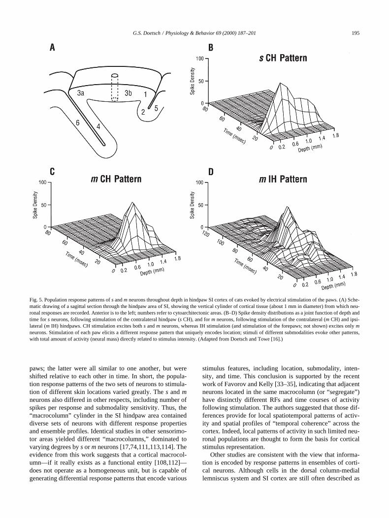
neurons predominated in motor (MI) areas andwere located in deeper layers. Earlier, Mountcastle [77] hadproposed the idea that radially oriented cortical “columns”(later called “macrocolumns” [75]) are the fundamentalunits of somatosensory cortical operation—initially, all neu-rons in a column were thought to have very similar responseproperties, including submodality sensitivity, RF location,and response latencies. Doetsch and Towe [15] examinedthis notion, and reconstructed the spatiotemporal responseprofiles of sequentially recorded neurons located in a verti-cal cylinder of tissue (approximately equivalent to a macro-column) in the SI hindpaw cortex of the cat. They con-structed spike density profiles as a joint function of depth inthe cortex and time after electrical stimulation of each paw.The population response patterns of
s
and
m
neurons werestudied separately to examine their differences and assesstheir contribution to overall ensemble activity (Fig. 5).
The authors found that
s
neurons responded only to stim-ulation of the contralateral hindpaw, and yielded an ensem-ble profile that was sharply demarcated in depth and time.The
m
neurons responded to stimulation of all four paws,but the ensemble profile for the contralateral hindpaw wasmuch more circumscribed than those for the other three
Fig. 4. Across-neuron response patterns (ANRPs) produced in samples ofmedian (A) and tibial (B) nerve fibers of raccoons by stimulation of thesame skin locations at two different intensities. The fibers are arrangedalong the abscissa in order of decreasing responsiveness to stimulation atintensity 1. For each sample of fibers, an increase in stimulus intensity(intensity 2) produces a greater total number of spikes (increase in neuralmass) with relatively little change in the profile or shape of the ANRP(compare with a change in location; Fig. 3). Together, Figs. 3 and 4 showthat stimulus quality (including location) is encoded by the ANRP profile,whereas stimulus intensity is encoded by the ANRP neural mass. (Adaptedfrom Ray and Doetsch [99].)
G.S. Doetsch / Physiology & Behavior 69 (2000) 187–201
195
paws; the latter were all similar to one another, but wereshifted relative to each other in time. In short, the popula-tion response patterns of the two sets of neurons to stimula-tion of different skin locations varied greatly. The
s
and
m
neurons also differed in other respects, including number ofspikes per response and submodality sensitivity. Thus, the“macrocolumn” cylinder in the SI hindpaw area containeddiverse sets of neurons with different response propertiesand ensemble profiles. Identical studies in other sensorimo-tor areas yielded different “macrocolumns,” dominated tovarying degrees by
s
or
m
neurons [17,74,111,113,114]. Theevidence from this work suggests that a cortical macrocol-umn—if it really exists as a functional entity [108,112]—does not operate as a homogeneous unit, but is capable ofgenerating differential response patterns that encode various
stimulus features, including location, submodality, inten-sity, and time. This conclusion is supported by the recentwork of Favorov and Kelly [33–35], indicating that adjacentneurons located in the same macrocolumn (or “segregate”)have distinctly different RFs and time courses of activityfollowing stimulation. The authors suggested that those dif-ferences provide for local spatiotemporal patterns of activ-ity and spatial profiles of “temporal coherence” across thecortex. Indeed, local patterns of activity in such limited neu-ronal populations are thought to form the basis for corticalstimulus representation.
Other studies are consistent with the view that informa-tion is encoded by response patterns in ensembles of corti-cal neurons. Although cells in the dorsal column-mediallemniscus system and SI cortex are still often described as
Fig. 5. Population response patterns of s and m neurons throughout depth in hindpaw SI cortex of cats evoked by electrical stimulation of the paws. (A) Sche-matic drawing of a sagittal section through the hindpaw area of SI, showing the vertical cylinder of cortical tissue (about 1 mm in diameter) from which neu-ronal responses are recorded. Anterior is to the left; numbers refer to cytoarchitectonic areas. (B–D) Spike density distributions as a joint function of depth andtime for s neurons, following stimulation of the contralateral hindpaw (s CH), and for m neurons, following stimulation of the contralateral (m CH) and ipsi-lateral (m IH) hindpaws. CH stimulation excites both s and m neurons, whereas IH stimulation (and stimulation of the forepaws; not shown) excites only mneurons. Stimulation of each paw elicits a different response pattern that uniquely encodes location; stimuli of different submodalities evoke other patterns,with total amount of activity (neural mass) directly related to stimulus intensity. (Adapted from Doetsch and Towe [16].)
196
G.S. Doetsch / Physiology & Behavior 69 (2000) 187–201
being “place and modality specific,” there now seems to beagreement that a population code is required to accuratelyrepresent stimulus location and spatial stimulus features.Furthermore, the total amount of activity in a respondingneuronal population is a generally accepted code for stimu-lus intensity [9,76,78]. For instance, Mountcastle and Dar-ian-Smith [78] proposed that local stimuli applied to theskin produce spatially distributed patterns of activity thatare limited in their extent by surround inhibition; such pat-terns represent “. . . the transformation in neural space of theintensity, contour, and location of the peripheral stimulus.”Moreover, they suggested that spatial tactile discriminationis mediated by differences in spatial profiles of activity, andthat intensity discrimination is based on the size of the ac-tive neuronal population and levels of neuronal firing. How-ever, Mountcastle [76] has continued to adhere to the notionthat submodality or sensory quality is encoded by distinctlabeled lines, suggesting that submodality-specific neuralchannels can be incorporated into other types of codes.
Direct evidence for cortical ensemble coding was ob-tained by Gardner and colleagues, who reconstructed the re-sponse profiles of populations of SI neurons in cats [43] andmonkeys [42] to shearing mechanical stimuli delivered tothe skin. They demonstrated that each of three stimuli, de-livered separately to different locations on the skin, eliciteda spatially distributed pattern of activity with a sharp peakand rapidly decreasing activity on the sides; the patternsoverlapped, but the peaks were shifted relative to each otheracross the cortex. In contrast, when the three stimuli werepresented simultaneously to those locations, they evokedonly one fused population profile, with a peak higher thanthat of the individual profiles. This “funneling” of activityinto one cortical pattern can account for the perceptual illu-sion of one tactile sensation centered over the middle ofthree appropriately placed stimuli on the skin. More gener-ally, the findings support the view that patterns of SI activ-ity encode information about the location and spatial fea-tures of tactile stimuli, and account for the sensoryphenomena associated with those stimuli. Interestingly,Gardner [41] later described the ability of single corticalneurons to extract specific features of cutaneous stimuli,such as contact area, edge orientation, and direction ofmovement (and even defined a set of “haptic neurons”),with little emphasis on population coding.
A different strategy for studying cortical population cod-ing involves the use of metabolic mapping and various imag-ing techniques. Whitsel, Juliano, and colleagues [61,109,121]employed
14
C-2-deoxyglucose mapping to study the globalprofiles of SI activation evoked by repetitive stimulation ofrestricted skin regions on the limbs of monkeys. They foundthat vertical displacement or brushing of the skin producedcomplex spatial patterns of patchy metabolic labeling thatwere distributed across surprisingly large regions of SI. Thepatches extended from cortical layers II through V, andbroad spatial patterns elicited in different animals by thesame stimulus were very similar. The findings indicate that
even very localized stimuli engage widespread ensemblesof neurons, generating spatial patterns of activation that areunique for each stimulus. Whitsel et al. [121] concluded thatthe “. . . S-I cortical network 2DG labeling pattern evokedby somatic stimulation reflects both the place and the modeof somatic stimulation—that is, it is stimulus-specific.”These kinds of studies paved the way for subsequent workon the cortical representation of various sensory-perceptual,motor, and other functions using modern imaging techniques[6,7,37,97,100,110].
3.6.1. Population studies based on simultaneous recordings
Perhaps the most direct approach to population coding isrepresented by the elegant work of Nicolelis et al. [82,83],based on simultaneous recordings from many neurons atseveral levels of the somatosensory system. These investi-gators showed that the RFs of neurons in the ventral poste-rior medial (VPM) nucleus in the thalamus of the rat weremuch larger and overlapped more extensively than gener-ally thought, and often shifted considerably in location overtime (within about 35 ms) following stimulation. Nicoleliset al. [83] suggested that “. . . VPM contains a dynamic anddistributed representation of the face, in which stimulus in-formation is coded in both spatial and temporal domains.”Furthermore, simultaneous recordings made from up to fivedifferent relays of the trigeminal system (including VPMand SI cortex) in freely behaving rats revealed that neuronsat those levels developed widespread 7–12-Hz synchronousoscillations related to rhythmic whisker twitching (WT).Moreover, tactile stimulation of individual whiskers eliciteddistributed spatiotemporal patterns of activity in neuronalensembles located within the same areas displaying oscilla-tory behavior. Nicolelis et al. [82] concluded that “Dynamicpatterns of neural ensemble activity in this sensory systemwere found not only to code tactile stimulus attributes butalso to anticipate the occurrence of stereotyped WT behav-iors associated with active tactile exploration of the sur-rounding environment.” This view is entirely consistentwith the ANRP theory, and is a far cry from “labeled lines”or “place- and modality-specific neurons.”
3.7. Distributed neural networks
Information processing in the brain involves both serial(hierarchical) and parallel circuits that are all part of largerinterconnected networks [36,50,123]. For example, localstimulus-induced patterns of activity (ANRPs) produced inSI areas 3a, 3b, 1, and 2 are transmitted to SII and to parietalareas 5 and 7b (Fig. 6). Furthermore, different cortical lami-nae project selectively to different targets—supragranularlayers transmit information primarily to cortical areas in theipsilateral and contralateral hemisphere; infragranular layerstransfer information mainly to various subcortical targets,including the thalamus [60]. Hence, the patterns of outputfrom different cortical layers to their respective targets mayvary significantly in detail. For instance, Hsiao et al. [51]suggested that processing of information about tactile form
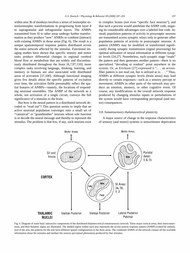
G.S. Doetsch / Physiology & Behavior 69 (2000) 187–201
197
within area 3b of monkeys involves a series of isomorphic-to-nonisomorphic transformations in progressing from layer 4to supragranular and infragranular layers. The ANRPstransmitted from SI to other areas undergo further transfor-mation as they produce “new” ANRPs or combine (interact)with existing ANRPs in those areas (Fig. 6). The result is aunique spatiotemporal response pattern distributed acrossthe entire network affected by the stimulus. Functional im-aging studies have shown that specific sensory and motortasks produce differential changes in regional cerebralblood flow or metabolism that are widely and discontinu-ously distributed throughout the brain [6,7,97,110]; morecomplex tasks involving language, thinking, learning, andmemory in humans are also associated with distributedareas of activation [37,100]. Although functional imaginggives few details about the specific patterns of excitationover time, the activation fields presumably reflect the spa-tial features of ANRPs—namely, the locations of respond-ing neuronal ensembles. The ANRP of the network as awhole, not activation of a single circuit, conveys the fullsignificance of a stimulus to the brain.
But how is the neural pattern in a distributed network de-coded or “read out”? This question seems to imply that anactive neuronal population converges onto a small set of“canonical” or “grandmother” neurons whose sole functionis to decode the neural message and thereby to represent thestimulus. The problem is that few, if any, such neurons exist
in complex brains (not even “specific face neurons”), andthat such a process would annihilate the ANRP code, negat-ing its considerable advantages over a labeled line code. In-stead, population patterns of activity in presynaptic neuronsare transmitted across synaptic relays only to generate otherpopulation patterns of activity in postsynaptic neurons. Apattern (ANRP) may be modified or transformed signifi-cantly during synaptic transmission (signal processing) foroptimal utilization of neural information at different synap-tic levels [24,27]. Nonetheless, each synaptic stage “reads”the pattern and then generates another pattern—there is nospecialized “decoding or readout” point anywhere in thesystem. Or, as Erickson [27] expressed it “. . . an across-fiber pattern is not read out, but is utilized as is. . . .” Thus,ANRPs at different synaptic levels (brain areas) may leaddirectly to certain responses—such as a sensory percept ormovement; ANRPs in other parts of the network may pro-duce an emotion, memory, or other cognitive event. Ofcourse, any modifications in the overall network responseproduced by changing stimulus inputs or perturbations ofthe system would have corresponding perceptual (and mo-tor) consequences.
3.8. Somatosensory thalamocortical plasticity
A major source of change in the response characteristicsof sensory (and motor) systems is sensorimotor deprivation
Fig. 6. Diagram of some basic interactive components of the distributed thalamocortical somatosensory network. Three major cortical areas, their interconnec-tions, and their thalamic inputs are illustrated. The shaded region within each area represents the across-neuron response pattern (ANRP) evoked by stimula-tion of the arm; the patterns for the arm have different spatial configurations in the three areas. The combined ANRPs of the network contain all the availableinformation about the stimulus and mediate the sensory-perceptual phenomena produced by that stimulus.
198
G.S. Doetsch / Physiology & Behavior 69 (2000) 187–201
and learning. Twenty years ago, the sensory systems of theadult mammalian brain were generally considered to bestatic, hard-wired entities. Since then, many studies haveshown that selective loss of sensory input or specific sen-sorimotor training can modify the physiological propertiesof single neurons and cause reorganization of topographicsensory (and motor) maps (see reviews [62,63,65,71,72]).However, with several exceptions, most studies did not spe-cifically examine the effects of sensorimotor perturbationon neuronal population behavior.
Nicolelis et al. [84] studied the changes produced in en-sembles of simultaneously recorded neurons in the VPMnucleus of rats by blocking afferent input from localizedwhisker regions of the face. They found that injections oflidocaine caused single VPM neurons to show dramatictemporal and spatial shifts in the organization of their RFs,resulting in immediate (but reversible) reorganization oflarge portions of the face representation in VPM. Most im-portantly, the investigators reconstructed the neuronal popu-lation response patterns produced by stimulation, and com-pared those patterns before and after sensory block. Theresults showed that the block abolished responses to stimu-lation of whiskers in the anesthetized area and permittednew, short-latency responses to appear after stimulation offar-surround whiskers. Long-latency responses to stimula-tion of near-surround whiskers remained or increased instrength. These changes were qualitatively similar to depri-vation-induced plasticity observed in SI cortex (see reviews[62,63,65,71,72]), but the findings revealed neuronal popu-lation dynamics that are not evident from static topographicmaps. Nicolelis et al. [84] suggested that “. . . sensory depri-vation disrupts the normal dynamic state of equilibrium be-tween excitation and inhibition within the network thatcomprises the entire somatosensory system, producing reor-ganization at multiple levels of this pathway.”
Consistent with that approach, Doetsch [13] examinedthe perceptual significance of denervation-induced reorga-nization of somatotopic cortical maps, using the ANRPmodel as a guide. He discussed two competing hypotheses:(1) the idea of functional respecification suggests that exci-tation of partially deafferented neurons by inputs from newRFs is associated with a change in function (peripheral ref-erence) to signal the new skin fields; (2) the notion of func-tional conservation suggests, instead, that the activity ofthose neurons continues to signal the original skin fields de-spite the responsiveness to stimulation of the new RFs. Theweight of the behavioral evidence supports the idea of con-servation—stimulation of skin regions adjoining an area ofdenervation or amputation in humans typically evokes sen-sation referred to the original (now phantom) skin regions.
This phenomenon can be explained by the ANRP model,with reference to the hand and arm. Stimulation of the intacthand and arm evokes two distinct ANRPs in slightly over-lapping sets of SI neurons; the ANRPs constitute the neuralbasis for sensations projected to the hand and arm. If thehand is lost, stimulation of the arm still elicits the original
“arm ANRP,” but simultaneously evokes responses in theneurons of the SI hand area that have developed new RFs onthe arm. Because the new “hand ANRP” resembles the orig-inal pattern for the hand, sensation is referred to the (phantom)hand. Thus, stimulation of the arm typically yields two sensa-tions—one projected to the arm, and the other to the phantom.
The idea that one population of neurons can generatesimilar ANRPs from stimulation of different skin fields (be-fore and after reorganization) is supported by the findings ofNicolelis et al. [84] in VPM of the rat. Close inspection ofthe ensemble patterns shown in their Fig. 3B reveals thatstimulation of whiskers 1–3 after reorganization evokedpatterns (across neurons 1–11) that were similar to the pat-terns elicited before reorganization by stimulation of whis-kers 4–6. Thus, stimulation of whiskers 1–3 may be inter-preted by the rat as originating from the stimulatedwhiskers, but also from phantom whiskers 4–6. This line ofargument is consistent with the idea that the functional sig-nificance of a given ANRP is not altered.
If the meaning (perceptual consequence) of activity inone brain area were to change due to plasticity, one wouldexpect that the meaning of activity in the entire networkmust change to preserve perceptual and behavioral order.Indeed, Merzenich and deCharms [71] have argued that thisprobably occurs during cortical reorganization. They furthersuggested that the altered meaning of activity patterns mustultimately spread to involve the entire brain. Such radicalchanges in the functional significance of ANRPs—the neu-ral code—are highly problematical. Continuously changingmeanings attached to a given ANRP would appear to be in-compatible with reliable transmission of information. Howcould the brain recognize when a particular pattern in agiven circuit has acquired new meaning, i.e., when the codehas changed? Such functional respecification would seem torequire information other than, or in addition to, neural in-put—information that is not available to the brain. From thisperspective, it is very unlikely that the functional meaningof ANRPs in neural networks can change significantly. Insummary, population response patterns constitute the basiclanguage of the brain, and the functional significance ofthose brain patterns appears to be highly conserved.
Acknowledgments
I thank Douglas D. Rasmusson, Richard H. Ray, and S.David Stoney, Jr. for very helpful comments on this manu-script. I am especially grateful to Robert P. Erickson formany stimulating discussions over the years, for his contin-ued support of my work, and for his enduring friendship.
References
[1] Abbott L, Sejnowski TJ, editors. Neural Codes and Distributed Rep-resentations: Foundations of Neural Computation. Cambridge, MA:M.I.T. Press, 1999.
G.S. Doetsch / Physiology & Behavior 69 (2000) 187–201
199
[2] Abbott LF, Rolls ET, Tovee MJ. Representational capacity of facecoding in monkeys. Cereb Cortex 1996;6:498–505.
[3] Adrian ED, Cattell M, Hoagland H. Sensory discharges in single cu-taneous nerve fibers. J Physiol 1931;72:377–91.
[4] Barlow HB. Single units and sensations: a neuron doctrine for per-ceptual psychology. Perception 1972;1:371–94.
[5] Bergenheim M, Johansson J, Pedersen J, Ohberg F, Sjolander P. En-semble coding of muscle stretches in afferent populations contain-ing different types of muscle afferents. Brain Res 1996;734:157–66.
[6] Bonda E, Petrides M, Evans A. Neural systems for tactual memo-ries. J Neurophysiol 1996;75:1730–7.
[7] Burton H, MacLeod AMK, Videen TO, Raichle ME. Multiple fociin parietal and frontal cortex activated by rubbing embossed gratingpatterns across fingerpads: a positron emission tomography study inhumans. Cereb Cortex 1997;7:3–17.
[8] Churchland PS, Sejnowski TJ. The Computational Brain. Cam-bridge, MA: M.I.T. Press, 1992.
[9] Darian–Smith I. The sense of touch: performance and peripheralneural processes. In: Brookhart JM, Mountcastle VB, editors. Hand-book of Physiology: The Nervous System, vol. 3. Bethesda, MD:American Physiological Society, 1984. pp. 739–88.
[10] Darian–Smith I, Oke LE. Peripheral neural representation of thespatial frequency of a grating moving at different velocities acrossthe monkey’s finger pad. J Physiol 1980;309:117–33.
[11] Darian–Smith I, Davidson I, Johnson KO. Peripheral neural repre-sentation of the two spatial dimensions of a textured surface movingacross the monkey’s finger pad. J Physiol 1980;309:135–46.
[12] Deadwyler SA, Hampson RE. The significance of neural ensemblecodes during behavior and cognition. Annu Rev Neurosci 1997;20:217–44.
[13] Doetsch GS. Perceptual significance of somatosensory cortical reor-ganization following peripheral denervation. Neuroreport 1998;9:R29–35.
[14] Doetsch GS, Erickson RP. Synaptic processing of taste-quality in-formation in the nucleus tractus solitarius of the rat. J Neurophysiol1970;33:490–507.
[15] Doetsch GS, Towe AL. Population response patterns of distinct neu-ronal subsets in hindlimb sensorimotor cerebral cortex of the do-mestic cat. Exp Neurol 1976;53:548–66.
[16] Doetsch GS, Towe AL. Response properties of distinct neuronalsubsets in hindlimb sensorimotor cerebral cortex of the domesticcat. Exp Neurol 1976;53:520–47.
[17] Doetsch GS, Escobar N, Norman HL. Population response charac-teristics of neurons in anterior motorsensory cerebral cortex (field 6)of the domestic cat. Brain Res 1977;137:277–89.
[18] Doetsch GS, Ganchrow JJ, Nelson LM, Erickson RP. Informationprocessing in the taste system of the rat. In: Pfaffmann C, editor. Ol-faction and Taste. New York: Rockefeller University Press, 1969.pp. 492–511.
[19] Dykes RW. Parallel processing of somatosensory information: Atheory. Brain Res Rev 1983;6:47–115.
[20] Eisenman LM. Neural encoding of sound location: an electrophysio-logical study in auditory cortex (AI) of the cat using free field stim-uli. Brain Res 1974;75:203–13.
[21] Erickson RP. A neural metric. Neurosci Biobehav Rev 1986;10:377–86.
[22] Erickson RP. On the intensive aspect of the temperature sense.Brain Res 1973;61:113–8.
[23] Erickson RP. On the neural bases of behavior. Am Sci 1984;72:233–41.
[24] Erickson RP. Parallel “population” neural coding in feature extrac-tion. In: Schmitt FO, Worden FW, editors. The Neurosciences.Third Study Program. Cambridge, MA: M.I.T. Press, 1974. pp.155–69.
[25] Erickson RP. Sensory neural patterns and gustation. In: ZottermanY, editor. Proceedings of the First International Symposium on Ol-faction and Taste. New York: Pergamon Press, 1963. pp. 205–13.
[26] Erickson RP. Stimulus coding in topographic and non-topographicafferent modalities: on the significance of the activity of individualsensory neurons. Psychol Rev 1968;75:447–65.
[27] Erickson RP. The “across-fiber pattern” theory: an organizing prin-ciple for molar neural function. In: Neff WD, editor. Contributionsto Sensory Physiology, vol. 6. New York: Academic Press, 1982.pp. 79–110.
[28] Erickson RP, Poulos DA. On the qualitative aspect of the tempera-ture sense. Brain Res 1973;61:107–12.
[29] Erickson RP, Schiffman SS. The chemical senses: a systematic ap-proach. In: Gazzaniga MS, Blakemore C, editors. Handbook of Psy-chobiology. New York: Academic Press, 1975. pp. 393–426.
[30] Erickson RP, Covey E, Doetsch GS. Neuron and stimulus typolo-gies in the rat gustatory system. Brain Res 1980;196:513–9.
[31] Erickson RP, Doetsch GS, Marshall DA. The gustatory neural re-sponse function. J Gen Physiol 1965;49:247–63.
[32] Erickson RP, Schiffman SS, Doetsch GS, Di Lorenzo PM, Wood-bury MA. A fuzzy set approach to the organization of the gustatorysystem. Prim Sensory Neuron 1995;1:65–80.
[33] Favorov OV, Kelly DG. Minicolumnar organization within soma-tosensory cortical segregates: I. Development of afferent connec-tions. Cereb Cortex 1994;4:408–27.
[34] Favorov OV, Kelly DG. Minicolumnar organization within soma-tosensory cortical segregates: II. Emergent functional properties.Cereb Cortex 1994;4:428–42.
[35] Favorov OV, Kelly DG. Stimulus-response diversity in local neu-ronal populations of the cerebral cortex. Neuroreport 1996;7:2293–301.
[36] Felleman DJ, Van Essen DC. Distributed hierarchical processing inthe primate cerebral cortex. Cereb Cortex 1991;1:1–47.
[37] Frackowiak RSJ. Functional mapping of verbal memory and lan-guage. Trends Neurosci 1994;17:109–15.
[38] Frank M. The classification of mammalian afferent taste nerve fi-bers. Chem Senses Flavor 1974;1:53–60.
[39] Frey M von. Beiträge zur Physiologie des Schmerzsinns, III. Ber kglsächs Ges Wiss Math phys Kl 1895;47:166–84.
[40] Ganchrow JR, Erickson RP. Neural correlates of gustatory intensityand quality. J Neurophysiol 1970;33:768–83.
[41] Gardner EP. Somatosensory cortical mechanisms of feature detec-tion in tactile and kinesthetic discrimination. J Physiol Pharmacol1988;66:439–54.
[42] Gardner EP, Costanzo RM. Spatial integration of multiple-pointstimuli in primary somatosensory cortical receptive fields of alertmonkeys. J Neurophysiol 1980;43:444–68.
[43] Gardner EP, Spencer WA. Sensory funneling: cortical neuronal rep-resentation of patterned cutaneous stimuli. J Neurophysiol 1972;35:954–77.
[44] Georgopoulos AP, Caminiti R, Kalaska JF, Massey JT. Spatial codingof movement: a hypothesis concerning the coding of movement direc-tion by motor cortical populations. Exp Brain Res 1983;7:327–36.
[45] Gibson JM, Beitel RE, Welker W. Diversity of coding profiles ofmechanoreceptors in glabrous skin of kittens. Brain Res 1975;86:181–203.
[46] Goodwin AW, Pierce ME. Population of quickly adapting mechan-oreceptive afferents innervating monkey glabrous skin: representa-tion of two vibrating probes. J Neurophysiol 1981;45:227–42.
[47] Goodwin AW, Browning AS, Wheat HE. Representation of curvedsurfaces in responses of mechanoreceptive afferent fibers innervat-ing the monkey’s fingerpad. J Neurosci 1995;15:798–810.
[48] Helmholtz HLF von. Die Lehre von den Tonempfindungen alsphysiologische Grundlage für die Theorie der Musik. Braun-schweig: Viewig und Sohn, 1863.
[49] Helmholtz HLF von. Handbuch der physiologischen Optik. Ham-burg: Leopold Voss, 1860.
[50] Hinton GE, McClelland JL, Rumelhart DE. Distributed representa-tions. In: Rumelhart DE, McClelland JL, editors. Parallel Distrib-uted Processing. Cambridge, MA: M.I.T. Press, 1986. pp. 77–109.
200
G.S. Doetsch / Physiology & Behavior 69 (2000) 187–201
[51] Hsiao SS, Johnson KO, Twombly A, DiCarlo J. Form processingand attention effects in the somatosensory system. In: Franzen O,Johansson R, Terenius L, editors. Somesthesis and the neurobiologyof the somatosensory cortex. Basel, Switzerland: Birkhäuser Verlag,1996. pp. 229–47.
[52] Hubel DH, Wiesel TN. Receptive fields, binocular interaction, andfunctional architecture in the cat’s visual cortex. J Physiol 1962;160:106–54.
[53] Innocenti GM. Exuberant development of connections, and its pos-sible permissive role in cortical evolution. Trends Neurosci 1995;18:397–402.
[54] Johansson H, Bergenheim M, Djupsjobacka M, Sjolander P. Amethod for analysis of encoding of stimulus separation in ensemblesof afferents. J Neurosci Methods 1995;63:67–74.
[55] Johansson RS, Vallbo AB. Tactile sensibility in the human hand:relative and absolute densities of four types of mechanoreceptiveunits in glabrous skin. J Physiol 1979;286:283–300.
[56] John ER. Switchboard vs. statistical theories of learning and mem-ory. Science 1972;177:850–64.
[57] Johnson KO. Reconstruction of population response to a vibratorystimulus in quickly adapting mechanoreceptive afferent fiber popu-lation innervating glabrous skin of the monkey. J Neurophysiol1974;37:48–72.
[58] Johnson KO, Hsiao SS. Neural mechanisms of tactual form and tex-ture perception. Annu Rev Neurosci 1992;15:227–50.
[59] Johnson KO, Phillips JR. Tactile spatial resolution: I. Two-pointdiscrimination, gap detection, grating resolution, and letter recogni-tion. J Physiol 1981;46:1177–91.
[60] Jones EG. Laminar distribution of cortical efferent cells. In: PetersA, Jones EG, editors. Cerebral Cortex. Cellular Components of theCerebral Cortex, vol. 1. New York: Plenum Press, 1984. pp. 521–53.
[61] Juliano SL, Hand PJ, Whitsel BL. Patterns of increased metabolicactivity in somatosensory cortex of monkeys (
Macaca fascicularis
)subjected to controlled cutaneous stimulation: A 2-deoxyglucosestudy. J Neurophysiol 1981;46:1260–84.
[62] Kaas JH. Plasticity of sensory and motor maps in adult mammals.Annu Rev Neurosci 1991;14:137–67.
[63] Kaas JH. The reorganization of sensory and motor maps in adultmammals. In: Gazzaniga MS, editor. The Cognitive Neurosciences.Cambridge, MA: M.I.T. Press, 1995. pp. 51–71.
[64] Katsuki Y. Neural mechanism of auditory sensation in cats. In:Rosenblith WA, editor. Sensory Communication. Cambridge, MA:M.I.T. Press, 1961. pp. 561–83.
[65] Kelahan AM, Doetsch GS. Time-dependent changes in the func-tional organization of somatosensory cerebral cortex following digitamputation in adult raccoons. Somatosens Res 1984;2:49–81.
[66] Kuffler SW. Discharge patterns and functional organization ofmammalian retina. J Neurophysiol 1953;16:37–68.
[67] LaMotte RH, Lu C, Srinivasan MA. Tactile neural codes for theshapes and orientation of objects. In: Franzen O, Johansson R, Tere-nius L, editors. Somesthesis and the Neurobiology of the Soma-tosensory Cortex. Basel, Switzerland: Birkhäuser Verlag, 1996. pp.113–22.
[68] Marshall DA. A comparative study of neural coding in gustation.Physiol Behav 1968;3:1–15.
[69] McConnell SK. The determination of neuronal fate in the cerebralcortex. Trends Neurosci 1989;12:342–9.
[70] Melzack R, Wall PD. On the nature of cutaneous sensory mecha-nisms. Brain 1962;85:331–56.
[71] Merzenich MM, deCharms RC. Neural representations, experience,and change. In: Llinas R, Churchland P, editors. The Mind–BrainContinuum. Cambridge, MA: M.I.T. Press, 1996. pp. 61–81.
[72] Merzenich MM, Recanzone G, Jenkins WM, Allard TT, Nudo RJ.Cortical representational plasticity. In: Rakic P, Singer W, editors.Neurobiology of Neocortex. New York: John Wiley & Sons; 1988:41–67.
[73] Molnar, Z.; Blakemore, C. How do thalamic axons find their way tothe cortex? Trends Neurosci 1995;18:389-397.
[74] Morse, R.W.; Adkins, R.J.; Towe, A.L. Population and modalitycharacteristics of neurons in the coronal region of somatosensoryarea I of the cat. Exp Neurol. 1965;11:419–440.
[75] Mountcastle VB. An organizing principle for cerebral function: theunit module and the distributed system. In: Edelman GM, Mount-castle VB, editors. The Mindful Brain. Cambridge, MA: M.I.T.Press, 1978. pp. 7–50.
[76] Mountcastle VB. Central nervous mechanisms in mechanoreceptivesensibility. In: Brookhart JM, Mountcastle VB, editors. Handbookof Physiology: The Nervous System, vol. 3. Bethesda, MD: Ameri-can Physiological Society, 1984. pp. 789–878.
[77] Mountcastle VB. Modality and topographic properties of single neu-rons of cat’s somatic sensory cortex. J Neurophysiol 1957;20:408–34.
[78] Mountcastle VB, Darian–Smith I. Neural mechanisms in somesthe-sia. In: Mountcastle VB, editor. Medical Physiology, vol. 2. St.Louis: C.V. Mosby, 1968. pp. 1372–423.
[79] Müller J. Zur vergleichenden Physiologie des Gesichtssinnes desMenschen und der Tiere. Leipzig: C. Cnobloch, 1826.
[80] Nafe JP. The pressure, pain, and temperature senses. In: MurchisonC, editor. A Handbook of General Experimental Psychology.Worcester, MA: Clark University Press, 1934. pp. 1037–87.
[81] Newsome WT. Deciding about motion: linking perception to action.J Comp Physiol A 1997;181:5–12.
[82] Nicolelis MAL, Baccala LA, Lin RCS, Chapin JK. Sensorimotorencoding by synchronous neural ensemble activity at multiple levelsof the somatosensory system. Science 1995;268:1353–8.
[83] Nicolelis MAL, Lin RCS, Woodward DJ, Chapin JK. Dynamic anddistributed properties of many-neuron ensembles in the ventral pos-terior medial thalamus of awake rats. Proc Natl Acad Sci USA1993;90:2212–6.
[84] Nicolelis MAL, Lin RCS, Woodward DJ, Chapin JK. Induction ofimmediate spatiotemporal changes in thalamic networks by periph-eral block of ascending cutaneous information. Nature 1993;361:533–6.
[85] Ochoa J, Torebjork E. Sensations evoked by intraneural microstim-ulation of single mechanoreceptor units innervating the humanhand. J Physiol 1983;342:633–54.
[86] O’Leary DDM. Do cortical areas emerge from a protocortex?Trends Neurosci 1989;12:400–6.
[87] Palm G. Cell assemblies as a guideline for brain research. ConceptsNeurosci 1990;1:133–47.
[88] Parker AJ, Newsome WT. Sense and the single neuron: probing thephysiology of perception. Annu Rev Neurosci 1998;21:227–77.
[89] Perkel DH, Bullock TH. Neural coding. Neurosci Res Prog Bull1968;6:405–527.
[90] Perl ER, Boivie JG. Neural substrates of somatic sensation, vol. 3.In: Guyton AC, editor. M.T.P. International Review of Science,1975. pp. 303–411.
[91] Pfaffmann C. Gustatory nerve impulses in rat, cat, and rabbit. J Neu-rophysiol 1955;18:429–40.
[92] Pfaffmann C. The afferent code for sensory quality. Am Psychol1959;14:226–32.
[93] Pfaffmann C, Frank M, Bartoshuk LM, Shell TC. Coding gustatoryinformation in the squirrel monkey chorda tympani. Prog Psycho-biol Physiol Psychol 1976;6:1–27.
[94] Powell TPS, Mountcastle VB. Some aspects of the functional orga-nization of the cortex of the post-central gyrus of the monkey: a cor-relation of findings obtained in a single unit analysis with cytoarchi-tecture. Bull Johns Hopkins Hosp 1959;105:133–62.
[95] Pubols LM, Leroy RF. Orientation detectors in the primary soma-tosensory neocortex of the raccoon. Brain Res 1977;129:61–74.
[96] Rakic P. Specification of cerebral cortical areas. Science 1988;241:170–6.
[97] Rao SM, Binder JR, Bandettini PA, Hammeke TA, Yetkin FZ, Jes-manowicz A, Lisk LM, Morris GL, Mueller WM, Estkowski LD,
G.S. Doetsch / Physiology & Behavior 69 (2000) 187–201
201
Wong EC, Haughton VM, Hyde JS. Functional magnetic resonanceimaging of complex human movements. Neurology 1993;43:2311–18.
[98] Ray RH, Doetsch GS. Coding of stimulus location and intensity inpopulations of mechanosensitive nerve fibers of the raccoon: I. Sin-gle fiber response properties. Brain Res Bull 1990;25:517–32.
[99] Ray RH, Doetsch GS. Coding of stimulus location and intensity inpopulations of mechanosensitive nerve fibers of the raccoon: II.Across-fiber response patterns. Brain Res Bull 1990;25:533–50.
[100] Roland PE. Brain Activation. New York: Wiley-Liss, 1993.[101] Rowe MW, Stone J. Naming of neurons. Brain Behav Evol 1977;
14:185–216.[102] Sakurai Y. Population coding by cell assemblies—What it really is
in the brain. Neurosci Res 1996;26:1–16.[103] Schady WJL, Torebjork HE, Ochoa JL. Peripheral projections of
nerve fibers in the human median nerve. Brain Res 1983;277:249–61.[104] Schiffman SS, Erickson RP. A theoretical review: a psychophysical
model for gustatory quality. Physiol Behav 1971;7:617–33.[105] Scott TR Jr, Erickson RP. Synaptic processing of taste-quality infor-
mation in thalamus of the rat. J Neurophysiol 1971;34:868–84.[106] Sinclair DC. Cutaneous Sensation. London: Oxford University
Press, 1967.[107] Smith GE. Some problems relating to the evolution of the brain.
Lancet 1910;Jan. 1, 15, and 22.[108] Swindale NV. Is the cerebral cortex modular? Trends Neurosci
1990;13:487–92.[109] Tommerdahl M, Favorov OV, Whitsel BL, Nakhle B, Gonchar YA.
Minicolumnar activation patterns in cat and monkey SI cortex.Cereb Cortex 1993;3:99–411.
[110] Tommerdahl M, Delemos KA, Favorov OV, Metz CB, Vierck CJ,Whitsel BL. Response of anterior parietal cortex to different modesof same-site skin stimulation. J Neurophysiol 1998;80:3272–83.
[111] Towe AL. Neuronal population behavior in the somatosensory sys-tems. In: Kenshalo DR, editor. The Skin Senses. Springfield, IL:Thomas, 1968. pp. 552–74.
[112] Towe AL. Notes on the hypothesis of columnar organization in so-matosensory cerebral cortex. Brain Behav Evol 1975;11:16–47.
[113] Towe AL, Patton HD, Kennedy TT. Response properties of neuronsin the pericruciate cortex of the cat following electrical stimulationof the appendages. Exp Neurol 1964;10:325–44.
[114] Towe AL, Whitehorn D, Nyquist JK. Differential activity amongwide-field neurons of the cat postcruciate cerebral cortex. Exp Neu-rol 1968;20:497–521.
[115] Tyner CF. The naming of neurons: applications of taxonomic theoryto the study of cellular populations. Brain Behav Evol 1975;12:75–96.
[116] Vallbo AB. Sensations evoked from the glabrous skin of the humanhand by electrical stimulation of unitary mechanosensitive afferents.Brain Res 1981;215:359–63.
[117] Vallbo AB, Olsson KA, Westberg KG, Clark FJ. Microstimulationof single tactile afferents from the human hand. Brain 1984;107:727–49.
[118] Weddell G. Somesthesis and the chemical senses. Annu Rev Psy-chol 1955;6:119–36.
[119] Wheat HE, Goodwin AW, Browning AS. Tactile resolution: periph-eral neural mechanisms underlying the human capacity to determinepositions of objects contacting the fingerpad. J Neurosci 1995;15:5582–95.
[120] Whitfield IC. The object of the sensory cortex. Brain Behav Evol1979;16:129–54.
[121] Whitsel BL, Favorov OV, Tommerdahl M, Diamond ME, JulianoSJ, Kelly DG. Dynamic processes governing the somatosensory cor-tical response to natural stimulation. In: Lund JS, editor. SensoryProcessing in the Mammalian Brain. New York: Oxford UniversityPress, 1989. pp. 84–116.
[122] Young MP, Yamane S. Sparse population coding of faces in the in-ferotemporal cortex. Science 1992;256:1327–31.
[123] Young MP, Scannell JW, Burns GAPC, Blakemore C. Analysis ofconnectivity: neural systems in the cerebral cortex. Rev Neurosci1994;5:227–49.
[124] Young T. On the theory of light and colours. Philos Trans R SocLond 1802;92:12–48.