Embed Size (px)
Citation preview

HAL Id: tel-02062629https://tel.archives-ouvertes.fr/tel-02062629
Submitted on 9 Mar 2019
HAL is a multi-disciplinary open accessarchive for the deposit and dissemination of sci-entific research documents, whether they are pub-lished or not. The documents may come fromteaching and research institutions in France orabroad, or from public or private research centers.
L’archive ouverte pluridisciplinaire HAL, estdestinée au dépôt et à la diffusion de documentsscientifiques de niveau recherche, publiés ou non,émanant des établissements d’enseignement et derecherche français ou étrangers, des laboratoirespublics ou privés.
Paysage épigénétique du cancer du seinAslihan Seda Dagdemir
To cite this version:Aslihan Seda Dagdemir. Paysage épigénétique du cancer du sein. Médecine humaine et pathologie.Université d’Auvergne - Clermont-Ferrand I, 2014. Français. �NNT : 2014CLF1MM14�. �tel-02062629�

UNIVERSITÉ BLAISE PASCAL UNIVERSITÉ D’AUVERGNE
2014 N° d’ordre
ECOLE DOCTORALE
DES SCIENCES DE LA VIE ET DE LA SANTE N° d’ordre :
Thèse
Présentée à l’Université d’Auvergne
pour l’obtention du grade de DOCTEUR
Spécialité : Nutrition, Biologie Celulaire et Moléculaire
soutenue le 29 Septembre 2014
DAGDEMIR Aslihan
THE EPIGENETIC LANDSCAPE OF BREAST CANCER Pr és id en t :Pr. Yves-Jean Bignon Université d’Auvergne, EA 4677, Centre Jean Perrin, Clermont Ferrand
Rap p or t eu r s :Dr. Pascale Rio INSERM U 1069, Université François-Rabelais, Tours
Pr. Catherine Bennetau Université Bordeaux, Neurocentre Magendie, INSERM U 862
Directrice de Thèse :Dr. Dominique Bernard-Gallon Centre Jean Perrin, EA 4677, Université d’Auvergne Clermont Ferrand
M emb r e :Dr. Altan Yalciner Laboratoire Duzen, Istanbul, 34383Turquie
Département d’Oncogénétique du Centre Jean Perrin-CBRV, Ertica-EA 4677

Acknowledgments
This study was carried out by the Department of Oncogenetics, CBRV; Centre Jean
Perrin, during 2011-2014.
I would like to express my deepest gratitude to my advisor, Dr. Dominique Bernard-
Gallon. Her support, patience, enthusiasm, broad experience and talent in every aspect of
science have made this thesis possible. Her continued support has led me in the right
direction. I highly appreciate her systematic way of working and understanding of the
essentials in in the field of science. She has continuously provided guidance throughout the
duration of this project. I am grateful to her and all members of Bernard and Gallon’s
families for all of their support.
I would also like to extend a special thanks to Prof. Catherine Bennetau and Dr.
Pascal Rio who were the official reviewers of this thesis. Their valuable comments greatly
enriched this work.
In addition, my sincere thanks to Prof. Penault-Llorca, the present head of the Centre
Jean Perrin, for giving me the opportunity to work with her great ERTICA team, and for
always supporting my project from start to finish.
I also extend my appreciation to my committee member of Prof. Yves-Jean Bignon.
Also, I would like to thank Tim Gunnels for his skillful review of the English
translation of my work.
I owe special thanks to my friends and colleagues, PhD Seher Karsli-Ceppioglu and
MSc. Gaëlle Judes, for their friendship and for sharing their project on breast cancer with
me.
Thanks are extended to Julie Durif, Nicolas Sonnier and all my colleagues in
Department of Oncogenetics CBRV.
Finally, I would like to express my immeasurable appreciation to five unconditionally
devoted people for their love, trust, inspiration and understanding; my father, my mother, Idil,
Sevda and Sonat. This thesis is dedicated to them. Without their encouragement and support,
I would not have had the opportunity to be in France for my studies.
I want to thank my dear friends outside the laboratory for always being supportive.
Finally, and most of all, a special thanks Chief; Dr. Altan Yalciner. His wonderful
sense of support has helped me to see the positive side of the most difficult moments. Without
his continuous support and belief in me, this thesis would have never been possible.

To Dr. Altan Yalciner…

Abbreviations
AdoHcy : S-adenosylhomocysteine hydrolase inhibitor
AdoMet : S-adenosyl-methionine
AF-1 : Activation Function 1
AI : Aromatase Inhibitors
CBP : CREB-Binding Protein
CH3 : Methyl
ChIP : Chromatin Immunoprecipitation
CpG : Cytosine Phosphate Guanine Islands
CREB : cAMP Response Element-Binding Protein
DBDs : DNA Binding Domains
DNA : Deoxyribonucleic Acid
DNMT : DNA Methyltransferase
DZNep : 3-Deazaneplanocin A hydrochloride
E2 : Mammalian estrogen 17 -estradiol
EGFR : Epidermal Growth Factor Receptor
ER : Estrogen Receptor
ErbBB : Epidermal Growth Factor Receptor (EGFR/ERBB) Family
ERE : Estrogen Response Element
EZH2 : Enhancer of Zeste Homolog 2
GFR : Growth Factor Receptor
GPR30 : G Protein-coupled Receptor
H2A : Histone 2A
H3 : Histone 3
H3K4ac : Histone 3 Lysine 4 Acetylation
H3K9ac : Histone 3 Lysine 9 Acetylation
H3K27me3 : Histone 3 Lysine 27 Methylation
HAT : Histone Acetyltransferase
HDAC : Histone Deacetylase
HDACi : Histone Deacetylase Inhibitor
HER2 : Human Epidermal Growth Factor Receptor 2
HKMT : Histone Lysine Methyltransferase
HMTi : Histone Methylation Inhibitor

HRMT : Histone Arginine Methyltransferase
miRNA : MicroRNAs
IGFR : Insulin-like Growth Factor Receptor
LBDs : Ligand Binding Domains
NaBu : Sodium Butyrate
NF- B : Nuclear Factor Kappa-Light-Chain-Enhancer of Activated B Cells
NR3C3 : Nuclear Receptor Subfamily 3, group C, member 3
NST : No Special Type
QC : Quality Control
Q-PCR : Quantitative Polymerase Chain Reaction
O-DMA : O-desmethylangolensin
PcG : Polycomb Group Proteins
PR : Progesterone Receptor
PRC2 : Polycomp Repressive Complex 2
R : Arginine
RNA : Ribonucleic Acid
SAHA : Suberoyl Anilide Hydroxamic Acid
SBR : Scarff-Bloom-Richardson
SERMs : Selective ER Modulators
SIRT : Sirtuin
TNBC : Triple Negative Breast Cancer

1
TABLE OF CONTENTS
1. INTRODUCTION ........................................................................................................................... 3
2. BIBLIOGRAPHY ........................................................................................................................... 5
A. Breast Cancer ............................................................................................................................. 5
i. General description ................................................................................................................ 5
ii. Incidence and Mortality ......................................................................................................... 7
iii. Breast cancer risk factors ....................................................................................................... 9
Gender: ............................................................................................................... 9
Advanced age ..................................................................................................... 9
Age at first birth and parity .............................................................................. 10
Benign breast disease ....................................................................................... 10
Family history .................................................................................................. 11
Breast cancer genes: ......................................................................................... 11
Alcohol use ....................................................................................................... 11
Race and ethnicity ............................................................................................ 11
Dense breast tissue ........................................................................................... 11
Size of a woman ............................................................................................... 12
iv. Tumor markers ..................................................................................................................... 12
1. Estrogen receptor (ER): ........................................................................................ 13
2. Progesterone receptor (PR): ................................................................................. 15
3. HER2/neu: ............................................................................................................ 16
4. Grade: ................................................................................................................... 16
5. Lymph node metastasis: ....................................................................................... 17
6. Ki67: ..................................................................................................................... 17
v. Classification of breast cancer ............................................................................................. 18
1. Luminal A: ....................................................................................................... 19
2. Luminal B: ........................................................................................................ 19
3. HER2 Positive: ................................................................................................. 20
4. Basal-like: ......................................................................................................... 21
5. Claudin-low: ..................................................................................................... 22
B. Phytoestrogens ......................................................................................................................... 24
i. General description .............................................................................................................. 24
ii. Phytoestrogen Signaling Pathways ...................................................................................... 25
iii. Similarities and interaction between soy phytoestrogens and estradiol ............................... 25

2
iv. The Predominant Isoflavones: Genistein, Daidzein and Equol............................................ 27
1. Genistein: ............................................................................................................. 27
2. Diadzein: .............................................................................................................. 29
3. Equol: ................................................................................................................... 30
v. Studies with Phytoestrogens on breast cancer ..................................................................... 31
Presentation of Publication 1: .............................................................................. 35
Conclusion of Publication1: ................................................................................. 36
C. Epigenetic Changes .................................................................................................................. 37
i. General description .............................................................................................................. 37
Presentation of Publication 2: .............................................................................. 39
Conclusion of Publication2: ................................................................................. 40
ii. Epigenetic modifications in breast cancer and epigenetic therapy ...................................... 41
Presentation of Publication 3: .............................................................................. 43
Conclusion of Publication3: ................................................................................. 45
3. OBJECTIVES OF THE STUDY ................................................................................................... 46
4. RESULTS ...................................................................................................................................... 47
A. Effects of Phytoestrogens and Estrogen on Breast Cancer Cell Lines ..................................... 47
Presentation of Publication 4 ................................................................................ 47
Conclusion of Publication 4 ................................................................................. 48
B. Epigenetic Therapy in Breast Cancer Cell Lines ..................................................................... 49
Presentation of Publication 5 ................................................................................ 49
Conclusion of Publication 5 ................................................................................. 50
C. Epigenetic Modifications between Tumor and Normal Tissue in Sporadic Breast Cancer According to Clinicopathological Parameters ................................................................................... 51
D. Promoter Genome-Wide Analysis Tumor and Normal Tissue in Sporadic Breast Cancer ...... 60
5. DISCUSSION ............................................................................................................................... 63
6. CONCLUSION ............................................................................................................................. 74
7. REFERENCES .............................................................................................................................. 75
8. REFERENCES of web .................................................................................................................. 83

3
1. INTRODUCTION
Breast cancer remains the leading cause of cancer-related deaths in women. It is
malignant cell growth in the breast. Breast cancer is noted for conflicting clinical behaviors
and patient outcomes, despite common histopathological features at diagnosis. This can be
explained by the high histological and molecular heterogeneity of the disease, making it hard
to choose a therapy adapted uniquely to each patient.
In biology, epigenetics is study of heritable changes in gene expression or cellular
phenotype caused by mechanisms, other than changes in the underlying DNA sequence. It
refers to functionally relevant modifications to the genome that do not involve a change in the
nucleotide sequence. Epigenetics refer to changes in phenotype and gene expression.
Epigenetic modifications of the genome can be acquired de novo and are potentially inherited.
Epigenetic mechanisms work to change the accessibility of chromatin to
transcriptional regulation locally and globally via modifications of the DNA and by
modifications or rearrangements of nucleosomes. Epigenetics consist in several molecular
mechanisms: histone modifications, small non-coding or antisense RNAs and DNA
methylation; that are closely interconnected.
The incidence and mortality of breast cancer is high in the Western world as
compared with countries in Asia. There are also differences in the regional cancer incidence
rates in Western countries. Several studies involving immigrants to Western countries suggest
that lifestyle and diet are two of the main causes of these differences. In Eastern countries, the
incidence of breast cancer is approximately one-third that of Western countries, whilst their
high dietary intake of phytoestrogens, mainly in the form of soy products, can produce
circulating levels of phytoestrogens that are known experimentally to have estrogenic effects
[1].
Phytoestrogens are plant-derived xenoestrogens functioning like the primary female
sex hormone. They are not generated within the endocrine system, but consumed by eating
phytoestrogenic plants. Also called “dietary estrogens”, they are a diverse group of naturally
occurring nonsteroidal plant compounds that, because of their structural similarity to 17- -
estradiol, have the ability to cause estrogenic and antiestrogenic effects. An increasing
number of epidemiological and experimental studies have suggested that the consumption of a

4
phytoestrogen-rich diet may have protective effects on estrogen-related conditions, such as
breast cancer [2].
Based upon this information, we studied the effects of treatment phytoestrogens;
genistein, daidzein and 17- -estradiol on the post-translational modification of histones such
as lysine methylation and acetylation of histones H3 and H4 in breast cancer cell lines.
Subsequently, we studied the effects of histone methylation inhibitor and histone
deacetylase inhibitor on histone lysine trimethylation and acetylation in breast cancer cell
lines. For this study, we used two breast cancer cell lines MCF-7 and MDA-MB-231. Each
cell line was treated respectively with 3-Deazaneplanocin A hydrochloride (DZNep) [5 µM]
(HMTi), Sodium Butyrate (NaBu) [2 mM] (HDACi) and Suberoylanilide Hydroxamic acid
(SAHA) [1 M] (HDACi) for 48 hours.
Finally, we completed studies in all cell lines with breast tumors to assess Chromatin
ImmunoPrecipitation (ChIP) of selected histone modifications in cancer. The relative levels
of three modified histones, including H3K27me3 (Histone 3 Lysine 27 Methylation), H3K9ac
(Histone 3 Lysine 9 Acetylation), and H3K4ac (Histone 3 Lysine 4 Acetylation) will be
determined in breast tumors compared to matched normal tissue according to the
classification of Saint Gallen.
Today, ChIP has been coupled with promoter DNA microarrays to evaluate the
mechanisms of human gene regulation on a genome-wide scale. ChIP-on-chip technology
could be used to investigate the alterations of global gene expression in tumorigenesis. Here,
we investigated differentially expressed genes associated with modified histones H3K27me3,
H3K9ac and H3K4ac in breast tumors by Agilent SurePrint G3 400kX2 microarrays
containing approximately 21,000 of human transcripts. We will scan the enriched regions at
each gene promoter in thirty breast tumors compared with normal tissue samples. Breast
tumor samples will be classified according to their clinical profiles, especially hormone
receptor status.

5
2. BIBLIOGRAPHY
A. Breast Cancer
i. General description
Breast cancer is a malignant tumor that begins in the cells of the breast. A malignant
tumor is a group of cancer cells that can grow into adjacent tissues or metastasize to distant
areas of the body. The breast cancer occurs almost in women, but men can get it as well [3].
The female breasts are comprised mainly of milk-producing glands, ducts, and stroma
(fatty tissue and connective tissue surrounding the ducts and lobules, blood vessels, and
lymphatic vessels). Most breast cancers begin in the cells that line the ducts (ductal cancers).
Some begin in the cells that line the lobules (lobular cancers), while a small number start in
other tissues [4] (Figure 1).
The most common histologic type of breast cancer is ductal (70-80% of all breast
cancer types) followed by the lobular type of cancer (5-10% of all types). Cancer can also be
ductal-lobular, tubular, medullary and mucinous, and these class only a small minority of all
breast cancers. Ductal cancer is easily detected by mammogram, but for the prognosis, the
lobular type of breast cancer is better than ductal cancer.
It is also important to understand the lymph system because it is one way breast
cancers can spread. This system has several parts. Lymph nodes are small, bean-shaped
collections of immune system cells that are connected by lymphatic vessels. Lymphatic
vessels are like small veins, except that they carry a clear fluid called lymph (instead of blood)
away from the breast. Lymph contains tissue fluid and waste products, as well as immune
system cells. Breast cancer cells can enter lymphatic vessels and begin to grow in lymph
nodes [4].
Most lymphatic vessels in the breast connect to lymph nodes under the arm (axillary
nodes). Some lymphatic vessels connect to lymph nodes inside the chest (internal mammary
nodes) and those either above or below the collarbone (supraclavicular or infraclavicular
nodes) (Figure 2).

6
If the cancer cells have spread to lymph nodes, there is a higher chance that the cells
could have spread into the blood stream and spread (metastasized) to other sites in the body.
Because of this, finding cancer in one or more lymph nodes often affects the treatment plan.
Still, not all women with cancer cells in their lymph nodes develop metastases, and some
women can have no cancer cells in their lymph nodes and later develop metastases.
Figure1. Section of Breast Imaging (a)
Figure 2. Section of Breast Imaging with Nodes and Vessels (b)
Breast profile: A Ducts B Lobules C Dilated section of duct to hold milk D Nipple E Fat F Pectoralis major muscle G Chest wall/rib cage
Enlargement A Normal duct cells B Basement membrane C Lumen (center of duct)

7
ii. Incidence and Mortality
Recent data show that breast cancer incidence and mortality in Europe are a key
resource in both planning and assessing the impact of cancer control programs at the country
and regional level. Europe carries a significant load of the global burden, with one quarter of
the global burden of cancer observed in Europe in 2013 despite a total population that
comprises one-ninth of the world’s population. The International Agency for Research on
Cancer (IARC), through its programs of collaboration with population-based cancer registries
in Europe members of the European Network of Cancer Registries (ENCR,
http://www.encr.com.fr/), has provided estimates of cancer burden at the European and
European Union (EU) member state level over the last 25 years (Figure3-4) [1].
The breast cancer was the leading cancer site in women in all countries of Europe
and also the leading cause of death of cancer in women in Europe. It is likely that the
variation observed in breast cancer incidence across European countries maybe attributable to
the variable extent and type of screening activities in operation, a differential in the
prevalence and distribution of known risk factors for breast cancer. There is a 3- fold variation
(49–148/100,000) with a clear geographical pattern. High incidence rates were estimated in
Western European countries, notably in Belgium (147), France (137) and The Netherlands
(131) and in Northern Europe, particularly in the United Kingdom (129) and in the Nordic
countries, Denmark (143), Iceland (131) and Finland (121). In comparison, incidence rates in
Eastern European countries such as the Ukraine (54) and Moldova (53) were much lower. The
range of mortality rates varies two fold (15–36 per 100,000). Mortality rates were highest in
the North (e.g. Belgium, 29 and Denmark, 28) and in the South (e.g. Serbia, 31 and
Macedonia, 36). The high mortality rates in the northern countries reflect the high incidence,
while in the south, there is a high mortality to incidence ratio, a proxy of low survival [5, 6].

8
Figure 3. Incidence of breast cancer in the world (c)
Figure 4. Incidence and mortality of cancer types (c)
Incidence Mortality

9
iii. Breast cancer risk factors
The causes of breast cancer are not fully known and we know that breast cancer is
multi-factorial disease. However, researchers have identified a number of factors that increase
or decrease the chances of getting breast cancer. These are called risk factors. Breast cancer is
complex and likely to be caused by a combination of factors.
Women have many different breast cancer risk factors and mechanisms over the
course of their life. Some of these factors, like a family history cannot be changed, but many
others are modifiable.
Well-established risk factors for breast cancer include reproductive factors such as;
early age at menarche, late age at first birth, nulliparity, and late age at menopause; family
history of breast cancer; alcohol intake; exposure to ionizing radiation; use of combined
estrogen plus progestin postmenopausal hormone therapy; recent use of oral contraceptives;
physical inactivity; and leanness in early life and obesity in later life.
Gender: Female gender is a strong risk factor for breast cancer. Women have a 150-
fold higher breast cancer risk than men. This is evident due to female sex hormones.
Conditions which lead to high estrogens levels in men are also associated with male breast
cancer. This is probably because men have less of the female hormones, estrogen and
progesterone, which can promote breast cancer cell growth [7].
Advanced age: Breast cancer incidence increases rapidly after age of 40, but after the
age of 65 the incidence decreases. The risk of developing breast cancer increases with age.
About 1 out of 8 invasive breast cancers are found in women younger than 40, while about 2
of 3 invasive breast cancers are found in women age 55 or older.
Age at menarche and menopause: Women experiencing menarche before 12 years
have a 50% higher risk for breast compared to women who experience menarche at 14 years
or older. Likewise, delayed menopause is associated with a risk elevation of 3% for each
delayed year. Both early menarche and late menopause increase the length of lifetime
exposure to endogenous female sex hormones which indicates the importance of these
hormones in the development of breast cancer. The mechanisms underlying this relationship

10
are not well understood, but may involve higher levels of estrogen both earlier and later in life
in girls with earlier menarche. Estrogen is thought to promote the growth of estrogen receptor-
positive (ER+) breast cancer and may also have a role in the early development of ER+ and
ER- breast cancers [8].
Many studies have found that age at menarche was associated with both hormone
receptor-positive and hormone receptor-negative breast cancers, with one of the study
reporting a stronger effect on hormone receptor-positive cancer [9]. In a pooled analysis of
breast cancer patients from 34 studies, early age at menarche was less common among cases
with progesterone receptor-negative (PR-) breast cancer than among cases with PR+ breast
cancer. In the Multiethnic Cohort Study, age at menarche was associated with ER+/PR+
breast cancer, but not with ER-/PR- breast cancer [10].
Age at menarche is determined in part by hereditary factors, but body size, nutrition,
and physical activity can also play a role.
Age at first birth and parity: Full-term pregnancy has a protective effect against breast
cancer risk. During pregnancy, both estrogen and progesterone cause proliferation and
differentiation of the ductal and lobular-alveolar epithelium, which ultimately reduces the risk
for malignant transformation of the breast tissue. Human breast tissue also contains receptors
for human chorionic gonadotropin and luteinizing hormones. Human chorionic gonadotropin
and pregnancy may affect the expression of certain genes and growth factors which inhibit
cell proliferation. Human chorionic gonadotropin may be the most important protective factor.
The earlier the first full-term pregnancy occurs, the lower the risk. Women older than 30 at
first delivery have a 2- 3.5 –fold higher risk for breast cancer, compared to women whose first
delivery was before 21. The risk of breast cancer decreases by approximately 10% per birth.
Even if the first birth is at age 30 or later, multi-party (5 deliveries) has a protective effect
against breast cancer [7].
Benign breast disease: Heterogeneous groups of proliferative and non-proliferative
breast lesions are defined as benign breast diseases. Non-proliferative lesions are not
associated with breast cancer risk, but proliferative lesions, either with (3.5-5-fold) or without
atypical (1.5-2-fold), are associated with an increased risk for breast cancer. Proliferative
diseases account for 25-30% of all benign breast diseases, of which 5-10% show proliferative
lesions with cellular atypia. Both benign and malignant breast disease can present similar
symptoms with a palpable mass or an abnormal screening mammogram with no clinical
findings [11].

11
Family history: Approximately 30% of all breast cancer patients have relatives with
breast cancer. If a first-degree relative has breast cancer, the risk is elevated approximately 2-
fold. The risk increases with the number of relatives affected and is greater for women with
relatives affected at young age. The overall lifetime breast cancer risk for women without a
family history of breast cancer is 7.8%. For those who have one first degree-relative affected,
the risk is 13.3%, and for those having two, the risk is 21.1% [12].
Breast cancer genes: It has been determined that 5-10% of all breast cancers are
caused by mutations in well-identified breast cancer susceptibility genes. The two most
important mutations are the high-risk breast cancer genes BRCA1 and BRCA2. However,
these mutations only account for a part of the genetic susceptibility of breast cancer [13].
Alcohol use: Alcohol use is associated with an increased risk for breast cancer. The
IARC classifies alcoholic beverages as carcinogenic to humans; alcohol causes cancers of the
female breast, oral cavity, pharynx, larynx, oesophagus, liver, and colon and rectum.
Relatively few studies have evaluated the impact of alcohol intake at young ages on risk of
breast cancer. This elevation may be 9-11% with a daily consumption of one alcoholic drink
(10 g/d), and the risk increase is linear up to 6 drinks. Further analysis of one of these studies,
however, focused on alcohol intake during the interval between two important reproductive
events: menarche and first full-term pregnancy. Among women with a longer interval
between menarche and first pregnancy (10 years or longer), each 10 g/day increase in alcohol
intake increased the risk of breast cancer by 21 %, independent of alcohol intake after first
pregnancy. Among women with a shorter interval between menarche and first pregnancy,
alcohol intake did not increase the risk of breast cancer. This suggests that a prolonged period
of exposure at a stage when breast tissue is most vulnerable may increase the risk of breast
cancer. The mechanism of alcohol-induced elevation in breast cancer risk is unknown, but
increased levels of estrogen and androgen appear to be important. Alcohol may also enhance
the susceptibility of mammary cells to carcinogenesis and increase the metastatic potential of
breast cancer cells [14].
Race and ethnicity: Generally, white women are slightly more likely to develop breast
cancer than are African- American women, but African-American women are more likely to
die of this cancer. In women under 45 years of age, however, breast cancer is more common
in African- American women. Asian, Hispanic, and Native American women have a lower
risk of developing and dying from breast cancer [15].
Dense breast tissue: Women with dense breasts have a higher risk of breast cancer
than women with less dense breasts. Unfortunately, dense breast tissue can also make

12
mammograms less accurate. However a number of factors can affect breast density, such as
age, menopausal status, the use of drugs (such as menopausal hormone therapy), pregnancy,
and genetics [16].
Size of a woman: Obesity is associated with a risk for breast cancer. However, obesity
in childhood has not proven to have an effect on the risk of breast cancer later in life, but
weight gain after the age of 18 or after menopause is associated with increased risk of breast
cancer among postmenopausal women. On the contrary, a higher body mass index (BMI) at
18 years is associated with a lower risk of breast cancer in premenopausal life and, in some
studies, in postmenopausal life as well. A high BMI (>31 vs. < 21) is also associated with a
46% lower risk for breast cancer in premenopause. One explanation for the increased risk for
breast cancer after menopause in obese women is the high amount of endogenous estrogens
produced in adipose tissue. Furthermore, obesity increases the circulating concentrations of
insulin, which may be associated with the risk for breast cancer. Tall women appear to have a
higher risk for breast cancer. Childhood energy intake, the cumulative exposure to growth
hormone and insulin-like growth factor-I, or the number of ductal stem cells in the mammary
gland have been proposed as potential biologic mechanisms associated with an increased
breast cancer risk among tall women.
iv. Tumor markers
Breast cancer is a complicated, miscellaneous disease of presence that shows
appreciable variation in morphological, clinical and molecular charges. Traditional
classifications including histological assessment and clinical staging are used to guide patient
management [17].
Historically, breast cancer classification systems have been based on histopathological
assessment including histological type and grade.
At the present time, we use many markers for classification of breast cancer; such as,
expression of estrogen receptor (ER), expression of progesterone receptor (PR) and over-
expression and/or amplification of the human epidermal growth factor receptor 2 (HER2),
tumor grade, lymp node metastasis and Ki-67. They have been included to set the
classification for predicting prognosis as well as the potential response to endocrine treatment

13
and the humanized monoclonal antibody trastuzumab (Herceptin). ER and PR tests are
usually done by immunohistochemistry whereas HER2/neu is accessed by FISH. This protein
profiling of tumors helps to predict the eventual prognosis and can assist in the determination
of the most appropriate treatment for the individual [18].
1. Estrogen receptor (ER):
The nuclear hormone family is activated by the hormone 17 -estradiol, and ER is an
intra-cellular receptor which is a member of this family. The main function of ER is as a
DNA-binding transcription factor which regulates gene expression. There are two classes of
estrogen receptor: ER, which is a member of the nuclear hormone family of intracellular
receptors, and GPR30, which is a member of the rhodopsin-like family of G protein-coupled
receptors. In addition, there are two different forms of ER; alpha ( ) and beta ( ), each
encoded by a separate gene. Hormone-activated ERs form dimers. These two forms of ERs
are co-expressed in various cell types including thyroid, bone, adrenals and female rat brain.
This may lead to the formation of homodimer ER ( ) or ER ( ) or heterodimer ER
( ). Estrogen receptors and show significant overall sequence homology, and both are
composed of five domains. They share about 96% homology between their DNA binding
domains (DBDs), but only 56% homology between their ligand binding domains (LBDs) and
28% homology between their amino-terminal activation functions 1 (AF-1s). ER and ER
can homo- or hetero-dimerize, indicating that the two isoforms can act together or separately.
The two ER isoforms share overlapping functions due, in part, to the significant homology of
their DNA binding domains (Figure 5) [19].
The isoform is encoded by the ESR1 and the isoform is encoded by the ESR2
gene. The two ER isoforms are encoded from two separate genes in two different
chromosomal locations.ESR1 is encoded on chromosome 6 (6q25.1) and ESR2 is encoded on
chromosome 14 (14q). Both ERs are widely expressed in different tissue types, however,
there are some differences in their expression patterns. ER is expressed in endometrial,
breast cancer cells, ovarian stroma cells and in the hypothalamus. ER is expressed in kidney,
brain, bone, heart, lungs, intestinal mucosa, prostate, and endothelial cells. The ER's helix 12
domain plays an important role in determining interactions with co-activators and co-
repressors, thereby affecting the respective agonist or antagonist effect of the ligand. The ER
proteins are attributed to existence cytoplasmic receptors in their unliganded state, but

14
scanning research has shown that there are many ER fractions in the nucleus of ER-negative
breast cancer.
ERs are expressed in approximately 70% of breast cancer cases which are attended to
as "ER-positive" tumors [20]. Binding of estrogen to ER stimulates proliferation of mammary
cells, resulting in an increase in cell division and DNA replication and increases mutation
rate. For these reasons, disruption of the cell cycle and apoptosis and DNA repair processes
that eventually lead to tumor formation. Additionally, estrogen metabolism leads to the
production of genotoxic by-products that could directly damage DNA, resulting in point
mutations. ER expression is associated with more differentiated tumors, while evidence that
ER is involved is controversial. However, recent research suggests that ER is associated
with a poor prognosis and proliferation. Different versions of the ESR1 gene have been
identified and are associated with different risks of developing breast cancer [21].
Patients with high levels of ER are treated with endocrine therapy. Endocrine therapy
for breast cancer involves Selective ER Modulators (SERMS) which act as ER antagonists in
breast tissue or as aromatase inhibitors. ER status is used to determine sensitivity of breast
cancer lesions to tamoxifen and aromatase inhibitors. Raloxifene, which has anti-estrogenic
behavior, has been used as a preventative chemotherapy for women determined to have a high
risk of developing breast cancer [19].
Figure 5: Domains of ER and ER (d)

15
2. Progesterone receptor (PR):
The human progesterone receptor (PR), also known as NR3C3 (nuclear receptor
subfamily 3, group C, member 3), is an intracellular steroid receptor that binds progesterone.
PR is encoded by the PGR gene which exists on chromosome 11 (11q22). PR is expressed as
two isoforms, PR-A (94 kD) and PR-B (114 kD), which function as ligand-activated
transcription factors. These two isoforms are transcribed from distinct ER-inducible
promoters within a single copy PR gene [22].
The PRA form is a truncated version of the PRB form, lacking the first 164 N-terminal
amino acids. In humans, PRA acts as a trans-dominant repressor of the transcriptional activity
of PRB, glucocorticoid receptor, ER, androgen receptor and mineralocorticoid receptor. PRB
functions mainly as a transcriptional activator. PRB is expressed strongly in endometrial
glandular and stromal nuclei in the proliferative phase of the menstrual cycle and weakly
during the secretory phase and early pregnancy (Figure 6) [23].
PR is expressed in reproductive tissue and has important roles in folliculogenesis,
ovulation, implantation and pregnancy. Estrogen is necessary to induce the PRs activity. PRs
become hyperphosphorylated upon binding of the steroid ligand. PR phosphorylation is
complex, occurring in different cellular compartments and perhaps requiring multiple serine
kinases. After progesterone binds to the receptor, restructuring with dimerization follows and
the complex enters the nucleus and binds to DNA. There, transcription takes place, resulting
in formation of messenger RNA that is translated by ribosomes to produce specific proteins
[24].
Figure 6: Domains of PRA and PRB (e)

16
3. HER2/neu:
Receptor tyrosine-protein kinase erbB-2, also known as CD340 (cluster of
differentiation 340), proto-oncogene Neu, Erbb2 (rodent), or ERBB2 (human) is a protein that
in humans is encoded by the ERBB2 gene. The HER2 gene is a proto-oncogene located at the
long arm of chromosome 17 (17q11.2-q12).The ERBB2 gene is also frequently called HER2
(from human epidermal growth factor receptor 2) or HER2/neu.
HER2 is a member of the epidermal growth factor receptor (EGFR/ERBB) family.
HER2/neu plays a significant part in the pathogenesis of breast cancer and as a target of
treatment. It is a cell membrane surface-bound receptor tyrosine kinase and is normally
involved in the signal transduction pathways leading to cell growth and differentiation. HER2
is thought to be an orphan receptor, with none of the EGF family of ligands able to activate it.
However, ErbB receptors dimerise on ligand binding, and HER2 is the preferential
dimerisation partner of other members of the ERBB family [25].
Today this protein has become an important biomarker and target of therapy for
around 30% of breast cancer patients. Amplification or over-expression of this gene has been
shown to play an important role in the development and progression of certain aggressive
types of breast cancer and also associated with increased disease recurrence and worse
prognosis. The poor prognosis may be due to global genomic instability as cells with high
frequencies of chromosomal alterations have been associated with increased cellular
proliferation and aggressive behaviour [25].
4. Grade:
Over the last years, histological grading has become extensively accepted as a
powerful indicator of prognosis in breast cancer, especially of tumor grading systems
currently missionary for breast cancer associated with nuclear grade, tubule formation and
mitotic rate. On the whole, each element is given a score of 1 to 3; that is 1 being the best and
3 the worst, and the score of all three components are added together to derive the "grade".
The lowest possible score (1+1+1=3) is given to well differentiated tumors that all form
tubules and have a low mitotic rate. The highest possible score is 9 (3+3+3=9). The exact
criteria for each component differ in each system and the systems are evolving as more

17
detailed data becomes available. Some studies even suggest that mitotic rate alone can be as
predictive as the grading systems.
The United States uses the most common grading systems as described above; the
original Scarff-Bloom-Richardson (SBR) system and the Black method, which accentuates
nuclear grading and excludes consideration of tubules as criteria. In Europe, the Elston-Ellis
modification of the SBR grading system is preferred and is becoming increasingly popular in
the US. This modification provides somewhat more objective criteria for the three component
elements of grading and specifically addresses mitosis counting in a more precision form. For
example, hyperchromatic nuclei and apoptotic cells which are counted in the original SBR
system are excluded in the Elston-Ellis modification and the area being assessed is
specifically defined in square millimeters. These modifications have enhanced reproducibility
of grading among pathologists and, to a considerable extent, have stimulated acceptance of
grading by clinicians [26].
Criteria for grading is an active area of investigation, particularly in defining more
objective criteria for assessing nuclear grade and we should expect image analysis to greatly
contribute to this area in the future.
5. Lymph node metastasis:
Lymph node metastasis is considered an important prognostic parameter for use in
determining treatment for breast cancer patients. The sentinel node is the first lymph node
reached by metastasizing cells from a primary tumor. A sentinel node biopsy is a minimally
invasive technique to identify lymph node metastases. Involvement of a lymph node in breast
cancer significantly correlates with worse prognosis compared with no lymph node
involvement. Such patients have a higher incidence of death due to disease and should
therefore be treated more aggressively [27].
6. Ki67:
The choice of adjuvant systemic therapy is based on targeted therapy in line with the
St. Gallen consensus meeting. In addition to the traditional parameters, the panel
recommended the use of proliferation markers and multigene assays. The purpose of the
present study was to evaluate the clinical significance of proliferative activity using the Ki-67

18
index as a prognostic marker and as a predictor of recurrence time in breast cancer patients.
Ki-67 is present in all proliferating cells, and there is great interest in its role as a proliferation
marker [28].
The Ki-67 antibody reacts with 395 kDa, which is a nuclear non-histone protein that is
present in all active phases of the cell cycle, except the G0 phase. Proliferation is a key feature
of the progression of tumors and is now widely estimated by the immunohistochemical
assessment of the nuclear antigen Ki-67. The expression of Ki-67 correlates with other
markers of proliferation, including S-phase and bromodeoxyuridine uptake. High Ki-67 is a
sign of poor prognosis associated with a good chance of clinical response to chemotherapy,
but its independent significance is modest and does not rate measurements in most routine
clinical scenari. However, its application as a pharmacodynamic intermediate marker of the
effectiveness of medical therapy holds great promise for rapid evaluation of new drugs [29].
v. Classification of breast cancer
Breast cancer is a complex disease with distinctive properties such as clinical,
morphological and molecular. This heterogeneity cannot be explained just by clinical
parameters such as tumor size, lymph node involvement, histological grade, age; or by
biomarkers like ER, PR and HER2 routinely used in the diagnosis and treatment of patients
[30].
There are more than 21 subtypes of invasive breast carcinoma defined in the fourth
edition of the WHO (World Healthy Organisation) Classification of Tumours of the Breast.
The most frequent is Invasive Carcinoma of No Special Type (NST), also known as invasive
ductal carcinoma NST, and it comprises 40–75 % of cases. The remaining tumor types are
morphologically distinct “special” types including invasive lobular, tubular, mucinous and
metaplastic carcinoma and carcinoma with medullary, neuroendocrine or apocrine features.
The less common subtypes include mucinous, cribriform, micropapillary, papillary,
tubular, medullary, metaplastic, and inflammatory carcinomas. These morphological subtypes
of breast cancer can be further sub-divided into classifications based on their molecular
signatures (ie, expression of protein biomarkers or gene expression profiles).
19
Routine histopathological subclassification of invasive ductal carcinomas is
accomplished by immunostaining cancer tissues to detect expression of the estrogen receptor,
the progesterone receptor and the human epidermal growth receptor 2, as well as HER1 and
various cytokeratins (eg, CK5/6). The differential expression of these protein biomarkers
provides a clinical classification for breast cancer [31].
1. Luminal A:
The luminal A breast cancer is the most common subtype, representing 50–60% of
the total. It is characterized by the expression of genes activated by the ER transcription
factors that are typically expressed in the luminal epithelium lining the mammary ducts. It
also presents a low expression of genes related to cell proliferation. Based on their molecular
profile, all cases of lobular carcinoma in situ are luminal A tumors, as are most of the
infiltrating lobular carcinomas. The luminal A immunohistochemistry profile is characterized
by the expression of ER, PGR and cytokeratin CK8/18, an absence of HER2 expression, a
low rate of proliferation measured by Ki67 and a low histological grade [32].
Patients with this subtype of cancer have a good prognosis; the relapse rate is 27.8%
being significantly lower than that for other sub-types. In addition, survival from the time of
relapse is also longer. They have a distinct pattern of recurrence with a higher incidence of
bone metastases and with respect to other localizations such as central nervous system, liver
and lung which represent less than 10%. The treatment of this sub-group of breast cancer is
mainly based on third-generation hormonal aromatase inhibitors (AI) in postmenopausal
patients, SERMs like tamoxifen and pure selective regulators of ER like fulvestrant [33].
2. Luminal B:
Luminal B breast cancer occurs less frequently, approximetly 10% and 20% of all
breast cancers correlate with the luminal A. Luminal B breast cancers have a more aggressive
phenotype, higher histological grade and proliferative index and worse prognosis. The pattern
of regression also differs, and although the bone is still the most common site of recurrence
(30%), this subtype has a higher recurrence rate in sites such as the liver (13.8%).
Additionally, the survival from time of relapse is lower. Luminal A and B both express ER,

20
but since luminal B’s prognosis is very different, a strong effort to find biomarkers that
distinguish between these two subtypes has been made [30].
From the immunohistochemical point of view, the luminal B subtype has tumors
with ER+/HER2- and high Ki67 or ER+/HER2+. It is worth noting that this definition does
not include all luminal B subtype tumors (up to 6% of the luminal B tumors are clinically ER-
/HER2-). Moreover, the technique used to determine Ki67 (cut-off point to distinguish
luminal A and B set at 13.25%) has not been standardized adding a variability factor in the
assessment of this marker. However, considering that this marker is the most widely used to
measure cell proliferation; efforts are being made to reach a consensus on how to evaluate it.
In fact, an international consortium has recently published a set of recommendations for Ki67
assessment in breast cancer [34].
Luminal B tumors have a worse prognosis than do luminal A tumors despite
treatment with tamoxifen. However, they respond better to neoadjuvant chemotherapy
achieving pathological complete response in 17% of the luminal B tumors (7% in luminalA).
This is clearly lower than for the HER2+ and basal-like tumors with values of 36% and 43%,
respectively. For these reasons, treatment of this subtype of breast cancer is currently
challenging [35].
3. HER2 Positive:
Fifteen to twenty percent of all breast cancers correspond to this molecular subtype.
They are characterized by a high expression of the HER2 gene and other genes associated
with the HER2 pathway and/or HER2 amplicon located in the 17q12 chromosome. These
cancers exhibit an over-expression of genes related to cellular proliferation. Although this
sub-type does not express genes of the basal-like cluster, it may show a low expression of
characteristic luminal genes. Morphologically, these tumors are highly proliferative, with 75%
having a high histological grade and more than 40% have p53 mutations. The
immunohistochemical profile ER-/HER2+ does not correspond perfectly with the intrinsic
subtype, since only 70% of HER2+ tumors by microarray have the protein over-expressed by
immunohistochemical [36].
Conversely, not all tumors with HER2 amplification or over-expression are included
in the cluster of HER2 in the analysis of microarrays. In addition, a significant number of

21
tumors clinically ER+/HER2+ are classified molecularly as luminal B. HER2 amplified
tumors have been further sub-classified into three separate subtypes; one with a clearly worse
prognosis (12% of 10 years survival compared to the 50–55% survival in the other two
groups). In addition, it also had a strong prognostic value in tumors that over-expressed HER2
inside other subgroups of breast cancer [37].
From the clinical point of view, the HER2 subtype is characterized by a poor
prognosis, although in the last decade, anti-HER2 treatment has substantially improved
survival in, not only the metastatic diseases, but also in the initial stages. In neoadjuvant
studies, this sub-type, as well as the basal-like subgroup, has a high chemosensitivity with
higher response rates than that for luminal A and B tumors.
4. Basal-like:
The basal-like subtype represents 10–20% of all breast carcinomas. The term was
coined because they express genes usually present in normal breast myoepithelial cells,
including high molecular weight cytokeratins CK5 and CK17, P-cadherin, caveolin 1 and 2,
nestin, CD44 and EGFR. They also express genes characteristic of luminal epithelium such as
CK8/18 and Ki67, but at level significantly lower levels than those of luminal carcinomas.
Clinically, they are characterized by their appearance at an early age, predominantly in
women of African origin having a large tumor size at diagnosis, a high histological grade and
a high frequency of lymph node affectation. One of the most relevant features of this type of
tumor is the absence of expression of the three key receptors in breast cancer: ER, PGR and
HER2. Therefore, in clinical practice, the terms basal-like and Triple Negative (TN) are often
interchanged. They are not, however, equivalent terms since a discordance of up to 30%
between the two groups has been described. Attempts to identify the basal-like group by an
imminohistochemically profile have led to the selection of five markers (Basal Core Group):
ER, PGR, HER2, EGFR and CK5/6. These markers classify this sub-type with a specificity of
100% and a sensitivity of 76%. Basal-like tumors have a worse prognosis than do luminal
ones. Therefore, it is critical to identify new therapeutic targets and design appropriate
treatment strategies [38].

22
5. Claudin-low:
After the initial molecular classification into sub-types of breast cancer, another
intrinsic subtype was identified in 2007. It is characterized by a low expression of genes
involved in tight junctions and intercellular adhesion; including claudin-3, -4, -7 cingulin,
ocludin, and E-cadherin hence the name claudin-low. This subtype is located in the
hierarchical clustering near the basal-like tumors, suggesting that both subtypes share some
characteristic gene expressions such as low expression of HER2 and luminal gene cluster. In
contrast to the basal-like subtype, this new group over-expresses a set of 40 genes related to
immune response indicating a high infiltration of tumor immune system cells [39].
Claudin-low tumors have a poor prognosis, albeit presenting a low expression of
genes related to cell proliferation. Immunohistochemically, they are normally triple negative.
Like basal-like tumors, the concordance triple negative/claudin-low is not 100%, and about
20% of claudin-low tumors are positive for hormone receptors. These tumors show poor long-
term prognosis and an insufficient response to neoadjuvant chemotherapy with intermediate
values between basal and luminal tumors [40].
The implications of the molecular classification in the therapeutic approach have
been progressively accepted by some international panels. The St. Gallen International Expert
Consensus for Early Breast Cancer 2013 recognized the usefulness of this classification in the
therapeutic decision process. Note that the panel accepted that the different breast cancer sub-
types can be defined, not only by genetic array testing, but by approximations to this
classification using inmmunohistochemistry. This Expert Consensus established five clinico-
pathological definitions, luminal A, luminal B – HER2 negative, luminal B – HER2 positive,
HER2 positive – non luminal and Triple Negative (ductal) ( Table1).

23
CATEGORIES CHARACTERISTICS
Luminal A
• ER and PgR positive
• HER2 negative
• Ki-67 low
• Recurrence risk ‘low’ based on
• Multi-gene-expression assay (if available)
Luminal B
HER2 negative
• ER positive
• HER2 negative
and at least one of:
• Ki-67 ‘high’
• PgR ‘negative or low’
HER2 positive
• ER positive
• HER2 over-expressed or amplified
• Any Ki-67
• Any PgR
HER2 Positive Non-Luminal
• HER2 over-expressed or amplified
• ER and PgR absent
Basal like
• ER and PgR absent
• HER2 negative
Table 1. Classification of Breast Cancer with Saint Gallen Criterias

24
B. Phytoestrogens
i. General description
When we investigated of the incidence and mortality of breast cancer, we saw that
the breast cancer levels are high in the Western world compared with countries in Asia. There
are also differences in the regional cancer-incidence rates in Western countries. Several
studies involving immigrants suggest that lifestyle and diet are two of the main causes of
these differences. This could be related to phytoestrogens. There are many hypotheses that
phytochemicals in Asian diets, which are vegetarian or semi-vegetarian compared with
western diets which are rich in animal proteins and fats, may affect cancer incidence by
altering production, metabolism, and action of steroid hormones. We believed that the
intestines and microflora had a central role in mediating the effects of diet on the disease
pattern in western countries, whilst their high dietary intake of phytoestrogens, mainly in the
form of soy products, can produce circulating levels of phytoestrogens that are known
experimentally to have estrogenic effects [41].
Phytoestrogens are plant-derived formulates that structurally or functionally mimic
mammalian estrogens, and are therefore under consideration to play an important role in the
preclusion of cancers, heart disease, menopausal symptoms and osteoporosis. Phytoestrogens
substitute a heterogeneous group of herbal substances, the structure of which is similar to that
of 17- -estradiol. They are called estrogen-like molecules or non-steroidal estrogens. On the
other hand the structural similarity with estradiol; phytoestrogens are diphenolic yet non-
steroidal compounds [41].
Currently the group of phytoestrogens include more than 100 molecules divided
according to their chemical structure into;
1) Isoflavones (genistein, daidzein, biochanin A, formonetin)
2) Lignans (matairesinol, secoisolariciresinol-diglucoside)
3) Coumestans (coumestrol,4-methoxycoumestrol)
4) Stilbens (resveratrol)

25
ii. Phytoestrogen Signaling Pathways
Phytoestrogens are able to interact with enzymes and receptors, and also because of
their stable structure and low molecular weight they can pass through cell membranes. These
interactions allow them to bind to ERs, induce specific estrogen-responsive gene products,
stimulate ER-positive breast cancer cell growth, interfere with steroid hormone metabolism or
action and alter ER structure and affect transcription. Some genomic mechanisms of action
include estrogenic and antiestrogenic effects on ERs, while other effects may not involve
direct interaction with ERs. Nongenomic effects that do not involve ERs include: induction of
cancer cell differentiation, inhibition of tyrosine kinase and DNA topoisomerase activities,
suppression of angiogenesis and antioxidant effects of phytoestrogens. Other effects can take
place at the cellular and molecular level and potentially influence the biosynthesis and
metabolism of steroids and fatty acids, the serum steroid carrier proteins (sex steroid binding
proteins and -fetoprotein), and the intracellular and transmembrane transfer of hormones to a
membrane and to nuclear receptors [42].
Phytoestrogens inhibit the enzymes needed for hormone conversions, which may
reduce cancers by lowering the biological activity of sex hormones in target organs. As
estrogen-like compounds, some phytoestrogens are able to induce estrus in mammals. The
different activities and the bioavailability of phytoestrogens alter depending on such factors as
the form of administration, dosage, individual metabolism and the ingestion of other
pharmacological substances. Target tissue, concentration addiction, number and type of ER,
and the presence or absence of endogenous estrogens also influence the effect of
phytoestrogen. Not only do phytoestrogens differ in their biological activity, but they also
differ structurally because they come from diverse chemical classes, which may affect their
influence on tissues and receptors. For these reasons the dissimilarity of chemicals that show
estrogenic effects, it appears that estrogenic activity is often emphasized over chemical
structure in defining phytoestrogens [43].
iii. Similarities and interaction between soy phytoestrogens and estradiol
There are lots of studies shown that phytoestrogens defined functionally are
substances that promote estrogenic actions in mammals and structurally are similar to
mammalian estrogen 17 -estradiol (E2) (Figure7) [44].
26
Other mammalian endogenous estrogens are estriol and estrone, which are weakly
estrogenic compared with their mammalian counterpart, E2. The diverse biological activity of
phytoestrogens is due in part to their ability to act estrogenically as estrogen agonists and
antiestrogenically as antagonists. As estrogen agonists, phytoestrogens mimic endogenous
estrogens and cause estrogenic effects. As estrogen antagonists, they may block or alter ER
and prevent estrogenic activity, causing antiestrogenic effects.
As estrogen agonists and antagonists, phytoestrogens can also be classified as
SERMs. SERMs are non-steroidal chemicals with a similar structure to E2 and an affinity
toward estrogen receptors. They are unique in that they can function as agonists or antagonists
depending on the tissue, ER and concentration of circulating endogenous estrogens.
Tamoxifen and raloxifene are well-known SERMs. Tamoxifen has been used in clinical
practice for breast cancer patients because it acts as an estrogen antagonist in breast tissue,
slowing cancer cell proliferation and an estrogen agonist in bone tissue and in the
cardiovascular system to prevent osteoporosis and heart disease. However, tamoxifen has
shown estrogenic activity in the uterus and therefore may increase the risk of endometrial
cancer.
Mechanistically phytoestrogens have been shown to bind to two types of estrogen
receptors: as we talked about previously ER and ER , which was cloned in rats and in
humans. The two receptors differ in their tissue distribution and affinity to ligands, yet there is
some overlap. In rats, ER and ER both are clearly expressed in ovary and uterus tissue.
ER has been shown to have ligand specificity toward phytoestrogens and is distributed in
humans in ovary, spleen, testis and thymus tissue and in rats in bladder, brain, lung, ovary,
prostate, testis and uterus tissue. Phytoestrogens show a lower binding affinity than E2 and
some show a higher binding affinity for ER than for ER , which may suggest different
pathways for their actions and explain tissue specific variability of phytoestrogenic action.
The complexity of phytoestrogens and ERs appears to be further compounded
because different transcriptional activities in vitro are activated depending on the ligands, as
well as the environment of the promoter region of specific genes for translated ER and ER
receptors. [45, 46].

27
iv. The Predominant Isoflavones: Genistein, Daidzein and Equol
Along with daidzein (4’, 7dihydroxyisoflavone), genistein (4’, 5,
7trihydroxyisoflavone) is the most widely studied isoflavone, universally found in soybeans.
Genistein and daidzein are the main soybean phytoestrogens that have a specific chemical
structure very close to the human estrogen and therefore to the estrogenic activity.
Figure 7. Molecular structure of 17- -estradiol and isoflavones genistein and
daidzéine [47].
1. Genistein:
Genistein is one of the most active natural flavonoids and exercises various biological
effects including chemoprevention, antioxidation, antiproliferation and anticancer. More than
30 clinical trials of genistein with various disease indications have been conducted to evaluate
its clinical effectiveness. Based on many animals and human pharmacokinetic studies, it is
well known that the most challenging issue for developing genistein as a chemoprevention
agent is the low oral bioavailability, which may be the major reason for its ambiguous
therapeutic effects and large inter individual variations in clinical trials [48].
In unfermented soy foods, genistein is the result of sugars forming -glycosides that
cannot be absorbed as such by human intestine. Following ingestion, -glycoside of genistein
is hydrolyzed by intestinal -glucosidases to respective aglycone genistein, which is then
17- -estradiol Genistein Daidzein

28
absorbed, probably by nonionic passive diffusion from the jejunum. Fermented soy products
(miso, tempeh, natto) contain larger amounts of isoflavone aglycones due to bacterial
hydrolysis, which may influence the bioavailability. Isoflavone aglycones are glucuronidated
and, to a smaller extent, sulfated in the intestinal wall and liver.
A fraction of isoflavones undergo enterohepatic circulation in analogy to endogenous
estrogens. In blood, the majority of isoflavones are in the form of glucuronide and sulfate
conjugates, and a small amount of aglycones are present as both free as well as bound to
plasma proteins. The main metabolites of genistein in humans are 7-OH-glucuronic acid and
4-OH-sulfate. These conjugates are eventually excreted in the urine in analogy of human
estrogens [49].
Genistein has a diphenol structure that resembles stereo chemically human
endogenous E2. The similar distance between the OH groups on the opposite sides of
genistein and E2 molecules makes genistein capable of binding to ER subtypes and .
Genistein, however, binds with higher affinity to ER than ER and also increases the
binding of ER to a genomic estrogen response element (ERE) more strongly than with ER .
Nevertheless, in human ER subtype specific reporter cells, genistein is a stronger agonist of
ER . Recently, some studies have shown genistein may promote carcinogenesis in mammary
tissues. Therefore, it is crucial to understand the distribution of genistein in breast tissue in
order to correlate exposure with its protective or adverse response to breast cancer [50].
Genistein showed relatively low concentrations in the breast tissue, as compared to plasma,
after the intake of a high dosage of soy isoflavones in humans which indicates a weak
estrogenic response on the breast. In addition to the differential tissue expression of the ER
subtypes, ER often has the opposite effect when compared to ER . This may explain why
the physiological net effect of genistein seems to be partly agonist, initiating estrogen-like
actions, partly antagonist, inhibiting estrogen action; this is the case even though genistein has
been classified to be a pure estrogen agonist in human cells.
Determining the actual biological net effect of phytoestrogens is complicated by
different factors: the route of administration, bioavailability and metabolism, timing and level
of exposure, endogenous estrogen state and the various non-hormonal effects. Moreover,
genistein combined with DNA methylation inhibitors or other DNMTs can enhance the
reactivation of genes silenced by methylation. As evidence of this, Li et al. found that

29
genistein inhibits DNMT1, 3a and 3b and inhibits the expression of hTERT. Genistein also
increases acetylation by enhancing HAT activity [45].
2. Diadzein:
Daidzein belongs to the isoflavone family and is the most commonly ingested and
most intensely studied type of phytoestrogen. It is often found in nuts, fruits, soybeans, and
soy-based products. Previously, daidzein has garnered interest as a non-toxic compound
capable of inducing tumor cell death in a variety of cancer types [51]. Also some studies have
shown that daidzein causes cell cycle arrest at the G1 and G2/M phases in human breast
cancer cells. They have also shown that, while caspase-9 activity was significantly increased
by daidzein, cyclin D expression decreased [52, 53]. In vivo, 9, 10-dimethyl-1, 2-
dibenzanthracene-induced mammary tumors in rats were notably inhibited by daidzein and
tumor latency was significantly increased in mouse mammary tumor virus-neu mice.
Moreover, one study has revealed that daidzein induced MCF-7 cell proliferation was blocked
by treatment with antiestrogen antibody Faslodex (ICI 182780), demonstrating that daidzein-
induced stimulatory effect was estrogen receptor (ER) mediated [54]. All of this data and the
evidence from in vitro and in vivo studies indicate that the anti-cancer activity of daidzein in
breast cancer is mediated through cell cycle arrest and apoptosis. However, the specific
apoptosis mechanisms at work are not yet well understood. Because caspase-9 (an apoptosis
biomarker) activity was significantly increased by daidzein, it prompted us to hypothesize that
daidzein induces apoptosis through the intrinsic pathway, which regulates apoptotic cascades
by signaling convergence in the mitochondrion. Therefore, the aim of the present study was to
thoroughly authenticate the original research article of daidzein-induced apoptosis and
explore the potential mechanisms at work.
Daidzein can cross the placenta and has been found in breast milk. It is unknown
whether daidzein influences early onset of puberty in girls. In vitro and in vivo studies have
found that daidzein stimulates the growth of estrogen-sensitive breast cancer cells.
Epidemiologic studies have found conflicting evidence. Some studies have found an
association between soy exposure and decreased breast cancer risk while others have found no
association [55]. Some epidemiological evidence indicates that soy intake may be more
protective when the exposure occurs prior to puberty. More research needs to be conducted on

30
the association between breast cancer risk and daidzein specifically before conclusions can be
drawn. This fact sheet provides information about daidzein, one of three phytoestrogens being
measured and examined by the Breast Cancer and the Environment Research Centers
(BCERC) epidemiology studies, sources of exposures, effects on puberty, effects in the body,
and research studies looking at daidzein as being associated with breast cancer risk [56].
Daidzein is an isoflavone aglycone and is produced in the body from plant
isoflavones. Isoflavones are contained in soybean or soy foods in two chemical forms, i.e.,
aglycones (uncongugated form) and glucosides (bound to a sugar molecule). The main dietary
source of daidzein is the biologically active glucoside daidzin. Fermentation or digestion of
soybeans or soy products results in the release of the sugar molecule from the isoflavone
glycoside, daidzin, leaving the isoflavone aglycone, daidzein [57]. Before daidzein can act it
first needs to be released from daidzin. This normally happens in the stomach (acid
hydrolysis) and intestine (action of bacterial enzymes). After daidzein is released from
daidzin, it may be absorbed into the blood or it may be further metabolized by intestinal
bacteria into the metabolites equol and O-desmethylangolensin (O-DMA) [56]. The extent of
this metabolism appears to be highly variable among individuals and is influenced by the
specific bacteria present in the intestine and other components of the diet. After consuming
soy or daidzein, approximately 30%-50% of the population produces equol, and
approximately 80%-90% produces O-DMA [56].
Daidzein is also an antioxidant. It is thought that daidzein is a less potent antioxidant
than genistein; however, there are few studies comparing the antioxidant activity of the two
isoflavones. Equol is a more potent antioxidant than daidzein.
3. Equol:
Equol is a metabolite of daidzein that has gained interest due to its possible effects on
cancer risk. In vitro studies of equol found it to be more biologically active than daidzein,
with a higher affinity for the estrogen receptor, and more potent antioxidant activity. This
suggests that it may be advantageous to convert daidzein to equol to enhance its estrogenic
potency. Equol is a chiral molecule and can exist as two isomers, R- and S-equol. S-(-)-equol
is the metabolite of daidzein by intestinal bacteria. Equol is expected to prevent hormone-
dependent diseases, including breast cancer, due to its ability to bind both ER and ER . S-(-)
-equol is especially known to have a much stronger affinity for ER compared to R-(+)-equol

31
[58], and moreover due to its superior anti-oxidative potential to all the isoflavones [59]. In
addition, equol binds to sex hormone binding globulin and competitively inhibits estradiol
and testosterone binding in a dose-dependent manner. Very little research focusing on equol
specifically has been conducted, whereas mechanisms of soy isoflavone on breast cancer have
been well done. In vitro studies have demonstrated that equol, both racemic and S-equol
inhibited the growth of the breast cancer cell line MDA-MB-231 at higher concentrations
( 10 M) but in contrast, equol at lower concentrations ( 10 M) stimulated the proliferation
of ER positive breast cancer cells. The compounds also showed effects in inhibiting the
invasion of MDA-MB-231 cancer cells through matrigel [60]. Another study reported that
(±)-equol had proliferative effects on MCF-7 cell growth in vitro within the concentration of
plasma equol 2.10-3.21 M.
v. Studies with Phytoestrogens on breast cancer
Considering the mechanism of action, the phytoestrogens on breast cancer may be
mediated via many different mechanisms. Several isoforms of the estrogen receptor can be
involved by mechanisms, such as ER heterodimerisation with ER , and a consequent
reduction in estrogen effects. There are also many studies that have shown that genistein, soy,
and rye bran can also cause apoptosis in cancer cells both in vitro and in vivo.
In addition, we now know that the chemical structure of phytoestrogen resembles
that of E2 suggesting that ER-activated genomic and/or nongenomic signaling pathways and
might mediate the principal function of phytoestrogen.
The signalling pathways play a role of the apoptosis, invasion, metastasis and
proliferation of breast cancer cells. These pathways include nuclear ER (genomic ER)-
initiated, membrane ER (non-genomic ER)-mediated, growth factor (GF)-transduction, G
protein receptor (GPR)-directed and apoptotic signaling pathways.
To induce the modification of gene expression in breast cancer, nuclear ER and/or
ER must be activated. Although encoded by unique genes, ER and ER have certain
functional domains with a highly similar affinity to ligand- and DNA-binding sites. This
suggests that these two receptors might play redundant roles. The mammary cell has a
predominant distribution of ER .

32
In the classic pathway to activate transcriptome, the activated ERs dimerizes and
forms a complex for binding to a specific DNA sequence, the ERE.
In non-classic pathway, E2-ER complex interacts with several transcription factors
such as Activator Protein 1 (AP1), Specificity Protein 1 (SP1) and NFkB to modulate gene
expression without ERE. Another genomic pathway is through a ligand-independencer,
showing that Growth Factor (GF) activates in intracellular kinase pathway to phosphorylate
ER at ERE-containing promoters [61]. Separate from nuclear genomic action, the membrane
ER has been demonstrated as a G protein-coupled receptor. The activated membrane ER
induces the discretion of the G-protein subunit to trigger the activation of Src kinases and
PI3 kinase (PI3-K), respectively followed by downstream protein kinase C (PKC) and kinase
cascade to extra-cellular regulated protein kinase (Erk), which are involved in proliferation
and survival of breast cancer cells. The PI3-K activation also activates the recruitment of
AKT, a serine-threnine kinase to control cell survival by activating downstream anti-apoptotic
signals (e.g. Bcl-2) and transcription factors (e.g. NFkB and CREB) [62, 63]. In addition,
growth factor receptors (GFR), including insulin-like growth factor receptor (IGFR) and
epidermal growth factor receptor (EGFR), are the other E2-membrane signalling pathways
within breast cancer cells. IGFR mediates the signal pathway through connecting ER to
plasma membrane, activating EGFR, and initiating PI3-K and Erk signalling [64].
In the ER-negative breast cancer cell, to induce the activation of downstream Scr-
coupled Erk and cAMP/PKA signaling networks via G g subunit protein, E2 directly
interacted with GPR30 [62]. The apoptotic signalling pathway is inactivated in most cancer
cells, including a group of proteases such as caspase-8 and caspase-3 that cross-talk with EGF
survival signalling network [63].
Finally, these mechanisms may occur as protection against cellular proliferation of
breast cancers by intracellular mechanisms of phytoestrogen;
1. Binding to nuclear ER and inhibiting genomic ER-mediated gene expression
2. Interaction with membrane ER, blocking protein kinases and suppressing
transcription factors
3. Inhibiting GFR activation and its downstream signalling networks
4. Activating caspases to initiate cellular apoptosis
5. Reducing the G-protein mediated signalling pathway in the ER-negative
mammary cancer cell.

33
The nuclear ER interaction is the most completely studied mechanism of the effects
of phytoestrogen. The phytoestrogen-rich pueraria mirifica showed a strong competitive
binding ability to ER and/or possibly synthesized suppressor of ER in the therapy of rat
mammary tumor [65]. Phytoestrogen has recently demonstrated to modulate ER through the
activation of ligand-independent pathway.
To summarize, breast cancer is a common malignancy with a high mortality rate in
Western population. However, the people in the Asian societies have lower incidences of this
cancer than the individuals from the Western societies. This speculation is supported by the
finding that the higher the soy food consumption in adolescence, the lower is the risk of breast
cancer. Phytoestrogens exhibit a wide array of pharmacologic properties, and recently,
interest in the potential benefits of diets high in phytoestrogens has intensified, especially
those related to chemoprevention. The link between phytoestrogens and breast cancer
prevention has been the subject of numerous studies, and the epidemiology of breast cancer in
relation to phytoestrogen consumption has recently been extensively reviewed. Generally,
epidemiologic studies have been inconclusive, and the relationship between phytoestrogens
and breast cancer prevention remains uncertain. Some studies have revealed the modest
protective effects of phytoestrogens; others have detected no association between
phytoestrogen intake and breast cancer risk; and a few have reported marked protective
effects.
Nevertheless, some evidence suggests that soy intake must be high during certain
windows of development, specifically pre-pubescence, in order to gain the protective effects
of phytoestrogens. Despite intense investigation, it remains unclear whether phytoestrogens
are actually chemo-protective agents or whether their presence is simply a biomarker
indicative of a healthy diet.
Additionally, there are many studies about the phytoestrogens on breast cancer in our
laboratory. Some of them, after genistein and daidzein 72h exposures, Satih et al. observed a
restored expression of RAR in MCF-7, but this was not the case in MDA-MB-231 cells. This
observation may be due to the different ER status exhibited by these two cell lines. [66].
Several studies have shown that phytoestrogens may have an impact on the
expression of BRCA2. To focus on these processes, they set up the BRCA2 specific

34
knockdown by RNA interference in two breast tumor cell lines (MCF-7 and MDA-MB-231)
and also in a non-tumorigenic breast cell line (MCF-10a). After inhibition of BRCA2
expression, cells were maintained in different conditions and treated with either daidzein or
genistein or left untreated. Microarray analysis of mRNAs isolated from the BRCA2 knocked
down MCF-7, MDA-MB-231, and MCF-10a cell lines after being treated with phytoestrogens
showed 35 differentially expressed genes between positive-ER cells and negative-ER cells.
After genistein or daidzein treatments, BRCA1 was found to be up-regulated when knocked
down with BRCA2-siRNA MCF-7 and BRCA2 was found to be up-regulated when knocked
down with BRCA2-siRNA MDA-MB 231 cells. In MCF-10a, they observed a significant
decrease in BAX and BCL2 expressions with a greater effect of daidzein [44].
Also Bosviel et al. investigated the effects of equol on DNA methylation of BRCA1
and BRCA2 in breast cancer cell lines (MDA-MB-231 and MCF-7) and in a dystrophic breast
cell line (MCF-10a). They demonstrated by quantitative analysis of methylated alleles that a
significant decrease in the methylation of the cytosine phosphate guanine (CpG) islands in the
promoters of BRCA1 and BRCA2 after the S-equol treatment in MCF-7 and MDA-MB-231
cells and a trend in MCF-10a cells. They also showed that S-equol increases BRCA1 and
BRCA2 protein expression in the nuclei and the cytoplasm in MCF-7, MDA-MB-231 and
MCF-10a cell lines by immunohistochemistry. They demonstrated the demethylating effect of
S-equol on the CpG islands inside the promoters of BRCA1 and BRCA2 genes, resulting in an
increase in the level of expressed oncosuppressors in breast cancer cell lines [13].
Additionally, there has been a second study by Bosviel et al. about the putative
effects on DNA methylation by two naturally occurring isoflavones, genistein and daidzein, in
a study of the BRCA1 and BRCA2 oncosuppressor genes in breast cancer cell lines (MCF-7,
MDA-MB 231, and MCF10a) by methylated DNA immunoprecipitation coupled with PCR.
Their results suggest that treatment with genistein or daidzein might reverse DNA
hypermethylation and restore the expression of the oncosuppressor genes BRCA1 and BRCA2.
These observations, while requiring replication, provide new evidence on potential epigenetic
mechanisms by which genistein and daidzein might contribute to regulation of the BRCA1 and
BRCA2 [67].

35
Presentation of Publication 1:
Dagdemir A, Durif J, Ngollo M, Bignon Y-J, Bernard-Gallon D. Breast Cancer:
Mechanisms Involved in Action of Phytoestrogens and Epigenetic Changes in Vivo. 2013 Jan-
Feb; 27(1):1-9.
Before we began our first study, we considered the effects of phytoestrogen soybean
on breast cancer. We present in this review a summary of the action of phytoestrogens in
biological mechanisms and regulations of epigenetic breast cancer.
A high intake of dietary phytoestrogens has been suggested to account for the lower
rates of climacteric complaints, cardiovascular diseases, breast and endometrial cancers, and
osteoporosis-related fractures in Asian than in Western countries.
Phytoestrogens display an array of pharmacologic properties, and in recent
investigation of their potential as anticancer agents has increased dramatically. In this article
we review the published literature related to phytoestrogens and breast cancer as well as
suggest the possible mechanisms that may underlie the relationship between phytoestrogens
and breast cancer.

Abstract. In this review, we consider phytoestrogens and
different epigenetic modifications in breast cancer.
Epigenetic phenomena are mediated by several molecular
mechanisms comprising histone modifications, small non-
coding or anti-sense RNA and DNA methylation. These
different modifications are closely interrelated. Deregulation
of gene expression is a hallmark of cancer. Although genetic
lesions have been the focus of cancer research for many
years, it has become increasingly recognized that aberrant
epigenetic modifications also play major roles in breast
carcinogenesis. The incidence and mortality of breast cancer
are high in the Western world compared with countries in
Asia. There are also differences in the breast cancer
incidence rates in different Western countries. This could be
related to phytoestrogens.
Breast cancer remains the first cause of cancer-related
mortality in women. This can be explained by the high
histological and molecular heterogeneity of the disease,
making it hard to choose a therapy adapted to each patient.
Over this past year, several groups have evaluated the
epigenetic component of breast cancer, as epigenetics
appears to be important in carcinogenesis.
The progression through the multiple steps of breast cancer
from epithelial hypertrophy to highly invasive breast
carcinoma involves multiple coordinated changes in gene
expression programming. Such coordinated changes are
bound to be controlled by global mechanisms of gene
expression programming. The genome is programmed by the
epigenome, which consists of the chromatin structure, a
pattern of modification of DNA by DNA methylation, and a
profile of expression of noncoding RNAs, such as microRNA.
The revolution of epigenetics has revitalized cancer research,
shifting focus away from somatic mutation towards a more
global perspective involving the dynamic states of chromatin.
Disruption of chromatin organization can directly and
indirectly precipitate genomic instability and transformation.
Epigenetic changes are reversible and may lead to loss or gain
of biological functions. More importantly, many of the
elucidated epigenetic changes are linked to the pathogenesis
of human diseases, including cancer. Epigenetic aberrations
arise early in carcinogenesis, before gene mutations in DNA
supplies targets for early detection.
The incidence and mortality of breast cancer are high in
the Western world compared with countries in Asia. There
are also differences in the regional cancer incidence rates in
Western countries. Several studies involving immigrants to
Western countries suggest that lifestyle and diet are two of
the main causes of these differences. In Eastern countries,
the incidence of breast cancer is approximately one-third that
of Western countries, whilst their high dietary intake of
phytoestrogens, mainly in the form of soy products, can
produce circulating levels of phytoestrogens that are known
experimentally to have estrogenic effects.
Phytoestrogens
Phytoestrogens are plant-derived xenoestrogens functioning
like the primary female sex hormone; however, not generated
within the endocrine system but consumed by eating
phytoestrogenic plants. Also called ‘dietary estrogens’, they
3
Correspondence to: Yves-Jean Bignon, Centre Jean Perrin,
Laboratory of Molecular Biology GeneAuvergne, 58 Rue
Montalembert, BP 392, 63011 Clermont-Ferrand, France, E-mail:
Key Words: Breast cancer, phytoestrogens, DNA methylation,
histone modifications, review.
in vivo 27: xxx-xxx (2013)
Review
Breast Cancer: Mechanisms Involved in Action ofPhytoestrogens and Epigenetic Changes
ASLIHAN DAGDEMIR 1,2,3, JULIE DURIF 1,2,3, MARJOLAINE NGOLLO 1,2,3,
YVES-JEAN BIGNON 1,2,3* AND DOMINIQUE BERNARD-GALLON1,2,3
1Centre Jean Perrin, Laboratory of Molecular Biology GeneAuvergne, Clermont-Ferrand, France;2Research Team on Individualized Treatments of Cancer (EA 4677),
University of Auvergne Clermont-Ferrand, France;3Center of Research in Human Nutrition, Clermont-Ferrand, France
No: 2636-D
Please mark the appropriate
section for this paper
Experimental
Clinical
Epidemiological
0258-851X/2013 $2.00+.40
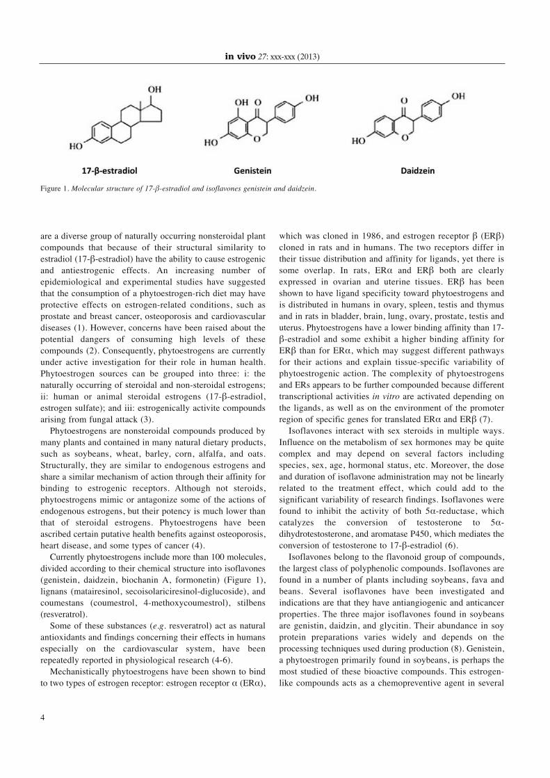
are a diverse group of naturally occurring nonsteroidal plant
compounds that because of their structural similarity to
estradiol (17- -estradiol) have the ability to cause estrogenic
and antiestrogenic effects. An increasing number of
epidemiological and experimental studies have suggested
that the consumption of a phytoestrogen-rich diet may have
protective effects on estrogen-related conditions, such as
prostate and breast cancer, osteoporosis and cardiovascular
diseases (1). However, concerns have been raised about the
potential dangers of consuming high levels of these
compounds (2). Consequently, phytoestrogens are currently
under active investigation for their role in human health.
Phytoestrogen sources can be grouped into three: i: the
naturally occurring of steroidal and non-steroidal estrogens;
ii: human or animal steroidal estrogens (17- -estradiol,
estrogen sulfate); and iii: estrogenically activite compounds
arising from fungal attack (3).
Phytoestrogens are nonsteroidal compounds produced by
many plants and contained in many natural dietary products,
such as soybeans, wheat, barley, corn, alfalfa, and oats.
Structurally, they are similar to endogenous estrogens and
share a similar mechanism of action through their affinity for
binding to estrogenic receptors. Although not steroids,
phytoestrogens mimic or antagonize some of the actions of
endogenous estrogens, but their potency is much lower than
that of steroidal estrogens. Phytoestrogens have been
ascribed certain putative health benefits against osteoporosis,
heart disease, and some types of cancer (4).
Currently phytoestrogens include more than 100 molecules,
divided according to their chemical structure into isoflavones
(genistein, daidzein, biochanin A, formonetin) (Figure 1),
lignans (matairesinol, secoisolariciresinol-diglucoside), and
coumestans (coumestrol, 4-methoxycoumestrol), stilbens
(resveratrol).
Some of these substances (e.g. resveratrol) act as natural
antioxidants and findings concerning their effects in humans
especially on the cardiovascular system, have been
repeatedly reported in physiological research (4-6).
Mechanistically phytoestrogens have been shown to bind
to two types of estrogen receptor: estrogen receptor (ER ),
which was cloned in 1986, and estrogen receptor (ER )
cloned in rats and in humans. The two receptors differ in
their tissue distribution and affinity for ligands, yet there is
some overlap. In rats, ER and ER both are clearly
expressed in ovarian and uterine tissues. ER has been
shown to have ligand specificity toward phytoestrogens and
is distributed in humans in ovary, spleen, testis and thymus
and in rats in bladder, brain, lung, ovary, prostate, testis and
uterus. Phytoestrogens have a lower binding affinity than 17-
-estradiol and some exhibit a higher binding affinity for
ER than for ER , which may suggest different pathways
for their actions and explain tissue-specific variability of
phytoestrogenic action. The complexity of phytoestrogens
and ERs appears to be further compounded because different
transcriptional activities in vitro are activated depending on
the ligands, as well as on the environment of the promoter
region of specific genes for translated ER and ER (7).
Isoflavones interact with sex steroids in multiple ways.
Influence on the metabolism of sex hormones may be quite
complex and may depend on several factors including
species, sex, age, hormonal status, etc. Moreover, the dose
and duration of isoflavone administration may not be linearly
related to the treatment effect, which could add to the
significant variability of research findings. Isoflavones were
found to inhibit the activity of both 5 -reductase, which
catalyzes the conversion of testosterone to 5 -
dihydrotestosterone, and aromatase P450, which mediates the
conversion of testosterone to 17- -estradiol (6).
Isoflavones belong to the flavonoid group of compounds,
the largest class of polyphenolic compounds. Isoflavones are
found in a number of plants including soybeans, fava and
beans. Several isoflavones have been investigated and
indications are that they have antiangiogenic and anticancer
properties. The three major isoflavones found in soybeans
are genistin, daidzin, and glycitin. Their abundance in soy
protein preparations varies widely and depends on the
processing techniques used during production (8). Genistein,
a phytoestrogen primarily found in soybeans, is perhaps the
most studied of these bioactive compounds. This estrogen-
like compounds acts as a chemopreventive agent in several
in vivo 27: xxx-xxx (2013)
4
Figure 1. Molecular structure of 17- -estradiol and isoflavones genistein and daidzein.

types of cancer (9). The affinity of genistein for ER is about
20-30 times higher than that for ER and is comparable to
the affinity of 17- -estradiol (10). The affinity of other
isoflavones is approximately 100-500 times lower than that
of 17- -estradiol. Isoflavones act as agonists of ERs, but
their activity is lower than that of 17- -estradiol. At
sufficiently high levels (over about 100 nmol/l for genistein),
the effects of isoflavones may approach the effect of
endogenous 17- -estradiol at its physiological level.
Genistein has estrogenic properties in receptor-binding
assays, cell culture and uterine weight assays. Genistein
inhibits topisomerase II, platelet-activating factor and
epidermal growth factor-induced expression of c-Fos,
diacylglycerol synthesis and tyrosine kinases. It also inhibits
microsomal lipid peroxidation and angiogenesis. Most of these
mechanistic data were derived from in vitro studies (11).
Not only do phytoestrogens differ in their biological
activity, but they also differ structurally because they come
from diverse chemical classes, which may affect their
influence on tissues and receptors. Due to the diversity of
chemicals that exhibit estrogenic effects, it appears that
estrogenic activity is often emphasized over chemical
structure in defining phytoestrogens (7).
Phytoestrogens and Breast Cancer
The weak estrogenic action of soy isoflavones and other
phytoestrogens suggested the possibility that they could
lessen the deleterious effects of more potent endogenous
estrogens on breast and endometrial cancer. This hypothesis
came from the low incidence of breast and endometrial
cancer in Asian countries where soy products are prevalent in
the diet and from certain animal models of breast and
endometrial cancer showing the benefit of soy isoflavones (8).
In Western countries, breast cancer is the most common
type of cancer affecting women. Historically, the risk of
breast cancer was much higher in American women than in
Asian women prior to the influence of the Western diet on
Asian cultures (7). It is known that some types of tumors,
such as breast, prostate and colon cancer, have a lower
incidence in Asian countries compared to the population of
Western countries (6).
At the beginning of the 1980s, it was suggested that
lignans and isoflavonoids may prevent breast cancer. This
idea led to numerous epidemiological, experimental,
casecontrol, and prospective studies investigating the
hypothesis (12). In epidemiological studies, associations
varied between intake of soy foods and isoflavones and
incidence of breast cancer. Much of the epidemiology of
breast cancer can be explained by reproductive and hormonal
factors; in relation to diet, the only factors definitely related
to breast cancer risk are obesity in postmenopausal women
and alcohol consumption. Several large prospective studies
have investigated whether high intakes of fruit and
vegetables might be associated with a reduced risk of breast
cancer, but overall the results are close to null. It seems
unlikely that high intakes of fruit and vegetables in general
have a significant protective effect, but it is still possible that
specific vegetables rich in isoflavones, especially soya beans,
might have a protective effect by reducing the estrogenic
stimulation of breast cells (13).
Several studies have shown that in breast cancer cells
treated with a low concentration of genistein, the promoter of
the Glutathione S-transferase Pi 1 (GSTP1) gene was
demethylated. Moreover, genistein combined with DNA
methylation inhibitors or other DNA methyltransferases
(DNMTs) can enhance the reactivation of genes silenced by
methylation (14). As a result of research in that genistein
inhibits DNMT1, 3a -3b and inhibits the expression of
Telomerase reverse transcriptase (hTERT). Genistein also
increases acetylation by enhancing histone acetyltransferas
(HAT) activity. Furthermore, investigations have demonstrated
that genistein-mediated hypomethylation and hyperacetylation
reactivate the expression of tumor suppressor genes in prostate
and breast cancer cells. Genistein and other isoflavones have
also been found to regulate miRNA expression in several
cancer cell lines (14, 15).
Animals fed with high doses of soybeans exhibited a
lower incidence of breast and mammary gland cancer (16).
In postmenopausal women, consumption of isoflavones was
found to be associated with reduction of breast cancer
incidence, mammary gland density and proliferative ability
of mammary gland cells (17). These effects have been
associated with the ability of isoflavones to increase the sex
hormone-binding globulin (SHBG) concentration in serum,
thereby reducing the bioavailability of sexual hormones in
hormone-dependent tissues (17). Moreover, in peripheral
tissues, isoflavones inhibit enzymes involved in the processes
of cell proliferation (e.g. tyrosine kinase) and reduce
estradiol availability through an inhibitory effect on
aromatase P450 (6). On the other hand, it has been reported
that high doses of genistein may activate cell proliferation in
estrogen-dependent tumors (18).
However, despite the observation of inhibition of estradiol-
forming enzymes in vitro, adding coumestrol or genistein to
breast cancer cells in culture does not reduce or prevent
estradiol formation (19). The conversion of estrogens to
androgens in breast cells is thought to be important for the
development of breast cancer. Genistein stimulates several
antioxidative enzymes, such as catalase, superoxide
dismutase, glutathione peroxidase and reductase, and it is
also an inducer of tumor cell differentiation. Genistein down-
regulates the epidermal growth factor receptor (EGFR) and
(ERBB2/Neu) receptors in cancer cells, and may also inhibit
tumor-cell invasion by inhibiting (MMP9) (92 kDa type IV
collagenase) and up-regulating tissue inhibitor of
Dagdemir et al: Breast Cancer: Mechanisms Involved in Action of Phytoestrogens and Epigenetic Changes (Review)
5

metalloproteinases (TIMP1), and various trypsin inhibitors.
Daidzein may also enhance immune function (12).
At the present time, there is a growing number of studies
showing that a high soy intake during childhood is associated
with reduced breast cancer risk. But there is no convincing
evidence to suggest that soy or isoflavone consumption in
Western countries during adult life is protective against
breast cancer (3, 8, 12). Soy consumption before puberty
may have the same risk-lowering effects as an early
pregnancy. It is suggested that phytoestrogens promote cell
differentiation in the mammary gland, resulting in
enhancement of mammary gland maturation.
Epigenetic Changes in Breast Cancer
The molecular mechanisms underlying the development and
progression of breast cancer are far from understood. It is
evident that the initiation of breast cancer, as well as its
transition towards distinct breast cancer subtypes, is triggered
by the accumulation of pathologically altered gene functions.
As in other types of cancer, an increasing number of
deregulated genes subsequently affect virtually all important
cellular networks, such as cell cycle control, apoptosis, DNA
repair, detoxification, inflammation, cell adhesion and
migration. According to the somatic mutation theory, cancer
has long been considered as a genetic disorder arising from
the fatal acquisition of multiple mutations in key genes
which coordinate these functional networks. Such mutations
can result either in inactivation of tumor-suppressor genes
[e.g. TP53, (BRCA1)] or activation of protooncogenes [e.g.
(MYC)], both of which contribute to the malignant state of a
transformed cell.
The somatic mutation theory of cancer has recently been
revolutionized for it became evident that epigenetic
misfunction also plays a role in cancer development. The
concept of epigenetics describes mitotically stable states and
changes of gene activity that do not involve alterations of the
primary DNA sequence, thus providing a second layer of
information over that of the pure genomic plan (20, 21).
Epigenetic alterations in transformed cells involve changes
in DNA methylation including global hypomethylation and
locus-specific hypermethylation, altered histone tail
modification patterns and nucleosomal remodeling. DNA
methylation is an enzyme-driven chemical change to the
DNA sequence that most commonly occurs at CpG
dinucleotides in mammals (22). DNA hypomethylation can
be associated with gene reactivation and chromosomal
instabilities and might lead to the up-regulation or
overexpression of proto-oncogenes, increased recombination
and mutation rates, skewed or loss of X-chromosomal
inactivation and loss of imprinting (23). DNA
hypermethylation is frequently associated with gene
repression and genomic instability and can lead to the
suppression of tumor-suppressor genes and compaction of
chromatin (20).
Associated with DNA methylation are post-translational
histone tail modifications, another epigenetic mechanism that
can modulate chromatin structure to regulate gene expression
(24). In addition, it has been shown that some regulators that
control nucleosomal remodeling are also involved in the
regulation of DNA methylation and histone modification (25,
26). An understanding of all these epigenetic changes and
their contributions to breast tumorigenesis is very important
for further progress in the field of diagnosis, prognosis and
therapy of breast cancer.
DNA Methylation in Breast Cancer
DNA methylation patterns highlight significant differences
in breast cancer between tumor tissues and corresponding
normal tissues (27). A common paradox observed in
carcinomas is that despite regional hypermethylation of
tumor-suppressor genes, the global 5-methylcytosine content
is drastically-reduced in the bulk of the tumor genome (26).
Less frequent than regional DNA hypermethylation, regional
DNA hypomethylation can also occur in cancer, resulting in
the activation of potential oncogenes (27, 28).
DNA hypermethylation. DNA hypermethylation indicates
which genes are turned off in breast tumors and a unique
pattern is observed in breast tumors. Interestingly, 40% loss
of methylated cytosine is observed in breast tumors. Important
genes in familial breast cancer are also epigenetically silenced.
In sporadic tumors, BRCA1 expression has been shown to be
suppressed by a combination of gene deletion and epigenetic
silencing via DNA hypermethylation (29).
Ectopic cytosine hypermethylation is generally associated
with transcriptional repression and ultimately tumor formation.
Collaboration between genetic and epigenetic breast cancer
causes has been directly demonstrated by example in which
one tumor-suppressor allele is inactivated by mutation and the
other allele is transcriptionally silenced because of
hypermethylation (30). In addition hypermethylation of tumor-
suppressor genes may be an early event in cancer development
(31, 32), suggesting that epigenetic and mutational cancer
causes may collaborate from an early time point in disease
progression (32). The list of tumor-suppressor genes found
transcriptionally inactivated by hypermethylation in cancer is
long and steadily growing (33), and includes genes that are
part of every cancer-related pathway, including important
genes such as (CDKN2A), (pRB), (APC), (PTEN), (BRCA1),
(VHL) and (CDH1). By extension, epigenetic silencing may
underlie genetic cancer causes. Epigenetic induction of a
classical mutator phenotype via transcriptional inactivation of
the DNA mismatch repair gene (MLH1) has been proposed to
account for microsatellite instability in colorectal cancer and
in vivo 27: xxx-xxx (2013)
6

silencing of the DNA repair gene coding for O-6-
methylguanine-DNMT has been associated with specific
mutations in (K-RAS) and p53 (34, 35).
DNA Hypomethylation. Among solid tumor types, global
DNA hypomethylation is most evident in breast cancer with
up to 50% of cases showing reduced 5-methylcytosine
content when compared with normal tissue counterparts (36).
Hypomethylation in breast cancer mainly affects iterative
DNA sequences and pericentromeric satellite DNA, which
are normally heavily methylated in non-malignant cells (35).
Although a relatively rare event, DNA hypomethylation
can also affect individual genes. In breast cancer, this is the
case for the melanoma-associated cancer/testis antigens
MAGE. The MAGE gene family encodes for (HLA)-
restricted tumor-associated rejection antigens recognized by
cytotoxic T-lymphocytes. Some of these target antigens may
be potentially useful for cancer-specific immunotherapy. The
expression of MAGE genes has been reported not only in
melanoma but also in various other malignant tumors, such
as hepatocellular carcinoma and germ cell tumors. Genes for
MAGE antigens are methylated and silenced in adult tissues,
but hypomethylated and expressed in several tumors and
breast cancer cells (37). Other hypomethylated genes in
breast tumors include the gene encoding the plasminogen
activator uPA (PLAU), the breast cancer-specific protein
1/synuclein- gene (SNCG), and more recently reported, the
multidrug-resistance 1 gene (MDR1) (38, 39).
Global DNA hypomethylation is a hallmark in human
cancer, but its functional consequences are unclear (39). The
mechanism of global hypomethylation is a long-standing
question in cancer epigenetics (40).
Histone Modifications in Breast Cancer
A number of studies have investigated the use of histone
modifications as biomarkers in tumors. High relative levels
of global histone acetylation and methylation were associated
with a favorable prognosis, and were detected almost
exclusively in luminal-like breast tumors (93%). Clustering
analysis identified three groups of histone status patterns
which correlate with clinical outcome (41).
Histone acetylation. Histon acetylation is a dynamic process
directed by HATs and histone deacetylases (HDACs).
Normally, transcription factors recruit coactivators with HAT
activity to regulatory DNA sites, whereas transcriptional
repressors recruit co-repressors with HDAC activity.
Many HATs have also been shown to be involved in breast
cancer. Among of them, p300/CREB-binding protein (CBP)
and nuclear receptor coactivator family (NCOAs) are the
most important and well-characterized HAT proteins
associated with breast cancer (42).
p300 and its close homolog CBP are often referred to as a
single entity. A role for p300 in tumor suppression has been
proposed by the fact that disturbance of p300 function by
viral oncoproteins is essential for the transformation of
rodent primary cells and, consistent with this hypothesis,
mutations of p300 have been identified in certain types of
human cancer, including breast carcinoma (43). Both the
localization of p300 and the recruitment to aggresomes differ
between breast cancer and normal mammary glands. The
expression level of p300 in breast cancer epithelia is higher
than that in normal mammary gland (44).
Cytoplasmic localization of p300 was also observed in
tumor epithelia whereas nuclear localization was found in
normal mammary glands in both animal models and in non-
malignant adjacent areas of human breast cancer specimens.
Proteasomal inhibition induced p300 redistribution to
aggresomes in tumor but not in normal mammary gland-
derived cells (45). The regulation of gene expression by nuclear
receptors (NRs) controls the phenotypic properties and diverse
biologies of target cells. In breast cancer cells, ER is a master
regulator of transcriptional stimulation and repression (46).
Upon 17- -estradiol treatment, gene transcription is
widely impacted, creating highly complex regulatory
networks whose ultimate goal is the stimulation or
suppression of specific biological processes. p300/CBP can
function as a transcriptional cofactor of ERs and other
nuclear hormone receptors (47).
The NCOA family, also named as p160 or steroid receptor
coactivator, contains three homologous members: NCOA1
(SRC-1), NCOA2 (SRC-2, GRIP1 or TIF2) and NOCA3
(SRC-3, p/CIP, RAC3, ACTR, AIB1 or TRAM-1). These
three members have an overall sequence similarity of 50-
55% and sequence identity of 43-48%. As well as being
NRs, NCOAs also serve as coactivators for many other
transcription factors associated with breast cancer, such as
(HIF1), (NF- B), (E2F1), p53, RB and (MRTFs) (48, 49).
By regulating a broad range of gene expression controlled by
NRs and non-NR transcription factors, NCOAs regulate
diverse events in the development of breast cancer. NCOA1
is overexpressed in 19% to 29% of breast cancer cases and
plays important roles in cell proliferation, lymph node
metastasis, disease recurrence and poor disease-free survival
(DFS) (50). Therefore, elevated expression of NCOA1 has
been regarded as an independent predictor of breast cancer
recurrence following therapy (51). Although the evidence is
not conclusive, NCOA2 overexpression might also promote
proliferation and invasion of breast cancer cells. In addition,
NCOAs play important roles in the chemotherapy resistance
of breast cancer. Increased expression levels of the ER-
NCOA3 complex were found in tamoxifen-resistant cells,
and such overexpression of NCOA3 could enhance the
agonist activity of tamoxifen and, therefore, reduce its
antitumor activity in patients with breast cancer (52).
Dagdemir et al: Breast Cancer: Mechanisms Involved in Action of Phytoestrogens and Epigenetic Changes (Review)
7

The 18 HDACs identified so far can be categorized into
four classes: class I (HDAC 1-3, HDAC 8), class II (HDAC
4-7, 9-10), class III (Sirtuin 1-7) and class IV (HDAC 11).
Class I, II and IV HDACs share homology in both sequence
and structure and all require a zinc ion for catalytic activity
(53). HDACs remove the acetyl groups from histone lysine
tails and are thought to facilitate transcriptional repression
by reducing the level of histone acetylation. Like HATs,
HDACs also have non-histone targets. Several HDACs have
been found to be involved in breast cancer. In ER-positive
MCF-7 breast cancer cells, expression of HDAC6 was
increased after treatment by estradiol, and elevated
expression of HDAC6 increased deacetylation of alpha-
tubulin and increased cell motility (54).
In vivo assays showed that patients with high levels of
HDAC6 mRNA tended to be more responsive to endocrine
treatment than those with low levels, indicating that the level
of HDAC6 expression might be used as both as a marker of
endocrine responsiveness and also as a prognostic indicator
in breast cancer (48, 55). However, HDAC1, Sirtuin3
(SIRT3) and SIRT7 are all overexpressed in breast cancer
(56, 57). HDAC4 overexpression and mutations have also
been found in breast cancer samples (57).
Histone methylation. Histones can be mono-, di-, or tri-
methylated at lysine or arginine residues by histone
methyltransferases (HMTs). Many HMTs, including both
lysine-specific HMTs (e.g. SMYD3) and arginine-specific
HMTs (e.g. PRMT1 and CARM1), have been shown to act
as ER coactivators and be involved in breast cancer.
Many histone lysine metyltransferase (HKMTs) have been
isolated and characterized. Except for (Dot1), all HKMTs
contains a conserved Su(var), enhancer of zeste, trithorax
(SET) domain that is responsible for catalysis and binding
of cofactor S-adenosyl methionine, and many have been
shown to play roles in breast cancer (58).
Other methyltransferase mechanism attack of the breast
cancer is arginine methyltransferase (HRMT). The protein
arginine methyltransferase (PRMT) family is the major
family of HRMTs to date. The PRMTs are classified into
four groups depending on the type of methylarginine they
generate: Type I (PRMT1, PRMT2, PRMT3, PRMT4,
PRMT6 and PRMT8), type II (PRMT5, PRMT7 and
PRMT9), type III (remained unclear) and type IV (only
found in Saccharomyces cerevisiae as yet) (59).
Compared to HKMTs, the evidence for the involvement of
HRMTs in human cancer is not forthcoming. However,
underexpression of PRMT1 has been observed in breast
cancer (60). PRMT4, also known as coactivator-associated
arginine methyltransferase-1 (CARM1), is a coactivator for
nuclear receptors and is overexpressed in prostate and breast
cancer (61). PRMT4 plays an important role in estrogen-
induced cell cycle progression in the MCF-7 breast cancer
cell line. Upon estrogen stimulation, the E2F1 promoter is
subject to PRMT4-dependent dimethylation on H3R17, and
this recruitment of PRMT4 by ER is dependent on the
presence of NCOA3 (62).
Histone phosphorylation. Phosphorylation of H3 on S10 and
S28 is important not only during mitotic chromosome
condensation but also in transcriptional activation of
immediate early genes and growth factors stimulating the
(RAS)/(MAPK) and increasing H3 pS10 at transcriptionally
active loci may contribute to aberrant gene expression and
breast cancer progression (63).
Other histone modifications in breast cancer. Besides
acetylation, methylation and phosphorylation, there are some
other modifications which occur of histone. These epigenetic
changes include ubiquitination/sumoylation, ADP-ribosylation,
deamination, and proline isomerization. Although the
functions and mechanisms have not been demonstrated, some
studies have shown that these modifications are also associated
with breast cancer and other types of human cancer.
Recent studies revealed that E3 ubiquitin ligases play
important roles in breast carcinogenesis. Ubiquitin-mediated
protein degradation plays an important role in many cancer-
related cellular processes. E3 ubiquitin ligases play critical
roles because they control substrate specificity of histone.
Accumulating evidence suggests that genetic and expression
alteration of E3 ubiquitin ligases contributes to breast
carcinogenesis (64).
(miRNAs) in Breast Cancer
miRNAs have been shown to play a critical role in the
regulation of a wide range of biological and pathological
processes. Recent large-scale profiling approaches have
revealed that miRNAs are globally down-regulated in several
cancer types, including breast cancer. Moreover, panels of
miRNAs in breast carcinomas characteristic for the HER2/neu
or ER status of the analyzed tumor have been detected (65).
There is now increasing evidence that signatures of miRNA
expression may not only be used in the future as tumor
biomarkers for diagnosis and patient risk stratification, but
since hypermethylation was identified as an important
mechanism of miRNA silencing, deregulated miRNAs may
also represent novel targets for an anticancer therapy. One
study describing differential expression patterns of miRNAs in
breast cancer also investigated their expression changes in
relation to chromosomal localization. Interestingly, the authors
found several miRNA candidates that reside in chromosomal
regions which are either frequently deleted or amplified in
breast cancer, e.g. down-regulation of miR-125b in the
frequently deleted region 11q-23-24, or overexpression of miR-
21 in 17q23, which is commonly amplified in breast cancer
in vivo 27: xxx-xxx (2013)
8

(66). Another mechanism by which miRNA profiles may be
altered in tumors lies within abnormalities in the miRNA-
processing machinery. It was the same study that observed
significant changes in expression of Dicer and (AGO1), both
being involved in miRNA maturation processes (67).
Decreased Dicer expression was recently observed in breast
cancer, where loss of expression represented an independent
prognostic factor in metastatic disease, and reduced expression
of Dicer was associated with the highly aggressive
mesenchymal phenotype (68). Whether genetic lesions such as
(TARBP2) mutations in colorectal cancer also account for
impaired miRNA processing in breast cancer remains to be
determined in future studies. It is, however, conclusive that
besides DNA hypermethylation of miRNA genes, structural
genetic alterations also contribute to the observed dramatic
changes of miRNA expression profiles in human cancer.
Conclusion
Phytoestrogens seem to protect against breast cancer if
consumed throughout life, particularly before and during
adolescence, and a low plasma enterolactone concentration
is known to increase risk of breast cancer. Whether
phytoestrogens are actually responsible for the protection is
not known, it is more likely that the soybean products or
grain-fiber complexes are protective in their entirety.
Research in phytoestrogens has increased dramatically in
recent years as seen by the numerous publications. However,
many questions remain. Research is still needed to evaluate
the safety of phytoestrogens on human systems, beneficial
and harmful doses, gender differences in response to
phytoestrogens, differences in the chemical classes of
phytoestrogens and the effects phytoestrogens may have with
other drugs or dietary products. Due to the functional and
structural differences of phytoestrogens, their biological
activities are also highly variable and there may be other
effects that have not yet been studied.
In summary, epigenetic modifications provide crucial
regulatory functions in the process of gene transcription, and
they play very important roles in the proliferation,
metastasis, chemotherapy and other aspects of breast cancer,
as well as in many other types of human cancer. An
understanding of all these epigenetic changes and their
contribution to breast cancer might allow great progress in
the field of diagnosis, prognosis and therapy of breast cancer.
We may thus hope that many of the open questions about the
impact of phytoestrogen and epigenetics will be answered in
the near future.
Acknowledgements
We thank The Ligue Against Cancer and committee of the region
Auvergne.
References
1 Cos P, De Bruyne T, Apers S, Vanden Berghe D, Pieters L and
Vlietinck AJ: Phytoestrogens: recent developments. Planta Med
69(7): 589-599, 2003.
2 Bakhit RM, Klein BP, Essex-Sorlie D, Ham JO, Erdman JW Jr.
and Potter SM: Intake of 25 g of soybean protein with or without
soybean fiber alters plasma lipids in men with elevated
cholesterol concentrations. J Nutr 124(2): 213-222, 1994.
3 Rice S and Whitehead SA: Phytoestrogens and breast cancer –
promoters or protectors? Endocr Relat Cancer 13(4): 995-1015,
2006.
4 Sacks FM, Lichtenstein A, Van Horn L, Harris W, Kris-Etherton
P and Winston M: Soy protein, isoflavones, and cardiovascular
health: a summary of a statement for professionals from the
american heart association nutrition committee. Arterioscler
Thromb Vasc Biol 26(8): 1689-1692, 2006.
5 Le Corre L, Chalabi N, Delort L, Bignon YJ and Bernard-Gallon
DJ: Resveratrol and breast cancer chemoprevention: molecular
mechanisms. Mol Nutr Food Res 49(5): 462-471, 2005.
6 Pilsakova L, Riecansky I and Jagla F: The physiological
actions of isoflavone phytoestrogens. Physiol Res 59(5): 651-
664, 2010.
7 Ososki AL and Kennelly EJ: Phytoestrogens: a review of the
present state of research. Phytother Res 17(8): 845-869, 2003.
8 Sacks FM, Lichtenstein A, Van Horn L, Harris W, Kris-Etherton
P and Winston M: Soy protein, isoflavones, and cardiovascular
health: an American Heart Association Science Advisory for
professionals from the Nutrition Committee. Circulation 113(7):
1034-1044, 2006.
9 Hardy TM and Tollefsbol TO: Epigenetic diet: impact on the
epigenome and cancer. Epigenomics 3(4): 503-518, 2011.
10 Morito K, Hirose T, Kinjo J, Hirakawa T, Okawa M, Nohara T,
Ogawa S, Inoue S, Muramatsu M and Masamune Y: Interaction
of phytoestrogens with estrogen receptors alpha and beta. Biol
Pharm Bull 24(4): 351-356, 2001.
11 Lamartiniere CA, Cotroneo MS, Fritz WA, Wang J, Mentor-
Marcel R and Elgavish A: Genistein chemoprevention: timing
and mechanisms of action in murine mammary and prostate. J
Nutr 132(3): 552S-558S, 2002.
12 Adlercreutz H: Phyto-oestrogens and cancer. Lancet Oncol 3 (6):
364-373, 2002.
13 Key TJ: Fruit and vegetables and cancer risk. Br J Cancer
104(1): 6-11, 2011.
14 Li Y, Liu L, Andrews LG and Tollefsbol TO: Genistein depletes
telomerase activity through cross-talk between genetic and
epigenetic mechanisms. Int J Cancer 125(2): 286-296, 2009.
15 Majid S, Dar AA, Ahmad AE, Hirata H, Kawakami K,
Shahryari V, Saini S, Tanaka Y, Dahiya A V, Khatri G and
Dahiya R: BTG3 tumor suppressor gene promoter
demethylation, histone modification and cell cycle arrest by
genistein in renal cancer. Carcinogenesis 30(4): 662-670, 2009.
16 Atkinson C, Warren RM, Sala E, Dowsett M, Dunning AM,
Healey CS, Runswick S, Day NE and Bingham SA: Red-clover-
derived isoflavones and mammographic breast density: a double-
blind, randomized, placebo-controlled trial [ISRCTN42940165].
Breast Cancer Res 6(3): R170-179, 2004.
17 Ingram D, Sanders K, Kolybaba M and Lopez D: Case-control
study of phyto-oestrogens and breast cancer. Lancet 350(9083):
990-994, 1997.
Dagdemir et al: Breast Cancer: Mechanisms Involved in Action of Phytoestrogens and Epigenetic Changes (Review)
9

18 Kao YC, Zhou C, Sherman M, Laughton CA and Chen S:
Molecular basis of the inhibition of human aromatase (estrogen
synthetase) by flavone and isoflavone phytoestrogens: A site-
directed mutagenesis study. Environ Health Perspect 106(2): 85-
92, 1998.
19 Makela S, Poutanen M, Lehtimaki J, Kostian ML, Santti R and
Vihko R: Estrogen-specific 17 beta-hydroxysteroid oxidoreductase
type 1 (E.C. 1.1.1.62) as a possible target for the action of
phytoestrogens. Proc Soc Exp Biol Med 208(1): 51-59, 1995.
20 Nicoloso MS, Spizzo R, Shimizu M, Rossi S and Calin GA:
MicroRNAs – the micro steering wheel of tumour metastases.
Nat Rev Cancer 9(4): 293-302, 2009.
21 Holliday R: Epigenetics: a historical overview. Epigenetics 1(2):
76-80, 2006.
22 Hinshelwood RA and Clark SJ: Breast cancer epigenetics:
normal human mammary epithelial cells as a model system. J
Mol Med (Berl) 86(12): 1315-1328, 2008.
23 De Smet C, Loriot A and Boon T: Promoter-dependent
mechanism leading to selective hypomethylation within the 5’
region of gene MAGE-A1 in tumor cells. Mol Cell Biol 24(11):
4781-4790, 2004.
24 Baylin SB and Ohm JE: Epigenetic gene silencing in cancer – a
mechanism for early oncogenic pathway addiction? Nat Rev
Cancer 6(2): 107-116, 2006.
25 Jovanovic J, Ronneberg J A, Tost J and Kristensen V: The
epigenetics of breast cancer. Mol Oncol 4 (3): 242-254, 2010.
26 Jones PA and Baylin SB: The epigenomics of cancer. Cell
128(4): 683-692, 2007.
27 Klein CB and Costa M: DNA methylation, heterochromatin and
epigenetic carcinogens. Mutat Res 386(2): 163-180, 1997.
28 Wilson AS, Power BE and Molloy PL: DNA hypomethylation
and human diseases. Biochim Biophys Acta 1775(1): 138-162,
2007.
29 Nowsheen S, Aziz K, Tran PT, Gorgoulis VG, Yang ES and
Georgakilas AG: Epigenetic inactivation of DNA repair in breast
cancer. Cancer Lett 2012 (in press).
30 Myohanen S K, Baylin S B and Herman J G: Hypermethylation
can selectively silence individual p16ink4A alleles in neoplasia.
Cancer Res 58(4): 591-593, 1998.
31 Waki T, Tamura G, Sato M, Terashima M, Nishizuka S and
Motoyama T: Promoter methylation status of DAP-kinase and
RUNX3 genes in neoplastic and non-neoplastic gastric epithelia.
Cancer Sci 94(4): 360-364, 2003.
32 Suter CM, Martin DI and Ward RL: Germline epimutation of
MLH1 in individuals with multiple cancers. Nat Genet 36(5):
497-501, 2004.
33 Jones PA and Baylin SB: The fundamental role of epigenetic
events in cancer. Nat Rev Genet 3(6): 415-428, 2002.
34 Esteller M: Epigenetic gene silencing in cancer: the DNA
hypermethylome. Hum Mol Genet 16 Spec No 1 R50-59, 2007.
35 Hedenfalk I, Duggan D, Chen Y, Radmacher M, Bittner M,
Simon R, Meltzer P, Gusterson B, Esteller M, Kallioniemi O P,
Wilfond B, Borg A, Trent J, Raffeld M, Yakhini Z, Ben-Dor A,
Dougherty E, Kononen J, Bubendorf L, Fehrle W, Pittaluga S,
Gruvberger S, Loman N, Johannsson O, Olsson H and Sauter G:
Gene-expression profiles in hereditary breast cancer. N Engl J
Med 344(8): 539-548, 2001.
36 Bernardino J, Roux C, Almeida A, Vogt N, Gibaud A, Gerbault-
Seureau M, Magdelenat H, Bourgeois CA, Malfoy B and
Dutrillaux B: DNA hypomethylation in breast cancer: an
independent parameter of tumor progression? Cancer Genet
Cytogenet 97(2): 83-89, 1997.
37 Weber J, Salgaller M, Samid D, Johnson B, Herlyn M, Lassam
N, Treisman J and Rosenberg SA: Expression of the MAGE-1
tumor antigen is up-regulated by the demethylating agent 5-aza-
2’-deoxycytidine. Cancer Res 54(7): 1766-1771, 1994.
38 Guo Y, Pakneshan P, Gladu J, Slack A, Szyf M and Rabbani SA:
Regulation of DNA methylation in human breast cancer. Effect
on the urokinase-type plasminogen activator gene production and
tumor invasion. J Biol Chem 277(44): 41571-41579, 2002.
39 Sharma G, Mirza S, Parshad R, Srivastava A, Datta Gupta S,
Pandya P and Ralhan R: CpG hypomethylation of MDR1 gene
in tumor and serum of invasive ductal breast carcinoma patients.
Clin Biochem 43(4-5): 373-379, 2010.
40 Hon GC, Hawkins RD, Caballero OL, Lo C, Lister R, Pelizzola
M, Valsesia A, Ye Z, Kuan S, Edsall LE, Camargo AA,
Stevenson BJ, Ecker JR, Bafna V, Strausberg RL, Simpson AJ
and Ren B: Global DNA hypomethylation coupled to repressive
chromatin domain formation and gene silencing in breast cancer.
Genome Res 22(2): 246-258, 2012.
41 Elsheikh SE, Green AR, Rakha EA, Powe DG, Ahmed RA,
Collins HM, Soria D, Garibaldi JM, Paish CE, Ammar AA,
Grainge MJ, Ball GR, Abdelghany MK, Martinez-Pomares L,
Heery DM and Ellis IO: Global histone modifications in breast
cancer correlate with tumor phenotypes, prognostic factors, and
patient outcome. Cancer Res 69(9): 3802-3809, 2009.
42 Parrella P: Epigenetic Signatures in Breast Cancer: Clinical
Perspective. Breast Care (Basel) 5(2): 66-73, 2010.
43 Gayther SA, Batley SJ, Linger L, Bannister A, Thorpe K, Chin
SF, Daigo Y, Russell P, Wilson A, Sowter HM, Delhanty JD,
Ponder BA, Kouzarides T and Caldas C: Mutations truncating
the EP300 acetylase in human cancers. Nat Genet 24(3): 300-
303, 2000.
44 Kim JH and Workman JL: Histone acetylation in
heterochromatin assembly. Genes Dev 24(8): 738-740, 2010.
45 Fermento ME, Gandini NA, Lang CA, Perez JE, Maturi HV,
Curino AC and Facchinetti MM: Intracellular distribution of
p300 and its differential recruitment to aggresomes in breast
cancer. Exp Mol Pathol 88(2): 256-264, 2010.
46 Frasor J, Danes JM, Komm B, Chang KC, Lyttle CR and
Katzenellenbogen BS: Profiling of estrogen up- and down-
regulated gene expression in human breast cancer cells: insights
into gene networks and pathways underlying estrogenic control
of proliferation and cell phenotype. Endocrinology 144(10):
4562-4574, 2003.
47 Hanstein B, Eckner R, DiRenzo J, Halachmi S, Liu H, Searcy
B, Kurokawa R and Brown M: p300 is a component of an
estrogen receptor coactivator complex. Proc Natl Acad Sci USA
93(21): 11540-11545, 1996.
48 Zhang H, Yi X, Sun X, Yin N, Shi B, Wu H, Wang D, Wu G and
Shang Y: Differential gene regulation by the SRC family of
coactivators. Genes Dev 18(14): 1753-1765, 2004.
49 Xu J, Wu RC and O’Malley BW: Normal and cancer-related
functions of the p160 steroid receptor co-activator (SRC) family.
Nat Rev Cancer 9(9): 615-630, 2009.
50 Fleming FJ, Myers E, Kelly G, Crotty TB, McDermott EW,
O’Higgins NJ, Hill AD and Young LS: Expression of SRC-1,
AIB1, and PEA3 in HER2 mediated endocrine resistant breast
cancer; a predictive role for SRC-1. J Clin Pathol 57(10): 1069-
1074, 2004.
in vivo 27: xxx-xxx (2013)
10

51 Redmond AM, Bane FT, Stafford AT, McIlroy M, Dillon MF,
Crotty TB, Hill AD and Young LS: Coassociation of estrogen
receptor and p160 proteins predicts resistance to endocrine
treatment; SRC-1 is an independent predictor of breast cancer
recurrence. Clin Cancer Res 15(6): 2098-2106, 2009.
52 Zhao W, Zhang Q, Kang X, Jin S and Lou C: AIB1 is required
for the acquisition of epithelial growth factor receptor-mediated
tamoxifen resistance in breast cancer cells. Biochem Biophys
Res Commun 380(3): 699-704, 2009.
53 Bolden JE, Peart MJ and Johnstone RW: Anticancer activities of
histone deacetylase inhibitors. Nat Rev Drug Discov 5(9): 769-
784, 2006.
54 Wang GG, Allis CD and Chi P: Chromatin remodeling and
cancer, Part II: ATP-dependent chromatin remodeling. Trends
Mol Med 13(9): 373-380, 2007.
55 Saji S, Kawakami M, Hayashi S, Yoshida N, Hirose M,
Horiguchi S, Itoh A, Funata N, Schreiber S L, Yoshida M and
Toi M: Significance of HDAC6 regulation via estrogen signaling
for cell motility and prognosis in estrogen receptor-positive
breast cancer. Oncogene 24(28): 4531-4539, 2005.
56 Zhang Z, Yamashita H, Toyama T, Sugiura H, Ando Y, Mita K,
Hamaguchi M, Hara Y, Kobayashi S and Iwase H: Quantitation
of HDAC1 mRNA expression in invasive carcinoma of the
breast*. Breast Cancer Res Treat 94(1): 11-16, 2005.
57 Saunders LR and Verdin E: Sirtuins: critical regulators at the
crossroads between cancer and aging. Oncogene 26(37): 5489-
5504, 2007.
58 Lee DY, Teyssier C, Strahl BD and Stallcup MR: Role of protein
methylation in regulation of transcription. Endocr Rev 26(2):
147-170, 2005.
59 Niewmierzycka A and Clarke S: S-Adenosylmethionine-
dependent methylation in Saccharomyces cerevisiae.
Identification of a novel protein arginine methyltransferase. J
Biol Chem 274(2): 814-824, 1999,
60 Scorilas A, Black MH, Talieri M and Diamandis E P: Genomic
organization, physical mapping, and expression analysis of the
human protein arginine methyltransferase 1 gene. Biochem
Biophys Res Commun 278(2): 349-359, 2000.
61 El Messaoudi S, Fabbrizio E, Rodriguez C, Chuchana P,
Fauquier L, Cheng D, Theillet C, Vandel L, Bedford MT and
Sardet C: Coactivator-associated arginine methyltransferase 1
(CARM1) is a positive regulator of the Cyclin E1 gene. Proc
Natl Acad Sci USA 103(36): 13351-13356, 2006.
62 Frietze S, Lupien M, Silver PA and Brown M: CARM1 regulates
estrogen-stimulated breast cancer growth through up-regulation
of E2F1. Cancer Res 68(1): 301-306, 2008.
63 Espino PS, Li L, He S, Yu J and Davie JR: Chromatin
modification of the trefoil factor 1 gene in human breast cancer
cells by the Ras/mitogen-activated protein kinase pathway.
Cancer Res 66(9): 4610-4616, 2006.
64 Chen C, Seth AK and Aplin AE: Genetic and expression
aberrations of E3 ubiquitin ligases in human breast cancer. Mol
Cancer Res 4(10): 695-707, 2006.
65 Mattie MD, Benz CC, Bowers J, Sensinger K, Wong L, Scott G
K, Fedele V, Ginzinger D, Getts R and Haqq C: Optimized high-
throughput microRNA expression profiling provides novel
biomarker assessment of clinical prostate and breast cancer
biopsies. Mol Cancer 5: 24, 2006.
66 Iorio MV, Ferracin M, Liu C G, Veronese A, Spizzo R, Sabbioni
S, Magri E, Pedriali M, Fabbri M, Campiglio M, Menard S,
Palazzo JP, Rosenberg A, Musiani P, Volinia S, Nenci I, Calin
GA, Querzoli P, Negrini M and Croce CM: MicroRNA gene
expression deregulation in human breast cancer. Cancer Res
65(16): 7065-7070, 2005.
67 Zhang L, Huang J, Yang N, Greshock J, Megraw MS,
Giannakakis A, Liang S, Naylor T L, Barchetti A, Ward MR, Yao
G, Medina A, O’Brien-Jenkins A, Katsaros D, Hatzigeorgiou A,
Gimotty PA, Weber BL and Coukos G: microRNAs exhibit high
frequency genomic alterations in human cancer. Proc Natl Acad
Sci USA 103(24): 9136-9141, 2006.
68 Grelier G, Voirin N, Ay AS, Cox DG, Chabaud S, Treilleux I,
Leon-Goddard S, Rimokh R, Mikaelian I, Venoux C, Puisieux
A, Lasset C and Moyret-Lalle C: Prognostic value of Dicer
expression in human breast cancers and association with the
mesenchymal phenotype. Br J Cancer 101(4): 673-683, 2009.
Received August 9, 2012
Revised October 19, 2012
Accepted October 23, 2012
Dagdemir et al: Breast Cancer: Mechanisms Involved in Action of Phytoestrogens and Epigenetic Changes (Review)
11

36
Conclusion of Publication1:
In this review we reported which phytoestrogens were more important for breast
cancer and what are the possible mechanisms of phytoestrogen on breast cancer. We reported
that phytoestrogens soy, genistein and daidzein exert protective effects vis-a-vis breast cancer.
However, some studies focus on the ambivalent role of phytoestrogens. It has indeed been
shown that phytoestrogens can induce both proliferation and inhibition of the growth of breast
tumor cells. Research is still needed to evaluate the safety of phytoestrogens on human
systems, beneficial and harmful doses, gender differences in response to phytoestrogens,
differences in the chemical classes of phytoestrogens and the effects phytoestrogens may have
with other drugs or dietary products. Due to the functional and structural differences of
phytoestrogens, their biological activities are also highly variable and there may be other
effects that have not yet been studied.
After understanding the effects of phytoestrogens on breast cancer, we started to
study epigenetics mechanisms and we also investigated the interaction between epigenetics
and breast cancer. Different mechanisms are involved in the maintenance of epigenetic states.
Studies discussed herein have shown that dietary factors are likely to contribute to epigenetic
alterations and in some cases may be able to reverse abnormal epigenetic states. We saw that
there are all three mechanisms are very important for breast cancers which are DNA
methylation, histone modification and miRNAs. Nutriepigenomic studies focusing on
personal responses to bioactive elements and personalized epigenetic diets consisting of
bioactive dietary factors mentioned herein will be of particular interest in the future.

37
C. Epigenetic Changes
i. General description
The term epigenetics was first used by Conrad Hal Waddington as "the causal
interactions between genes and their products, which bring the phenotype into being". This
definition initially referred to understanding chromatin structure and its impact on gene
function. The current definition of epigenetics is "the study of heritable changes in gene
expression that occur independent of changes in the primary DNA sequence". Epigenetic
mechanisms that occur these expression patterns have been divided into three main
categories: DNA methylation, covalent histone modifications and non-coding RNAs
including microRNAs (miRNAs). Aberrant epigenetic alterations in the genome such as DNA
methylation and chromatin remodeling play a significant role in breast cancer development
[68].
DNA methylation is the most-studied epigenetic factor referring to the modification
of cytosine residues by methylation at their carbon-5 position. In mammalian genomes, this
modification primarily occurs on cytosine residues that are located 5' to guanine i.e. CpG
dinucleotides. DNA methylation is catalyzed by DNMTs. In humans, at least, three DNMTs
are involved in establishment and maintenance of DNA methylation patterns; de novo DNA
methyltransferases DNMT3A and DNMT3B and maintenance DNA methyltransferase
DNMT1 . A fourth enzyme previously know as DNMT 2 is not a DNA methyltransferase.
CpG dinucleotides are not equally distributed across the human genome but are concentrated
in distinct CpG-rich regions called “CpG islands” and in regions of large repetitive sequences
[69].DNA methylation also provide to other X-chromosome inactivation and genome stability
[70]. DNA methylation patterns are reliably hereditary during both mitosis and meiosis. Also
some studies have shown that while some CpG regions are stably methylated, a small number
of dynamic methylated regions could play a major role in controlling the transcription
network of cells [71]. Failure to maintain correct methylation patterns leads to aberrant DNA
methylation, often observed in human diseases including neurodevelopmental defects,
neurodegenerative, neurological and autoimmune diseases, and cancers.
38
Chromatin is a highly dynamic structure and must be kept in balance by being folded
as much as needed and being accessible whenever necessary to cope with genome templated
processes such as replication, transcription and DNA repair. The functional state of chromatin
is partially regulated through posttranslational modifications of histones [72]. The amino-tails
of histones in the nucleosomes can be modified in a number of modifications, for example;
methylation, acetylation, phosphorylation, ubiquitination and sumoylation. Thereby these
modifications are involved in regulating the gene expression.
The interactions between DNA methylation and histone modifications mediate gene
silencing through recruitment of repressive complexes and methylated DNA binding proteins.
miRNAs are small non-coding RNAs of 20–22 nucleotides that inhibit gene
expression at the posttranscriptional level. miRNAs are involved in the regulation of key
biological processes, including development, differentiation, apoptosis and proliferation and
play a critical role in the regulation of a wide range of biological and pathological processes.
Each miRNA is expected to control several hundred genes. They have been implicated in
cancer initiation and progression, and their expression is often down-regulated during
carcinogenesis. Major mechanisms of miRNA deregulation include genetic and epigenetic
alterations as well as defects in the miRNA processing machinery

39
Presentation of Publication 2:
Dagdemir A, Durif J, Ngollo M, Bignon Y-J, Bernard-Gallon D. Epigenetic
Mechanisms that Operate in Mammals. Applied Cell Biology. 2013; 1(1):1-9.
Understanding the process of epigenetic reprogramming in development is important
for studies of breast cancer. This landscape will summarize current knowledge on interactions
between cancer cell metabolism and epigenetic modulation of gene regulation, and how both
processes can be affected by dietary components. A greater understanding of these processes
is also of clinical importance.
In this review, we will discuss the different layers of epigenetic regulation, including
writer enzymes for DNA methylation, histone modifications, non-coding RNA, and chromatin
conformation. We will highlight the combinatorial role of these structural and chemical
modifications along with their partners in various cellular processes. The fate of any given
cell is determined by epigenetic mechanisms. Each cell type in an organism has its own
epigenetic signature that depicts genotype, developmental history and environmental
influences and in the end leads to the phenotype of the organism. Various cell types, including
neurons, muscle cell and lymphocytes, derive from a fertilized oocyte all carrying the same
DNA sequence but are obviously distinct from each other. Once acquired a certain identity it
is important for a cell to remember its status.
Although pluripotent stem cells or progenitor cells can be used in cell-replacement
therapy to treat degenerative diseases, new drugs that target epigenetic regulators can be
developed to treat developmental disorders or cancers that are caused by altered epigenetic
states.

Epigenetic mechanisms that operate in mammals
Dagdemir Aslihan1,2,3, Durif Julie1,2,3, Ngollo Marjolaine1,2,3, Bignon Yves-Jean1,2,3*,
Bernard-Gallon Dominique1,2,3
1Centre Jean Perrin, LBM GenAuvergne, CBRV, 28 place Henri Dunant, BP 38, 63001 Clermont-Ferrand, (FRANCE)2ERTICA, EA 4677, Universit
3CRNH, 58 Rue Montalembert, BP 321, 63009 Clermont-Ferrand, , (FRANCE)
E-mail: [email protected]
Received: 9th October, 2012 ; Accepted: 10th November, 2012
Epigenetics;DNA methylation;
Histone modification;Non coding RNAs;
miRNA.
Epigenetic mechanisms act to change the accessibility of chromatin totranscriptional regulation locally and globally via modifications of theDNA and by modification or rearrangement of nucleosomes. Epigeneticchanges can be defined as stable molecular alterations such as the geneexpression they are heritable during cell divisions but do not involvechanges in the DNA sequence. Epigenetics consist in several molecularmechanisms: histone modifications, small non-coding or antisense RNAsand DNA methylation; that are closely interconnected.
2012 Trade Science Inc. - INDIA
INTRODUCTION
Historically, the word scribe events that could not be explained by geneticprinciples.
Conrad Waddington (1905credit for coining the term, defined epigenetics as branch of biology which studies the causal interactionsbetween genes and their products, which bring the phe-notype into being [1].
Much of today epigenetic research is converging onthe study of covalent and noncovalent modifications ofDNA and histone proteins and the mechanisms by whichsuch modifications influence overall chromatin structure.Chromatin, the complex of DNA and its intimately asso-ciated proteins, provides an attractive candidate for shap-ing the features of a cell epigenetic landscape.
Epigenetic phenomena have recently led research-ers toconserved molecular mechanisms involving chro-matin modification, a theme reinforced throughout thisspecial issue. We favor the view that the macromolecu-lar entities described below all significantly contributeto the physiologically relevant organization of most eu-karyotic genomes. These entities, and possibly othersyet unknown, should be considered collectively whenexploring epigenetic mechanisms.
EPIGENETICS
In biology and specifically genetics, epigenetics isthe study of heritable changes in gene expression orcellular phenotype caused by mechanisms, other thanchanges in the underlying DNA sequence. It refers tofunctionally relevant modifications to the genome that
Manuscript Information: App1129160Publish in Journal: Applied Cell BiologyManuscript Title: Epigenetic Mechanisms that Operate in MammalsDate of MS Started: 09-10-2012Date of MS Submitted: 09-10-2012Corresponding Author: Dominique Bernard-GallonManuscript Type: Review

Bignon Yves-Jean et al.
DNAMETHYLATION
DNA methylationprovides suppression of
development, transcription, chromatin structure,chromosome inactivation, genomic imprintingand
In mammals, methylated cytosine is predominantlyobserved in the context of CpG dinucleotides and isinvolved in a range of processes including embryogen-esis, genomic imprinting and tumorigenesis.
DNA methylation is an essential component of theepigenetic machinery in regulating gene expression andinteracting with nucleosomes that control DNA pack-aging, affecting entire domains of DNA[2].
The only known epigenetic modification of DNA inmammals is methylation of cytosine at position C
5 in
CpG dinucleotides[3]. By contrast, the other main groupof epigenetic modifications (the post-translational modi-fication of histones) shows a high level of diversity andcomplexity[4]. The mammalian DNA methylation ma-chinery is composed of two components, the DNAmethyltransferases (DNMTs), which establish and main-tain DNA methylation patterns, and the methyl-CpGbinding proteins (MBDs), which are involved in read-ing methylation marks[5]. DNA methyltransferases(DNMTs) catalyze genome-wide DNA methylation andare associated with histone modifying enzymes (e.g.
histone deacetylases (HDACs)), histone methyltrans-ferases (SUV (39) H1/2 and EZH2), and ATP depen-dent chromatin remodeling enzymes[6] (Figure 2). Themethyl moiety of methyl cytosine resides in the majorgroove of the DNA helix, where many DNA-bindingproteins make contact with DNA, and exerts its effectby attracting or repelling various DNA-binding proteins.A family of proteins that can bind to DNA containingmethylated CpG dinucleotides, known as methyl-CpG-binding proteins, have been shown to recruit repressorcomplexes to methylated promoter regions and therebycontribute to transcriptional silencing. Certain transcrip-tion factors bind to CpG-containing DNA sequencesonly when they are unmethylated.
DNA methylation may affect the transcription ofgenes in two ways. First, the methylation of DNA itselfmay physically impede the binding of transcriptionalproteinsmethylated DNA may be bound by proteins knownasMBDlocus, such astin remodeling
do not involve a change in the nucleotide sequence.Epigenetics refer to changes in phenotype and geneexpression that occur without alterations in DNA se-quence. Epigenetic modifications of the genome can beacquired de novo and are potentially heritable. Thesechanges may remain through cell divisions for the re-mainder of the cell life and may also last for multiplegenerations. However, there is no change in the under-lying DNA sequence of the organism, instead of non-genetic factors cause the organism genes to behave dif-ferently.
There are three major mechanisms of epigeneticregulation, including methylation of CpG islands, me-diated by DNA methyltransferases, modification of hi-stone proteins, and microRNAs. There are substan-tial interactions between these epigenetic mechanisms(Figure 1).
Figure 1 : Schematic of the mechanisms of epigenetic regu-
lation. DNA methylation, histone modification and RNA-me-
diated gene silencing constitute three distinct mechanisms
of epigenetic regulation. DNA methylation is a covalent modi-
fication of the cytosine (C) that is located 5
in a CpG dinucleotide. Histone (chromatin) modifications
refer to covalent post-translational modifications of N-ter-
minal tails of four core histones (H3, H4, H2A, and H2B).
The most recent mechanism of epigenetic inheritance in-
volves RNAs[2].

Epigenetic mechanisms that operate in mammals
forming compact, inactive chromatin, termed hetero-chromatin[7] (Figure 2).
Maintenance methylation activity is necessary to pre-serve DNA methylation after every cellular DNA repli-cation cycle. Without the(DNMT), the replication machinery itself would pro-duce daughter strands that are unmethylated and, overtime, would lead to passive demethylation.
DNMT1 is the proposed maintenance methyltrans-ferase that is responsible for copying DNA methylationpatterns to the daughter strands during DNA replica-tion. Mouse models with both copies of DNMT1 de-leted are embryonic lethal at approximately day 9, dueto the requirement of DNMT1 activity for developmentin mammalian cells[7].
It is thought that DNMT3a and DNMT3b are the de
novo methyltransferases that set up DNA methylationpatterns early in development. DNMT3L is a proteinthat is homologous to the other DNMT3s but has nocatalytic activity. Instead, DNMT3L assists the de novo
methyltransferases by increasing their ability to bind toDNA and stimulating their activity. Finally,
(TRDMT1)methyltransferase homolog, containing all 10 sequencemotifs common to all DNA methyltransferases. How-ever, DNMT2 (TRDMT1) does not methylate DNAbut instead methylates cytosine-38 in the anticodon loopof aspartic acid transfer RNA[7-9].
HISTONE MODIFICATION
Chromatin proteins serve as building blocks topackage eukaryotic DNA into higher order chromatinfibers. Each nucleosome encompasses approxi-mately146 bp of DNA wrapped around an octamer ofhistone proteins. These octamers consist of double sub-units of H2A, H2B, H3, and H4 core histone proteins(Figure 1). The histone proteins coordinate the changesbetween tightly packed DNA (heterochromatin) that isinaccessible to transcription and exposed DNA (eu-chromatin) that is available for binding and regulation oftranscription factors[10,11] (Figure 3). These changesoccur due to structural characteristics of the nucleo-some that are known as from the core octamer. These tails consist of N-terminiof the histone proteins and are the major sites for post-translational modifications.
Chromatin is a highly dynamic structure and mustkeep the balance between being folded as much asneeded and being accessible whenever necessary tocope with genome templated processes such as repli-cation, transcription and DNA repair. The functionalstate of chromatin is partially regulated through post-translational modifications (PTMs) of histones. Therebythese modifications are involved in regulating the geneexpression. Numerous types of histone modificationsexist and they are divided into two groups. First groupFigure 2 : Schema of the DNA methylation components[35]
(a)
Figure 3 : Euchromatin and Heterochromatin. Histone tails have three types of modification including acetylation (Ac),
phosphorylation (p), and methylation (Me)[14].
(b)

Bignon Yves-Jean et al.
belong acetylation of lysines, phosphorylation of serinesand threonines and methylation of arginines and lysinesas they convey small chemical groups. Second, thereare larger peptides such as ubiquitination andsumoylation of lysines and ADP-ribosylation of glutamicacid (TABLE 1). There are several mechanisms howhistone posttranslational modifications can influencechromatin. First, histones and their modifications canalter the chromatin structure and thus regulate DNAaccessibility. The other one is PTMs on histones facili-tates the binding of a protein to chromatin by creating aspecific binding site[12,13].
Acetylation of histone proteins correlates with tran-scriptional activation and a dynamic equilibrium of his-tone acetylation is governed by the opposing actions ofhistone acetyltransferases (HATs) and histonedeacetylases (HDACs). Histone acetylation status re-sults from an intricate cross-talk between HATs andHDACs[2]. HDACs are a class of enzymes catalyzingthe opposite action to HATs. They influence a myriadof cell processes including signal transduction,apoptosis, cell cycle regulation, and cell growth[15].HDACs catalyze deacetylation of both histone and non-histone proteins and, similar to HATs, can be eithernuclear or cytoplasmic. Aside from histones, many tran-scriptional regulators, chromatin modifiers, and intrac-ellular signal transducers are posttranslational modifiedby acetylation. Importantly, HDACs are associated witha number of other epigenetic repression mechanisms,including histone methylation[16], PcG-mediated repres-sion[17], and DNA methylation. Importantly, HDACactivity is often crucial to prepare the histone templatefor methyltransferases by removing acetyl groups ob-structing methylation[11].
Histone methylation also plays a major role in geneexpression regulation[14]. Histone methylation is associ-ated with transcriptional repression or activation de-pending on the affected specific amino acid. For ex-ample, methylation of histone H3 lysines 4 and 36 isassociated with active gene expression. However me-thylation of histone H3 lysines 9 and 27 is associatedwith gene silencing. Histone methylation is catalyzed bya large number of enzymes. Similar to acetylation/deacetylation, histone methylation is reversible and cata-lyzed by 2 families of histone demethylases (HDMTs),namely the lysine-specific demethylase 1 (LSD1) andthe Jumonji domain-containing enzymes[18,19]. Histone
methylases and HDMTs are usually part of large pro-tein complexes that regulate gene transcription[2,3,11].
Phosphorylation is in addition to acetylation andmethylation of histone. Important progress has also beenmade in the studies of other types of covalent modifica-tions including phosphorylation of histone H3 at Ser10(H3-S10)[20]. This has important implications regardingsignal transduction. It suggests that the mainly cytoplas-mic protein phosphorylation cascades that have domi-nated signal transduction processes for many years mayhave a more direct effect on gene expression throughthe phosphorylation of chromatin[10]. Also phosphory-lation in chromatin generates a barrier for the repair ofDNA damage. Two phosphorylation sites on this his-tone have a role in doublestrand break repair vianonhomologous end joining: H2AS129 mediated byMec1 and H4S1 mediated by Caesin kinase II[21].
Ubiquitylation very large modification has beenfound on H2A (K119) and H2B (K20 in human andK123 in yeast)[22]. A role for this modification has beendemonstrated in transcriptional elongation by the his-tone chaperone fact[23]. How ubiquitylation functions isunclear. It is likely to recruit additional factors to chro-matin but may also function to physically keep chroma-tin open by a [10,24].
Sumoylation is like ubiquitylation. Sumoylation isa very large modification and shows some low similar-ity to ubiquitylation. This modification has been shownto take place on all four core histones, and specific siteshave been identified on H4, H2A, and H2B[25].Sumoylation antagonizes both acetylation andubiquitylation, which occur on the same lysine residue,and consequently this modification is a repressive fortranscription in yeast.
ADP Ribosylation this histone modification is badlydefined with respect to function. ADP ribosylation canbe mono- or poly-, and the enzymes that mediate it areMARTs (Mono-ADP-ribosyltransferases) or PARPs(poly-ADP-ribosepolymerases), respectively[26]. Inaddition the Sir families of NAD-dependent histonedeacetylases have been shown to have low levels ofthis activity, so they may represent another class of thisfamily. There are many reports of ADP ribosylation ofhistones, but only one site, H2BE2ar1, has been defini-tively mapped. Although the function of the enzymeshas often been linked to transcription, evidence that thecatalytic activity is involved has been lacking.
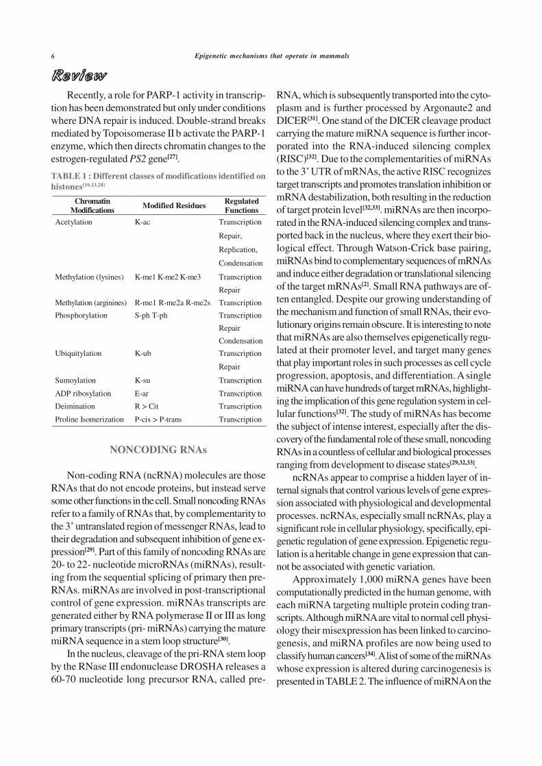
Epigenetic mechanisms that operate in mammals
RNA, which is subsequently transported into the cyto-plasm and is further processed by Argonaute2 andDICER[31]. One stand of the DICER cleavage productcarrying the mature miRNA sequence is further incor-porated into the RNA-induced silencing complex(RISC)[32]. Due to the complementarities of miRNAsto the 3target transcripts and promotes translation inhibition ormRNA destabilization, both resulting in the reductionof target protein level[32,33]. miRNAs are then incorpo-rated in the RNA-induced silencing complex and trans-ported back in the nucleus, where they exert their bio-logical effect. Through Watson-Crick base pairing,miRNAs bind to complementary sequences of mRNAsand induce either degradation or translational silencingof the target mRNAs[2]. Small RNA pathways are of-ten entangled. Despite our growing understanding ofthe mechanism and function of small RNAs, their evo-lutionary origins remain obscure. It is interesting to notethat miRNAs are also themselves epigenetically regu-lated at their promoter level, and target many genesthat play important roles in such processes as cell cycleprogression, apoptosis, and differentiation. A singlemiRNA can have hundreds of target mRNAs, highlight-ing the implication of this gene regulation system in cel-lular functions[32]. The study of miRNAs has becomethe subject of intense interest, especially after the dis-covery of the fundamental role of these small, noncodingRNAs in a countless of cellular and biological processesranging from development to disease states[29,32,33].
ncRNAs appear to comprise a hidden layer of in-ternal signals that control various levels of gene expres-sion associated with physiological and developmentalprocesses. ncRNAs, especially small ncRNAs, play asignificant role in cellular physiology, specifically, epi-genetic regulation of gene expression. Epigenetic regu-lation is a heritable change in gene expression that can-not be associated with genetic variation.
Approximately 1,000 miRNA genes have beencomputationally predicted in the human genome, witheach miRNA targeting multiple protein coding tran-scripts. Although miRNA are vital to normal cell physi-ology their misexpression has been linked to carcino-genesis, and miRNA profiles are now being used toclassify human cancers[34]. A list of some of the miRNAswhose expression is altered during carcinogenesis ispresented in TABLE 2. The influence of miRNA on the
Recently, a role for PARP-1 activity in transcrip-tion has been demonstrated but only under conditionswhere DNA repair is induced. Double-strand breaksmediated by Topoisomerase II b activate the PARP-1enzyme, which then directs chromatin changes to theestrogen-regulated PS2 gene[27].
TABLE 1 : Different classes of modifications identified on
histones[10,13,28]
Chromatin
Modifications Modified Residues
Regulated
Functions
Acetylation K-ac Transcription
Repair,
Replication,
Condensation
Methylation (lysines) K-me1 K-me2 K-me3 Transcription
Repair
Methylation (arginines) R-me1 R-me2a R-me2s Transcription
Phosphorylation S-ph T-ph Transcription
Repair
Condensation
Ubiquitylation K-ub Transcription
Repair
Sumoylation K-su Transcription
ADP ribosylation E-ar Transcription
Deimination R > Cit Transcription
Proline Isomerization P-cis > P-trans Transcription
NONCODING RNAs
Non-coding RNA (ncRNA) molecules are thoseRNAs that do not encode proteins, but instead servesome other functions in the cell. Small noncoding RNAsrefer to a family of RNAs that, by complementarity tothe 3their degradation and subsequent inhibition of gene ex-pression[29]. Part of this family of noncoding RNAs are20- to 22- nucleotide microRNAs (miRNAs), result-ing from the sequential splicing of primary then pre-RNAs. miRNAs are involved in post-transcriptionalcontrol of gene expression. miRNAs transcripts aregenerated either by RNA polymerase II or III as longprimary transcripts (pri- miRNAs) carrying the maturemiRNA sequence in a stem loop structure[30].
In the nucleus, cleavage of the pri-RNA stem loopby the RNase III endonuclease DROSHA releases a60-70 nucleotide long precursor RNA, called pre-

Bignon Yves-Jean et al.
epigenetic machinery and the reciprocal epigenetic regu-lation of miRNA expression suggest that its deregula-
tion during carcinogenesis has important implicationsfor global regulation of epigenetics and cancer.
TABLE 2 : MicroRNA alterations in various human cancers[4,31,33]
MicroRNAs Target Gene(s) Cancer Type
miR-125 AKT, ERBB2-4, FGF, FGFR, IGF, MAPKs, breast
MMP11,
SP1, TNF, VEGF
miR-205 VEGF-A, ErbB3 breast
miR-10b Rho C breast
miR-335 SOX4, TNC breast
miR-29a TTP breast
miR-9 CDH1 breast cancer
miR-520 CD44 breast cancer
miR-146 NF-kB breast, pancreas and prostate cancers
miR-10b HOXD10 metastatic breast cancer
miR-372,miR-373 RAS, p53, CD44 testicular germ cell tumor and breast
cancer
miR-342 ER, PR and HER2 breast and colon cancer
miR-145 ER colon and breast cancer
miR-126 CRK1,PIK3R2,SPRED1, VCAM1 breast and lung cancer
miR-200 family ZEB1, ZEB2 NCI-60 cell lines; breast, ovary
miR-218, miR-145 PXN breast, lung and prostate cancer
miR-155 RHOA Burkitt
and lung cancers
miR-21 PDCD4,PTEN,TPM1,RECK, TIMP3,BCL2 glioblastoma, breast, lung, prostate,
colon and cervical cancer
miR-15a, miR-16-1 CCND1, Wnt3A prostate
miR-101 Fos, EZH2 HCC, prostate
miR-127 Bcl-6 bladder cancer
miR-124 CDK6 colon cancer
miR-223 NFI-A, MEF2C acute myeloid leukemia
miR-34b/34c p53 network, CDK6, E2F3 colon cancer
miR-17, miR-92 c-MYC lung cancer
miR-92b PRMT5 brain primary tumors
miR-29c ECM proteins NPC
miR-127, miR-199a BCL6, E2F1 cervical cancer
miR-421 CBX7, RBMXL1 gastric cancer
miR-32, miR345, miR-1228,miR-195, CDKN2A,NF2,andJUN Malignant mesothelioma (MM)
miR30b
miR-190, miR-196 HGF pancreatic cancer
miR-34a c-Met HCC
miR-146a, miR-146b ROCK1, IRAK1,TRAF6 prostate cancer and papillary thyroid
carcinomas
miR-340, miR-421, miR-658 MYC, RB, PTEN lymph node metastasis and gastric cancer
let-7a-3 RAS, IGF-II lung and ovarian cancer
miR-9 NF-_B ovarian and lung cancer

Epigenetic mechanisms that operate in mammals
MicroRNAs Target Gene(s) Cancer Type
miR-221, miR-222 CDKN1C/P57 and CDKN1B/P27 hepatocellular carcinoma
miR-25, miR-32, miR-142 ITGA_1 lung cancer and solid tumor
miR- 124, miR-183 ITGB_1 lung cancer
miR-143 ERK5 cervical cancer
miR-372, miR-373 LATS2 testicular germ cell cancer
miR-370 MAP3K8 MzChA-1, KMCH-1, cholangiocarcinoma downregulation
miR- 124, miR-183 ITGB1 _ lung cancer
Abbreviations: CDK6, cyclin D kinase 6; MEF2C, myocyte enhancer factor 2C; NFIA, Nuclear factor 1 A-type; p53, tumor
protein 53; RAS, Rat Sarcoma; CD44, cluster differentiation 44; PDcD4, programmed cell death 4; TPM1, tropomyosin 1;
PTEN, phosphatase and tensin homolog ; BCL2, B-cell lymphoma 2 protein; RECK, reversion Inducing cysteine rich protein
kazal motif; ROHA, ras homolog gene family member A; NF-_B, nuclear factor-_appaB; PRNT5, protein arginine N-
methyltransferase 5; HOXD10, homeobox D10; CDH1, Cadherin-1; CBX7, chromobox 7; RBMX L1, RNA binding motif
protein X-linked; CDNK2A, cyclin-dependent kinase inhibitor 2A; NF2, neurofibromatosis, type 2; HGF, hepatocyte growth
factor; ERBB2-4= or (HER4), human epidermal growth factor Receptor 4; JUN, janus N-terminal kinases; FGFR, fibroblast
growth factor receptor; MAPKs, mitogen-activated protein kinase; MMP11, matrix metalloproteinase11; VEGF, vascular
endothelial growth factor; TNF_, tumor necrosis factor-alpha; CRK1, Cdc2-related kinase1; PIK3R2, phosphatidylinositol 3-
kinase egulatory subunit beta; SPRED1,sprouty-related, EVH1 domain containing 1; VCAM, vascular cell adhesion mol-
ecule; ROCK1, rho-associated, coiled-coil containing protein kinase 1; IRAK1, interleukin-1 receptor associated kinase-1;
TRAF6, TNF receptor associated factor 6; Rb, retinoblastoma; IGF-II, insulin-like growth factor 2; PXN, paxilin; ITG_1,
integrin beta-1; ERK5, extracellular signal-regulated kinase 5; LATS2, large tumor suppressor homolog 2; ER, estrogen
receptor; PR, progesterone receptor, TNC, tenascin C; HCC, hepatocellular carcinoma; BT-IC, breast tumor initiating cells;
NPC, nasopharyngeal carcinoma.
CONCLUSION
In summary, recent studies revealed that deregula-tion of a number of epigenetic mechanisms may favourappearance of genetic alterations. Understanding thecomplexity of the epigenome and the actors involved inmodulating its interactions within genomic sequences, itwill open new horizons in our search to know all themechanisms governing cellular fate.
REFERENCES
[1] C.H.Waddington; Int.J.Epidemiol., 41, 10-3 (2012).[2] R.Taby, J.P.Issa; CA Cancer J.Clin., 60, 376-92
(2010).[3] R.Kanwal, S.Gupta; J.Appl.Physiol., 109, 598-605
(2010).[4] R.Kanwal, S.Gupta; Clin.Genet., 81, 303-11 (2012).[5] K.D.Robertson; Nat.Rev.Genet., 6, 597-610
(2005).[6] H.Cedar, Y.Bergman; Nat.Rev.Genet., 10, 295-304
(2009).[7] A.Bird; Genes Dev., 16, 6-21 (2002).[8] S.Gopalakrishnan, B.O.Van Emburgh,
K.D.Robertson; Mutat.Res., 647, 30-8 (2008).[9] M.Leeb, A.Wutz; Chromosoma., 121, 251-62
(2012).
[10] T.Kouzarides; Cell, 128, 693-705 (2007).[11] A.H.Lund, M.van Lohuizen; Genes Dev., 18, 2315-
35 (2004).[12] A.Imhof; Curr.Biol., 13, R22-4 (2003).[13] S.Henikoff, A.Shilatifard; Trends Genet., 27, 389-
96 (2011).[14] T.Jenuwein, C.D.Allis; Science, 293, 1074-80
(2001).[15] X.J.Yang; Nucleic Acids Res., 32, 959-76 (2004).[16] H.Ogawa, K.Ishiguro, S.Gaubatz, D.M.Livingston,
Y.Nakatani; Science, 296, 1132-6 (2002).[17] J.van der Vlag, A.P.Otte; Nat.Genet., 23, 474-8
(1999).[18] Y.Shi, F.Lan, C.Matson, P.Mulligan, J.R.Whetstine,
P.A.Cole, R.A.Casero; Cell, 119, 941-53 (2004).[19] Y.Tsukada, J.Fang, H.Erdjument-Bromage,
M.E.Warren, C.H.Borchers, P.Tempst, Y.Zhang;Nature, 439, 811-6 (2006).
[20] Y.Zhang, D.Reinberg; Genes Dev., 15, 2343-60(2001).
[21] W.L.Cheung, F.B.Turner, T.Krishnamoorthy,B.Wolner, S.H.Ahn, M.Foley, J.A.Dorsey,C.L.Peterson, S.L.Berger, C.D.Allis; Curr.Biol., 15,656-60 (2005).
[22] H.Wang, L.Zhai, J.Xu, H.Y.Joo, S.Jackson,H.Erdjument-Bromage, P.Tempst, Y.Xiong,Y.Zhang; Mol.Cell, 22, 383-94 (2006).
[23] R.Pavri, B.Zhu, G.Li, P.Trojer, S.Mandal,A.Shilatifard, D.Reinberg; Cell, 125, 703-17 (2006).

Bignon Yves-Jean et al.
[24] K.Zhang, W.Lin, J.A.Latham, G.M.Riefler,J.M.Schumacher, C.Chan, K.Tatchell, D.H.Hawke,R.Kobayashi, S.Y.Dent; Cell, 122, 723-34 (2005).
[25] D.Nathan, K.Ingvarsdottir, D.E.Sterner,G.R.Bylebyl, M.Dokmanovic, J.A.Dorsey,K.A.Whelan, M.Krsmanovic, W.S.Lane,P.B.Meluh, E.S.Johnson, S.L.Berger; Genes Dev.,20, 966-76 (2006).
[26] P.O.Hassa, S.S.Haenni, M.Elser, M.O.Hottiger;Microbiol.Mol.Biol.Rev., 70, 789-829 (2006).
[27] M.C.Haffner, A.M.De Marzo, A.K.Meeker,W.G.Nelson, S.Yegnasubramanian; Clin.CancerRes., 17, 3858-64 (2011).
[28] P.S.Espino, L.Li, S.He, J.Yu, J.R.Davie; CancerRes., 66, 4610-6 (2006).
[29] M.Ghildiyal, P.D.Zamore; Nat.Rev.Genet., 10, 94-108 (2009).
[30] T.Kunej, I.Godnic, J.Ferdin, S.Horvat, P.Dovc,G.A.Calin; Mutat.Res., 717, 77-84 (2011).
[31] H.Zhou, H.Hu, M.Lai; Biol.Cell, 102, 645-55(2010).
[32] M.Malumbres; Mol.Aspects Med., (2012).[33] G.Egger, G.Liang, A.Aparicio, P.A.Jones; Nature,
429, 457-63 (2004).[34] M.S.Nicoloso, R.Spizzo, M.Shimizu, S.Rossi,
G.A.Calin; Nat.Rev.Cancer, 9, 293-302 (2009).[35] http://www.kapplerlab-munich.de/research.htm

40
Conclusion of Publication2:
Latest approaches in epigenetic research have helped us with a better understanding
of the dynamic of DNA methylation, histone modification and non-coding RNA mediated
gene expression and mammalian cell development.
This review focused on DNA methylation, histone modification and non-coding
RNA because these epigenetic events are widely implicated in cancer development and
progression. In this review, we want to emphasize of epigenetic mechanisms to help for breast
cancer therapy.

41
ii. Epigenetic modifications in breast cancer and epigenetic therapy
Epigenetic changes are critical for development and progression of cancers,
including breast cancer. Significant progress has been made in the basic understanding of how
various epigenetic changes such as DNA methylation, histone modification, miRNA
expression, and higher order chromatin structure affect gene expression. Subsequently, it
became clear that epigenetic modifications play important roles in diseases, including breast
cancer. There is thus a pressing need to understand the functional genome; that is, the changes
defined by regulatory mechanisms overlaying the genetic structure [73], [74].
It has been increasingly recognized over the past several years that CpG islands of a
large number of genes, which are mostly unmethylated in normal tissue, are methylated to
varying degrees in human cancers, thus representing tumour-specific alterations. In cancer
cells, there is a deregulation of DNA methylation patterns that lead to genome-wide
hypomethylation and hypermethylation of CpG islands associated with cancer-related genes.
DNA methylation plays an essential role in development, chromosomal stability, and for
maintaining gene expression states. DNA methylation occurs when methyl groups are added
to cytosines in CpG dinucleotides, leading to a closed chromatin conformation and gene
silencing. CpGs are often found at increased frequencies in promoter regions, forming CpG
islands. Hypermethylation of CpG islands affects genes involved in cell cycle control, DNA
repair, cell adhesion, signal transduction, apoptosis and cell differentiation. In tumour cells,
local promoter hypermethylation is often accompanied by global hypomethylation. This
results in more global patterns of methylation as compared with mutation spectra, which
differ greatly in extent and patterns between tumours [75].
In addition, a large group of cancers are also associated with aberrant Histone
deacetylases (HDAC) expression. Gene silencing and maintenance of cellular identity can
also be mediated by histone modifications carried out by polycomb group (PcG) proteins.
Enhancer of zeste homolog 2 (EZH2) is a core member of the polycomb repressive complex 2
(PRC2) that catalyses the histone mark characteristic for PcG-mediated silencing:
trimethylation of lysine 27 on histone H3 (H3K27me3), which leads to the blocking of
transcriptional activation factors and thereby gene silencing independent of promoter
methylation. In cancer cells, the presence of PRC2 can lead to recruitment of DNMTs

42
resulting in de novo DNA methylation and more permanent repression of PRC2 target genes
[76].
Moreover, many of the genes that undergo promoter methylation in breast cancer are
already expressed at low levels in corresponding normal cells, suggesting that a large fraction
of de novo methylation events in breast cancer cells are not subject to growth selection but
instead reflect an instructive mechanism inherent of the normal cell from which the tumour
originated.
Improving knowledge about epigenetic mechanisms is important for understanding
prognosis and predicting of breast cancer and also promising for epigenetic therapy research.
When we investigated therapeutic agents, we found that HDAC inhibitors (HDACi) have
potential as an anticancer therapeutics. HDACi are a new group of anti-tumor agents as a
future function in the treatment of breast cancer. Currently, seven groups of HDACis have
been reported; short-chain fatty acids, hydroxamic acids, benzamides, cyclic tetrapeptides
containing a 2-amino-8-oxo-9, 10-epoxy-decanoyl (AOE) moiety, cyclic peptides without the
AOE moiety, epoxides and psammaplins. Until now, two HDACi vorinostat (suberoylanilide
hydroxamic acid-SAHA, Zolinza) and depsipeptide (romidepsin, Istodax) have been approved
by the US Food and Drug Administration (FDA) for treatment of cutaneous T-cell lymphoma
[77],[78].

43
Presentation of Publication 3:
Karsli-Ceppioglu S, Dagdemir A, Judes G, Ngollo M, Penault-Llorca
F, Pajon A, BignonY-J, Bernard-Gallon D. Epigenetic Mechanisms of Breast
Cancer: Update of Current Knowledge. Epigenomics (December 2014, in
press).
In this review, we defined some epigenetic mechanisms and their effects of
correlation with breast cancer.
Estrogen receptors are over-expressed in around 70% of breast cancer cases, referred
to as "ER-positive", and can be demonstrated in such tissues using immunohistochemistry.
Two hypotheses have been proposed to explain why this causes tumorigenesis, and the
available evidence suggests that both mechanisms contribute. First, binding of estrogen to the
ER stimulates proliferation of mammary cells, with the resulting increase in cell division and
DNA replication, leading to mutations. Second, estrogen metabolism produces genotoxic
waste. The result of both processes is disruption of cell cycle, apoptosis and DNA repair, and,
therefore, tumour formation.
The PcG proteins are in charge of for maintaining the inactive expression of many
genes. In the last days, three different polycomb repressive complexes have been identified:
polycomb repressive complex 1 (PRC1), polycomb repressive complex 2 (PRC2), and
pleiohomeotic-repressive complex (PhoRC). Enhancer of zeste homolog 2(EZH2), is the most
important subunit of the PRC2. Recent studies have shown that overexpression of EZH2 is
associated with breast cancer. EZH2 over expression may promote cancer due to an increase
in histone methylation which silences the expression of tumor suppressor genes. An EZH2-
targeting drug inhibits the progression of breast cancer.
BRCA1 is human gene that produces tumor suppressor proteins. These proteins help
repair damaged DNA and, therefore, play a role in ensuring the stability of the genetic
material of cell. When either of these genes is mutated, or altered, such that its protein product
is not made or does not function correctly, DNA damage may not be repaired properly.

44
As a result, cells are more likely to develop additional genetic alterations that can lead
to cancer. Specific inherited mutations in BRCA1 increase the risk of female breast and
ovarian cancers, and they have been associated with increased risks of several additional types
of cancer.
They are important key factors for cancer development and prognosis. Investigations
related with aberrant epigenetic regulations in breast cancer focus on the initiating molecular
mechanisms in cancer development, identification of new biomarkers to predict breast cancer
aggressiveness and potential epigenetic therapy.

1
Epigenetic Mechanisms of Breast Cancer: Update of current knowledge.
Seher Karsli-Ceppioglu1,2,3, Aslihan Dagdemir1,2, Gaëlle Judes1,2, Marjolaine Ngollo1,2, Frédérique
Penault-Llorca 1,2, Amaury Pajon1,2, Yves-Jean Bignon1,2*, Dominique Bernard-Gallon 1,2.
1Department of Oncogenetics, Centre Jean Perrin, CBRV, 28 place Henri Dunant, BP 38, 63001 Clermont-Ferrand,
France. 2EA 4677 "ERTICA", University of Auvergne, 28 place Henri Dunant, BP 38, 63001 Clermont-Ferrand,
France. 3Department of Toxicology, Faculty of Pharmacy, Marmara University, Tibbiye Cad. No:49 34668 Istanbul,
Turkey.
*Author for correspondence: Yves-Jean Bignon, Department of Oncogenetics, Centre Jean
Perrin, CBRV, 28 place Henri Dunant, BP 38, 63001 Clermont-Ferrand, France.
E-mail: [email protected]
ABSTRACT
Epigenetic alterations are heritable changes in gene expression that occur without changes in DNA
sequence. They are important key factors for cancer development and prognosis. Breast cancer is
induced by the accumulation of altered gene regulation. Beside of genetic mutations, epigenetics
mechanisms have an important role in breast cancer tumorigenesis. Investigations related with
aberrant epigenetic regulations in breast cancer focus on the initiating molecular mechanisms in
cancer development, identification of new biomarkers to predict breast cancer aggressiveness and
potential epigenetic therapy. In this review, we will summarize the recent knowledge about the role
of epigenetic alterations related with DNA methylation and histone modification in breast cancer. In
addition, altered regulation of breast cancer specific genes and potential epigenetic therapy will be
discussed according to epigenetic mechanisms.
Key Words: epigenetics; breast cancer; DNA methylation; histone modification; estrogen; EZH2;
BRCA1; epigenetic therapy

2
INTRODUCTION
Cancer is a heterogeneous disease which is caused by multi-factorial processes. A vast majority of
investigations are studying sequence alterations in the genome; however, thanks to the increased
knowledge about gene expression mechanisms at last decades, it is found that epigenetic alterations
are important key factors for cancer development and prognosis [1]. The term epigenetics describes
the heritable changes in gene expression that occur without changes in DNA sequence and provides
information about the effect of chromatin organization on transcriptional regulation.
Nucleosome is the structural subunit of chromosomes. It consists of 147 base pairs of DNA
wrapped around a histone core octamer and comprised of two copies of H2A, H2B, H3 and H4.
Nucleosome is composed of two parts; a highly structured part called the core domain, and the
unstructured N- and C-terminal parts that stretch from the nucleosome core. The histone amino
terminal tails are subject to many post-translational modifications. There are three main epigenetic
modifications: DNA methylation, histone modifications and RNA-mediated gene silencing (FIGURE
1). DNA methylation is a covalent modification that takes place at the 5’ position of cytosine rings
in the CpG dinucleotide. Histone modifications are covalent post-translational modifications of N-
terminal tails of four core histones. Small-non-coding RNA molecules can alter gene expression
states by targeting mRNAs for degradation or by preventing their translation [2, 3]. Epigenetic
alterations due to DNA methylation and histone modifications have gained particular importance
for cancer prognosis and therapy.
Breast cancer is a prominent cause of cancer-related deaths in women. Deaths caused by female
breast cancer comprise 6.1% of total deaths in women. According to The American Cancer
Society’s predictions of breast cancer for 2013; about 232,340 new cases of invasive breast cancer
will be diagnosed in women and 39,620 women will die from breast cancer in United States [4].
Breast cancer is induced by the accumulation of altered gene regulation and function which cause
abnormal cell growth and expansion. In addition to genetic mutations, it has become obvious that
epigenetics play an important role in breast cancer tumorigenesis based upon increasing amount of
3
research. These studies focus on initiating molecular mechanisms in cancer development;
identification of new biomarkers to predict breast cancer aggressiveness and potential epigenetic
therapy [5-7]. In this review, we aim to discuss the recent knowledge about the role of epigenetic
alterations related with DNA methylation and histone modification in breast cancer.
DNA Methylation
DNA methylation is a covalent chemical modification, occurring with the addition of a methyl
(CH3) group from S-adenosylmethionine (SAM) at cytosine residues of the DNA template [1].
DNA methyltransferases (DNMT) are the main activators of DNA methylation. They either
catalyze the de n ova methylation or establish methylation in hemimethylated DNA. The discovered
DNMT protein family in mammals are DNMT1, DNMT1b, DNTM1o, DNTMT1p, DNMT2,
DNMT3A, DNMT3B with its isoforms, and DNMT3L, however just three of them have catalytic
methyltransferase activity (DNMT1, DNMT3A, and DNMT3B) [8, 9]. The mammalian DNMTs
comprise of two parts: the catalytic C-terminal domain which is responsible for cofactor binding
and catalysis; and the regulatory N-terminal part which is important for differentiation between
hemimethylated and unmethylated DNA [3]. DNMT1 is the most abundant DNMT in mammalian
cells that mainly catalyzes methylation for hemimethylated CpG sites during DNA replication. The
DNMT2 shows tRNA methyltransferase activity and furthermore, it detects DNA damage, DNA
recombination, and mutation repair [10, 11]. De n ova methyltransferases DNMT3A and DNMT3B
are mainly expressed during embryo development [12]. DNMT3A can catalyze methylation of
hemimethylated and unmethylated CpGs at the same rate; its DNA methyl-transferase activity is
lower than DNMT1, but greater than DNMT3B. DNMT3A and DNMT3B are mainly considered as
de n ova methyltransferases, due to knock-out of these genes in mice they die early in development
or shortly after birth [10, 13]. DNA methylation takes place at mechanisms associated with genomic
integrity and gene expression. It was often assumed as an epigenetic alteration responsible for gene
repression, which results in transcriptional silencing. However, increasing research in this field has
clarified that DNA methylation plays an important role in transcriptional activation as well [10].

4
Regions of high CpG content, called CpG islands, are located in the promoters of 60% of protein-
coding genes in the human genome. Methylation of CpG islands was the first evidence for aberrant
epigenetic change in cancers. Whereas most CpG islands are unmethylated in healthy cells; CpG
islands become hypermethylated in cancer cells which repressed the transcription of tumor
suppressor genes. On the other hand, CpG sites may become hypomethylated, leading to
transcriptional activation of normally repressed genes like oncogenes and retrotranspons [3].
The family of methyl-CpG-binding proteins (MBDs) mediates silencing of tumor suppressor genes
besides DNMTs. These proteins recognize and bind specifically to methylate DNA containing
regions of CpG islands with their methyl-CpG-binding domain. MBDs establish a connection
between DNA methylation and histone modification enzymes associated with transcriptional
repression. The family of MBD proteins consists of five members: MeCP2, MBD1, MBD2, MBD3
and MBD4. These proteins are related with hypermethylation of tumor-supressor gene promoters at
CpG islands and their silencing [3, [14].
DNA Methylation and Breast Cancer
Aberrant methylation plays an important role in breast cancer development. DNA hypomethylation
has been generally demonstrated on a genome-wide scale in cancer researches while
hypermethylated DNA regions of certain genes, especially normally unmethylated CpG islands,
were frequently reported. A growing number of studies have indicated hypermethylated genes in
breast cancer which have crucial roles in cell-cycle regulation, apoptosis, tissue invasion and
metastasis, angiogenesis and hormone signaling [6, 15-17]. Aberrant methylation profiles of these
genes are associated with breast cancer stage and prognosis; therefore, they have been proposed to
be a diagnostic marker. In a study, Esteller et al. investigated the hypermethylation profiles of 12
tumor suppressor genes which are undergoing epigenetic inactivation in human tumors [6].
According to their results, tumor suppressor genes BRCA1 and p16, DNA repair genes GSTP1 and
CHD1, which all are linked with metastasis and invasion, were found hypermethylated in breast
tumor samples. In addition, the methylation status of ADAM23 gene, which is a member of surface

5
molecules and involved in the cell adhesion process, was analyzed. It was shown that, the degree of
ADAM23 promoter hypermethylation gets higher along with elevated tumor grade [15]. Oshiro et
al. evaluated the epigenetic silencing of DSC3 expression in 32 breast tumor samples [16].
Desmocollins belong to cadherin superfamily and DSC3 is mainly expressed in the basal layers.
They demonstrated that DSC3 expression was downregulated in tumor samples and DSC3
silencing was related with cytosine methylation in 41% of the tumor samples. Wnt signaling plays
an essential role in cell differentiation and proliferation; however, it is strongly associated with
tumorigenesis. The SFRP family consists of five glycoproteins which are putative inhibitors of Wnt
signaling. Several studies emphasized aberrant methylation of Wnt antagonist genes in breast
cancer. Their results indicated that frequent promoter methylation of SFRP family genes was the
main factor for silencing in breast cancer. Moreover, SFRP1, SFRP2 and SFRP5 were
methylated in both cell lines and breast tumor samples at a quite higher frequency. They asserted
that majority of breast tumors represent aberrant methylation of at least one Wnt antagonist gene
and silencing of these genes would be the main factor of Wnt signaling in breast cancer [18-20].
Holm et al. determined the methylation status of 807 selected cancer-related genes according to
molecular subtypes of 189 breast tumors [17]. They found that basal-like, luminal A and luminal B
tumors have different methylation profiles, however, tumors of normal-like and HER2-enriched
subtype represent similar methylation profiles. The methylation frequency of genes, especially
RASS1 and GSTP1, was significantly higher in luminal B tumors, but low methylation frequency
was seen in basal-like tumors. Moreover, among BRCA1 and BRCA2 mutation carriers, tumors of
BRCA2 mutation carriers were significantly more methylated than BRCA1 mutation carriers.
Epigenetic silencing of RAS-association domain family 1 (RASS1F1) gene by promoter
hypermethylation is observed in a considerable number of cancers and it is widely investigated in
breast cancer. In recent studies, high frequency of RASSF1A promoter methylation was found in
breast cancer tumors [21-24]. Moreover, downregulated expression of RASSF1A gene was reported
concordant with its methylation status by Alvarez et al [21]. Significant association between

6
hypermethylated RASSF1A and hormone receptor status was demonstrated. Especially, ER positive
tumors were found to be more frequently methylated than ER negative tumors [21, 22, 24].
Genome-wide analyses of aberrant methylation in breast cancer take care of clarifying the entire
genomic distribution and the underlying molecular mechanisms. These studies also give the
opportunity to understand the methylation profiles of breast cancer subtypes. Li et al. investigated
the DNA methylation status of 12 pairs of ER+/PR+ and ER-/PR- breast cancer samples by
genome-wide profiling and different methylation profiles of FAM124B, ST6GALNAC1, NAV1 and
PER1 was represented according with receptor status [25]. In another study, extensive
hypermethylation was determined in ER positive tumors in comparison with ER negative tumors
[26]. In addition, 40 CpG loci were found to be associated with ER-subtype. Accordingly Fang et
al. observed that CpG island methylation was more prevalent in ER+/PR+ tumors and those tumors
demonstrated better prognosis than ER+/PR+ tumors which did not possess CpG island methylator
phenotype [27]. Hill et al. showed the impact of methylation profile on hormone receptor status in
various breast cell lines [28]. They pronounced that, hypermethylation of RECK, SFRP2, ITR,
UGT3A1, ACADL and UAP1L1 was significantly related to the worst survival and 18 genes/loci
was correlated with ER+/PR+ status.
Global DNA hypomethylation is prevalent in breast tumors and up to 50% of the cases represent
reduced 5-methylcytosine content when compared with matched-control normal tissues [29, 30].
Whole genome distribution of aberrant DNA methylation in eight breast cancer cell lines and
normal human mammary epithelial cells were analyzed by Ruike et al [31]. It was shown that
hypomethylation was distributed throughout the entire genome and simultaneous hypermethylation
occur at CpG-rich regions. In addition, hypomethylation was three to five times more frequent than
hypermethylation. Genome-wide DNA-methylation was investigated in familial breast cancer cases
to demonstrate methylation profiles of the different mutation groups (BRCA1, BRCA2 and non-
BRCA1/2) [30]. In this study, genome-wide correlation was not observed between gene expression
and methylation, however, expression of ~800 genes were inversely correlated with DNA

7
methylation. In addition to these studies, which demonstrated the genome-wide hypometylation
status of breast cancer, there are several investigations that reported hypomethylated genes. Paredes
et al. found that methylation of P-cadherin gene exists in all normal breast epithelial cells while
unmethylation was demonstrated in 42% of invasive carcinomas [32]. Furthermore, promoter
hypomethylation of FEN1 gene, which is associated with DNA replication, repair and apoptosis,
was reported by Singh et al. [33] and hypomethylation of N-acetyltransferase NAT1 promoter was
found at significantly higher levels in primary breast tumors by Kim et al. [34].
Histone Modification
Histone modifications are another main epigenetic mechanism, which are responsible for post-
translational modifications at N-terminal tails of histones in order to regulate chromatin structure
and gene expression. These modifications are carried out by acetylation, methylation,
phosphorylation, sumoylation, poly (ADP)-ribosylation and ubiquitination. Each histone
modification constitutes a particular mark that affects the chromatin structure [2]. Chromatin can be
found in two states: heterochromatin or closed, and euchromatin, also called open or noncondensed.
Genes in open chromatin are transcriptionally active; genes in heterochromatin are transcriptionally
silenced (FIGURE 2).
Histone acetylation, the most studied histone modification, catalyzes the transfer of acetyl group
from acetyl-CoA to the amino group of lysine residues. This modification alters the electrostatic
charge by neutralizing positive charges of histones, which induces open chromatin structure.
Acetylation of histones catalyzed by enzymatic actions of histone acetyl transferases (HATs) and
acetyl groups are removed by histone deacetylases (HDACs), which leads to gene inactivation and
silencing. HATs can be categorized in five families: the GNAT family; the MYST family; the
p300/CBP family; the SRC family and the TAFII250 family. In addition to histone proteins, HATs
catalyze the acetylation of a number of nonhistone proteins such as GATA1, E2F1, PRB or TP53
[3]. Eighteen HDACs have been identified in human and classified into four groups according to
their homology with yeast HDACs and enzymatic activities. Class I HDACs (HDACs 1, 2, 3 and 8)

8
are nuclear proteins and associated with various transcriptional repressors and cofactors. Class II
HDACs are divided into two sub-classes based on their protein structure; Class IIa with one
catalytic domain (HDACs 4, 5,7 and 9) and Class IIb with two catalytic domains (HDACs 6 and
10). Class III HDACs contain seven HDACs (SIRT1 to SIRT7) and they require co-factor
nicotinamide adenine dinucleotide (NAD+) for catalytic activity. Recently identified HDAC11 is
the only member of Class IV HDAC group [35].
Histone methylation induces the methylation of arginine and lysine residues and does not change
histone’s charge. Catalytic activity of histone methylation conducts with histone methyltransferases
(HMTs). The HMTs enzyme family has a conserved catalytic domain called as SET (Suppressor of
variegation, Enhancer of Zeste, Trithorax). Histone lysine methylation exists within the tails of
histone H3 and H4 either in a mono-, di- or trimethylation state [36]. According to their effects on
gene regulation, H3K4me2/3 and H3K79me3 are generally associated with transcriptional activity,
while methylation of H3K9me2/3, H3K27me2/3 and H4K20me3 are associated with repression [37,
38].
Histone Modifications in Breast Cancer
In breast cancer, investigations carried out on histone modifications are relatively newer than DNA
methylation studies. Post-translational histone modifications have a critical role in breast
tumorigenesis and aggressiveness of prognosis. In addition, they are potential therapeutic targets.
Since different breast cancer subtypes represent distinct gene expression profiles, it is important to
clarify the effect of histone marks on gene expression levels at breast tumors. However, there are
limited studies about histone modification patterns in breast cancer tumors, though the numbers of
investigations are increasing in this field.
Elsheikh et al. investigated the frequency of global histone modifications in breast cancer tumors by
using microarray technology and immunohistochemistry [39]. They evaluated the relative levels of
histone lysine acetylation (H3K9ac, H3K18ac, H4K12ac and H4K16ac), lysine methylation
(H3K4me2 and H4K20me3) and arginine methylation (H4R3me2) in 880 primary operable

9
invasive breast carcinoma cases. According to their results, levels of all seven histone marks were
low compared to the high tumor grade. Particularly, levels of H4R3me2, H3K9ac, and H4K16ac
were detected at significantly low levels relevant to large tumor size. Also, levels of H4R3me2 and
H3K9ac were detected at high levels when the lymph node stage was low. Histone modification
levels were significantly higher at steroid receptor (estrogen receptor, progesterone receptor, and
androgen receptor)-positive tumors. They have concluded that reduced detection of these histone
marks would be correlated with poor prognostic characteristics.
Suziki et al. evaluated the expression levels of acetylated histone H4 (ac-H4), ac-H4K12, ac-
tubulin, HDAC1, HDAC2 and HDAC6 in 58 breast surgical samples of normal mammary
epithelium, ductal carcinoma in situ and invasive ductal carcinoma by immunohistochemistry [7].
They have showed that acetylation of ac-H4, ac-H4K12, ac-tubulin, HDAC1, HDAC2, and HDAC6
were decreased in ductal carcinoma in situ, and invasive ductal carcinoma in comparison with
normal breast epithelium. The levels of ac-H4 and ac-H4K12 were decreased extremely at ductal
carcinoma in situ compared to normal epithelium.
Guendel et al. investigated the arginine and lysine methylation of BRCA 1 both in breast cancer cell
lines and 4 breast tumor tissue samples [40]. Their results indicate that BRCA 1 is methylated at
arginine and lysine residues both in cell lines and tumor samples. However, lysine methylation was
only detected in MDA-MB-231 cells, which are triple negative and possess a mutant p53, while
arginine methylation was detected in MDA-MB-231 and MCF-7 cells. MCF-7 cells are estrogen,
progesterone positive, HER2 negative and show wild-type 53. Further, BRCA1 was methylated at
both K and R residues in four breast tumor patient samples. In vivo BRCA1 binding to the APEX,
ARHG and GADD45G promoters was increased along with methylation inhibition. According to
these results, they proposed that methylation may affect the ability of BRCA1 binding to specific
promoters or affects protein-protein interactions that alter the recruitment of BRCA1 to these
promoters.

10
Choe et al. demonstrated the histone modification patterns of H3K4me1, H3K4me3 and H3K9/14ac
in breast cancer cell line MCF-7 and normal mammary epithelial cell line, MCF-10A by ChIP-seq
analysis [41]. No significant differences were seen in studied regions. This study showed that the
H3K4me1 distribution, which covers the entire human genome, was 53% of the gene body, 42% of
the intergenic region, and 5% of the promoter. The promoter regions covered 54% and 52% of total
sequence reads in H3K4me3 and H3K9/14ac, respectively. As a result, H3K4me3 and H3K9/14ac
are greatly co-localized in the same cells, but their correlation in different cells is moderate. In
another study of the same group, they analyzed the histone modification profiles of histone
H3K4me1, H3K4me3, H3K9/16ac, and H3K27me3 in MCF-7 cells in human chromosomes 8, 11
and 12 [42]. The results demonstrated that the promoters were the most abundant regulatory
elements and H3K4me3 and H3K9/ 14ac were highly enriched within ±1 kb from transcription start
sites.
It is clear that modified histones gain importance as biomarkers of breast cancer prognosis. The
investigations carried out on mechanisms of histone modifications are also promising for the
development of efficient HDAC inhibitor therapies.
Estrogen and Epigenetic Mechanisms
Expression level of estrogen receptors (ERs) in tumors is a crucial factor for predicting the
prognosis and the response to therapy in breast cancer patients. Two isoforms of ER, ER and ER ,
have been characterized. ER is encoded by ESR1 and ER is encoded by ESR2. These isoforms
show similar affinities for estradiol and have different roles in the regulation of gene expression.
Estrogens normally regulate growth, reproduction and neural development. However, exposure to
elevated levels of estrogen increases the breast cancer incidence and proliferation [43]. ER negative
tumors establish more aggressive progression with higher histological grade and are associated with
a higher recurrence rate and decreased overall survival. About 80% of breast cancers are ER
positive, however, a considerable part of them lose ER expression during tumor progression and
become resistant to drug therapies [44]. ER complexes regulate gene transcription by the

11
recruitment of co-regulator and transcription factors like HDAC1 and DNMT1 [45]. Several studies
focused on hypermethylation of ESR1 promoter silencing, which is resulted in decreased
expression of ER protein. Ramos et al. investigated the methylation of ESR1 and CXCL12 genes in
breast cancer cell lines and 69 breast tumor samples of Brazilian women. According to their results,
ESR1 promoter methylation was shown in samples with methylated CXCL12 islands [46]. Wei et
al. evaluated the promoter methylation patterns of ER and the ER expression levels of 113
familial breast cancer patients in Han Chinese Population [47]. ER methylation was demonstrated
in 41.6% samples of familial breast cancer patients. Moreover, ER promoter methylation was
significantly correlated with tumor size, progesterone expression, p53 nuclear accumulation,
BRCA1 and BRCA2 statuses. Prabhu et al. demonstrated the hypermetylation of ER promoter in 98
breast cancer tumor specimens of Indian women [48]. They found that ER promoter was
methylated in 27% of triple-negative tumors. On the other hand, less than 2% of ER-positive tumors
were hypermethylated.
There are limited studies about epigenetic regulation of ESR2. In a recent study, Zhao et al.
researched hypermethylation of ER in 178 sporadic breast cancer and 14 benign breast
hyperplasia cases of Chinese women [49]. They determined ER promoter methylation at 44.9% of
studied tumors, which was a higher frequency in comparison with methylation status of benign
breast diseases.
Enhancer of Zetse Homolog 2 (EZH2) and Breast Cancer
Enhancer of zeste homolog 2 (EZH2) is a catalytic subunit of Polycomp Repressive Complex 2
(PRC2). Expression levels of EZH2 are upregulated in variety of cancers including prostate,
bladder, gastric, lung, hepatocellular and breast. As a histone methyltransferase, EZH2 catalyzes
lysine 27 tri-methylation of histone H3 which results in a repressive histone mark [50].
Expression level of EZH2 is associated with aggressiveness of breast cancer and overexpression
affects the tumor invasion and cancer progression. Kleer et al. analyzed the expression levels of

12
EZH2 at 917 samples from 280 patients by tissue microarray and they reported that EZH2 levels
were higher in patients with invasive carcinoma and breast cancer metastases in comparison with
normal breast tissues [51]. At their follow-up study, Dig et al. assessed the EZH2 protein expression
levels to detect precancerous state in 13 women carrying BRCA 1 heterozygous mutation and 25
breast cancer patients with no personal or family history of breast cancer. According to their results,
EZH2 levels were increased in BRCA1 mutation carriers and they suggested that elevated EZH2
levels would be associated with higher risk for breast cancer.
EZH2 expression mostly increases in basal-like breast cancers, as well as in ER-negative breast
cancer cell lines. Besides, EZH2 has a crucial role in the regulation of progesterone signaling
through histone methylation. Doherty et al. demonstrated that after exposing endocrine-disrupting
chemicals such as diethylstilbestrol and Bisphenol-A in mice utero, mammary tissue expression of
EZH2 was increased [52]. Moreover, Pal et al. investigated the epigenetic mechanisms associated
with EZH2 in mammary physiology by determining genome-wide histone methylation profiles
[53]. They have found that EZH2 regulates mammary progenitor activity and alveolar development,
and coordinates in this way the progesterone signaling with global changes.
Holm et al. assessed the global levels of H3K27me3 and EZH2 protein expression in various
molecular subtypes of breast tumors [54]. They have found that EZH2 levels were statistically high
in tumors whose histological grade were 3, ER/PR negative, and with high S-phase fraction; while
H3K27me3 levels were low in these tumor groups. Furthermore, small tumor size was correlated
with high H3K27me3 levels. Protein levels of EZH2 were highest in triple negative tumors and
basal-like tumors, while expression was lowest in ER+/HER2-/Ki67 low and luminal A and
normal-like tumors. The trimethylation of H3K27 was low at triple negative and HER2+ tumors;
also it was low at triple negative and ER+/ HER2-/Ki67 high tumors. In conclusion, they indicated
that high levels of EZH2 and low levels of H3K27me3 were associated with poor survival.
EZH2 affects the invasion and progression of breast cancer by regulating a group of genes which
are linked with cell proliferation and invasion. These genes; RAD51, RUNX3 and CKD1C (p57KIP2
)

13
are associated with cell proliferation and FOX, CDH1 (E-cadherin) are related with metastasis.
RUNX3 was reported as tumor suppressor gene in gastric cancer [55]. It was also shown that its
expression is inactivated by DNA methylation in various cancers, such as prostate, lung and
pancreas. H3K27 trimethylation through EZH2 plays an important role in downregulation of
RUNX3. RUNX3 expression was recovered along with the reduced expression level of EZH2 in
MCF-7 cells, [56]. Moreover, a decline in the RUNX3 concentration reduces CKD1C (p57KIP2
)
expression levels [57]. Over-expression of EZH2 tends to transcriptional repression of FOX C1,
which is a member of the Forkhead box transcription factor family. Increasing levels of FOX C1
expression lead to reduced cell migration and invasion in breast cancer [58]. In addition, it is
reported that elevated levels of EZH2 reduce E-cadherin expression levels, which is related with the
invasiveness and malignant progression of epithelial tumors [59].
Breast Cancer 1 (BRCA1) and Breast Cancer
Inherited germline mutations in the BRCA1 tumor suppressor gene lead to development of
aggressive breast tumors at an early age with high histological grade, expression of proliferation
markers and hormone receptor negativity. In addition, promoter methylation of the BRCA1 gene is
reported in 10-30% of sporadic early-onset breast cancer, which is associated with BRCA1 gene
silencing and aggressive pathological features of breast tumors [47, 60, 61, 62].
Esteller et al. investigated the methylation patterns of BRCA1 promoter in series of breast cancer
cell lines and 215 breast and ovarian tumor samples [63]. Promoter hypermethylation of the BRCA1
gene was detected in 13% of 84 primary breast carcinomas; on the other hand, abnormal
methylation was not determined in breast cancer cells. Turner et al. analyzed promoter methylation
of BRCA1 to investigate BRCA1 silencing and downregulation in basal-like cancers [64]. They have
demonstrated that methylation-induced BRCA1 silencing was frequent in metaplastic tumors. Also,
BRCA1 expression levels were significantly lower in basal-like cancers. Stefansson et al.
investigated the relationship between subtype-specific markers and promoter methylation of BRCA1
in sporadic breast cancer by tissue microarrays [65]. Their results revealed that CpG island

14
hypermethylation plays a critical role at the development of basal/triple-negative breast cancers in
sporadic cases. Moreover, they showed that there is a correlation between the expression levels of
BRCA1, pRb, p16, PTEN and p53. These markers determine a subgroup of triple-negative breast
cancers related with defects in BRCA1 gene. Wong et al. assessed the BRCA1 promoter
methylation in peripheral blood and tumor samples obtained from 255 women to predict early-onset
breast cancers [66]. They demonstrated that approximately 4% of subjects have detectable BRCA1
promoter methylation and their tumor samples were highly methylated at the BRCA1 promoter, so
they have approximately 3.5-fold increased risk of breast cancer before 40 years-old. Jung et al.
examined the promoter methylation of 24 tumor suppressor genes in tumor and normal tissue
samples of 60 Korean patients with primary breast cancer [67]. They found that hypermethylation
of BRCA1 was linked with negative estrogen and progesterone receptor expression. Iwamoto et al.
investigated the relationship between BRCA1 promoter methylation and breast cancer pathogenesis
[68]. For this reason, they analyzed promoter methylation of BRCA1 in peripheral blood samples of
200 cases, 200 controls and 162 tumor tissues. They determined BRCA1 promoter methylation in
peripheral blood samples of 21.5% of breast cancer patients and 13.5% of controls ultimately; and
significantly higher risk of breast cancer was shown in women with BRCA1 promoter methylation.
In addition, BRCA1 promoter methylation was found in 31 of 162 breast tumors. Al-Moghrabi et al.
analyzed the promoter methylation of BRCA1 in 47 breast tumor samples from Arab female patients
and 73 peripheral blood samples of healthy controls by methylation-specific PCR [69]. They
demonstrated that frequency of hypermethylated BRCA1 promoter was high in primary breast
carcinomas of study population. Besides, BRCA1 promoter methylation was strongly associated
with the early-onset of cancer ( 40 years) and high-grade tumors, but no correlation was shown
with estrogen status. Sturgenon et al. evaluated the expression levels of 12 tumor suppressor genes,
which were frequently methylated in breast cancers, in serum of 325 breast cancer cases and 249
benign breast disease controls [70]. Their results showed that the median methylation level of
BRCA1, CCND2, CDH1, ESR1, HIN1, P16 and TWIST were higher in breast cancer cases with
15
negative lymph nodes than benign breast disease subjects. Bosviel et al. investigated the clinical
and prognostic role of BRCA1 gene in sporadic breast cancer patients [71]. They analyzed the site-
specific DNA methylation of BRCA1 in peripheral blood DNA of patients and demonstrated
increasing trend toward BRCA1 promoter hypermethylation in breast cancer patients compared with
controls, however the differences were not statistically significant. In addition, they have assessed
the methylation of the CpG islands present at the BRCA 2 promoter region in the same breast
cancer and control population [72]. They did not find a statistically significant difference between
methylation statuses of study populations. All these results support the aspect of BRCA 1 promoter
methylation being an important risk factor for breast cancer and thus, its affiliation with hormone
expression.
Relationship between EZH2 and BRCA1
As mentioned above, linked with somatic mutations or promoter methylation, BRCA 1 gene
silencing cause basal-like breast carcinomas. In addition, elevated EZH2 concentrations are
associated with the aggressive basal-like tumors. Gonzalez et al. demonstrated that BRCA1 is
regulated by EZH2 expression in benign breast cells and ER negative breast cancer cells [73]. They
also observed that EZH2 over-expression in MCF10A cells resulted in aberrant mitoses and
genomic instability. Wang et al. recently reported that BRCA1 directly binds to EZH2 in both
human breast cancer and mouse embryonic stem cells [74]. They found that genetic deletion of one
allele of BRCA1 or transient knockdown of BRCA1 in mouse embryonic stem cells increases the
EZH2 activity and H3K27me3 levels at PRC2 target loci concordantly, which causes the inhibition
of embryonic stem cell differentiation and result in aggressive breast cancer phenotype. As a result,
decreased expression level of BRCA1 leads to an enhancement in EZH2 activity which induces
aggressive progression of breast carcinomas.
Developments of Epigenetic Therapy in Breast Cancer
Improving knowledge about epigenetic mechanisms is important for prognosis and predicting the
course of breast cancer. Moreover, it is also promising for epigenetic therapy research. Since

16
modifications at HDAC activity have been reported in various tumor types, HDAC inhibitors
(HDACi) have potential as an anticancer therapeutics. HDACi cause transcriptional upregulation of
relevant genes by interfering with the catalytic domain of HDACs to block substrate recognition of
these enzymes. HDACi inhibit proliferation of breast cancer cells by activating genes that produce
cell-cycle arrest and inducing the apoptosis. HDACi can be classified into four groups: hydroxamic
acids, cyclic peptides, short-chain fatty acids and benzamids [75]. Until now, two HDACi
vorinostat (suberoylanilide hydroxamic acid-SAHA, Zolinza) and depsipeptide (romidepsin,
Istodax) have been approved by the US Food and Drug Administration (FDA) for treatment of
cutaneous T-cell lymphoma. In addition, depsipeptide has obtained FDA approval for treatment of
peripheral T-cell lymphoma in 2011 [76, 77].
Various studies have showed that, vorinostat, inhibitor of HDAC I and II, suppress the growth of
both ER positive and negative breast cancer cell lines by stimulating G1, G2/M cell cycle arrest
and apoptosis. HDACis are more effective on transformed cells in comparison with normal cells
and the expression levels of more than 10% of genes in transformed cells are altered with vorinostat
treatment [78]. The evidence from clinical trials demonstrated that vorinostat has limited
therapeutic activity with single treatment. However, it has the potential to modulate ER expression
and provides novel opportunity to reverse the resistance to hormone therapy in breast cancer. In
breast cancer therapy, the mechanisms of vorinostat action are investigated with various studies. For
instance, Zhou et al. demonstrated that ER expression was reactivated with vorinostat treatment and
EGR expression was inhibited due to impairment of its mRNA stability in ER-negative human
breast cancer cells [79]. In another study it was reported that vorinostat stimulates apoptosis by
elevating expression of genes related with p53 signaling in MCF7 cells. On the other, hand its
treatment in MDA-MB-231 cell line induces genes which take part in cell death pathways such as
TNFSF10 (TRAIL) caspase. Lauricella et al. found that vorinostat treatment sensitized both MCF7
and MDA-MB-231 cells to TRAIL-induced apoptosis and decreased the level of both EGFR and its
phosphorylated form [80]. ER-negative breast cells are resistant to apoptosis and after treatment

17
with vorinostat/TRAIL combination, MDA-MB-231 cell lines became sensitive to apoptosis
associated with EGFR downregulation. Vorinostat treatment caused cytotoxicity in tamoxifen-
resistant human breast cancer cells and induced G2/M phase arrest. Moreover, the acetylated
histone H3 and H4 levels were increased and expression of HDAC 1, 2, 4 and 7 were reduced with
the treatment.
Clinical trials that were held on breast cancer epigenetic therapy have focused on combination of
vorinostat and hormone therapy [81]. As a result of this phase II trial, tumor regression or
prolonged disease stabilization was observed in 40% of the patients with hormone receptor-positive
breast cancer. In another phase I–II clinical trial, combination therapy of vorinostat with paclitaxel
and bevacizumab was evaluated in metastatic breast cancer patients [82]. In addition, effects of
vorinostat on acetylated histones and non-histone proteins were detected. It was reported that
combination of vorinostat with paclitaxel and bevacizumab did not increase the toxicity of
paclitaxel-bevacizumab. Moreover, vorinostat was effective inducing hyperacetylation of histone
and non-histone proteins. According the early results of clinical trials, it could be concluded that
vorinostat has potential to recover hormone therapy resistance in breast cancer. The
hyperacetylation effects of HDACis were observed in breast cancer patients and evaluation of
vorinostat-induced acetylation on histone and non-histone proteins would be predictors for
biological activity and clinical benefit.
Various HDACis are being evaluated for their therapeutic potential. For instance, panobinostat
(LBH489) was reported to activate hyperacetylation of histones H3 and H4, decrease proliferation
and induce apoptosis and G2/M cell cycle arrest in triple negative cell lines with minimal toxicity
[83]. HDACi sodium butyrate has crucial functions on inhibition of cell growth and apoptosis. It
was shown that sodium butyrate induced the activity of caspase-3,-8 and -10 in a time- and dose-
dependent manner in human breast cancer cell line MRK-nu-1. Moreover, sodium butyrate
treatment caused DNA fragmentation in a dose-dependent manner [84].

18
With the increasing interest among epigenetic therapy, various research projects are reviewing the
combination treatment of HDACis to sensitize breast cancer cell lines to radiotherapy, hormone
therapy or chemotherapy. Cho et al. investigated the combined effects of sodium butyrate and
demethylation agent, 5-Aza-2'deoxycytidine (5-aza-dC) on radiosensivity in MCF-7 breast cell
lines [85]. They reported that sodium butyrate was more effective for enhancement of
radiosensivity in MCF-7 cell lines. Moreover, combination therapy with sodium butyrate and 5-aza-
DC increased the effect. Several studies were focused on combination treatment of trichostatin A/5-
aza-DC in triple negative cell lines that could activeate the expression of silenced genes in cancer
[86-88]. They also demonstrated that ER-negative cells became sensitive to tamoxifen therapy after
combine treatment with trichostatin A/5-aza-dC [86, 87]. In another study, entinostat showed
stimulation of aromatase expression by upregulating ER expression in ER -negative cell lines
[89].
Epigenetic drugs, which are effective on HAT mechanism, were also tested for their therapeutic
potential. 3-Deazaneplanocin A (DZNep) is an inhibitor of S-adenosylhomocysteine (AdoHcy)
hydrolase and inhibits histone methylation by downregulation of PRC2 complex [90, 91]. Tan et al.
demonstrated that DZNep treatment regulates expression of genes which are transcriptionally
repressive in breast cancer, by inversion of PRC2 and histone methylation-mediated gene silencing.
They clarified that DZNep induces apoptosis of PRC2 target genes [92].
Consequently, it is clear that evolving investigations about epigenetic therapy will represent further
opportunity to treat particularly basal-like tumors by repairing ER expression. HDACis have
critical ability to restore the resistance to hormonal therapy. Further investigations will elucidate
underlying mechanisms of those synergistic interactions.
Conclusion & future perspective
Breast cancer is a significantly heterogeneous disease in histology, genetics and prognosis. It is
clear that epigenetic alterations have crucial role in breast cancer tumorigenesis. In the last two
decades, with increased knowledge about epigenetic mechanisms, we could provide insight into

19
molecular mechanisms of breast cancer. According to the investigations carried out in epigenetic
field, novel approaches are propounded about new biomarkers to predict breast cancer
aggressiveness and potential epigenetic therapies. In addition to the genome-wide analyses that
identify epigenetic regulation of multiple genes; detection of specific genes such as EZH2, BRCA1
and RASSF1A, whose expression levels are altered epigenetically, will be beneficial for determining
prognosis and treatment in breast cancer patients. Technological advancements in epigenetic
researches, which clarify the role of additional mechanisms and complex epigenetic regulations,
lead to a better understanding of underlying mechanisms of breast tumorigenesis and the
approaches to the prevention and treatment of breast cancer. In the near future, finding correlations
between the early epigenetic changes and clinical profiles could provide a better clinic outcome.
Difficulties in the availability of suitable tissue material limit the research; however, investigating
the aberrant epigenetic patterns in primary tumors is essential to demonstrate epigenetic profiles of
breast cancer and to correlate them with clinical status. In conclusion, further investigations could
improve our aspects in epigenetic mechanisms of breast cancer tumorigenesis and its clinical
outcome.

20
Acknowledgements
Seher Karsli-Ceppioglu was supported by The Scientific and Technology Research Council of
Turkey (TUBITAK-2219) project grants and Aslihan Dagdemir was funded by Protema Saglik
Hizm. A.S. We thank Tim Gunnels for corrections to the English language in the manuscript.
21
Executive summary
Breast Cancer
Breast cancer is a significantly heterogeneous disease in histology, genetics, and prognosis.
Epigenetic alterations gained importance at reflecting the molecular mechanisms of
tumorigenesis.
DNA methylation and breast cancer
DNA methylation is one of the most common molecular alterations in cancer, refers to the
covalent addition of a methyl (CH3) group from S-adenosylmethionine (SAM) onto
cytosine residues of the DNA template.
Genome-wide DNA methylation analysis demonstrates that global DNA hypomethylation is
highly frequent in breast cancer. In addition, hypermethylation of breast cancer specific
genes such as BRCA 1, RA SSF1A and Cadher i n super f ami l y genes are also reported
very often.
Histone Modifications in Breast Cancer
Histone modifications are post-translational modifications at N-terminal tails of histones in
order to regulate chromatin structure and gene expression, which are carried out by
acetylation, methylation, phosphorylation, sumoylation, poly (ADP)-ribosylation and
ubiquitination.
Post-translational histone modifications are associated with breast tumorigenesis,
aggressiveness of prognosis, and they are potential therapeutic targets.
Estrogen, EZH2 and BRCA1
Hypermethylation of estrogen receptor genes ESR1 and ESR2 promoters results in with
gene silencing, which is associated with decreased expression levels of ER and ER
proteins in cancer cell lines. However, there are limited number of studies that demonstrated
ER and ER gene silencing in breast tumors.

22
Enhancer of zeste homolog 2 (EZH2) is a histone methyltransferase and its overexpression
is associated with the tumor invasion and cancer progression.
In addition to inherited germline mutations, promoter methylation of BRCA 1 tumor
suppressor gene is also associated with developing aggressive breast tumors.
Developments of Epigenetic Therapy in Breast Cancer
Epigenetic therapy will represent further opportunity to treat breast cancer tumors by
reversing the resistance to hormonal therapy.

23
Figures and Legends
Figure 1. Epigenetic alterations in breast cancer [93] (Reproduced by permission of the
publisher).
TSG: tumor suppressor gene; miRNAs: microRNAs; DNMTs: DNA methyltransferases;
HDACs: histone deaceteylases; PRC: polycomb repressor complexes.

Figure 2. DNA methylation, histone modifications and chromatin remodeling are occuring in tumor
suppressor genes during tumorigenesis. a. Most CpG islands of tumor suppressor genes are
unmethylated in healthy cells, and are transcriptionally active associated with the active histone 3
lysine 4 (H3K4) methylation and histone 3 lysine 9 (H3K9) acetylation chromatin marks. b.The
family of methyl-CpG-binding proteins (MBDs) recognize and bind specifically to methylated
DNA containing regions of CpG islands with their methyl-CpG-binding domain. Repressive
proteins, such as histone deacetylases (HDAC) and histone methyltransferases (HMT) which
deacetylate H3K9, methylate H3K9 and histone H3 lysine 27 (H3K27) residues, respectively, are
recruited by MBDs and lead to gene inactivation and silencing [8] (Reprinted by permission of the
publisher).
DNMT: DNA methyltransferases; HAT: Histone acetyltransferases; HDAC: Histone deacetylases;
HMT: histone methyltransferases; HP1: Heterochromatin protein 1; MBP: Methyl-CpG binding
proteins; PcG: Polycomb group; trxG: Trithorax group.

REFEREN CES
1. Dworkin AM, Huang TH, Toland AE: Epigenetic alterations in the breast: Implications for
breast cancer detection, prognosis and treatment. Semin Cancer Biol 19(3), 165-171 (2009). 2. Cortez CC, Jones PA: Chromatin, cancer and drug therapies. Mutat Res 647(1-2), 44-51
(2008). 3. Khan SI, Aumsuwan P, Khan IA, Walker LA, Dasmahapatra AK: Epigenetic events
associated with breast cancer and their prevention by dietary components targeting the epigenome. Chem Res Toxicol 25(1), 61-73 (2012).
4. Siegel R, Naishadham D, Jemal A: Cancer statistics, 2013. CA Cancer J Clin 63(1), 11-30 (2013).
5. Dagdemir A, Durif J, Ngollo M, Bignon YJ, Bernard-Gallon D: Histone lysine trimethylation or acetylation can be modulated by phytoestrogen, estrogen or anti-HDAC in breast cancer cell lines. Epigenomics 5(1), 51-63 (2013).
6. Esteller M, Corn PG, Baylin SB, Herman JG: A gene hypermethylation profile of human cancer. Cancer Res 61(8), 3225-3229 (2001).
7. Suzuki J, Chen YY, Scott GK et al.: Protein acetylation and histone deacetylase expression associated with malignant breast cancer progression. Clin Cancer Res 15(9), 3163-3171 (2009).
8. Lo PK, Sukumar S: Epigenomics and breast cancer. Pharmacogenomics 9(12), 1879-1902 (2008).
9. Veeck J, Esteller M: Breast cancer epigenetics: from DNA methylation to microRNAs. J
Mammary Gland Biol Neoplasia 15(1), 5-17 (2010). 10. Herceg Z, Vaissiere T: Epigenetic mechanisms and cancer: an interface between the
environment and the genome. Epigenetics 6(7), 804-819 (2011). 11. Okano M, Xie S, Li E: Dnmt2 is not required for de novo and maintenance methylation of
viral DNA in embryonic stem cells. Nucleic Acids Res 26(11), 2536-2540 (1998). 12. Okano M, Bell DW, Haber DA, Li E: DNA methyltransferases Dnmt3a and Dnmt3b are
essential for de novo methylation and mammalian development. Cell 99(3), 247-257 (1999). 13. Bird A: DNA methylation patterns and epigenetic memory. Genes Dev 16(1), 6-21 (2002). 14. Wajed SA, Laird PW, Demeester TR: DNA methylation: an alternative pathway to cancer.
Ann Surg 234(1), 10-20 (2001). 15. Costa FF, Verbisck NV, Salim AC et al.: Epigenetic silencing of the adhesion molecule
ADAM23 is highly frequent in breast tumors. Oncogene 23(7), 1481-1488 (2004). 16. Oshiro MM, Kim CJ, Wozniak RJ et al.: Epigenetic silencing of DSC3 is a common event
in human breast cancer. Breast Cancer Res 7(5), R669-680 (2005). 17. Holm K, Hegardt C, Staaf J et al.: Molecular subtypes of breast cancer are associated with
characteristic DNA methylation patterns. Breast Cancer Res 12(3), R36 (2010). 18. Suzuki H, Toyota M, Carraway H et al.: Frequent epigenetic inactivation of Wnt antagonist
genes in breast cancer. Br J Cancer 98(6), 1147-1156 (2008). 19. 19. Veeck J, Niederacher D, An H et al.: Aberrant methylation of the Wnt antagonist
SFRP1 in breast cancer is associated with unfavourable prognosis. Oncogene 25(24), 3479-3488 (2006).
20. Veeck J, Noetzel E, Bektas N et al.: Promoter hypermethylation of the SFRP2 gene is a high-frequent alteration and tumor-specific epigenetic marker in human breast cancer. Mol
Cancer 7, 83 (2008). 21. Alvarez C, Tapia T, Cornejo V et al.: Silencing of tumor suppressor genes RASSF1A,
SLIT2, and WIF1 by promoter hypermethylation in hereditary breast cancer. Mol Carcinog
52(6), 475-487 (2013). 22. Kajabova V, Smolkova B, Zmetakova I et al.: RASSF1A Promoter Methylation Levels
Positively Correlate with Estrogen Receptor Expression in Breast Cancer Patients. Transl
Oncol 6(3), 297-304 (2013).

23. Li Y, Wei Q, Cao F, Cao X: Expression and promoter methylation of the RASSF1A gene in sporadic breast cancers in Chinese women. Oncol Rep 19(5), 1149-1153 (2008).
24. Xu J, Shetty PB, Feng W et al.: Methylation of HIN-1, RASSF1A, RIL and CDH13 in breast cancer is associated with clinical characteristics, but only RASSF1A methylation is associated with outcome. BMC Cancer 12, 243 (2012).
25. Li L, Lee KM, Han W et al.: Estrogen and progesterone receptor status affect genome-wide DNA methylation profile in breast cancer. Hum Mol Genet 19(21), 4273-4277 (2010).
26. Fackler MJ, Umbricht CB, Williams D et al.: Genome-wide methylation analysis identifies genes specific to breast cancer hormone receptor status and risk of recurrence. Cancer Res
71(19), 6195-6207 (2011). 27. Fang F, Turcan S, Rimner A et al.: Breast cancer methylomes establish an epigenomic
foundation for metastasis. Sci Transl Med 3(75), 75ra25 (2011). 28. Hill VK, Ricketts C, Bieche I et al.: Genome-wide DNA methylation profiling of CpG
islands in breast cancer identifies novel genes associated with tumorigenicity. Cancer Res
71(8), 2988-2999 (2011). 29. Dagdemir A, Durif J, Ngollo M, Bignon YJ, Bernard-Gallon D: Breast cancer: mechanisms
involved in action of phytoestrogens and epigenetic changes. In Vivo 27(1), 1-9 (2013). 30. Flanagan JM, Cocciardi S, Waddell N et al.: DNA methylome of familial breast cancer
identifies distinct profiles defined by mutation status. Am J Hum Genet 86(3), 420-433 (2010).
31. Ruike Y, Imanaka Y, Sato F, Shimizu K, Tsujimoto G: Genome-wide analysis of aberrant methylation in human breast cancer cells using methyl-DNA immunoprecipitation combined with high-throughput sequencing. BMC Genomics 11, 137 (2010).
32. Paredes J, Albergaria A, Oliveira JT, Jeronimo C, Milanezi F, Schmitt FC: P-cadherin overexpression is an indicator of clinical outcome in invasive breast carcinomas and is associated with CDH3 promoter hypomethylation. Clin Cancer Res 11(16), 5869-5877 (2005).
33. Singh P, Yang M, Dai H et al.: Overexpression and hypomethylation of flap endonuclease 1 gene in breast and other cancers. Mol Cancer Res 6(11), 1710-1717 (2008).
34. Kim SJ, Kang HS, Chang HL et al.: Promoter hypomethylation of the N-acetyltransferase 1 gene in breast cancer. Oncol Rep 19(3), 663-668 (2008).
35. Kristensen LS, Nielsen HM, Hansen LL: Epigenetics and cancer treatment. Eur J
Pharmacol 625(1-3), 131-142 (2009). 36. Albert M, Helin K: Histone methyltransferases in cancer. Semin Cell Dev Biol 21(2), 209-
220 (2010). 37. Hublitz P, Albert M, Peters AH: Mechanisms of transcriptional repression by histone lysine
methylation. Int J Dev Biol 53(2-3), 335-354 (2009). 38. Martin C, Zhang Y: The diverse functions of histone lysine methylation. Nat Rev Mol Cell
Biol 6(11), 838-849 (2005). 39. Elsheikh SE, Green AR, Rakha EA et al.: Global histone modifications in breast cancer
correlate with tumor phenotypes, prognostic factors, and patient outcome. Cancer Res 69(9), 3802-3809 (2009).
40. Guendel I, Carpio L, Pedati C et al.: Methylation of the tumor suppressor protein, BRCA1, influences its transcriptional cofactor function. PLoS One 5(6), e11379 (2010).
41. Choe MK, Hong CP, Park J, Seo SH, Roh TY: Functional elements demarcated by histone modifications in breast cancer cells. Biochem Biophys Res Commun 418(3), 475-482 (2012).
42. Hong CP, Choe MK, Roh TY: Characterization of Chromatin Structure-associated Histone Modifications in Breast Cancer Cells. Genomics Inform 10(3), 145-152 (2012).
43. Hervouet E, Cartron PF, Jouvenot M, Delage-Mourroux R: Epigenetic regulation of estrogen signaling in breast cancer. Epigenetics 8(3), 237-245 (2013).

44. Ottaviano YL, Issa JP, Parl FF, Smith HS, Baylin SB, Davidson NE: Methylation of the estrogen receptor gene CpG island marks loss of estrogen receptor expression in human breast cancer cells. Cancer Res 54(10), 2552-2555 (1994).
45. Sharma D, Blum J, Yang X, Beaulieu N, Macleod AR, Davidson NE: Release of methyl CpG binding proteins and histone deacetylase 1 from the Estrogen receptor alpha (ER) promoter upon reactivation in ER-negative human breast cancer cells. Mol Endocrinol 19(7), 1740-1751 (2005).
46. Ramos EA, Camargo AA, Braun K et al.: Simultaneous CXCL12 and ESR1 CpG island hypermethylation correlates with poor prognosis in sporadic breast cancer. BMC Cancer 10, 23 (2010).
47. Wei M, Grushko TA, Dignam J et al.: BRCA1 promoter methylation in sporadic breast cancer is associated with reduced BRCA1 copy number and chromosome 17 aneusomy. Cancer Res 65(23), 10692-10699 (2005).
48. Prabhu JS, Wahi K, Korlimarla A et al.: The epigenetic silencing of the estrogen receptor (ER) by hypermethylation of the ESR1 promoter is seen predominantly in triple-negative breast cancers in Indian women. Tumour Biol 33(2), 315-323 (2012).
49. Zhao L, Yu Z, Li Y et al.: Clinical implications of ERbeta methylation on sporadic breast cancers in Chinese women. Med Oncol 29(3), 1569-1575 (2012).
50. Shen X, Liu Y, Hsu YJ et al.: EZH1 mediates methylation on histone H3 lysine 27 and complements EZH2 in maintaining stem cell identity and executing pluripotency. Mol Cell 32(4), 491-502 (2008).
51. Kleer CG, Cao Q, Varambally S et al.: EZH2 is a marker of aggressive breast cancer and promotes neoplastic transformation of breast epithelial cells. Proc Natl Acad Sci U S A 100(20), 11606-11611 (2003).
52. Doherty LF, Bromer JG, Zhou Y, Aldad TS, Taylor HS: In utero exposure to diethylstilbestrol (DES) or bisphenol-A (BPA) increases EZH2 expression in the mammary gland: an epigenetic mechanism linking endocrine disruptors to breast cancer. Horm Cancer 1(3), 146-155 (2010).
53. Pal B, Bouras T, Shi W et al.: Global changes in the mammary epigenome are induced by hormonal cues and coordinated by Ezh2. Cell Rep 3(2), 411-426 (2012).
54. Holm K, Grabau D, Lovgren K et al.: Global H3K27 trimethylation and EZH2 abundance in breast tumor subtypes. Mol Oncol 6(5), 494-506 (2012).
55. Ito K, Lim AC, Salto-Tellez M et al.: RUNX3 attenuates beta-catenin/T cell factors in intestinal tumorigenesis. Cancer Cell 14(3), 226-237 (2008).
56. Fujii S, Ito K, Ito Y, Ochiai A: Enhancer of zeste homologue 2 (EZH2) down-regulates RUNX3 by increasing histone H3 methylation. J Biol Chem 283(25), 17324-17332 (2008).
57. Chi XZ, Yang JO, Lee KY et al.: RUNX3 suppresses gastric epithelial cell growth by inducing p21(WAF1/Cip1) expression in cooperation with transforming growth factor {beta}-activated SMAD. Mol Cell Biol 25(18), 8097-8107 (2005).
58. Du J, Li L, Ou Z et al.: FOXC1, a target of polycomb, inhibits metastasis of breast cancer cells. Breast Cancer Res Treat 131(1), 65-73 (2012).
59. Yoo KH, Hennighausen L: EZH2 methyltransferase and H3K27 methylation in breast cancer. Int J Biol Sci 8(1), 59-65 (2012).
60. Matros E, Wang ZC, Lodeiro G, Miron A, Iglehart JD, Richardson AL: BRCA1 promoter methylation in sporadic breast tumors: relationship to gene expression profiles. Breast Cancer Res Treat 91(2), 179-186 (2005).
61. Dobrovic A, Simpfendorfer D: Methylation of the BRCA1 gene in sporadic breast cancer. Cancer Res 57(16), 3347-3350 (1997).
62. Rice JC, Ozcelik H, Maxeiner P, Andrulis I, Futscher BW: Methylation of the BRCA1 promoter is associated with decreased BRCA1 mRNA levels in clinical breast cancer specimens. Carcinogenesis 21(9), 1761-1765 (2000).

63. Esteller M, Silva JM, Dominguez G et al.: Promoter hypermethylation and BRCA1 inactivation in sporadic breast and ovarian tumors. J Natl Cancer Inst 92(7), 564-569 (2000).
64. Turner NC, Reis-Filho JS, Russell AM et al.: BRCA1 dysfunction in sporadic basal-like breast cancer. Oncogene 26(14), 2126-2132 (2007).
65. Stefansson OA, Jonasson JG, Olafsdottir K et al.: CpG island hypermethylation of BRCA1 and loss of pRb as co-occurring events in basal/triple-negative breast cancer. Epigenetics 6(5), 638-649 (2011).
66. Wong EM, Southey MC, Fox SB et al.: Constitutional methylation of the BRCA1 promoter is specifically associated with BRCA1 mutation-associated pathology in early-onset breast cancer. Cancer Prev Res (Phila) 4(1), 23-33 (2011).
67. Jung EJ, Kim IS, Lee EY et al.: Comparison of methylation profiling in cancerous and their corresponding normal tissues from korean patients with breast cancer. Ann Lab Med 33(6), 431-440 (2013).
68. Iwamoto T, Yamamoto N, Taguchi T, Tamaki Y, Noguchi S: BRCA1 promoter methylation in peripheral blood cells is associated with increased risk of breast cancer with BRCA1 promoter methylation. Breast Cancer Res Treat 129(1), 69-77 (2011).
69. Al-Moghrabi N, Al-Qasem AJ, Aboussekhra A: Methylation-related mutations in the BRCA1 promoter in peripheral blood cells from cancer-free women. Int J Oncol 39(1), 129-135 (2011).
70. Sturgeon SR, Balasubramanian R, Schairer C, Muss HB, Ziegler RG, Arcaro KF: Detection of promoter methylation of tumor suppressor genes in serum DNA of breast cancer cases and benign breast disease controls. Epigenetics 7(11), 1258-1267 (2012).
71. Bosviel R, Garcia S, Lavediaux G et al.: BRCA1 promoter methylation in peripheral blood DNA was identified in sporadic breast cancer and controls. Cancer Epidemiol 36(3), e177-182 (2012).
72. Bosviel R, Durif J, Guo J et al.: BRCA2 promoter hypermethylation in sporadic breast cancer. OMICS 16(12), 707-710 (2012).
73. Gonzalez ME, Duprie ML, Krueger H et al.: Histone methyltransferase EZH2 induces Akt-dependent genomic instability and BRCA1 inhibition in breast cancer. Cancer Res 71(6), 2360-2370 (2011).
74. Wang L, Huang H: EZH2 takes the stage when BRCA1 loses. Cell Cycle 12(23), 3575-3576 (2013).
75. Ververis K, Hiong A, Karagiannis TC, Licciardi PV: Histone deacetylase inhibitors (HDACIs): multitargeted anticancer agents. Biologics 7, 47-60 (2013).
76. Mann BS, Johnson JR, Cohen MH, Justice R, Pazdur R: FDA approval summary: vorinostat for treatment of advanced primary cutaneous T-cell lymphoma. Oncologist 12(10), 1247-1252 (2007).
77. Campas-Moya C: Romidepsin for the treatment of cutaneous T-cell lymphoma. Drugs
Today (Barc) 45(11), 787-795 (2009). 78. Huang L, Pardee AB: Suberoylanilide hydroxamic acid as a potential therapeutic agent for
human breast cancer treatment. Mol Med 6(10), 849-866 (2000). 79. Zhou Q, Shaw PG, Davidson NE: Inhibition of histone deacetylase suppresses EGF
signaling pathways by destabilizing EGFR mRNA in ER-negative human breast cancer cells. Breast Cancer Res Treat 117(2), 443-451 (2009).
80. Lauricella M, Ciraolo A, Carlisi D, Vento R, Tesoriere G: SAHA/TRAIL combination induces detachment and anoikis of MDA-MB231 and MCF-7 breast cancer cells. Biochimie 94(2), 287-299 (2012).
81. Munster PN, Thurn KT, Thomas S et al.: A phase II study of the histone deacetylase inhibitor vorinostat combined with tamoxifen for the treatment of patients with hormone therapy-resistant breast cancer. Br J Cancer 104(12), 1828-1835 (2011).

82. Ramaswamy B, Fiskus W, Cohen B et al.: Phase I-II study of vorinostat plus paclitaxel and bevacizumab in metastatic breast cancer: evidence for vorinostat-induced tubulin acetylation and Hsp90 inhibition in vivo. Breast Cancer Res Treat 132(3), 1063-1072 (2012).
83. Tate CR, Rhodes LV, Segar HC et al.: Targeting triple-negative breast cancer cells with the histone deacetylase inhibitor panobinostat. Breast Cancer Res 14(3), R79 (2012).
84. Nohara K, Yokoyama Y, Kano K: The important role of caspase-10 in sodium butyrate-induced apoptosis. Kobe J Med Sci 53(5), 265-273 (2007).
85. Cho HJ, Kim SY, Kim KH et al.: The combination effect of sodium butyrate and 5-Aza-2'-deoxycytidine on radiosensitivity in RKO colorectal cancer and MCF-7 breast cancer cell lines. World J Surg Oncol 7, 49 (2009).
86. Sharma D, Saxena NK, Davidson NE, Vertino PM: Restoration of tamoxifen sensitivity in estrogen receptor-negative breast cancer cells: tamoxifen-bound reactivated ER recruits distinctive corepressor complexes. Cancer Res 66(12), 6370-5378 (2006).
87. Fan J, Yin WJ, Lu JS et al.: ER alpha negative breast cancer cells restore response to endocrine therapy by combination treatment with both HDAC inhibitor and DNMT inhibitor. J Cancer Res Clin Oncol 134(8), 883-890 (2008).
88. Yang X, Phillips DL, Ferguson AT, Nelson WG, Herman JG, Davidson NE: Synergistic activation of functional estrogen receptor (ER)-alpha by DNA methyltransferase and histone deacetylase inhibition in human ER-alpha-negative breast cancer cells. Cancer Res 61(19), 7025-7029 (2001).
89. Sabnis GJ, Goloubeva O, Chumsri S, Nguyen N, Sukumar S, Brodie AM: Functional activation of the estrogen receptor- and aromatase by the HDAC inhibitor entinostat
sensitizes ER-negative tumors to letrozole. Cancer Res 71(5), 1893-1903 (2011).
90. Chiang PK, Cantoni GL: Perturbation of biochemical transmethylations by 3-deazaadenosine in vivo. Biochem Pharmacol 28(12), 1897-1902 (1979).
91. Liu S, Wolfe MS, Borchardt RT: Rational approaches to the design of antiviral agents based on S-adenosyl-L-homocysteine hydrolase as a molecular target. Antiviral Res 19(3), 247-265 (1992).
92. Tan J, Yang X, Zhuang L et al.: Pharmacologic disruption of Polycomb-repressive complex 2-mediated gene repression selectively induces apoptosis in cancer cells. Genes Dev 21(9), 1050-1063 (2007).
93. Toland AE: Aberrant Epigenetic Regulation in Breast Cancer. In: Patho-Epigenetics of Disease (Volume 6). Minarovits J, Niller HH (Ed.), Springer, New York, USA, 92 (2012).

45
Conclusion of Publication3:
After the review we understood that ER, EZH2 and BRCA1 expressions are very
important development of breast cancer. They are very consequential and they have got truly
critical effect themselves and also its very significant correlation among each other.
Previous studies have shown that mutation in BRCA1 causes of increase EZH2
concentration particularly in the basal like breast carcinomas. Generally EZH2 is
overexpressed in ER-negative breast cancer and inhibits BRCA1 phosphorylation. For this
reason transmission from G2 to M is accelerated in cell cycle and increased cell proliferation.
Also, it has been shown that before BRCA1 inhibits ligand-induced ER signaling
and blocks subsequent ER transcriptional activation. In many breast cancers, estrogen-
mediated signaling has a very important function of adjusting to evolution and endurance
signaling pathways and can directly drive tumorgenesis by leading the expression of genes.
Some authors have reported the ER is down-regulated in breast tumorigenesis and another
recent study has shown the regulation of ER expression by promoter methylation. In vitro
studies point out ER plays a role in the modulator of proliferation and invasion of breast
cancer cells, so the loss of ER expression could be one of the reasons to breast cancer
development.
Two enzymes are associated with in histone deacetylation; histone acetyltransferase
(HAT) and histone deacetylase (HDAC). These enzymes catalyse the transfer of an acethyl
group from acethyl-CoA molecules to the lysine-amino groups on the N-terminal tails of
histones. HDACi induce G1-phase cell cycle arrest with downregulation of cyclin D1 and
upregulation of p21 in breast cancer cells. When HDAC activity is inhibited, expressions of
only 2% of the mammalian genes are affected.

46
3. OBJECTIVES OF THE STUDY
The first goal of the project was to understand the mechanisms by which
phytoestrogens act on chromatin in breast cancer cell lines. For this study, we used chromatin
immunoprecipitation (ChIP) coupled with QPCR to follow soy phytoestrogen effects on
specific histone marks with selected genes. For the second project, we investigated the effects
of histone methylation inhibitors (HMTi) and histone deacetylase inhibitors (HDACi) on
histone lysine trimethylation and acetylation in breast cancer cell lines.
Subsequently, further investigations in epigenetics were performed with breast tumor
and matched normal tissues. The aim was to find some epigenetic differences between breast
tumors classified according to Saint Gallen and normal tissue breast. For that analysis, ChIP-
QPCR technique also was used to follow the histone modification patterns associated with
breast cancer.
Finnally, histone modifications patterns in breast tumor and normal samples were
analyzed using promoter microarrays. After validation in independent sets of samples, the
identified methylation and acetylation histone signature would be helpful for the diagnosis of
breast cancer and/or for the assessment of risk to develop breast cancer.

47
4. RESULTS
A. Effects of Phytoestrogens and Estrogen on Breast Cancer Cell Lines
Presentation of Publication 4
Dagdemir A, Durif J, Ngollo M, Bignon Y-J, Bernard-Gallon D. Histone Lysine
Trimethylation or Acetylation Can be Modulated by Phytoestrogen, Estrogen or Anti-HDAC
in Breast Cancer Cell Lines. Epigenomics. 2013 Feb;5(1):51-63. doi: 10.2217/epi.12.74.
In this study, we studdied phytoestrogens because they have been investigated as
natural alternatives to hormone replacement therapy and their potential as chemopreventive
agents. The goal of the project was to understand the mechanisms by which phytoestrogens
which act on chromatin in breast cancer cell lines. We suggest that phytoestrogens possess
anti-estrogenic properties may be also responsible for their chemopreventive effects in
epigenetics.

1ISSN 1750-191110.2217/EPI.12.74 © 2013 Future Medicine Ltd Epigenomics (2013) 5(1), 1–xxx
ReseaRch aRticle
Histone lysine trimethylation or acetylation can be
modulated by phytoestrogen, estrogen or
anti HDAC in breast cancer cell lines
Breast cancer is the most common cancer for women in developed countries and urban zones from developing countries [1]. Breast cancer incidence is lower in Asian women in comparison with Westerners. This difference had been attributed to the fact that the traditional Asian diet contains important soy intakes compared with the western diet. Phytoestrogen chemicals found in soy share similarities with the natural human estrogen, 17b estradiol, and are able to tighten the estrogen receptors [2–4] with a higher af nity for ERb [3–5].
Breast cancer progression is not well understood, however, it is likely due to a great amount of genetic mutations leading to widespread changes in gene expression prof iles [6,7], especially the expression of tumor suppressors and oncogenes [8]. Besides the genetic mutations, we need to further investigate epigenetic modi cations in breast cancer, such as DNA methylation [9,10] and histone marks [11,12]. Many post translational modi cations of histones, lysine acetylation and methylation, and arginine methylation seem to be implicated in gene regulation [13]. A combinatorial histone code is able to recognize these and other modi cations on chromatin regions and create transcription activation or repression of genes [14]. Although the epigenetic code is has not been completely elucidated, lysine acetylation (H3K4ac, H3K9ac and H4K8ac)
and trimethylation (H3K4me3) modi cations are able to generate transcriptionally active gene promoters [15–17], whereas other marks, such as lysine methylation (H3K9me3 and H3K27me3), are associated with repressed chromatin [13,14]. Thus, there is evidence that epigenetic alterations in breast cancers can be followed using smallmolecule epigenetic modulators. Therefore, we assessed the effects of soy phytoestrogens, 17b estradiol or anti HDAC (suberoylanilide hydroxamic acid [SAHA]) in breast tumor cell lines using chromatin immunoprecipitation (ChIP) of selected histone modifications. The relative levels of ve modi ed histones, including H3K9me3, H3K27me3, H3K4me3, H4K8ac and H3K4ac, were determined in MCF7 and MDA MB 231 breast cancer cell lines exhibiting different estrogen receptor statuses. In order to identify the presence of variations in global levels of histone marks after treatment, we proposed to follow ChIP with qPCR of six genes implicated in breast cancer and for which the decrease or increase in their expression can be the result of bulk histone modi cations. We chose the EZH2 and P300 genes that code, respectively, for a HMT and a HAT, which are chromatin modifying enzymes implicated in gene regulation by catalyzing reversible post translational modi cations of histones [18–20]. SRC3, which is coded by the ncoa gene, is a steroid receptor coactivator
Aim: The iso avones genistein, daidzein and equol (daidzein metabolite) have been reported to interact with epigenetic modi cations, speci cally hypermethylation of tumor suppressor genes. The objective of this study was to analyze and understand the mechanisms by which phytoestrogens act on chromatin in breast cancer cell lines. Materials & methods: Two breast cancer cell lines, MCF-7 and MDA-MB-231, were treated with genistein (18.5 µM), daidzein (78.5 µM), equol (12.8 µM), 17b-estradiol (10 nM)] and suberoylanilide hydroxamic acid [1 µM] for 48 h. A control with untreated cells was performed. 17b-estradiol and an anti-HDAC were used to compare their actions with phytoestrogens. The chromatin immunoprecipitation coupled with qPCR was used to follow soy phytoestrogen effects on H3 and H4 histones on H3K27me3, H3K9me3, H3K4me3, H4K8ac and H3K4ac marks, and we selected six genes (EZH2, BRCA1, ERa, ERb, SRC3 and P300) for ana lysis. Results: Soy phytoestrogens induced a decrease of trimethylated marks and an increase of acetylating marks studied at six selected genes. Conclusion: We demonstrated that soy phytoestrogens tend to modify transcription through the demethylation and acetylation of histones in breast cancer cell lines.
K yw R : acet lation n breast cancer n histone n meth lation n so ph toestrogens Aslihan Dagdemir1,2,3,
Julie Durif‡1,2,3,
Marjolaine Ngollo1,2,3,
Yves-Jean Bignon*1,2,3
& Dominique Bernard-
Gallon1,2,3
1Centre Jean Perrin, Département
d’Oncogéné que, CBRV, 28 place Henri
Dunant, BP 38, 63001 Clermont-
Ferrand, France 2ERTICA EA 4677, Université
d’Auvergne, 63001 Clermont-Ferrand,
France 3CRNH, 58 Rue Montalembert, 63009
Clermont-Ferrand, France
*Author for correspondence:
[email protected] ‡Authors contributed equally
part of
Auth
or Pro
of
. Breast cancer incidence is lower in Asian women in comparison with Westerners. This difference had been with Westerners. This difference had been attributed to the fact that the traditional Asian attributed to the fact that the traditional Asian diet contains important soy intakes compared diet contains important soy intakes compared with the western diet. Phytoestrogen chemicals with the western diet. Phytoestrogen chemicals
share similarities with the natural share similarities with the natural estradiol, and are able to estradiol, and are able to
tighten the estrogen receptors tighten the estrogen receptors [2–4][2–4] with a higher with a higher [3–5][3–5]..
Breast cancer progression is not well Breast cancer progression is not well understood, however, it is likely due to a understood, however, it is likely due to a great amount of genetic mutations leading great amount of genetic mutations leading to widespread changes in geneto widespread changes in gene
and trimethylation (H3K4me3) modiand trimethylation (H3K4me3) modiare able to generate transcriptionally active gene are able to generate transcriptionally active gene promoters promoters [15–17][15–17], whereas other marks, such as lysine methylation (H3K9me3 and H3K27me3), lysine methylation (H3K9me3 and H3K27me3), are associated with repressed chromatin are associated with repressed chromatin Thus, there is evidence that epigenetic alterations Thus, there is evidence that epigenetic alterations in breast cancers can be followed using smallin breast cancers can be followed using smallmolecule epigenetic modulators. Therefore, molecule epigenetic modulators. Therefore,
Auth
or Pro
of
Auth
or Pro
of
this study was to analyze and understand the mechanisms by which phytoestrogens act on chromatin in this study was to analyze and understand the mechanisms by which phytoestrogens act on chromatin in Two breast cancer cell lines, MCF-7 and MDA-MB-231, were Two breast cancer cell lines, MCF-7 and MDA-MB-231, were
treated with genistein (18.5 µM), daidzein (78.5 µM), equol (12.8 µM), 17treated with genistein (18.5 µM), daidzein (78.5 µM), equol (12.8 µM), 17
Auth
or Pro
of
bbbb-estradiol (10 nM)] and suberoylanilide hydroxamic acid [1 µM] for 48 h. A control with untreated cells was performed. 17suberoylanilide hydroxamic acid [1 µM] for 48 h. A control with untreated cells was performed. 17and an anti-HDAC were used to compare their actions with phytoestrogens. The chromatin and an anti-HDAC were used to compare their actions with phytoestrogens. The chromatin immunoprecipitation coupled with qPCR was used to follow soy phytoestrogen effects on H3 and H4 immunoprecipitation coupled with qPCR was used to follow soy phytoestrogen effects on H3 and H4 histones on H3K27me3, H3K9me3, H3K4me3, H4K8ac and H3K4ac marks, and we selected six genes (histones on H3K27me3, H3K9me3, H3K4me3, H4K8ac and H3K4ac marks, and we selected six genes (
Soy phytoestrogens induced a decrease of Soy phytoestrogens induced a decrease of trimethylated marks and an increase of acetylating marks studied at six selected genes. trimethylated marks and an increase of acetylating marks studied at six selected genes. demonstrated that soy phytoestrogens tend to modify transcription through the demethylation and demonstrated that soy phytoestrogens tend to modify transcription through the demethylation and
n meth meth lation lation n so ph

ReseaRch aRticle Dagdemir, Durif, Ngollo, Bignon & Bernard Gallon
Epigenomics (2013) 5(1)2 future science group
belonging to the p160 family (SRC1, SRC2 and SRC3), constituting a platform for other coactivators such as P300 [21], BRCA1, which is a transcription factor tumor suppressor gene [22], ERa, which is a nuclear receptor activated by 17b estradiol that stimulates proliferation, and ERb, which binds phytoestrogens and induces cellular proliferation and invasion [23].
Materials & methods n Cell linesMCF7 and MDA MB 231 breast tumor cell lines were collected from a pleural effusion of patients with invasive breast carcinoma [24,25]. Both human cell lines were provided by the American Type Culture Collection. MCF7 were cultured in RPMI 1640 medium supplemented with 20 µg/ml of l glutamine (Invitrogen), gentamycin (20 µg/ml; Panpharma), 10% fetal bovine serum (Invitrogen) and insulin (1–4 mg/ml; Novo Nordisk) in a humidi ed atmosphere at 37°C containing 5% CO
2. This
cell line had a positive ER status (ERa+/ERb+). MDA MB 231 cells were grown in LeibovitzL 15 medium with 15% fetal bovine serum (Invitrogen), gentamycin (20 µg/ml ; Panpharma) and l glutamine (20 µg/ml) in a 37°C humidi ed atmosphere without CO
2.
This cell line ERa /ERb+ status. ER status of cell lines has been previously con rmed by immunohistochemistry [26].
n TreatmentsCells (1 × 10⁶ per T75 f lask) were seeded in the medium and treated with 12.8 µM S equol synthesized by the ENITA Unité Micronutriments Reproduction Santé, genistein (18.5 µM; Sigma Aldrich), daidzein (78.5 µM; Sigma Aldrich), 17b estradiol (10 nM; SigmaAldrich) and suberoylanilide hydroxamic acid (SAHA; 1 µM; Sigma Aldrich) and dissolved in dimethyl sulfoxide and ethanol for 17b estradiol. For the controls, each cell line was conditioned in the medium without any treatment for 48 h. The doses of genistein and daidzein were previously determined as corresponding to the 50% inhibition of the proliferation (IC
50)
[26]. SAHA [27], 17b estradiol [28] and S equol concentrations were chosen according to studies by other authors and adaptated to our studies [5,29–31]
n Cell collection & DNA–protein cross-linking for the ChIP assayBefore 80% con uence was achieved, cells were trypsinised and counted by Millipore Scepter 2.0
Cell (Fisher Scienti c). We used 1 × 10⁶ cells for each treatment. Formaldehyde (36.5%; SigmaAldrich) was diluted to 1% in the culture media and cells were incubated at room temperature for 8 min for xation. Cross linking was nished at room temperature with 1.25 M glycine for 5 min. Hereafter, all chromatin preparations and ChIP reactions were handled at 4°C.
n Cell lysis & chromatin shearing by sonicationThe cross linked cells were washed with PBSInhibitor solution (NaBu 20 mM) and cell membranes were lyzed using HighCell ChIP Kit (Diagenode). The cross linked cells were then ready for chromatin shearing. The chromatin was prepared in TPX tubes (Diagenode) with shearing buffer S1 and 1X protease inhibitor (Diagenode). The samples could be submitted to sonication using Bioruptor UCD 200 (Diagenode) for 5 runs of 10 cycles (30 s ‘ON’, 30 s ‘OFF’) for MCF7 and 4 runs of 10 cycles for MDA MB 231 at 200 watt, at 4°C at all times during the sonication process. Between eachrun, samples were spun and vortexed. Fragments of sizes suitable for ChIP are of 100 at 500 bp. The sheared chromatin was frozen at 80°C for later use.
n Chromatin immunoprecipitationChIP reactions were carried out on SX 8X IP STAR compact automated system (Diagenode) and Diagenode Kits for all immunoprecipitation procedures. ChIP experiments were performed with 2 µg of rabbit polyclonal antibodies (anti H3K9me3 [pAb 056–050, Diagenode], H3K27me3 [pAb 069–050, Diagenode], H3K4me3 [pAb003–050, Diagenode], H4K8ac [pAb 103–050, Diagenode] or H3K4ac [pAb 165–050, Diagenode]) and nonimmune rabbit IgG (Kch504–250, Diagenode; e.g., negative control). ChIP reactions were brought up to 200 µl. The IP DNA was puri ed with DNA Isolation Buffer according to the HighCell ChIP kit protocol. Each Auto ChIP sample was performed using Auto Histone ChIP seq kit reagents and contained 1 µg of input chromatin. The reaction was incubated for 2 h for antibody coating with protein A coated magnetic beads, then for 10 h at 4°C for IP reactions (for eight strips).
n Quantitative real-time PCRReal time PCR was performed in triplicate using a 96 well optical tray with optical adhesive lm, at a nal reaction volume of 25 µl containing
Auth
or Pro
of
gentamycin (20 µg/ml; Panpharma), 10% fetal bovine serum (Invitrogen) and insulin (1–4 mg/ml; Novo Nordisk) in a humidi(1–4 mg/ml; Novo Nordisk) in a humidi ed ed atmosphere at 37°C containing 5% COatmosphere at 37°C containing 5% CO
2. This . This
cell line had a positive ER status (ERcell line had a positive ER status (ERaa+/ER/ERbb++). 231 cells were grown in Leibovitz231 cells were grown in Leibovitz
15 medium with 15% fetal bovine serum 15 medium with 15% fetal bovine serum (Invitrogen), gentamycin (20 µg/ml ; (Invitrogen), gentamycin (20 µg/ml ;
glutamine (20 µg/ml) in glutamine (20 µg/ml) in ed atmosphere without COed atmosphere without CO
This cell line ERa /ER/ERbb+ status. ER status of status. ER status of cell lines has been previously concell lines has been previously conimmunohistochemistry immunohistochemistry [26][26].
TreatmentsTreatmentsCells (1 × 10Cells (1 × 10⁶ per T75 f lask) were seeded per T75 f lask) were seeded in the medium and treated with 12.8 µM in the medium and treated with 12.8 µM SS equol synthesized by the ENITA Unité equol synthesized by the ENITA Unité MicronutrimentsMicronutriments(18.5 µM; Sigma(18.5 µM; SigmaSigmaSigmaAldrich) and suberoylanilide hydroxamic acid Auth
or Pro
of
linked cells were washed with PBSlinked cells were washed with PBSInhibitor solution (NaBu 20 mM) and cell Inhibitor solution (NaBu 20 mM) and cell membranes were lyzed using HighCell ChIP Kit membranes were lyzed using HighCell ChIP Kit (Diagenode). The cross(Diagenode). The cross linked cells were then ready for chromatin shearing. The chromatin ready for chromatin shearing. The chromatin was prepared in TPX tubes (Diagenode) with was prepared in TPX tubes (Diagenode) with shearing buffer S1 and 1X protease inhibitor shearing buffer S1 and 1X protease inhibitor (Diagenode). The samples could be submitted (Diagenode). The samples could be submitted to sonication using Bioruptor UCDto sonication using Bioruptor UCD(Diagenode) for 5 runs of 10 cycles (30 s ‘ON’, (Diagenode) for 5 runs of 10 cycles (30 s ‘ON’, 30 s ‘OFF’) for MCF7 and 4 runs of 10 cycles for 30 s ‘OFF’) for MCF7 and 4 runs of 10 cycles for MDAMDAduring the sonication process. Between each

Histon
e lysine trim
ethylation
& acetylation
in breast can
cer cell lines
Rese
aRch a
Rtic
le
ww
w.fu
ture
med
icine.co
m3
futu
re sc
ience g
roup
ControlSAHA
17Genistein
DaidzeinEquol
H3
K9
me
3
IgG
H3
K9
me
3
IgG
H3
K9
me
3
IgG
H3
K9
me
3
IgG
H3
K9
me
3
IgG
H3
K9
me
3
IgG
0
10
20
Fo
ld e
nri
ch
me
nt
ov
er C-FOS
TSH2B
C-FOS
Control H3K9me3-MCF7
ControlSAHA
17Genistein
Daidzein
Equol
H3
K9
me
3
IgG
H3
K9
me
3
IgG
H3
K9
me
3
IgG
H3
K9
me
3
IgG
H3
K9
me
3
IgG
H3
K9
me
3
IgG
02040
Fo
ld e
nri
ch
me
nt
ov
er C-FOS
TSH2B
C-FOS
Control H3K9me3-MDA-MB 231
Control H3K27me3-MDA-MB 231
Control H3K27me3-MCF7
Control H4K8ac-MCF7
Control H4K8ac-MDA-MB 231
ControlSAHA
17Genistein
Daidzein
Equol
H3
K4
me
3
IgG
H3
K4
me
3
IgG
H3
K4
me
3
IgG
H3
K4
me
3
IgG
H3
K4
me
3
IgG
H3
K4
me
3
IgG
020406080
Fo
ld e
nri
ch
me
nt
ov
er TSH2B
ControlSAHA
17Genistein
Daidzein
Equol
H3
K4
me
3
IgG
H3
K4
me
3
IgG
H3
K4
me
3
IgG
H3
K4
me
3
IgG
H3
K4
me
3
IgG
H3
K4
me
3
IgG
024
Fo
ld e
nri
ch
me
nt
ov
er TSH2B
C-FOS
TSH2B
C-FOS
TSH2B
Control H3K4me3-MCF7
Control H3K4me3-MDA-MB 231
Control H3K4ac-MCF7
Control H3K4ac-MDA-MB 231
ControlSAHA
17Genistein
DaidzeinEquol
H4
K8
ac
H4
K8
ac
H4
K8
ac
H4
K8
ac
H4
K8
ac
H4
K8
ac
IgG
IgG
IgG
IgG
IgG
IgG
01020304050
Fo
ld e
nri
ch
me
nt
ov
er TSH2B
C-FOS
TSH2B
C-FOS
TSH2B
ControlSAHA
17Genistein
DaidzeinEquol
H3K
27m
e3
IgG
H3K
27m
e3
IgG
H3K
27m
e3
IgG
H3K
27m
e3
IgG
H3K
27m
e3
IgG
H3K
27m
e3
IgG
02040
Fo
ld e
nri
ch
men
t
over C-FOS
TSH2B
C-FOS
ControlSAHA
17Genistein
DaidzeinEquol
H3K
27m
e3
IgG
H3K
27m
e3
IgG
H3K
27m
e3
IgG
H3K
27m
e3
IgG
H3K
27m
e3
IgG
H3K
27m
e3
IgG
0102030
Fo
ld e
nri
ch
men
t
over C-FOS
TSH2B
C-FOS
ControlSAHA
17Genistein
DaidzeinEquol
H4
K8
ac
H4
K8
ac
H4
K8
ac
H4
K8
ac
H4
K8
ac
H4
K8
ac
IgG
IgG
IgG
IgG
IgG
IgG
01020
4030
Fo
ld e
nri
ch
me
nt
ov
er TSH2B
C-FOS
TSH2B
C-FOS
TSH2B
ControlSAHA
17Genistein
DaidzeinEquol
H3
K4
ac
H3
K4
ac
H3
K4
ac
H3
K4
ac
H3
K4
ac
H3
K4
ac
IgG
IgG
IgG
IgG
IgG
IgG
0
5
10
15
Fo
ld e
nri
ch
me
nt
ov
er TSH2B
ControlSAHA
17Genistein
Daidzein
Equol
H3
K4
ac
H3
K4
ac
H3
K4
ac
H3
K4
ac
H3
K4
ac
H3
K4
ac
IgG
IgG
IgG
IgG
IgG
IgG
0
40302010
Fo
ld e
nri
ch
me
nt
ov
er TSH2B
Figure 1. Control of repressive and activating epigenetic marks ith TSH2B or C-FOS genes. MCF7 and MDA-MB 231 were treated by genistein (18.5 µM), daidzein (78.5 µM), equol (12.8 µM), 17b-estradiol (10 nM) or SAHA (1 µM) for 48 h and controls corresponded to untreated cells. Chromatin immunoprecipitation was controlled with rabbit IgG. Control of speci c antibody (A) H3K9me3; (B) H3K27me3; (C) H3K4me3; ( ) H4K8ac; ( ) H3K4ac. The vertical axis represents the fold enrichment of H3K9 and H3K27 trimethylated on positive gene TSH2B over negative gene C-FOS. The y-axis represents the fold enrichment of H3K9me3, H3K27me3, H4K8ac, H3K4me3 and H3K4ac over C-FOS or TSH2B genes, respectively. SAHA: Suberoylanilide hydroxamic acid.
Auth
or P
roof
Auth
or P
roof
Auth
or P
roof
Auth
or P
roof
Auth
or P
roof
Auth
or P
roof
Auth
or P
roof
Auth
or P
roof
Auth
or P
roof
Auth
or P
roof
Auth
or P
roof
Auth
or P
roof
Auth
or P
roof
Auth
or P
roof
Auth
or P
roof
Auth
or P
roof
Auth
or P
roof
Auth
or P
roof
Auth
or P
roof
Auth
or P
roof
Auth
or P
roof
Auth
or P
roof
Auth
or P
roof
Auth
or P
roof
Auth
or P
roof
GenisteinGenisteinDaidzeinDaidzein
EquolEquol
H3
K9
me
3H
3K
9m
e3
Auth
or P
roof
IgG
IgG
Control H3K9me3-MDA-MB 231Control H3K9me3-MDA-MB 231
Control H4K8ac-MCF7Control H4K8ac-MCF7
Control H4K8ac-MDA-MB 231Control H4K8ac-MDA-MB 231
ControlControlSAHA
Auth
or P
roof
H3
K4
me
3H
3K
4m
e3
H3
K4
me
3
002
Fo
ld e
nri
ch
me
nt
Fo
ld e
nri
ch
me
nt
ov
er
ov
er TSH2B
TSH2B
Auth
or P
roof
Auth
or P
roof
C-FOSC-FOS
TSH2BTSH2B
Auth
or P
roof
Auth
or P
roof
Auth
or P
roof
Auth
or P
roof
Auth
or P
roof
GenisteinGenisteinDaidzeinDaidzein
EquolEquol
H4
K8
ac
H4
K8
ac
H4
K8
ac
H4
K8
ac
H4
K8
ac
H4
K8
ac
IgG
IgG
IgG
IgG
IgG
IgG
Auth
or P
roof
Auth
or P
roof
Auth
or P
roof
Auth
or P
roof
H4
K8
ac
H4
K8
ac
IgG
IgG
Auth
or P
roof
MCF7 and MDA-MB 231 were treated by genistein (18.5 µM), daidzein

ReseaRch aRticle Dagdemir, Durif, Ngollo, Bignon & Bernard Gallon
Epigenomics (2013) 5(1)4 future science group
DNA IP (e.g., immunoprecipited DNA) or DNA Input (e.g., total DNA; 5 µl), 1X SYBR Green Supermix (Applied Biosystems) and 200 nM each of C-FOS (pp 1004–500, Diagenode; positive control for acetylation) or TSH2B (pp 1041–500, Diagenode; positive control for methylation) promoters. For other genes, Taqman Universal PCR Master Mix was used at 1X, 400 nM each of forward and
reverse primers and 250 nM of probe. Initial denaturation at 95°C for 10 min to activate DNA polymerase was followed by 40 cycles of denaturation at 95°C for 15 s, and annealing and extension at 60°C for 1 min (7900HT, RealTime PCR System; Applied Biosystems). Primer and probe sequences were selected with the help of Primer Express software (ABI). Primer and probe sequences are:
0.0
1.0
0.5
1.5
2.0
2.5
3.0
Fo
ld e
nri
ch
me
nt
ov
er
co
ntr
ol
co
nd
itio
n
SAHA 17 -estradiol
Treatments
Genistein Daidzein Equol
* * * * *
EZH2
BRCA1
ER
ER
SRC3
P300
EZH2
BRCA1
ER
ER
SRC3
P300
0.0
1.0
0.5
1.5
2.0
2.5
3.0
Fo
ld e
nri
ch
me
nt
ov
er
co
ntr
ol
co
nd
itio
n
SAHA 17 -estradiol Genistein Daidzein Equol
* * *
*
* * *
*
Treatments
Figure 2. ecrease in H3K9me3 marks in breast cancer cell lines after different treatments. Real-time PCR of EZH2, BRCA1, ERa, ERb, SRC3 and P300 after chromatin immunoprecipitation on repressive H3K9me3 marks. (A) MCF7 cell line after treatment with SAHA, 17b-estradiol, genistein, daidzein, equol and controls without any treatment. (B) MDA-MB 231 cell line. The y-axis represented the fold enrichment of H3K9me3 marks over the control condition without any treatment, was normalized to 1 on the graph (red bar). *p < 0.05. SAHA: Suberoylanilide hydroxamic acid.
Auth
or Pro
of
Auth
or Pro
of
Auth
or Pro
of
Auth
or Pro
of
Auth
or Pro
of
Auth
or Pro
of
0.00.00.00.0
1.01.0
0.50.5
Fo
ld e
nri
Fo
ld e
Auth
or Pro
of
Auth
or Pro
of
Auth
or Pro
of
Auth
or Pro
of
atmentsents
ein DaidzeinDaidze
Auth
or Pro
of
Auth
or Pro
of
Auth
or Pro
of
Auth
or Pro
of
Auth
or Pro
of
Auth
or Pro
of
Auth
or Pro
of
Auth
or Pro
of
Auth
or Pro
of
Auth
or Pro
of
Auth
or Pro
of
Auth
or Pro
of
Auth
or Pro
of
Auth
or Pro
of
Auth
or Pro
of
Auth
or Pro
of
Auth
or Pro
of
Auth
or Pro
of
Auth
or Pro
of
Auth
or Pro
of
Auth
or Pro
of
Auth
or Pro
of
Auth
or Pro
of
Auth
or Pro
of
Auth
or Pro
of
Auth
or Pro
of
Auth
or Pro
of
Auth
or Pro
of
Auth
or Pro
of
Auth
or Pro
of
Auth
or Pro
of
Auth
or Pro
of
Auth
or Pro
of
Auth
or Pro
of
Auth
or Pro
of
Auth
or Pro
of
Auth
or Pro
of
Auth
or Pro
of
Auth
or Pro
of
* ** *
Auth
or Pro
of
Auth
or Pro
of
Auth
or Pro
of
Auth
or Pro
of
Auth
or Pro
of
Auth
or Pro
of
Auth
or Pro
of
Auth
or Pro
of
Auth
or Pro
of
Auth
or Pro
of
Auth
or Pro
of
Auth
or Pro
of
Auth
or Pro
of
Auth
or Pro
of
Auth
or Pro
of
Auth
or Pro
of
Auth
or Pro
of
Auth
or Pro
of
Auth
or Pro
of
Auth
or Pro
of
Figure 2.

Histone lysine trimethylation & acetylation in breast cancer cell lines ReseaRch aRticle
www.futuremedicine.com 5future science group
for ERa, forward primer: 5´ CCC T A C A TTGGCTTAAACATCA 3´; reverse primer: 5´ T C T T TGG AT C G CT C C A A A T 3´; probe: 5´ 6FAM TCC AG G C A C A A C T CMGBNFQ 3 ;́
for ERb, forward primer: 5´ GA GA G G C TT T GGGTTTGTCAAAT 3 ;́ reverse primer: 5´ CCTCTAGTCCACGGCTTTGC 3´, probe: 5´ 6FAM CAGC A A A C G T A A CCTCGG G C C C T G TAMRA 3 ;́
for P300, forward primer: 5´ CGA TGG CAC A G G TTA GTT TCG 3´; reverse primer: 5´ GCG C A CC G A G T A GA A A A GAT TA
A 3 ;́ probe: 5´ 6FAM CA G C C C C G G C CT T C C A C GTT TAMRA 3 ;́
for SRC3, forward primer: 5´ AA AA TT AAGG GC AG GG CT AG GA 3 ;́ reverse primer: 5´ GT GC GG CC GC TT TCG 3´; probe: 5´ 6FAM TC CG GA TC CC GA GG GA GCTCC TAMRA 3 ;́
for EZH2, forward primer: 5́ CC CT CC A GA A AC AC AA TC AA TA GA 3 ;́ reverse primer: 5´ C C G C CT GG TC T G GC TT T A T 3´; probe: 5´ 6FAM CA GA GC AG CT CG AC TC TT CC CT CA AA CT T TAMRA 3 ;́
Figure 3. ecrease in H3K27me3 targets in breast cancer cell lines after treatments. Chromatin immunoprecipitation with H3K27me3 targets analyzed by quantitative PCR on EZH2, BRCA1, ERa and ERb, SRC3 and P300 genes. (A) MCF7 cell line. (B) MDA-MB 231 cell line. The y-axis represented the fold enrichment of studied marks over control condition without any treatment, was normalized to 1 (red bar). *p < 0.05. SAHA: Suberoylanilide hydroxamic acid.
0.0
1.0-
0.5
1.5
2.0
2.5
3.0
Fo
ld e
nri
ch
men
t o
ver
co
ntr
ol co
nd
itio
n
SAHA 17 -estradiol
Treatments
Treatments
Genistein Daidzein Equol
*
* * * * *
EZH2
BRCA1
ER
ER
SRC3
P300
0.0
1.0
0.5
1.5
2.0
2.5
3.0
Fo
ld e
nri
ch
men
t o
ver
co
ntr
ol co
nd
itio
n
SAHA 17 -estradiol Genistein Daidzein Equol
* * * * *
EZH2
BRCA1
ER
ER
SRC3
P300
Auth
or Pro
of
Auth
or Pro
of
ecrease in H3K27me3 targets in breast cancer cell lines after treatments.ecrease in H3K27me3 targets in breast cancer cell lines after treatments.Chromatin immunoprecipitation with H3K27me3 targets analyzed by quantitative PCR on Chromatin immunoprecipitation with H3K27me3 targets analyzed by quantitative PCR on
and ERERbb, , SRC3 SRC3 and and P300 P300
represented the fold enrichment of studied marks over control condition without any treatment, was represented the fold enrichment of studied marks over control condition without any treatment, was normalized to 1 (red bar). normalized to 1 (red bar).
SAHA: Suberoylanilide hydroxamic acid.SAHA: Suberoylanilide hydroxamic acid.Auth
or Pro
of
TreatmTreatm
Auth
or Pro
of
A 17
Auth
or Pro
of
-estradiol Genistei-estradiol Genistei
Auth
or Pro
of
Auth
or Pro
of
Auth
or Pro
of
Auth
or Pro
of
Auth
or Pro
of
Auth
or Pro
of
Auth
or Pro
of
Auth
or Pro
of
Auth
or Pro
of
Auth
or Pro
of
Auth
or Pro
of
Auth
or Pro
of
Auth
or Pro
of
Auth
or Pro
of
Auth
or Pro
of
Auth
or Pro
of
Auth
or Pro
of
Auth
or Pro
of
Auth
or Pro
of
Auth
or Pro
of
Auth
or Pro
of
Auth
or Pro
of
Auth
or Pro
of
Auth
or Pro
of
Auth
or Pro
of
Auth
or Pro
of
Auth
or Pro
of
Auth
or Pro
of
Auth
or Pro
of
Auth
or Pro
of
Auth
or Pro
of
Auth
or Pro
of
Auth
or Pro
of
Auth
or Pro
of
Auth
or Pro
of
Auth
or Pro
of
Auth
or Pro
of
Auth
or Pro
of
Auth
or Pro
of
Auth
or Pro
of
Auth
or Pro
of
Auth
or Pro
of
Auth
or Pro
of
Auth
or Pro
of
Auth
or Pro
of
Auth
or Pro
of
* ** *
Auth
or Pro
of
Auth
or Pro
of
Auth
or Pro
of
Auth
or Pro
of
Auth
or Pro
of
Auth
or Pro
of

ReseaRch aRticle Dagdemir, Durif, Ngollo, Bignon & Bernard Gallon
Epigenomics (2013) 5(1)6 future science group
for BRCA1, forward primer: 5́ C C C C G T CC A G G A AG T C T CA 3´; reverse primer: 5´ G C G C GG GA AT TA CA GA TA AA TT 3 ;́ probe: 5́ 6FAM CG AG CT CA CG CC GCGC AG TAMRA 3 .́
The amount of FAM uorescence released during the PCR was measured by the real time PCR system and is directly proportional to the amount of the PCR product generated. The cycle number at which the uorescence signal crosses a detection threshold is referred to as Ct. The level
of methylation or acetylation was disclosed by the rate of IP relative to input. The ef ciency of chromatin immunoprecipitation of a particular genomic locus can be calculated from qPCR data and reported as a percentage of starting material:
% ChIP/total input
2 ^ Ct x% inputlog
log (x%)Ct ChIP 100%
2
=
- - #
^^e
hh o= 6G @
All p values were calculated using Student’s test; p < 0.05 was considered to be statistically
EZH2
BRCA1
ER
ER
SRC3
P300
EZH2
BRCA1
ER
ER
SRC3
P300
0.0
1.0
0.5
1.5
2.0
2.5
3.0
Fo
ld e
nri
ch
men
t o
ver
co
ntr
ol co
nd
itio
n
SAHA 17 -estradiol Genistein
Treatments
Treatments
Daidzein Equol
*
*
*
*
* *
*
*
0.0
1.0
0.5
1.5
2.0
2.5
3.0
Fo
ld e
nri
ch
men
t o
ver
co
ntr
ol co
nd
itio
n
SAHA 17 -estradiol Genistein Daidzein Equol
*
* * * *
*
Figure 4. Global assessment of activation modi cation H3K4me3 in breast cancer cell lines after treatments. Data analyses after ChIP-qPCR with H3K4me3 activatrice marks. (A) MDA-MB 231 cell line. (B) MCF7 cell line. The y-axis represented the fold enrichment of the studied mark over controls without any treatment, were normalized to 1 (red bar). *p < 0.05. SAHA: Suberoylanilide hydroxamic acid.
(1)
Auth
or Pro
of
Auth
or Pro
of
Auth
or Pro
of
Auth
or Pro
of
Auth
or Pro
of
Auth
or Pro
of
Auth
or Pro
of
Auth
or Pro
of
Auth
or Pro
of
Auth
or Pro
of
Auth
or Pro
of
Auth
or Pro
of
Auth
or Pro
of
Auth
or Pro
of
Auth
or Pro
of
Auth
or Pro
of
tein
ssTreatmentsents
Daidzein EDaidzein E
Auth
or Pro
of
Auth
or Pro
of
Auth
or Pro
of
Auth
or Pro
of
Auth
or Pro
of
Auth
or Pro
of
Auth
or Pro
of
Auth
or Pro
of
Auth
or Pro
of
Auth
or Pro
of
Auth
or Pro
of
Auth
or Pro
of
Auth
or Pro
of
Auth
or Pro
of
0.00.00.0
0.50.5
Fo
ld e
nr
Auth
or Pro
of
Auth
or Pro
of
Auth
or Pro
of
Auth
or Pro
of
Auth
or Pro
of
Auth
or Pro
of
Auth
or Pro
of
Auth
or Pro
of
Auth
or Pro
of
Auth
or Pro
of
Auth
or Pro
of
Auth
or Pro
of
Auth
or Pro
of
Auth
or Pro
of
Auth
or Pro
of
Auth
or Pro
of
Auth
or Pro
of
Auth
or Pro
of
Auth
or Pro
of
Auth
or Pro
of
Auth
or Pro
of
Auth
or Pro
of
* ** *
Auth
or Pro
of
Auth
or Pro
of
**
Auth
or Pro
of
Figure 4. Global assessment of activation modi

Histone lysine trimethylation & acetylation in breast cancer cell lines ReseaRch aRticle
www.futuremedicine.com 7future science group
signi cant.
n ImmunohistochemistryA total of 4 µm alcohol–formalin–acetic acid fixed and paraffin embedded sections of MDA MB 231 human breast cancer cell pellets were cut using a microtome, mounted on silanized glass slides (Starfrost) and dried overnight at 37°C. An automated Benchmark XT immunohistochemical instrument (Roche) was used to process the slides. Deparaf nization and rehydration of sections using EZ Prep (Roche), and a heat induced antigen retrieval method was performed for 30 min with CC1 or CC2 Buffer (Roche). The slides were then incubated at 37°C for 40 min with anti EZH2 (1:100 Polyclonal Rabbit; Diagenode) or anti P300 (1:200 Polyclonal Rabbit; Santa Cruz) primary antibodies. Subsequent incubations with a secondary antibody were carried out with the UltraView universal DAB detection kit (Roche) according to the manufacturer’s protocol. The signal was ampli ed using the Roche amplif ication kit. Slides were thencounterstained with hematoxylin, cleaned in distilled water, and coverslipped with an aqueous Faramount mounting media (DAKO). A negative control was done with replacement of the primary polyclonal antibody with PBS.
Resultsn Soy phytoestrogen decrease trimethylation at H3K9First, we compared repressive histone modi cation of histone H3 with H3K9me3 marks in MCF7 and MDA MB 231 cell lines without any treatment, versus cell lines treated by soy phytoestrogens. We performed positive control of activating marks of C-FOS and positive control of repressive marks, TSH2B, and negative control of ChIP reaction as rabbit IgG (Figure 1A). We carried out ChIP assays using an antibody against H3K9me3 analyzed by realtime PCR performed with EZH2, BRCA1, ERa, ERb, SRC3 and P300 primers. When MCF7 and MDA MB 231 were treated by phytoestrogens the fold enrichment of H3K9me3 over control condition decreased significatively, such as 17b estradiol treatment whose action is the same as phytoestrogens (Figure 2A & 2B).
n Loss of repressive trimethylation at H3K27We performed the same control as previously and the fold enrichment over C-FOS increased with TSH2B (Figure 1B). ChIP on cells derived from
breast cancer without any treatment con rmed the previously described predominance of H3K27 alterations in cancer [32]. In MCF7 and MDA MB 231 cells that were treated with soy phytoestrogens and the natural hormone, 17b estradiol, the H3K27me3 marks were found to decrease signi catively over controls without any treatment on transcriptional genes (Figure 3A & 3B).
n Global decrease of activating H3K4me3 marks in MCF7 & MDA-MB 231Thus, we investigated the methylation of activating marks. We kept the controls with C-FOS, the TSH2B genes, and polyclonal rabbit IgG. The fold enrichment on C-FOS over TSH2B was found to be increased in all treatments in two cell lines (Figure 1C). Results of the treatment by the anti HDAC inhibitor (SAHA) showed a signif icant increase on activating mark H3K4me3. Despite activating action on the studied mark [33], the treatment by Genistein and Daidzein leads to signi cativeloss of methylation at K4 of H3 in MCF7 and MDA MB 231 cell lines, and by 17b estradiol and S equol in MCF7 cell lines (Figure 4A & 4B).
n Soy phytoestrogens & SAHA increase acetylation at H4K8 & H3K4We performed controls with C-FOS and TSH2B genes, and polyclonal nonimmune rabbit IgG. We examined the enrichment of acetylation modi cation on C-FOS (positive control) over TSH2B (negative control; Figure 1D & 1e). ChIP experiments revealed the acetylating effect of SAHA in MDA MB 231 and MCF7. Phytoestrogens and 17b estradiol increased acetylation signi catively at K8 and K4 of H4 and H3, respectively (Figure 5A, 5B,
6A & 6B).
n Immunochemistry correlates with histone methylation & acetylation at selected silenced or enhanced genesActivating marks were increased by treatment. Histone acetyltransferase P300 establishes the formation of the acetylated mark H4K8ac. An extensive increase in P300 staining was found by immunochemistry in the cytoplasm in MDA MB 231 cell line after 18.5 µM Genistein exposure for 48 h compared with untreated cells (Figure 7A). By contrast, repressive marks were found to be decreased by treatment with 17b estradiol and SAHA. The H3K27me3 mark, the most studied in breast cancer, was
Auth
or Pro
of
Auth
or Pro
of
A negative control was done with replacement of the primary polyclonal antibody with PBS.
Soy phytoestrogen decrease Soy phytoestrogen decrease
First, we compared repressive histone First, we compared repressive histone cation of histone H3 with H3K9me3 cation of histone H3 with H3K9me3
marks in MCF7 and MDAmarks in MCF7 and MDA MB 231 cell lines MB 231 cell lines without any treatment, versus cell lines treated without any treatment, versus cell lines treated
soy phytoestrogens. We performed positive soy phytoestrogens. We performed positive control of activating marks of control of activating marks of positive control of repressive marks, positive control of repressive marks, and negative control of ChIP reaction as rabbit and negative control of ChIP reaction as rabbit A
uth
or Pro
of
Thus, we investigated the methylation of Thus, we investigated the methylation of activating marks. We kept the controls with activating marks. We kept the controls with
genes, and polyclonal genes, and polyclonal rabbit IgG. The fold enrichment on rabbit IgG. The fold enrichment on C-FOSC-FOS
was found to be increased in all was found to be increased in all treatments in two cell lines (F(Figureigure 1C) 1C). Results . Results of the treatment by the antiof the treatment by the anti HDAC inhibitor HDAC inhibitor (SAHA) showed a signif icant increase on (SAHA) showed a signif icant increase on activating mark H3K4me3. Despite activating activating mark H3K4me3. Despite activating action on the studied mark action on the studied mark [33][33], the treatment
Genistein and Daidzein leads to signiGenistein and Daidzein leads to signiloss of methylation at K4 of H3 in MCF7 and loss of methylation at K4 of H3 in MCF7 and MDA MB 231 cell lines, and by 17MB 231 cell lines, and by 17and SS equol in MCF7 cell lines equol in MCF7 cell lines
nn Soy phytoestrogens & SAHA Soy phytoestrogens & SAHA increase acetylation at H4K8 & H3K4increase acetylation at H4K8 & H3K4We performed controls with We performed controls with TSH2BTSH2Brabbit IgG. We examined the enrichment of rabbit IgG. We examined the enrichment of

ReseaRch aRticle Dagdemir, Durif, Ngollo, Bignon & Bernard Gallon
Epigenomics (2013) 5(1)8 future science group
established via EZH2 protein. This result was con rmed by immunochemistry on MDA MB 231 treated with 17b estradiol and SAHA;a decrease in EZH2 staining has been shown in the cell nuclei (Figure 7B). A control was performed without the primary antibody and remained negative.
iscussionThe mutations in BRCA1 or BRCA2 tumor suppressor genes are important factors. The hormonal status changes in the female endocrine system that occurr at different stages of life are also important. To deal with
that, in the present work we chose to study two different ER status breast cancer cell lines, MDA MB 231 (negative ERa) and MCF7 (positive ERa). However, these two cell lines also exhibited positive ERb and further investigation, such as ERb gene knockdown, is necessary to demonstrate the role of ERb in phytoestrogen mediated histone modi cations.
In addition, environmental factors, such as diet, also affect the onset of breast cancer [34]. Breast cancer is a hormone dependent cancer. In Asian countries, the risk of developing breast cancer remains lower than western countries. This could be explained by a diet
1.0
0.5
1.5
2.0
2.5
3.0
3.5
4.5
4.0
Fo
ld e
nri
ch
men
t o
ver
co
ntr
ol co
nd
itio
n
**
* *
*
0.0
SAHA 17 -estradiol
Treatments
Treatments
Genistein Daidzein Equol
EZH2
BRCA1
ER
ER
SRC3
P300
EZH2
BRCA1
ER
ER
SRC3
P300
1.0
0.5
1.5
2.0
2.5
3.0
3.5
4.5
4.0
Fo
ld e
nri
ch
men
t o
ver
co
ntr
ol co
nd
itio
n
0.0
SAHA 17 -estradiol Genistein Daidzein Equol
*
* * * **
*
Figure 5. Histone modi cation enrichment ith H4K8ac in breast cancer cell lines after treatments. Chromatin immunoprecipitation was performed with anti-H4K8ac. (A) MDA-MB 231 cell line. (B) MCF7 cell line. The vertical axis represented the fold enrichment of the studied mark over controls condition without any treatment, were normalized to 1 (red bar). *p < 0.05. SAHA: Suberoylanilide hydroxamic acidAuth
or Pro
of
Auth
or Pro
of
Auth
or Pro
of
Auth
or Pro
of
Auth
or Pro
of
Auth
or Pro
of
Auth
or Pro
of
Auth
or Pro
of
Auth
or Pro
of
Auth
or Pro
of
Auth
or Pro
of
Auth
or Pro
of
Auth
or Pro
of
Auth
or Pro
of
Auth
or Pro
of
Auth
or Pro
of
Auth
or Pro
of
Auth
or Pro
of
Auth
or Pro
of
Auth
or Pro
of
Auth
or Pro
of
Auth
or Pro
of
Auth
or Pro
of
Auth
or Pro
of
Auth
or Pro
of
Auth
or Pro
of
Auth
or Pro
of
Auth
or Pro
of
Auth
or Pro
of
Auth
or Pro
of
Auth
or Pro
of
Auth
or Pro
of
Auth
or Pro
of
Auth
or Pro
of
Auth
or Pro
of
Auth
or Pro
of
Auth
or Pro
of
s
Daidzein EquoDaidzein Equo
Auth
or Pro
of
Auth
or Pro
of
Auth
or Pro
of
Auth
or Pro
of
Auth
or Pro
of
Auth
or Pro
of
Auth
or Pro
of
Auth
or Pro
of
Auth
or Pro
of
Auth
or Pro
of
Auth
or Pro
of
Auth
or Pro
of
Auth
or Pro
of
Auth
or Pro
of
Auth
or Pro
of
Auth
or Pro
of
Auth
or Pro
of
Auth
or Pro
of
Auth
or Pro
of
Auth
or Pro
of
Auth
or Pro
of
0.50.5
Auth
or Pro
of
0.00.0
SAHA 17SAHA 17
Auth
or Pro
of
Auth
or Pro
of
Auth
or Pro
of
Auth
or Pro
of
Auth
or Pro
of
* * * **
Auth
or Pro
of
Figure 5. Histone modiFigure 5. Histone moditreatments.treatments.cell line. cell line. over cover c*p < 0.05.

Histone lysine trimethylation & acetylation in breast cancer cell lines ReseaRch aRticle
www.futuremedicine.com 9future science group
high in soy and its derivatives. The iso avones from soy have been shown to exert an agonist or antagonist effect on breast tumor cell proliferation and have been determined as key in protecting against it [35]. Iso avones belong to the phytoestrogen family and act as estrogenlike compounds, having various biological effects, with some data leading to caution [36]. Indeed in humans, phytoestrogens from soy have ambivalent effects on breast cancer [37].
A growing number of studies have revealed the importance of histone modi cations in cancer, with a global hypermethylation at K27 of histone 3, leading to chromosomic instability
and loss of the expression of tumor suppressor genes [38]. In breast cancer, studies have shown variations of expression of GSTP1, BRCA1 and BRCA2 tumor suppressor genes, resulting from epigenetic changes such as hypermethylation of the promoters of these genes [9,39–41] by inhibiting DNMT [42], and acetylating action at histones via the recruitment of SRC2 and P300 after iso avone treatments [43].
Following studies that have shown epigenetic post translational modulation and DNA methylation play a key role in cancer. We decided to study the effects of phytoestrogens, estrogen and SAHA, as a positive control for
1.0
0.5
1.5
2.0
2.5
3.0
3.5
4.0
Fo
ld e
nri
ch
men
t o
ver
co
ntr
ol co
nd
itio
n
0.0
SAHA 17 -estradiol Genistein
Treatments
Daidzein Equol
EZH2
BRCA1
ER
ER
SRC3
P300
*
*
* * *
Fo
ld e
nri
ch
men
t o
ver
co
ntr
ol co
nd
itio
n
Treatments
0
SAHA 17 -estradiol Genistein Daidzein Equol
EZH2
BRCA1
ER
ER
SRC3
P300
1
2
3
4
5
6
7
8
9
*
*
* *
*
Figure 6. H3K4ac enrichment pro les in breast cancer cell lines, after different treatments. Chromatin immunoprecipitation was performed with H3K4ac antibodies and analyzed by qPCR with selected genes (EZH2, BRCA1, ERa and ERb, SRC3 and P300). (A) MCF7 cell line. (B) MDA-MB 231 line. The y-axis represented the fold enrichment of the studied mark over controls without any treatment, were normalized to 1 (red bar). *p < 0.05. SAHA: Suberoylanilide hydroxamic acid.A
uth
or Pro
of
Auth
or Pro
of
Auth
or Pro
of
Auth
or Pro
of
Auth
or Pro
of
Auth
or Pro
of
Auth
or Pro
of
Auth
or Pro
of
Auth
or Pro
of
Auth
or Pro
of
Auth
or Pro
of
Auth
or Pro
of
Auth
or Pro
of
Auth
or Pro
of
Auth
or Pro
of
Auth
or Pro
of
Auth
or Pro
of
Auth
or Pro
of
Auth
or Pro
of
Auth
or Pro
of
Auth
or Pro
of
TreatmTreatm
Auth
or Pro
of
Auth
or Pro
of
-estradiol Genistein Datradiol Genistei
Auth
or Pro
of
Auth
or Pro
of
Auth
or Pro
of
Auth
or Pro
of
Auth
or Pro
of
Auth
or Pro
of
Auth
or Pro
of
Auth
or Pro
of
Auth
or Pro
of
Auth
or Pro
of
Auth
or Pro
of
Auth
or Pro
of
Auth
or Pro
of
Auth
or Pro
of
Auth
or Pro
of
Auth
or Pro
of
Auth
or Pro
of
Auth
or Pro
of
Auth
or Pro
of
Auth
or Pro
of
Auth
or Pro
of
Auth
or Pro
of
Auth
or Pro
of
Auth
or Pro
of
Auth
or Pro
of
Auth
or Pro
of
Auth
or Pro
of
Auth
or Pro
of
Auth
or Pro
of
Auth
or Pro
of
Auth
or Pro
of
Auth
or Pro
of
Auth
or Pro
of
Auth
or Pro
of
Auth
or Pro
of
Auth
or Pro
of
Auth
or Pro
of
Auth
or Pro
of
Auth
or Pro
of
* *
*
Auth
or Pro
of
Figure 6. H3K4ac enrichment proFigure 6. H3K4ac enrichment pro les in breast cancer cell lines, after different treatments.les in breast cancer cell lines, after different treatments.Chromatin immunoprecipitation was performed with H3K4ac antibodies and analyzed by qPCR with Chromatin immunoprecipitation was performed with H3K4ac antibodies and analyzed by qPCR with selected genes (EZH2EZH2, , BRCA1BRCA1, , ERERa and and a
e. The y-axis represented the fold enrichment of the studied mark over controls without any e. The y-axis represented the fold enrichment of the studied mark over controls without any treatment, were normalized to 1 (red bar). treatment, were normalized to 1 (red bar).
SAHA: Suberoylanilide hydroxamic acid.SAHA: Suberoylanilide hydroxamic acid.

ReseaRch aRticle Dagdemir, Durif, Ngollo, Bignon & Bernard Gallon
Epigenomics (2013) 5(1)10 future science group
acetylation, in breast cancer cell lines (MCF7 and MDA MB 231) on selected genes (EZH2, BRCA1, ERa, ERb, SRC3 and P300) involved in transcription. Previously, the ERa, SRC3, P300 and EZH2 genes were described as overexpressed and promoted the activation of transcription in breast cancer cells [18,44]. By contrast, the BRCA1 gene is known to be repressed in sporadic breast cancers [22] and the ERb gene inhibits the proliferation induced by the hormone 17b estradiol in the breast cancer cells [45,46]
We studied the effects of treatments on the variation of post translational modi cation of histones, such as lysine methylation and
acetylation of histone H3 and H4. We provided evidence that phytoestrogens demethylated and acetylated histones of these selected genes in MCF7 and MDA MB 231 breast cancer cell lines, leads to unpacking of the chromatin and enhance transcription. SAHA acetylates histones in both cell lines to enhance transcription, and leads to cell apoptosis [27]. However, further exploration in the MCF7 and MDA MB231 breast cancer cell lines might use DZNep, a histone methyltransferase inhibitor of histone H3K27, for other positive controls corresponding to the demethylation of histones.In summary, this study showed that phytoestrogens have a demethylating and acetylating effects on histone modi cations in EZH2, BRCA1, ERa, ERb, SRC3 and P300 genes. These results suggest a bene cial effect for the genes upregulated in cancer but an adverse effect for genes downregulated because the action did not seem to be speci c for genes. With the support of previous studies, we can establish a hypothesis of themechanism of action of phytoestrogens in breast cancer cells, at the establishment of epigenetic modi cations. In cancer cells that were treated by soy phytoestrogens, the level of H3K27me3 decreased via Polycomb complex formation and increases the establishment of the H4K8ac mark via a transcriptional coactivator, such as P300, SRC3 and activates transcription (Figure 8A & 8B). Our data add a novel layer of complexity to epigenetic dysregulation in cancer and also established histone modi cation mediated silencing or enhancing for a promising therapeutic target.
Financial & competing interests disclosure
This work was supported by La Ligue Nationale Contre
le Cancer, Comité de la Région Auvergne. The authors
are indebted to Pierre Déchelotte who contributed to this
study. The authors also thank Christelle Picard for
immunohistochemical technique. Aslihan Dagdemir
received a grant from Protema Saglik Hizm. A.S. The
authors have no other relevant af liations or nancial
involvement with any organization or entity with a
nancial interest in or nancial con ict with the subject
matter or materials discussed in the manuscript apart
from those disclosed.
No writing assistance was utilized in the production
of this manuscript.
thical conduct of research
The authors state that they have obtained appropriate
insti tutional review board approval or have followed the
P300
MDA-MB 231 ×63 (+/- Cyt, ++ Nuclei)
MDA-MB 231 ×63 (+++ Cyt)
MDA-MB 231 ×40 (+ Nuclei, + Chromatin)
Anti-HDAC: SAHA
EZH2
MDA-MB 231 ×40 (++ Nuclei, + Chromatin)
MDA-MB 231 ×40 (+ Nuclei, + Chromatin)
N
N
N
Cyt
Cyt
Untreated cells Genistein
Antibody
Antibody
Untreated cells 17- estradiol
Anti-HDAC: SAHA
Figure 7. Immunohistochemistr staining of M A-MB-231 human breast cancer cell lines. (A) Cytoplasmic staining was exhibited with 1:200 P300 polyclonal antibody (Santa-Cruz). The P300 staining after 18.5 µM-Genistein treatment was considerably increased (×63). (B) Nuclear staining was exhibited with 1:100 EZH2 polyclonal antibody (anti-EZH2, Diagenode), shown by arrowheads and chromatin staining was exhibited. Nuclear staining was decreased in cells treated with 17b-estradiol and SAHA (×40). Cyt: Cytoplasm; N: Nuclei; SAHA: Suberoylanilide hydroxamic acid.A
uth
or Pro
of
Auth
or Pro
of
Auth
or Pro
of
inhibitor of histone H3K27, for other positive inhibitor of histone H3K27, for other positive controls corresponding to the demethylation controls corresponding to the demethylation of histones.In summary, this study showed that In summary, this study showed that phytoestrogens have a demethylating and phytoestrogens have a demethylating and acetylating effects on histone modiacetylating effects on histone modiin EZH2EZH2, BRCA1BRCA1P300P300 genes. These results suggest a bene genes. These results suggest a beneP300P300effect for the genes upregulated in cancer but effect for the genes upregulated in cancer but an adverse effect for genes downregulated an adverse effect for genes downregulated because the action did not seem to be specibecause the action did not seem to be specifor genes. With the support of previous for genes. With the support of previous studies, we can establish a hypothesis of the
Auth
or Pro
of
atin) atin)
MDA-MBMDA-MBMDANucleiNuclei(+ Nuc(+
Auth
or Pro
of
Auth
or Pro
of
NNNN
Auth
or Pro
of
Anti-HDAC: SAHAAnti-HDAC: SAHA
Auth
or Pro
of
Figure 7. Immunohistochemistr staining of M staining of M A-MB-231 human breast A-MB-231 human breast Cytoplasmic staining was exhibited with 1:200 P300 Cytoplasmic staining was exhibited with 1:200 P300
polyclonal antibody (Santa-Cruz). The P300 staining after 18.5 µM-Genistein polyclonal antibody (Santa-Cruz). The P300 staining after 18.5 µM-Genistein treatment was considerably increased (×63). treatment was considerably increased (×63). (B)with 1:100 EZH2 polyclonal antibody (anti-EZH2, Diagenode), shown by with 1:100 EZH2 polyclonal antibody (anti-EZH2, Diagenode), shown by arrowheads and chromatin staining was exhibited. Nuclear staining was decreased arrowheads and chromatin staining was exhibited. Nuclear staining was decreased
b-estradiol and SAHA (×40). -estradiol and SAHA (×40). Cyt: Cytoplasm; N: Nuclei; SAHA: Suberoylanilide hydroxamic acid.Cyt: Cytoplasm; N: Nuclei; SAHA: Suberoylanilide hydroxamic acid.
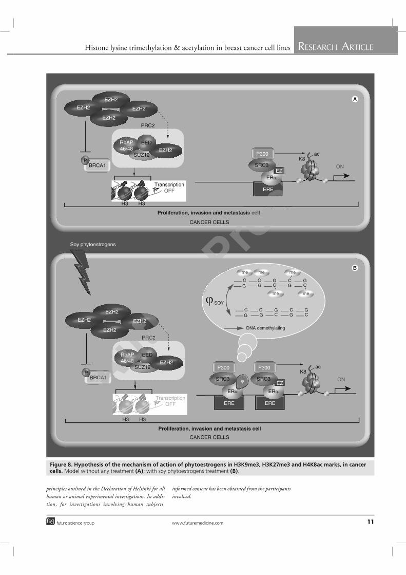
Histone lysine trimethylation & acetylation in breast cancer cell lines ReseaRch aRticle
www.futuremedicine.com 11future science group
princi ples outlined in the Declaration of Helsinki for all
human or animal experimental investigations. In addi-
tion, for investi gations involving human subjects,
informed consent has been obtained from the participants
involved.
ER
RbAP
46/48
EZH2
EZH2
EZH2
EZH2
EZH2
EED
SUZ12
PRC2
PBRCA1
RbAP
46/48
EZH2
EZH2
EZH2
EZH2
EZH2
EED
SUZ12
PRC2
PBRCA1
K27K27
XTranscription
OFF
XXXXXTranscription
OFF
Proliferation, invasion and metastasis cell
P300
SRC3EZ
ERE
K8ac
ON
K8ac
ON
CANCER CELLS
CANCER CELLS
H3 H3
Proliferation, invasion and metastasis cell
H3 H3
ER
P300
SRC3
ERE
ER
ERE
EZ
P300
SRC3
SOY
C
G G GC
C CCG G
C
G G GC
C CCG G
me me me
me me
DNA demethylating
Soy phytoestrogens
Figure 8. H pothesis of the mechanism of action of ph toestrogens in H3K9me3, H3K27me3 and H4K8ac marks, in cancer cells. Model without any treatment (A); with soy phytoestrogens treatment (B).
Auth
or Pro
of
Auth
or Pro
of
Auth
or Pro
of
Auth
or Pro
of
Auth
or Pro
of
Auth
or Pro
of
Auth
or Pro
of
Auth
or Pro
of
Auth
or Pro
of
Auth
or Pro
of
Auth
or Pro
of
Auth
or Pro
of
Auth
or Pro
of
Auth
or Pro
of
Auth
or Pro
of
Auth
or Pro
of
Auth
or Pro
of
Auth
or Pro
of
Auth
or Pro
of
Auth
or Pro
of
Auth
or Pro
of
Auth
or Pro
of
Auth
or Pro
of
Auth
or Pro
of
Auth
or Pro
of
Auth
or Pro
of
Auth
or Pro
of
Auth
or Pro
of
Auth
or Pro
of
Auth
or Pro
of
Auth
or Pro
of
Auth
or Pro
of
Auth
or Pro
of
Auth
or Pro
of
Auth
or Pro
of
Auth
or Pro
of
Auth
or Pro
of
Auth
or Pro
of
Auth
or Pro
of
Auth
or Pro
of
Auth
or Pro
of
Auth
or Pro
of
Auth
or Pro
of
Auth
or Pro
of
Auth
or Pro
of
Auth
or Pro
of
Auth
or Pro
of
Auth
or Pro
of
Auth
or Pro
of
Auth
or Pro
of
Auth
or Pro
of
Auth
or Pro
of
Auth
or Pro
of
Auth
or Pro
of
Auth
or Pro
of
Auth
or Pro
of
Auth
or Pro
of
Auth
or Pro
of
Auth
or Pro
of
Auth
or Pro
of
Auth
or Pro
of
Auth
or Pro
of
Auth
or Pro
of
Auth
or Pro
of
Auth
or Pro
of
Auth
or Pro
of
Auth
or Pro
of
Auth
or Pro
of
Auth
or Pro
of
Auth
or Pro
of
Auth
or Pro
of
Auth
or Pro
of
Auth
or Pro
of
Auth
or Pro
of
Auth
or Pro
of
Auth
or Pro
of
Auth
or Pro
of
Auth
or Pro
of
Auth
or Pro
of
Auth
or Pro
of
Auth
or Pro
of
Auth
or Pro
of
Auth
or Pro
of
Auth
or Pro
of
Auth
or Pro
of
Auth
or Pro
of
Auth
or Pro
of
Auth
or Pro
of
Auth
or Pro
of
Auth
or Pro
of
Auth
or Pro
of
Auth
or Pro
of
Auth
or Pro
of
Auth
or Pro
of
Auth
or Pro
of
Auth
or Pro
of
Auth
or Pro
of
Auth
or Pro
of
Auth
or Pro
of
Auth
or Pro
of
Auth
or Pro
of
Auth
or Pro
of
Auth
or Pro
of
Auth
or Pro
of
Auth
or Pro
of
Auth
or Pro
of
Auth
or Pro
of
Auth
or Pro
of
Auth
or Pro
of
Auth
or Pro
of
Auth
or Pro
of
Auth
or Pro
of
Auth
or Pro
of
Auth
or Pro
of
Auth
or Pro
of
Auth
or Pro
of
Auth
or Pro
of
Auth
or Pro
of
Auth
or Pro
of
Auth
or Pro
of
Auth
or Pro
of
Auth
or Pro
of
Auth
or Pro
of
Auth
or Pro
of
Auth
or Pro
of
Auth
or Pro
of
Auth
or Pro
of
Auth
or Pro
of
Auth
or Pro
of
Auth
or Pro
of
Auth
or Pro
of
Auth
or Pro
of
Auth
or Pro
of
Auth
or Pro
of
Auth
or Pro
of
Auth
or Pro
of
Auth
or Pro
of
Auth
or Pro
of
Auth
or Pro
of
Auth
or Pro
of
Auth
or Pro
of
Auth
or Pro
of
Auth
or Pro
of
Auth
or Pro
of
Auth
or Pro
of
Auth
or Pro
of
Auth
or Pro
of
Auth
or Pro
of
Auth
or Pro
of
Auth
or Pro
of
Auth
or Pro
of
Auth
or Pro
of
Auth
or Pro
of
Auth
or Pro
of
Auth
or Pro
of
Auth
or Pro
of
Auth
or Pro
of
Auth
or Pro
of
Auth
or Pro
of
Auth
or Pro
of
Auth
or Pro
of
Auth
or Pro
of
Auth
or Pro
of
Auth
or Pro
of
Auth
or Pro
of
Auth
or Pro
of
Auth
or Pro
of
Auth
or Pro
of
Auth
or Pro
of
Auth
or Pro
of
Auth
or Pro
of
Auth
or Pro
of
Auth
or Pro
of
Auth
or Pro
of
Auth
or Pro
of
Auth
or Pro
of
Auth
or Pro
of
Auth
or Pro
of
Auth
or Pro
of
Auth
or Pro
of
Auth
or Pro
of
Auth
or Pro
of
Auth
or Pro
of
Auth
or Pro
of
Auth
or Pro
of
Auth
or Pro
of
Auth
or Pro
of
Auth
or Pro
of
Auth
or Pro
of
Auth
or Pro
of
Auth
or Pro
of
Auth
or Pro
of
Auth
or Pro
of
Auth
or Pro
of
Auth
or Pro
of
Auth
or Pro
of
Auth
or Pro
of
Auth
or Pro
of
Auth
or Pro
of
Auth
or Pro
of
Auth
or Pro
of
Auth
or Pro
of
Auth
or Pro
of
Auth
or Pro
of
Auth
or Pro
of
Auth
or Pro
of
Auth
or Pro
of
Auth
or Pro
of
Auth
or Pro
of
Auth
or Pro
of
Auth
or Pro
of
Auth
or Pro
of
Auth
or Pro
of
Auth
or Pro
of
Auth
or Pro
of
Auth
or Pro
of
Auth
or Pro
of
Auth
or Pro
of
Auth
or Pro
of
Auth
or Pro
of
Auth
or Pro
of
Auth
or Pro
of
Auth
or Pro
of
Auth
or Pro
of
Auth
or Pro
of
Auth
or Pro
of
Auth
or Pro
of
Auth
or Pro
of
Auth
or Pro
of
Auth
or Pro
of
Auth
or Pro
of
Auth
or Pro
of
Auth
or Pro
of
Auth
or Pro
of
Auth
or Pro
of
Auth
or Pro
of
Auth
or Pro
of
Auth
or Pro
of
Auth
or Pro
of
Auth
or Pro
of
Auth
or Pro
of
Auth
or Pro
of
Auth
or Pro
of
Auth
or Pro
of
Auth
or Pro
of
Auth
or Pro
of
Auth
or Pro
of
Auth
or Pro
of
Auth
or Pro
of
Auth
or Pro
of
Auth
or Pro
of
Auth
or Pro
of
Auth
or Pro
of
Auth
or Pro
of
Auth
or Pro
of
Auth
or Pro
of
Auth
or Pro
of
Auth
or Pro
of
Auth
or Pro
of
Auth
or Pro
of
Auth
or Pro
of
Auth
or Pro
of
Auth
or Pro
of
Auth
or Pro
of
Auth
or Pro
of
Auth
or Pro
of
Auth
or Pro
of
Auth
or Pro
of
Auth
or Pro
of
Auth
or Pro
of
Auth
or Pro
of
Auth
or Pro
of
Auth
or Pro
of
Auth
or Pro
of
Auth
or Pro
of
Auth
or Pro
of
Auth
or Pro
of
Auth
or Pro
of
Auth
or Pro
of
Auth
or Pro
of
Auth
or Pro
of
Auth
or Pro
of
Auth
or Pro
of
Auth
or Pro
of
Auth
or Pro
of
Auth
or Pro
of
Auth
or Pro
of
Auth
or Pro
of
Auth
or Pro
of
Auth
or Pro
of
Auth
or Pro
of
Auth
or Pro
of
Auth
or Pro
of
Auth
or Pro
of
Auth
or Pro
of
Auth
or Pro
of
Auth
or Pro
of
Auth
or Pro
of
Auth
or Pro
of
Auth
or Pro
of
Auth
or Pro
of
Auth
or Pro
of
Auth
or Pro
of
Auth
or Pro
of
Auth
or Pro
of
Auth
or Pro
of
Auth
or Pro
of
Auth
or Pro
of
Auth
or Pro
of
Auth
or Pro
of
Auth
or Pro
of
Auth
or Pro
of
Auth
or Pro
of
Auth
or Pro
of
Auth
or Pro
of
Auth
or Pro
of
Auth
or Pro
of
Auth
or Pro
of
Auth
or Pro
of
Auth
or Pro
of
Auth
or Pro
of
Auth
or Pro
of
Auth
or Pro
of
Auth
or Pro
of
Auth
or Pro
of
Auth
or Pro
of
Auth
or Pro
of
Auth
or Pro
of
Auth
or Pro
of
Auth
or Pro
of
Auth
or Pro
of
Auth
or Pro
of
Auth
or Pro
of
Auth
or Pro
of
Auth
or Pro
of
Auth
or Pro
of
Auth
or Pro
of
Auth
or Pro
of
Auth
or Pro
of
Auth
or Pro
of
Auth
or Pro
of
Auth
or Pro
of
Auth
or Pro
of
Auth
or Pro
of
Auth
or Pro
of
Auth
or Pro
of
Auth
or Pro
of
Auth
or Pro
of
Auth
or Pro
of
Auth
or Pro
of
Auth
or Pro
of
Auth
or Pro
of
Auth
or Pro
of
Auth
or Pro
of
Auth
or Pro
of
Auth
or Pro
of
Auth
or Pro
of
Auth
or Pro
of
Auth
or Pro
of
Auth
or Pro
of
Auth
or Pro
of
Auth
or Pro
of
Auth
or Pro
of
Auth
or Pro
of
Auth
or Pro
of
Auth
or Pro
of
Auth
or Pro
of
Auth
or Pro
of
Auth
or Pro
of
Auth
or Pro
of
Auth
or Pro
of
Auth
or Pro
of
Auth
or Pro
of
Auth
or Pro
of
Auth
or Pro
of
Auth
or Pro
of
Auth
or Pro
of
Auth
or Pro
of
Auth
or Pro
of
Auth
or Pro
of
Auth
or Pro
of
Auth
or Pro
of
Auth
or Pro
of
Auth
or Pro
of
Auth
or Pro
of
Auth
or Pro
of
Auth
or Pro
of
Auth
or Pro
of
Auth
or Pro
of
Auth
or Pro
of
Auth
or Pro
of
Auth
or Pro
of
Auth
or Pro
of
Auth
or Pro
of
Auth
or Pro
of
Auth
or Pro
of
Auth
or Pro
of
Auth
or Pro
of
RbAPRbAPRbAPRbAPRbAPRbAPRbAPRbAPRbAPRbAPRbAPRbAPRbAPRbAPRbAPRbAPRbAPRbAPRbAPRbAPRbAPRbAPRbAPRbAPRbAPRbAPRbAPRbAPRbAPRbAPRbAPRbAPRbAPRbAPRbAPRbAPRbAPRbAPRbAPRbAPRbAPRbAPRbAPRbAPRbAPRbAPRbAPRbAPRbAPRbAPRbAPRbAP
46/4846/4846/4846/4846/4846/4846/4846/4846/4846/4846/4846/4846/4846/4846/4846/4846/4846/4846/4846/4846/4846/4846/4846/4846/4846/4846/4846/4846/4846/4846/4846/4846/4846/4846/4846/4846/4846/4846/4846/4846/4846/4846/4846/4846/4846/4846/4846/4846/4846/4846/4846/4846/4846/4846/4846/4846/4846/4846/4846/4846/4846/4846/4846/4846/4846/4846/4846/48
Auth
or Pro
of
Auth
or Pro
of
Auth
or Pro
of
Auth
or Pro
of
Auth
or Pro
of
Auth
or Pro
of
Auth
or Pro
of
Auth
or Pro
of
Auth
or Pro
of
Auth
or Pro
of
Auth
or Pro
of
Auth
or Pro
of
Auth
or Pro
of
Auth
or Pro
of
Auth
or Pro
of
Auth
or Pro
of
Auth
or Pro
of
Auth
or Pro
of
Auth
or Pro
of
Auth
or Pro
of
Auth
or Pro
of
Auth
or Pro
of
Auth
or Pro
of
Auth
or Pro
of
Auth
or Pro
of
Auth
or Pro
of
Auth
or Pro
of
Auth
or Pro
of
Auth
or Pro
of
Auth
or Pro
of
Auth
or Pro
of
Auth
or Pro
of
Auth
or Pro
of
Auth
or Pro
of
Auth
or Pro
of
Auth
or Pro
of
Auth
or Pro
of
Auth
or Pro
of
Auth
or Pro
of
Auth
or Pro
of
Auth
or Pro
of
Auth
or Pro
of
Auth
or Pro
of
Auth
or Pro
of
Auth
or Pro
of
Auth
or Pro
of
Auth
or Pro
of
Auth
or Pro
of
Auth
or Pro
of
Auth
or Pro
of
Auth
or Pro
of
Auth
or Pro
of
Auth
or Pro
of
Auth
or Pro
of
Auth
or Pro
of
Auth
or Pro
of
Auth
or Pro
of
Auth
or Pro
of
Auth
or Pro
of
Auth
or Pro
of
Auth
or Pro
of
Auth
or Pro
of
Auth
or Pro
of
Auth
or Pro
of
Auth
or Pro
of
Auth
or Pro
of
Auth
or Pro
of
Auth
or Pro
of
Auth
or Pro
of
Auth
or Pro
of
Auth
or Pro
of
Auth
or Pro
of
Auth
or Pro
of
Auth
or Pro
of
Auth
or Pro
of
Auth
or Pro
of
Auth
or Pro
of
Auth
or Pro
of
Auth
or Pro
of
Auth
or Pro
of
Auth
or Pro
of
Auth
or Pro
of
Auth
or Pro
of
Auth
or Pro
of
Auth
or Pro
of
Auth
or Pro
of
Auth
or Pro
of
Auth
or Pro
of
Auth
or Pro
of
Auth
or Pro
of
Auth
or Pro
of
Auth
or Pro
of
Auth
or Pro
of
Auth
or Pro
of
Auth
or Pro
of
Auth
or Pro
of
Auth
or Pro
of
Auth
or Pro
of
Auth
or Pro
of
Auth
or Pro
of
Auth
or Pro
of
Auth
or Pro
of
Auth
or Pro
of
Auth
or Pro
of
Auth
or Pro
of
Auth
or Pro
of
Auth
or Pro
of
Auth
or Pro
of
Auth
or Pro
of
Auth
or Pro
of
Auth
or Pro
of
Auth
or Pro
of
Auth
or Pro
of
Auth
or Pro
of
Auth
or Pro
of
Auth
or Pro
of
Auth
or Pro
of
Auth
or Pro
of
Auth
or Pro
of
Auth
or Pro
of
Auth
or Pro
of
Auth
or Pro
of
Auth
or Pro
of
Auth
or Pro
of
Auth
or Pro
of
Auth
or Pro
of
Auth
or Pro
of
Auth
or Pro
of
Auth
or Pro
of
Auth
or Pro
of
Auth
or Pro
of
Auth
or Pro
of
Auth
or Pro
of
Auth
or Pro
of
Auth
or Pro
of
Auth
or Pro
of
Auth
or Pro
of
Auth
or Pro
of
Auth
or Pro
of
Auth
or Pro
of
Auth
or Pro
of
Auth
or Pro
of
Auth
or Pro
of
Auth
or Pro
of
Auth
or Pro
of
Auth
or Pro
of
Auth
or Pro
of
Auth
or Pro
of
Auth
or Pro
of
Auth
or Pro
of
Auth
or Pro
of
Auth
or Pro
of
Auth
or Pro
of
Auth
or Pro
of
Auth
or Pro
of
Auth
or Pro
of
Auth
or Pro
of
Auth
or Pro
of
Auth
or Pro
of
EZH2EZH2EZH2EZH2EZH2EZH2EZH2EZH2EZH2EZH2EZH2EZH2EZH2EZH2EZH2EZH2EZH2EZH2EZH2EZH2EZH2EZH2EZH2EZH2EZH2EZH2EZH2EZH2EZH2EZH2EZH2EZH2EZH2EZH2EZH2EZH2EZH2EZH2EZH2EZH2EZH2EZH2EZH2EZH2EZH2EZH2EZH2EZH2EZH2EZH2EZH2EZH2EZH2EZH2EZH2EZH2EZH2EZH2EZH2EZH2EZH2EZH2EZH2EZH2EZH2EZH2EZH2EZH2
Auth
or Pro
of
Auth
or Pro
of
Auth
or Pro
of
Auth
or Pro
of
Auth
or Pro
of
Auth
or Pro
of
Auth
or Pro
of
Auth
or Pro
of
Auth
or Pro
of
Auth
or Pro
of
Auth
or Pro
of
Auth
or Pro
of
Auth
or Pro
of
Auth
or Pro
of
Auth
or Pro
of
Auth
or Pro
of
Auth
or Pro
of
Auth
or Pro
of
Auth
or Pro
of
Auth
or Pro
of
Auth
or Pro
of
Auth
or Pro
of
Auth
or Pro
of
Auth
or Pro
of
Auth
or Pro
of
Auth
or Pro
of
Auth
or Pro
of
Auth
or Pro
of
Auth
or Pro
of
Auth
or Pro
of
Auth
or Pro
of
Auth
or Pro
of
Auth
or Pro
of
Auth
or Pro
of
Auth
or Pro
of
Auth
or Pro
of
Auth
or Pro
of
Auth
or Pro
of
Auth
or Pro
of
Auth
or Pro
of
Auth
or Pro
of
Auth
or Pro
of
Auth
or Pro
of
Auth
or Pro
of
Auth
or Pro
of
Auth
or Pro
of
Auth
or Pro
of
Auth
or Pro
of
Auth
or Pro
of
Auth
or Pro
of
Auth
or Pro
of
Auth
or Pro
of
Auth
or Pro
of
Auth
or Pro
of
Auth
or Pro
of
Auth
or Pro
of
Auth
or Pro
of
Auth
or Pro
of
Auth
or Pro
of
Auth
or Pro
of
Auth
or Pro
of
Auth
or Pro
of
Auth
or Pro
of
Auth
or Pro
of
Auth
or Pro
of
Auth
or Pro
of
Auth
or Pro
of
Auth
or Pro
of
Auth
or Pro
of
Auth
or Pro
of
Auth
or Pro
of
Auth
or Pro
of
Auth
or Pro
of
Auth
or Pro
of
Auth
or Pro
of
Auth
or Pro
of
Auth
or Pro
of
Auth
or Pro
of
Auth
or Pro
of
Auth
or Pro
of
Auth
or Pro
of
Auth
or Pro
of
Auth
or Pro
of
Auth
or Pro
of
Auth
or Pro
of
Auth
or Pro
of
Auth
or Pro
of
Auth
or Pro
of
Auth
or Pro
of
Auth
or Pro
of
Auth
or Pro
of
Auth
or Pro
of
Auth
or Pro
of
Auth
or Pro
of
Auth
or Pro
of
Auth
or Pro
of
Auth
or Pro
of
Auth
or Pro
of
Auth
or Pro
of
Auth
or Pro
of
Auth
or Pro
of
Auth
or Pro
of
Auth
or Pro
of
Auth
or Pro
of
Auth
or Pro
of
Auth
or Pro
of
Auth
or Pro
of
Auth
or Pro
of
Auth
or Pro
of
Auth
or Pro
of
Auth
or Pro
of
Auth
or Pro
of
Auth
or Pro
of
Auth
or Pro
of
Auth
or Pro
of
Auth
or Pro
of
Auth
or Pro
of
Auth
or Pro
of
Auth
or Pro
of
Auth
or Pro
of
Auth
or Pro
of
Auth
or Pro
of
Auth
or Pro
of
Auth
or Pro
of
Auth
or Pro
of
Auth
or Pro
of
Auth
or Pro
of
Auth
or Pro
of
Auth
or Pro
of
Auth
or Pro
of
Auth
or Pro
of
Auth
or Pro
of
Auth
or Pro
of
Auth
or Pro
of
Auth
or Pro
of
Auth
or Pro
of
Auth
or Pro
of
Auth
or Pro
of
Auth
or Pro
of
Auth
or Pro
of
Auth
or Pro
of
Auth
or Pro
of
Auth
or Pro
of
Auth
or Pro
of
Auth
or Pro
of
Auth
or Pro
of
Auth
or Pro
of
Auth
or Pro
of
Auth
or Pro
of
Auth
or Pro
of
Auth
or Pro
of
Auth
or Pro
of
Auth
or Pro
of
Auth
or Pro
of
Auth
or Pro
of
Auth
or Pro
of
Auth
or Pro
of
Auth
or Pro
of
Auth
or Pro
of
Auth
or Pro
of
Auth
or Pro
of
Auth
or Pro
of
Auth
or Pro
of
Auth
or Pro
of
Auth
or Pro
of
Auth
or Pro
of
Auth
or Pro
of
Auth
or Pro
of
Auth
or Pro
of
Auth
or Pro
of
Auth
or Pro
of
Auth
or Pro
of
Auth
or Pro
of
Auth
or Pro
of
Auth
or Pro
of
Auth
or Pro
of
Auth
or Pro
of
Auth
or Pro
of
Auth
or Pro
of
Auth
or Pro
of
Auth
or Pro
of
Auth
or Pro
of
Auth
or Pro
of
Auth
or Pro
of
Auth
or Pro
of
Auth
or Pro
of
Auth
or Pro
of
Auth
or Pro
of
Auth
or Pro
of
Auth
or Pro
of
Auth
or Pro
of
Auth
or Pro
of
Auth
or Pro
of
Auth
or Pro
of
Auth
or Pro
of
Auth
or Pro
of
Auth
or Pro
of
Auth
or Pro
of
Auth
or Pro
of
Auth
or Pro
of
Auth
or Pro
of
Auth
or Pro
of
Auth
or Pro
of
Auth
or Pro
of
Auth
or Pro
of
Auth
or Pro
of
Auth
or Pro
of
Auth
or Pro
of
Auth
or Pro
of
Auth
or Pro
of
Auth
or Pro
of
Auth
or Pro
of
Auth
or Pro
of
Auth
or Pro
of
Auth
or Pro
of
Auth
or Pro
of
Auth
or Pro
of
Auth
or Pro
of
Auth
or Pro
of
Auth
or Pro
of
ERERERERERERERERERERERERERERERER
Auth
or Pro
of
Auth
or Pro
of
Auth
or Pro
of
Auth
or Pro
of
Auth
or Pro
of
Auth
or Pro
of
Auth
or Pro
of
Auth
or Pro
of
Auth
or Pro
of
Auth
or Pro
of
Auth
or Pro
of
Auth
or Pro
of
Auth
or Pro
of
Auth
or Pro
of
Auth
or Pro
of
Auth
or Pro
of
Auth
or Pro
of
Auth
or Pro
of
Auth
or Pro
of
Auth
or Pro
of
Auth
or Pro
of
Auth
or Pro
of
Auth
or Pro
of
Auth
or Pro
of
Auth
or Pro
of
Auth
or Pro
of
Auth
or Pro
of
Auth
or Pro
of
Auth
or Pro
of
Auth
or Pro
of
Auth
or Pro
of
Auth
or Pro
of
Auth
or Pro
of
Auth
or Pro
of
Auth
or Pro
of
Auth
or Pro
of
Auth
or Pro
of
Auth
or Pro
of
Auth
or Pro
of
Auth
or Pro
of
Auth
or Pro
of
Auth
or Pro
of
Auth
or Pro
of
Auth
or Pro
of
Auth
or Pro
of
Auth
or Pro
of
Auth
or Pro
of
Auth
or Pro
of
Auth
or Pro
of
Auth
or Pro
of
Auth
or Pro
of
Auth
or Pro
of
Auth
or Pro
of
Auth
or Pro
of
Auth
or Pro
of
Auth
or Pro
of
Auth
or Pro
of
Auth
or Pro
of
Auth
or Pro
of
Auth
or Pro
of
Auth
or Pro
of
Auth
or Pro
of
Auth
or Pro
of
Auth
or Pro
of
Auth
or Pro
of
Auth
or Pro
of
Auth
or Pro
of
Auth
or Pro
of
Auth
or Pro
of
Auth
or Pro
of
Auth
or Pro
of
Auth
or Pro
of
Auth
or Pro
of
Auth
or Pro
of
Auth
or Pro
of
Auth
or Pro
of
Auth
or Pro
of
Auth
or Pro
of
Auth
or Pro
of
Auth
or Pro
of
Auth
or Pro
of
Auth
or Pro
of
Auth
or Pro
of
Auth
or Pro
of
Auth
or Pro
of
Auth
or Pro
of
Auth
or Pro
of
Auth
or Pro
of
Auth
or Pro
of
Auth
or Pro
of
Auth
or Pro
of
Auth
or Pro
of
Auth
or Pro
of
Auth
or Pro
of
Auth
or Pro
of
Auth
or Pro
of
Auth
or Pro
of
Auth
or Pro
of
Auth
or Pro
of
Auth
or Pro
of
Auth
or Pro
of
Auth
or Pro
of
Auth
or Pro
of
Auth
or Pro
of
Auth
or Pro
of
Auth
or Pro
of
Auth
or Pro
of
Auth
or Pro
of
Auth
or Pro
of
Auth
or Pro
of
Auth
or Pro
of
Auth
or Pro
of
Auth
or Pro
of
Auth
or Pro
of
Auth
or Pro
of
Auth
or Pro
of
Auth
or Pro
of
Auth
or Pro
of
Auth
or Pro
of
Auth
or Pro
of
Auth
or Pro
of
Auth
or Pro
of
Auth
or Pro
of
Auth
or Pro
of
Auth
or Pro
of
Auth
or Pro
of
Auth
or Pro
of
Auth
or Pro
of
Auth
or Pro
of
Auth
or Pro
of
Auth
or Pro
of
Auth
or Pro
of
Auth
or Pro
of
Auth
or Pro
of
Auth
or Pro
of
Auth
or Pro
of
Auth
or Pro
of
Auth
or Pro
of
Auth
or Pro
of
Auth
or Pro
of
Auth
or Pro
of
Auth
or Pro
of
Auth
or Pro
of
Auth
or Pro
of
Auth
or Pro
of
EEDEEDEEDEEDEEDEEDEEDEEDEEDEEDEEDEEDEEDEEDEEDEEDEEDEEDEEDEEDEED
Auth
or Pro
of
Auth
or Pro
of
PRC2PRC2
PPPPPPPPPPPPPPPPPPPPPPP
Auth
or Pro
of
Auth
or Pro
of
BRCA1BRCA1Auth
or Pro
of
Proliferation, invasion and metastasis cellProliferation, invasion and metastasis cell
Auth
or Pro
of
Auth
or Pro
of
Auth
or Pro
of
Auth
or Pro
of
Auth
or Pro
of
Auth
or Pro
of
Auth
or Pro
of
Auth
or Pro
of
Auth
or Pro
of
Auth
or Pro
of
Auth
or Pro
of
Auth
or Pro
of
Auth
or Pro
of
Auth
or Pro
of
Auth
or Pro
of
Auth
or Pro
of
Auth
or Pro
of
Auth
or Pro
of
Auth
or Pro
of
Auth
or Pro
of
Auth
or Pro
of
Auth
or Pro
of
Auth
or Pro
of
Auth
or Pro
of
Auth
or Pro
of
Auth
or Pro
of
Auth
or Pro
of
Auth
or Pro
of
Auth
or Pro
of
Auth
or Pro
of
Auth
or Pro
of
Auth
or Pro
of
Auth
or Pro
of
Auth
or Pro
of
Auth
or Pro
of
Auth
or Pro
of
Auth
or Pro
of
Auth
or Pro
of
Auth
or Pro
of
Auth
or Pro
of
Auth
or Pro
of
Auth
or Pro
of
Auth
or Pro
of
Auth
or Pro
of
Auth
or Pro
of
Auth
or Pro
of
Auth
or Pro
of
Auth
or Pro
of
Auth
or Pro
of
Auth
or Pro
of
Auth
or Pro
of
Auth
or Pro
of
Auth
or Pro
of
Auth
or Pro
of
Auth
or Pro
of
Auth
or Pro
of
Auth
or Pro
of
Auth
or Pro
of
Auth
or Pro
of
Auth
or Pro
of
Auth
or Pro
of
Auth
or Pro
of
Auth
or Pro
of
Auth
or Pro
of
Auth
or Pro
of
Auth
or Pro
of
Auth
or Pro
of
Auth
or Pro
of
Auth
or Pro
of
Auth
or Pro
of
Auth
or Pro
of
Auth
or Pro
of
Auth
or Pro
of
Auth
or Pro
of
Auth
or Pro
of
Auth
or Pro
of
Auth
or Pro
of
Auth
or Pro
of
Auth
or Pro
of
Auth
or Pro
of
Auth
or Pro
of
Auth
or Pro
of
Auth
or Pro
of
Auth
or Pro
of
Auth
or Pro
of
Auth
or Pro
of
Auth
or Pro
of
Auth
or Pro
of
Auth
or Pro
of
Auth
or Pro
of
Auth
or Pro
of
Auth
or Pro
of
Auth
or Pro
of
Auth
or Pro
of
Auth
or Pro
of
Auth
or Pro
of
Auth
or Pro
of
Auth
or Pro
of
Auth
or Pro
of
Auth
or Pro
of
Auth
or Pro
of
Auth
or Pro
of
Auth
or Pro
of
Auth
or Pro
of
Auth
or Pro
of
Auth
or Pro
of
Auth
or Pro
of
Auth
or Pro
of
Auth
or Pro
of
Auth
or Pro
of
Auth
or Pro
of
Auth
or Pro
of
Auth
or Pro
of
Auth
or Pro
of
Auth
or Pro
of
Auth
or Pro
of
Auth
or Pro
of
Auth
or Pro
of
Auth
or Pro
of
Auth
or Pro
of
Auth
or Pro
of
Auth
or Pro
of
Auth
or Pro
of
Auth
or Pro
of
Auth
or Pro
of
Auth
or Pro
of
Auth
or Pro
of
Auth
or Pro
of
Auth
or Pro
of
Auth
or Pro
of
Auth
or Pro
of
Auth
or Pro
of
Auth
or Pro
of
Auth
or Pro
of
Auth
or Pro
of
Auth
or Pro
of
Auth
or Pro
of
Auth
or Pro
of
Auth
or Pro
of
EZEZ
EREERE
Auth
or Pro
of
Auth
or Pro
of
Auth
or Pro
of
Auth
or Pro
of
Auth
or Pro
of
Auth
or Pro
of
Auth
or Pro
of
Auth
or Pro
of
Auth
or Pro
of
Auth
or Pro
of
Auth
or Pro
of
Auth
or Pro
of
Auth
or Pro
of
Auth
or Pro
of
Auth
or Pro
of
Auth
or Pro
of
Auth
or Pro
of
Auth
or Pro
of
Auth
or Pro
of
Auth
or Pro
of
Auth
or Pro
of
Auth
or Pro
of
Auth
or Pro
of
Auth
or Pro
of
Auth
or Pro
of
Auth
or Pro
of
Auth
or Pro
of
Auth
or Pro
of
Auth
or Pro
of
Auth
or Pro
of
Auth
or Pro
of
Auth
or Pro
of
Auth
or Pro
of
Auth
or Pro
of
Auth
or Pro
of
Auth
or Pro
of
Auth
or Pro
of
Auth
or Pro
of
Auth
or Pro
of
Auth
or Pro
of
Auth
or Pro
of
Auth
or Pro
of
Auth
or Pro
of
Auth
or Pro
of
Auth
or Pro
of
Auth
or Pro
of
Auth
or Pro
of
Auth
or Pro
of
Auth
or Pro
of
Auth
or Pro
of
Auth
or Pro
of
Auth
or Pro
of
Auth
or Pro
of
Auth
or Pro
of
Auth
or Pro
of
Auth
or Pro
of
Auth
or Pro
of
Auth
or Pro
of
Auth
or Pro
of
Auth
or Pro
of
Auth
or Pro
of
Auth
or Pro
of
Auth
or Pro
of
Auth
or Pro
of
Auth
or Pro
of
Auth
or Pro
of
Auth
or Pro
of
Auth
or Pro
of
Auth
or Pro
of
Auth
or Pro
of
Auth
or Pro
of
Auth
or Pro
of
Auth
or Pro
of
Auth
or Pro
of
Auth
or Pro
of
Auth
or Pro
of
Auth
or Pro
of
Auth
or Pro
of
Auth
or Pro
of
Auth
or Pro
of
Auth
or Pro
of
Auth
or Pro
of
Auth
or Pro
of
Auth
or Pro
of
Auth
or Pro
of
Auth
or Pro
of
Auth
or Pro
of
Auth
or Pro
of
Auth
or Pro
of
Auth
or Pro
of
Auth
or Pro
of
Auth
or Pro
of
Auth
or Pro
of
Auth
or Pro
of
Auth
or Pro
of
Auth
or Pro
of
Auth
or Pro
of
Auth
or Pro
of
Auth
or Pro
of
Auth
or Pro
of
Auth
or Pro
of
Auth
or Pro
of
Auth
or Pro
of
Auth
or Pro
of
Auth
or Pro
of
Auth
or Pro
of
Auth
or Pro
of
Auth
or Pro
of
Auth
or Pro
of
Auth
or Pro
of
CANCER CELLS
Auth
or Pro
of
Auth
or Pro
of

ReseaRch aRticle Dagdemir, Durif, Ngollo, Bignon & Bernard Gallon
Epigenomics (2013) 5(1)12 future science group
ReferencesPapers of special note have been highlighted as:n of interestnn of considerable interest
1 Jemal A, Bray F, Center MM, Ferlay J,
Ward E, Forman D. Global cancer statistics.
CA Cancer J. Clin. 61(2), 69–90 (2011).
2 Kuiper GG, Lemmen JG, Carlsson B et al.
Interaction of estrogenic chemicals and
phytoestrogens with estrogen receptor beta.
Endocrinology 139(10), 4252–4263 (1998).
3 P tscher A, Reiter E, Jungbauer A. Receptor
binding and transactivation activities of red
clover iso avones and their metabolites.
J. Steroid Biochem. Mol. Biol. 112(1–3),
87–94 (2008).
4 Takeuchi S, Takahashi T, Sawada Y, Iida M,
Matsuda T, Kojima H. Comparative study on
the nuclear hormone receptor activity of
various phytochemicals and their metabolites
by reporter gene assays using Chinese hamster
ovary cells. Biol. Pharm. Bull. 32(2), 195–202
(2009).
5 Muthyala RS, Ju YH, Sheng S et al. Equol, a
natural estrogenic metabolite from soy
iso avones: convenient preparation and
resolution of R and S equols and their
differing binding and biological activity
through estrogen receptors alpha and beta.
Bioorg. Med. Chem. 12(6), 1559–1567 (2004).
6 Perou CM, Sorlie T, Eisen MB et al.
Molecular portraits of human breast tumours.
Nature 406(6797), 747–752 (2000).
7 Sorlie T, Perou CM, Tibshirani R et al. Gene
expression patterns of breast carcinomas
distinguish tumor subclasses with clinical
implications. Proc. Natl Acad. Sci. USA
98(19), 10869–10874 (2001).
8 Wood LD, Parsons DW, Jones S et al. The
genomic landscapes of human breast and
colorectal cancers. Science 318(5853),
1108–1113 (2007).
9 Bosviel R, Garcia S, Lavediaux G et al.
BRCA1 promoter methylation in peripheral
blood DNA was identi ed in sporadic breast
cancer and controls. Cancer Epidemiol. 36(3),
e177–e182 (2012).
10 Adjakly M, Bosviel R, Rabiau N et al. DNA
methylation and soy phytoestrogens:
quantitative study in DU 145 and PC 3
human prostate cancer cell lines. Epigenomics
3(6), 795–803 (2011).
11 Kurdistani SK. Histone modi cations as
markers of cancer prognosis: a cellular view.
Br. J. Cancer 97(1), 1–5 (2007).
12 Santos Rosa H, Caldas C. Chromatin
modi er enzymes, the histone code and
cancer. Eur. J. Cancer 41(16), 2381–2402
(2005).
13 Kouzarides T. Chromatin modi cations and
their function. Cell 128(4), 693–705 (2007).
14 Jenuwein T, Allis CD. Translating the histone
code. Science 293(5532), 1074–1080 (2001).
15 Bernstein BE, Kamal M, Lindblad Toh K
et al. Genomic maps and comparative analysis
of histone modi cations in human and
mouse. Cell 120(2), 169–181 (2005).
16 Pokholok DK, Harbison CT, Levine S et al.
Genome wide map of nucleosome acetylation
and methylation in yeast. Cell 122(4),
517–527 (2005).
17 Schneider R, Bannister AJ, Myers FA, Thorne
AW, Crane Robinson C, Kouzarides T.
Histone H3 lysine 4 methylation patterns in
higher eukaryotic genes. Nat. Cell Biol. 6(1),
73–77 (2004).
18 Yoo KH, Hennighausen L. EZH2
methyltransferase and H3K27 methylation in
breast cancer. Int. J. Biol. Sci. 8(1), 59–65
(2012).
19 Tang Y, Zhao W, Chen Y, Zhao Y, Gu W.
Acetylation is indispensable for p53
activation. Cell 133(4), 612–626 (2008).
20 Peterson CL, Laniel MA. Histones and
histone modi cations. Curr. Biol. 14(14),
R546–551 (2004).
21 Long W, Foulds CE, Qin J et al. ERK3
signals through SRC 3 coactivator to promote
human lung cancer cell invasion. J. Clin.
Invest. 122(5), 1869–1880 (2012).
22 Esteller M, Silva JM, Dominguez G et al.
Promoter hypermethylation and BRCA1
inactivation in sporadic breast and ovarian
tumors. J. Natl Cancer Inst. 92(7), 564–569
(2000).
23 Satih S, Rabiau N, Bignon YJ, Bernard
Gallon DJ. Soy phytoestrogens and breast
cancer chemoprevention: molecular
mechanisms. Curr. Nutr. Food Sci. 4,
259–264 (2008).
24 Cailleau R, Young R, Olive M, Reeves WJ Jr.
Breast tumor cell lines from pleural effusions.
J. Natl Cancer Inst. 53(3), 661–674 (1974).
25 Soule HD, Vazguez J, Long A, Albert S,
Brennan M. A human cell line from a pleural
effusion derived from a breast carcinoma.
J. Natl Cancer Inst. 51(5), 1409–1416 (1973).
26 Vissac Sabatier C, Bignon YJ, Bernard
Gallon DJ. Effects of the phytoestrogens
genistein and daidzein on BRCA2 tumor
suppressor gene expression in breast cell lines.
Nutr. Cancer 45(2), 247–255 (2003).
27 Yerbes R, Lopez Rivas A. Itch/AIP4
independent proteasomal degradation of
cFLIP induced by the histone deacetylase
inhibitor SAHA sensitizes breast tumour cells
to TRAIL. Invest. New Drugs 30(2), 541–547
(2012).
28 Pasqualini JR. The selective estrogen enzyme
modulators in breast cancer: a review.
Biochim. Biophys. Acta 1654(2), 123–143
(2004).
29 Hedlund TE, Johannes WU, Miller GJ. Soy
iso avonoid equol modulates the growth of
benign and malignant prostatic epithelial cells
in vitro. Prostate 54(1), 68–78 (2003).
30 Jackson RL, Greiwe JS, Desai PB, Schwen RJ.
Single dose and steady state pharmacokinetic
studies of S equol, a potent nonhormonal,
estrogen receptor beta agonist being
developed for the treatment of menopausal
symptoms. Menopause 18(2), 185–193 (2011).
31 Plomley JB, Jackson RL, Schwen RJ,
Greiwe JS. Development of chiral liquid
chromatography tandem mass spectrometry
isotope dilution methods for the
determination of unconjugated and total
S equol in human plasma and urine.
xecutive summar
Histone methylation or acetylation, an epigenetic event observed in cancer, occurs at lysine or arginine on histones.
This mechanism induces the opening or closing of chromatin and therefore gene enhancing or silencing.
In this study, an investigation of iso avones, 17b-estradiol and anti-HDAC (suberoylanilide hydroxamic acid) on EZH2, BRCA1, ERa, ERb,
SRC3 and P300 genes, was carried out by chromatin immunoprecipitation-qPCR.
Two breast cancer cell lines, MCF7 and MDA-MB 231, were treated with 18.5 µM genistein, 78.5 µM daidzein, 12.8 µM equol, 10 nM
17b-estradiol and 1 µM suberoylanilide hydroxamic acid (anti-HDAC) for 48 h.
Decrease trimethylation and increase acetylating marks were observed in cells treated with soy phytoestrogens compared with
untreated cells.
Soy phytoestrogens tend to modify the transcription through the demethylation and acetylation of the histones in breast cancer cell
lines.
Auth
or Pro
of
various phytochemicals and their metabolites
by reporter gene assays using Chinese hamster by reporter gene assays using Chinese hamster
32(2), 195–202 32(2), 195–202
et al. Equol, a Equol, a
natural estrogenic metabolite from soy natural estrogenic metabolite from soy
avones: convenient preparation and avones: convenient preparation and
S equols and their equols and their
differing binding and biological activity differing binding and biological activity
through estrogen receptors alpha and beta. through estrogen receptors alpha and beta.
12(6), 1559–1567 (2004).12(6), 1559–1567 (2004).
et al. et al.
Epigenomics
cations as
markers of cancer prognosis: a cellular view. markers of cancer prognosis: a cellular view.
97(1), 1–5 (2007).97(1), 1–5 (2007).
Rosa H, Caldas C. Chromatin Rosa H, Caldas C. Chromatin
er enzymes, the histone code and er enzymes, the histone code and
Eur. J. Cancer 41(16), 2381–2402 41(16), 2381–2402
Kouzarides T. Chromatin modiKouzarides T. Chromatin modi cations and cations and
their function. Cell Cell 128(4), 693–705 (2007).128(4), 693–705 (2007).
Jenuwein T, Allis CD. Translating the histone Jenuwein T, Allis CD. Translating the histone
code. Science Science 293(5532), 1074–1080 (2001).293(5532), 1074–1080 (2001).
15 Bernstein BE, Kamal M, LindbladBernstein BE, Kamal M, Lindblad
et al.et al. Genomic maps and comparative analysis Genomic maps and comparative analysis
of histone modiof histone modi
mouse. mouse. Cell Cell
16 16 Pokholok DK, Harbison CT, Levine SPokholok DK, Harbison CT, Levine S
GenomeGenome
and methylation in yeast. and methylation in yeast.
Auth
or Pro
of
Promoter hypermethylation and BRCA1 Promoter hypermethylation and BRCA1
inactivation in sporadic breast and ovarian inactivation in sporadic breast and ovarian
tumors. tumors. J. Natl Cancer Inst. J. Natl Cancer Inst.
(2000).(2000).
23 Satih S, Rabiau N, Bignon YJ, BernardSatih S, Rabiau N, Bignon YJ, Bernard
Gallon DJ. Soy phytoestrogens and breast Gallon DJ. Soy phytoestrogens and breast
cancer chemoprevention: molecular cancer chemoprevention: molecular
mechanisms. mechanisms.
259–264 (2008).259–264 (2008).
24

Histone lysine trimethylation & acetylation in breast cancer cell lines ReseaRch aRticleReseaRch aRticle
www.futuremedicine.com 13future science group
J. Pharm. Biomed. Anal. 55(1), 125–134
(2011).
32 Esteller M. Cancer epigenomics: DNA
methylomes and histone modi cation maps.
Nat. Rev. Genet. 8(4), 286–298 (2007).
33 Volkel P, Angrand PO. The control of histone
lysine methylation in epigenetic regulation.
Biochimie 89(1), 1–20 (2007).
34 Wolff MS, Weston A. Breast cancer risk and
environmental exposures. Environ. Health
Perspect. 105(Suppl. 4), 891–896 (1997).
35 Wu AH, Yu MC, Tseng CC, Pike MC.
Epidemiology of soy exposures and breast
cancer risk. Br. J. Cancer 98(1), 9–14 (2008).
36 Trock BJ, Hilakivi Clarke L, Clarke R. Meta
analysis of soy intake and breast cancer risk.
J. Natl Cancer Inst. 98(7), 459–471 (2006).
37 Keinan Boker L, van Der Schouw YT,
Grobbee DE, Peeters PH. Dietary
phytoestrogens and breast cancer risk. Am.
J. Clin. Nutr. 79(2), 282–288 (2004).
38 Kondo Y, Shen L, Cheng AS, Ahmed S,
Boumber Y, Charo C et al. Gene silencing in
cancer by histone H3 lysine 27 trimethylation
independent of promoter DNA methylation.
Nat. Genet. 40(6), 741–750 (2008).
39 Bosviel R, Dumollard E, Dechelotte P,
Bignon YJ, Bernard Gallon D. Can soy
phytoestrogens decrease DNA methylation in
BRCA1 and BRCA2 oncosuppressor genes in
breast cancer? OMICS 16(5), 235–244
(2012).
40 King Batoon A, Leszczynska JM, Klein CB.
Modulation of gene methylation by genistein
or lycopene in breast cancer cells. Environ.
Mol. Mutagen. 49(1), 36–45 (2008).
41 Bosviel R, Durif J, Dechelotte P, Bignon YJ,
Bernard Gallon D. Epigenetic modulation of
BRCA1 and BRCA2 gene expression by equol
in breast cancer cell lines. Br. J. Nutr. 108(7),
1187–1193 (2012).
42 Fang M, Chen D, Yang CS. Dietary
polyphenols may affect DNA methylation.
J. Nutr. 137(Suppl. 1), 223S 228S (2007).
43 Hong T, Nakagawa T, Pan W, Kim MY,
Kraus WL, Ikehara T et al. Iso avones
stimulate estrogen receptor mediated core
histone acetylation. Biochem. Biophys. Res.
Commun. 317(1), 259–264 (2004).
44 Zwart W, Theodorou V, Kok M, Canisius S,
Linn S, Carroll JS. Oestrogen receptor co
factor chromatin speci city in the
transcriptional regulation of breast cancer.
EMBO J. 30(23), 4764–4776 (2011).
45 Nadal Serrano M, Sastre Serra J, Pons DG,
Miro AM, Oliver J, Roca P. The
ERalpha/ERbeta ratio determines oxidative
stress in breast cancer cell lines in response to
17beta estradiol. J. Cell Biochem. 113(10),
3178–3185 (2012).
46 Strom A, Hartman J, Foster JS, Kietz S,
Wimalasena J, Gustafsson JA. Estrogen
receptor beta inhibits 17beta estradiol
stimulated proliferation of the breast cancer
cell line T47D. Proc. Natl Acad. Sci. USA
101(6), 1566–1571 (2004).
Auth
or Pro
of gene expression by equol
108(7),
Fang M, Chen D, Yang CS. Dietary
polyphenols may affect DNA methylation. polyphenols may affect DNA methylation.
137(Suppl. 1), 223S 228S (2007).228S (2007).
stress in breast cancer cell lines in response to stress in breast cancer cell lines in response to
17beta17beta
3178–3185 (2012).3178–3185 (2012).
46 46 Strom A, Hartman J, Foster JS, Kietz S, Strom A, Hartman J, Foster JS, Kietz S,
Wimalasena J, Gustafsson JA. Estrogen Wimalasena J, Gustafsson JA. Estrogen
receptor beta inhibits 17betareceptor beta inhibits 17beta
stimulated proliferation of the breast cancer stimulated proliferation of the breast cancer
cell line T47D. cell line T47D.

48
Conclusion of Publication 4
We assessed the effects of soy phytoestrogens, 17 -estradiol and SAHA for selected
histone modifications in breast cancer cell lines using chromatin immunoprecipitation and
immunohistochemistry. We determined the relative levels of five modified histones, including
H3K9me3, H3K27me3, H3K4me3, H4K8ac, and H3K4ac in MCF7 and MDA-MB 231 in
breast cancer cell lines.
In cancer cells treated by soy phytoestrogens the level of H3K27me3 decreased
through polycomb complex formation and the treatment increased the establishment of the
H4K8ac mark via transcriptional co-activator such as P300, SRC3 and activates transcription.

49
B. Epigenetic Therapy in Breast Cancer Cell Lines
Presentation of Publication 5
Dagdemir A, Karsli-Ceppioglu S, Judes G, Lebert A, Echegut M, Ngollo M, Penault-
Llorca F, Bignon YJ, Bernard-Gallon D. What are the Effects of Histone Methylation
Inhibitor and/or Histone Deacetylase Inhibitor on Histone Lysine Trimethylation or
Acetylation in Breast Cancer Cell Lines? (2014, submitted).
The aim of this study was an understanding of the latest ‘epi-drug’ discoveries
focusing on the chemical characterization and use of epigenetic modulators in pre-clinical and
clinical settings against breast cancer. We know that histone modifications are major
epigenetic modifications and several enzymes are responsible in this mechanism. For
example, the dynamic process of histone acetylation has been linked to gene transcription, and
histone deacetylation has been related to inactive chromatin. Under physiological conditions,
chromatin acetylation is regulated by the balanced action of histone acetyltransferases (HATs)
and deacetylases (HDACs).
For this reason, we investigated two HDAC inhibitors which are NaBu and SAHA;
and a HMT inhibitor which is DZNep. We focused on the chemical aspects of such
molecules, joined to their effective (or potential) application in breast cancer therapy.

1
What are the Effects of the Histone Methylation Inhibitor and/or the
Histone Deacetylase Inh b tor on Histone Lysine Trimethylation or
Acetylation in Breast Cancer Cell Lines?
Aslihan Dagdemir 1,2 , Seher Karsli-Ceppioglu1,2,3 , Gaëlle Judes1,2 , André Lebert4, Maureen
Echegut 1,2 , Amaury Pajon 1,2, Marjolaine Ngollo 1,2, Frédérique Penault-Llorca 2,5, Yves-Jean
Bignon 1,2*, Dominique Bernard-Gallon 1,2
1Department of Oncogenetics, Centre Jean Perrin, CBRV, 63001, Clermont-Ferrand, France, 2EA
4677 "ERTICA", University of Auvergne, 63011, Clermont-Ferrand, France, 3Department of
Toxicology, Faculty of Pharmacy, Marmara University, 34668, Istanbul, Turkey, 4University Blaise
Pascal, Institut Pascal UMR 6602 CNRS/UBP, 63177, Aubière, France,5Centre Jean Perrin,
Laboratory of Anatomopathology, 63011, Clermont-Ferrand, France.
*Correspondence to: Yves-Jean Bignon, Department of Oncogenetics, Centre Jean Perrin, CBRV,
28 place Henri Dunant, BP 38, 63001 Clermont-Ferrand, France
E-mail: [email protected]

2
Abstract
The Histone Deacetylase Inhibitors (HDACi) and Histone Methylation Inhibitors (HMTi) are known
to interact with epigenetic modifications. In our study we used two breast cancer cell lines MCF-7
and MDA-MB-231, treated with 3-Deazaneplanocin A hydrochloride (DZNep) [5 µM], Sodium
Butyrate (NaBu) [2 mM] and Suberoylanilide Hydroxamic acid (SAHA) [1 M] for 48 hours. We
used a culture control with untreated cells. Firstly, we applied chromatin immunoprecipitation (ChIP)
coupled with QPCR. This was to follow the impact of HDACis and HMTi on H3K27me3, H3K9ac
and H3K4ac marks, and we selected EZH2, BRCA1, ER , ER , SRC3, P300 genes for analysis.
Secondly, we examined Western Blot with correspondent proteins including PGR and RNA analyses
also.
HDACi induced a decrease of trimethylated marks and activates of acetylating marks studied with
selected genes. SAHA and NaBu increased acetylation at lysine 4 of histone H3 in MCF7 and
DZNep decreased on H3K27me3 marks a slight difference in MDA-MB-231. Moreover we found
that by Western blot analysis; DZNep acted as a global inhibitor of histone methylation and it was
not selective to BRCA1 for each cell line. Finally, when we investigated mRNA expression levels,
DZNep acted globally and it could inhibit both activating and repressive histone marks, conversely
NaBu and SAHA were increasing of all mRNA expression especially in MCF 7.
These results suggested of HDACi and HMTi have beneficial effects on the genes, which are
upregulated in cancer; however an adverse effects for downregulated genes, so the action did not
seem to be specific for genes. Our results add a new outlook of complication to epigenetic
dysregulation in cancer and also established histone modifications-mediated silencing or enhancing
for up-and-coming therapeutic target. Our data demonstrate that HDACi disposed to modify the
transcription in the demethylation and acetylation of the histones in breast cancer cell lines.
Key words
Breast cancer, HMTi, HDACi, Histone Methylation, Histone Acetylation.

3
Introduction
Breast cancer is a heterogeneous disease and it can impressionable by way of multi-factorial
processes and it is also a hormone-dependent cancer. Previously, many studies have shown that
breast cancer and epigenetic mechanisms have a very powerful interactive relation. In recent times,
epigenetic modifications like an abnormal methylation and acetylation of genes and histones have
been shown to play a critical role in breast cancer development. With this information, our study
focuses our investigation on this issue.
Histone methylation or acetylation is an epigenetic event observed in cancer. It occurs at lysine or
arginine on histones. This mechanism induces the opening or closing of chromatin, thus gene
enhancing or silencing. Breast cancer is a heterogeneous disease organizing from premalignant
hyperproliferation to incursive and metastatic carcinoma [1]. Chromatin-modifying enzymes are very
important for in gene regulation by catalyzing reversible post-translational modifications of histones
like lysine acetylation and methylation [2]. This histone modifications and other modifications
generate a synthesis of histone code that separate chromatin regions for transcription activation or
repression [3].
Histone deacetylases (HDAC) can catalyze the hydrolysis of acetyl groups on lysine residues of
histones, cause the wrapped and packing of chromosomal DNA around histones, and then regulate
gene expression. Deacetylation of histones, catalyzed by HDACs, is also reported to be associated
with gene silencing. In large group of HDAC there are 18 members classified into four groups. Class
I includes HDAC1, HDAC2, HDAC 3 and HDAC 8. Class II HDACs occurs two subtype HDACs
group; Class IIa comprises HDAC 4, HDAC 5, HDAC 7, HDAC 9 and Class IIb HDACs composed
HDAC 6 and HDAC 10. Class III uses a different mechanism of action as a call like NAD+-
dependent proteins; SIRT1, SIRT2, SIRT3, SIRT4, SIRT5, SIRT6 and SIRT7. The last group of
HDAC is Class IV and it involved HDAC11 [4,5]. Inhibitors of histone deacetylase (HDACi) de-
repress genes that afterwards developed in evolution the defense mechanism, differentiation and

4
apoptosis of cancer cells. This is especially true for HDAC1, HDAC2, HDAC3, HDAC4 and
HDAC6 associated with cancer cell proliferation. For instance, the role of HDAC3 is inducing G2/M
arrest and also HDAC1 and HDAC2 downregulation leads to p21Waf1/Cip1 upregulation that induce
apoptosis. HDACi is a significant group of anti-tumor agents as a future function in the treatment of
breast cancer. According to a recent study which was carried out in Estrogen (ER)-positive cells,
treatment with HDACi has been identified with a transcriptional down-regulation of ER and its
response genes [6]. In ER-negative cell lines, HDACi demonstrated the reconstruction of ER
expression. Furthermore, HDACi has been reported to adjust the progesterone receptor [7]. To date,
seven groups of HDACis have been reported; short-chain fatty acids, hydroxamic acids, benzamides,
cyclic tetrapeptides containing a 2-amino-8-oxo-9, 10-epoxy-decanoyl (AOE) moiety, cyclic
peptides without the AOE moiety, epoxides and psammaplins [8].
Histone methylation is mediated through histone methyltransferases (HMTs) and it appears mono-,
di- or tri- methylated at lysine or arginine [9]. Many HMTs (including histone lysine
methytransferase (HKMT) and histone arginine methyletransferase (HRMT)) effected with estrogen
coactivators are involved with breast cancer [10]. Recently, there are a lot of studies concerning
HKMTs because it can be an activator or repressor of gene expression, depending on the position of
the modified residue. The Polycombgroup (PcG) proteins are in charge of maintaining the inactive
expression of many genes. In the last days, three different polycomb repressive complexes have been
identified: polycomb repressive complex 1 (PRC1), polycomb repressive complex 2 (PRC2), and
pleiohomeotic-repressive complex (PhoRC) [11].
PRC1 and PRC2 are related to chromatin contraction. This is especially true for PRC1 which
catalyzes the monoubiquitylation of H2A. On the other hand, PRC2 provides the methylation of
H3K27. PRC2 is certainly important in the midst of PcG proteins because it has connected with stem
cell biology and cancer. PRC2 multi-protein complexes include 4 core proteins. Enhancer of zeste
homolog 2 (EZH2) is the most important subunit of the PRC2 and also PRC2 comprises Suppressor

5
of Zeste 12 Homolog (SUZ12), Embryonic Ectoderm Development (EED) and Retinoblastoma
binding protein 4 (RbAp46/48). EZH2 acts as an HKMTase [12].
There are a lot of modifications that occur on the histone N-terminal region in a manner like
acetylation, methylation, ubiquitination or like a phosphorylation. Primarily, the targets of HMTs are
K4, K9, K27 and K36 on the H3. Recent studies have shown that overexpression of EZH2 is
associated with breast cancer [13]. We have chosen that gene for its histone methylase acts,
specifically at lysine 27 of histone 3. There are 3 transcription factors that assist in the regulation of
EZH2. The first factor is E2F; the target of the retinoblastoma protein (pRb). It has a crucial role in
cell cycle process, especially activating genes that control for S phase in cell cycle. The other
transcription factor is the HIF response elements (HRE) which are controlled EZH2 expression
levels in gene promoter. Once and for all, MEK/ERk/Elk pathway provides to EZH2 overexpression
in ERBB2-overexpressing breast cancer cell line [14].
Previous study has shown that mutation in BRCA1 causes of increase EZH2 concentration
particularly in the basal like breast carcinomas [15]. Generally EZH2 is overexpressed in ER-
negative breast cancer and inhibits BRCA1 phosphorylation. For this reason, transmission from G2
to M is accelerated in cell cycle and increased cell proliferation.
DZNep expend the cellular levels of EZH2 and inhibits the associated histone H3 lysine 27
trimethylation. Recent studies have shown that DZNep inhibits cell proliferation by inducing G1
arrest and apoptosis. DZNep repressed S-adenosylhomocysteine hydrolase; a factor of metionin
cycle, resulting in increasing of the S-adenosylhomocysteine hydrolase repression with a knock-on
interruption of methylation of substrates by EZH2 [16,17] (Figure1).
Suberoylanilide hydroxamic acid (SAHA) and Sodium Butyrate (NaBu) are commonly used as a
HDACi [18]. SAHA shows strong anti-proliferative effects on various cancer cell lines and is
currently in clinical trial for the treatment of solid and hematological tumors [19]. However, the

6
mechanism by which SAHA induces autophagy cell death in acquired endocrine-therapy is not
clearly understood.
NaBu is sodium salt of butyric acid. It is short-chain fatty acid that aims at the activated region of
zinc in HDAC. It has a very short half-life [20]. It has many effects like an inhibition of proliferation,
induction or repression of gene expression and induction of differentiation [21]. Particularly, NaBu
has been a necessary instrument for identifying the role of histone acetylation in chromatin structure
[22]. Previous study has shown that it affected the expression of only 2% of mammalian genes by
inhibiting the HDAC activity [23]. Inhibition of HDAC1 and HDAC2 with NaBu, P300 activities
increase because HAT continued, therefore hyperacetylation of the histones and induction of
P21Waf/Cip1 gene expression. This gene plays key role in G1/S phase of the cell cycle (Figure 1).
P300 is a member of the mammalian histone acetyl transferase (HAT) family. HAT acts are
activating the induce of apoptosis in a breast cancer cell line [24]. EZH2 and P300 genes, chromatin
–modifying enzymes, implicated in gene regulation by catalyzing reversible post-translational
modifications of histones [14].
Steroid receptor coactivator-3 (SRC-3), a transcription coactivator, is frequently overexpressed in
breast cancer [25]. We chose that gene due to function of the coactivator for HAT [26]. BRCA1 is a
tumor suppressor gene and transcription factor. Also, it has been shown that before BRCA1 inhibits
ligand-induced ER signaling and blocks subsequent ER transcriptional activation [27]. In many
breast cancers, estrogen-mediated signaling is a very important function of adjusting to evolution and
endurance signaling pathways and can directly drive tumorgenesis by leading the expression of
genes. Some authors have reported the ER is down-regulated in breast tumorigenesis and another
recent study has shown the regulation of ER expression by promoter methylation [28]. In vitro
studies point out ER plays a role in the modulator of proliferation and invasion of breast cancer
cells, so the loss of ER expression could be one of the reasons for breast cancer development [29].
In addition to these 6 genes, we decided to add Progesterone Growth Factor gene (PGR) in

7
accordance with previous study which has shown that; progesterone uses its effects mainly via
estrogen-dependent PGR, the expression of progesterone effects may be dominated by the primer
effect of estrogen [30].
It becomes necessary to investigate epigenetic alterations in breast cancers to describe new
prognostic markers and therapeutic targets. For these reasons, we can determine the effects of HMTi
(DZNep) and HDACi (SAHA and NaBu) in cell lines on selected histone modifications.
Materials and Methods
Cell culture and cell treatment
We chose two breast cancer cell lines MCF7 and MDA-MB 231 derived from a pleural effusion
breast adenocarcinoma. The MCF-7 cell line an estrogen receptor (ER) positive control cell line that
we selected for this study [31]. On the other hand, we used MDA-MB-231 cells as a contrast to
MCF7 because MDA-MB-231 human breast cancer cell line is a model of ER-negative breast
cancers for both ER isoforms [32].
All cell lines supplied by the American Type Culture Collection. MCF7 were cultured in Roswell
Park Memorial Institute (RPMI)-1640 medium supplemented with 20 µg/ml of L-Glutamine
(Invitrogen), Gentamycin (20 g/ml; Panpharma), 10% fetal bovine serum (Invitrogen) and Insulin
(1-4 mg/ml; Novo Nordisk) in a humidified atmosphere at 37°C containing 5% CO2. MDA-MB-231
cells were cultured in Leibovitz L-15 medium with 15% fetal bovine serum (Invitrogen), gentamycin
(20 g/ml; Panpharma) and 20 µg/ml of L-glutamine in a 37°C humidified atmosphere without CO2.
Cells (1x10⁶ per T75 flask) were cultured in the medium and treated with 5 µM DZNep, 2 mM
NaBu and 1 M SAHA provided by the Sigma-Aldrich and dissolved them successively in water,
ethanol and dimethyl sulfoxide; all the concentrations have already been determined by our previous

8
studies [33], [34] . For controls, each cell line was conditioned in the medium without any treatment,
during 48 hours.
ChIP Assay
All the cell lines were trypsinized and counted by Millipore Scepter 2.0 Cell (Fisher Scientific) in the
event of 80% confluence. Before the cross linking, we used 1x10⁶ cells for each treatment. A 36.5%
concentration of formaldehyde (Sigma-Aldrich) was diluted to 1% in the culture media after that
cells were incubated 8 minutes at room temperature for fixation. To finish the cross-linked, we added
1.25 M Glycine and incubated it for 5 minutes at room temperature. Afterwards, all chromatin
preparations and ChIP reactions were handled at 4°C [33].
After the cross-linked, cells were washed with a PBS-Inhibitor and cell membranes were lysed by
HighCell ChIP Kit (Diagenode). Finally, the cross-linked cells were ready for chromatin shearing.
The chromatin was prepared in special tubes provided by the Diagenode (TPX tubes) with shearing
buffer (S1) and 1X volume protease inhibitor (Diagenode). The samples were sonicated by
Bioruptor® Standard, which was provided by the Diagenode, for 5 runs of 5 cycles. Each cycle
contained all 30 seconds “ON” and 30 seconds “OFF” at 200 Watt, and were held at 4°C at all times
during the sonication process. Between each run, samples were vortexed after a short spin. The
sheared chromatin could be frozen at -80°C for later use [33].
ChIP reactions were performed on SX-8G IP-Star® Compact Automated System supplied by
Diagenode. ChIP experiments were applied with anti-H3K27me3 (pAb-069-050, Diagenode), anti-
H3K9ac (pAb-103-050, Diagenode), anti-H3K4ac (pAb-165-050, Diagenode)] and non-immune
rabbit IgG (Kch-504-250, Diagenode) (e.g. negative control). ChIP reactions were created 200 µl
volumes each samples. All Auto-ChIP samples were exerted Auto Histone ChIP-seq kit reagents and
included 1µg of input chromatin. Reaction incubated for 2h for Antibodies-coating with protein A-
coated magnetic beads, and 10h for IP reactions for 8 strips, at 4°C [33].

9
Quantitative Real-Time PCR (Q-PCR)
After the ChIP reactions, a Quantitative Real-Time PCR (Q-PCR) was performed in triplicate. We
used a ninety-six-well optical tray with optical adhesive film. Two control genes: C-FOS (pp-1004-
500, Diagenode), which was the positive control for the acetylation marks, and TSH2B (pp-1041-
500, Diagenode), which was positive control for the methylation marks, was used. Final reaction
volume was 25 µl and it contained 5 µl DNA IP (e.g immunoprecipited DNA) or DNA Input (e.g
Total DNA), 1X SYBR Green Supermix (Applied Biosystem) and 200 nM each primer of control
genes (C-FOS- TSH2B). For the other genes (EZH2, BRCA1, ER , ER , SRC3, P300), we used
Taqman Universal PCR Master Mix at 1X, 400 nM each with forward and reverse primers and 250
nM of the probe for each gene. Initial denaturation was at 95°C for 10 min to activate DNA
polymerase. This was followed by 45 cycles of denaturation at 95°C for 15 sec and annealing and
extension at 60°C for 1 min. Q-PCR was performed using 7900HT, Real-Time PCR System from
Applied Biosystems. Primer and probe sequences were selected by Primer Express software (ABI)
(Table 1).
The efficiency of chromatin immunoprecipitation of particular genomic locus was calculated by
qPCR data and reported as a percentage of starting material:
%(ChIP/Total Input) = 2^[(Ct(x%input) – log(x%)/log2) – Ct(ChIP)]x 100%.
Western Blot
After the Q-PCR we examined the proteins for each cell line by Western Blot. MCF-7 and MDA-
MB-231 cells treated with DZNep, NaBu and SAHA were collected after 48 hours. All the protein
was extracted after cell lysis. Lysis buffer contained 20 mM Tris (pH 8), 50 mM EDTA, 0.8% NaCl,
0.1% Triton X-100 and 1% Glycerol. Protease Inhibitor Cocktail (Sigma) (1% Protease inhibitors)
and Phosphatase Inhibitor Cocktail 2 (Sigma) (1% phosphatase inhibitors) were added to the basic

10
buffer. Protein was quantified using the Bradford Method. Twenty five microgram proteins were
loaded onto 10% gels (for Anti BRCA1 and Anti P300 we used 4% gels, which are heavier than the
others) for SDS-PAGE and electrophoresed. When proteins were separated, they were transferred to
a polyvinylidene difluoride (PVDF) membrane. After one-hour, blocking in Tris Buffered Saline
Tween 0.1% (TBST) containing 5% milk membranes were incubated overnight at 4°C respectively
with Anti BRCA1 (1:50 [Ab-1] monoclonal Calbiochem), Anti P300 (1 :100 [N-15] polyclonal Santa
Cruz Biotechnology), Anti SRC-3 (1:500 NCOA3 [N1N2] polyclonal Gene Tex), Anti PGR (1:250
monoclonal ABNOVA), Anti EZH2 (1:1000 monoclonal MILLIPORE), Anti ER- (1:500 [F-10]
polyclonal Santa Cruz Biotechnology, 1:500 monoclonal [HC-20] Santa Cruz Biotechnologie), Anti
ER- (1:500 [H-150] polyclonal Santa Cruz Biotechnology, 1:500 polyclonal ZYMED) and Anti-
actin (1:120,000 Mouse [Ab-1], Calbiochem) antibodies. Membranes were washed three times with
TBST and incubated for one hour with alkaline phosphatase-conjugated secondary antibody (1:2000
anti-mouse IgG (H&L) AP conjugate or 1:2000 anti-rabbit IgG (Fc) AP conjugate, Promega).
Detection was performed with Western Blue® Stabilized Substrate for Alkaline Phosphatase
produced by Promega. Relative quantification of immunoblotted proteins was achieved using the
Bio-Rad Quantity One software with local background subtraction method. Membranes were re-
probed with actin antibody as a loading control.
Real-time quantitative RT-PCR
Finally, we applied Reverse Transcription and RT-QPCR on the RNA. To begin, we did an RNA
extraction. For this, after 48 hours of treatment, each of the cell lines were washed three times with
phosphate buffered saline (PBS). Afterwards, the cells were lysed in 5 mL of RNA B™ , according
to manufacturer's protocol of BioProbe Systems. All the RNA samples were verified using a
NanoDrop ND-8000 Spectrophotometer. The samples were then kept in liquid nitrogen.

11
Thereafter, 25 g of total RNA were reverse transcribed in a total volume of 15 µl using the First-
Strand DNA Synthesis Kit according to the manufacturer’s protocol (GE Healthcare Life Science).
Reverse transcriptase was inactivated for 10 minutes at 95°C. The resulting cDNA was then
quantified with the TaqMan® method supplied by ABI Prism® 7900 HT Applied Biosystems, USA.
Multiplex PCR was performed in 96-well plates: 5 ng of cDNA and 20 µl of reaction mix containing
12.5 µl TaqMan universal PCR Master Mix (dATP, dCTP, dGTP and dUTP, MgCl2, AmpliTaqGold,
Amperase uracil-N-glycosylase), 200 nM probe of each gene (EZH2, BRCA1, ER , ER , SRC3,
P300, PGR) assay-on-demand, 10 µM of 18S rRNA forward and reverse primers and 5 µM of 18S
rRNA TaqMan probe. For all genes, assay-on-demand, primers and Taqman® probes were
purchased from Applied Biosystems as follows: BRCA1:Hs01556193_m1, ER : Hs00174860_m1,
ER : Hs01003531_m1, P300: Hs00914223_m1, SRC3: Hs01105251_m1, EZH2: Hs01016789_m1,
PGR: Hs01556702_m1 ; 18S, forward: 5’-CGG CTA CCA CAT CCA AGG AA-3’, reverse: 5’-GCT
GGA ATT ACC GCG GCT-3’, probe: 5’-TGC TGG CAC CAG ACT TGC CCT C-3’ (VIC). Data
were collected using an ABI Prism® 7900 Sequence Detector System by Applied Biosystems.
Relative quantitation of mRNA level was done using the CT method [35], which is the
quantitative normalization of cDNA in each sample to an internal control (i.e., 18S rRNA) to
normalize quantity and quality of cDNA samples. All data were generated in triplicate and expressed
as mean +/- SD.
Statistical Analysis
All statistical analyses were performed using R 3.0.1 software and the statistical packages agricolae,
HH and multcomp [36]. All the data obtained were statistically analyzed by three-ways ANOVA to
test the level of statistical significance of cell lines, treatments (and their interactions) and marks on
methylation and acetylation status of the six genes. Post-hoc procedures were used when the F-test

12
was significant (p< 0.05). Multiple comparisons among means were examined by a Tukey test for
cell lines and treatments. The level of statistical significance was set at p< 0.05.
Results
Controls of activator and repressive marks for MCF7 and MDA-MB 231 cells
We verified expression levels of C-FOS and TSH2B genes by qPCR, and polyclonal non immune
rabbit. IgG was used as a control antibody for ChIP analyses. We examined the enrichment of
acetylation modification on C-FOS (positive control) over TSH2B (negative control). C-FOS
expression levels demonstrated that H3K4ac and H3K9ac marks were an activator in MCF7 and
MDA-MB 231 cell lines treated with anti-HDAC or HMTi and cell lines without any treatment
(Figure 2A). Likewise, for methylation mark of H3K27me3, we used over C-FOS gene as a negative
control and we investigated that TSH2B expression levels were increased associated with in both cell
lines (Figure 2B). We started to work with other genes by the way of these control results.
The Global results of ChIP experiments
The results were expressed for the 2 cell lines (MCF7 and MDA-MB-231) as a function of the three
studied marks H3Kac, H3K4ac and H3K27me3; the treatments, DZNep, NaBu and SAHA compared
to the untreated control; the 6 genes (EZH2, BRCA1, ER , ER , SRC3, P300) and for each, the
efficiency of ChIP was calculated by qPCR and reported as a percentage of starting material
%(ChIP/Total Input) on the Y-axis. When we examined the overall figure 3; for H3K9ac and
H3K4ac marks, we had demonstrated that the HMTi and HDACi treatments are more effective in
MCF7 than in MDA-MB-231 cell lines. Conversely, we demonstated that for H3K27me3 mark, the
treatments are more effective in MDA-MB-231 than in the MCF7 cell line. After the examination of
this figure we explained in detail for each gene.

13
Consequently, this analysis allowed determination of which genes seem to be more specific in the 2
cell lines with different treatments and inside the tested marks.
DZNep reduce EZH2 on H3K27me3 marks in MDA-MB-231
Then, the enlightened genes were statistically analyzed by three-ways ANOVA to test the level of
statistical significance. Post-hoc procedures were used when the F-test was significant (p<0.05).
Multiple comparisons among means were examined by a Tukey test. The level of statistical
significance was set at p< 0.05.
When we evaluated the global results of ChIP experiments, there is not a lot of distinct differences at
the expression levels of EZH2 gene between three treatments in all cell lines for H3K9ac mark
(Figure 3).
Conversely; EZH2 expression level showed diversity and more effective on H3K27me3 in MDA-
MB-231 cell line. Furthermore, when we estimated the results of EZH2 on H3K4ac mark, we
discerned that H3K4ac mark was very effective in MCF7 cell line (Figure 3).
We then examined in the cells in more details to understand the effects of treatments; especially
EZH2 level reduced on H3K27me3 in all cell line with DZNep treatment (Figure 4). This result was
supported by Western blot results (Figure 5). Western blot results have shown that; there is a
significant difference between control and treatment with DZNep for two cell lines.
After the western blot, we performed RT-PCR to clarify the DZNep effects on two cell lines (Figure
6). For level of DZNep, there was no significant difference between MCF7 and MDA-MB-231 cell
lines, but the rate of DZNep was lower than the other treatments (NaBu and SAHA).
Although it has clearly shown that DZNep acts globally and can inhibit both active and repressive
histone marks[37], DZNep may still be clinically useful [38]. We therefore demonstrated that further
investigation of the potential of DZNep as an epigenetic therapeutic was warranted, in addition with
using DZNep to further elucidate how chromatin structure affects gene expression [39].

14
HDACi allows the HAT activity of P300 to increase the histone acetylation levels
When we evaluated the P300 ChIP experiment results and there was no difference between three
treatments on H3K27me3 and H3K9ac marks (Figure 3). Furthermore, the expression level of P300
significantly increased with treatments of NaBu and SAHA on H3K4ac in MCF7 cell line when they
were statistically analyzed (p<0.05) (Figure 7A). When we investigated the difference in treatment
between m-RNA levels, P300 was increased when treated with NaBu and SAHA in all cell lines
(Figure 6). Also we found that in the Western blot results, there was an increase in P300 protein
expression following treatment with NaBu and SAHA for all cell lines. These results enforce our
knowledge that, P300 belongs to the HAT family.
High-throughput screening of SRC-3 treatments with HDACi
Similarly, SRC3 has shown that the like features as P300 (Figure 3). The Expression level of SRC3
increased after treatment with NaBu and SAHA in MCF7 cell line. Moreover, expression was
decreased in the MDA-MB-231 cell line on the H3K4ac mark associated with DZNep treatment
(Figure 7B). In addition, we found that after Tukey test, the difference between treatment was
statistically significant (p<0.05). In addition, the SRC3 protein expression increased in following
treatment with NaBu and SAHA (Figure 5). Likewise, when we examined western blot results, it was
observed that DZNep levels were very low for each cell line and they were supported by the RT-
PCR results (Figure 6).
HDACi induce BRCA1expression in MCF7 cell line on H3K4ac
ChIP experiments demonstrated that there were no difference at expression levels of BRCA1 between
H3K27me3 and H3K9ac for all treatments in two cell line (Figure 3). In spite of this, the expression
level of BRCA1 increased on H3K4ac in the MCF7 cell line treated with NaBu and SAHA and these

15
results were statically significant in accordance with Tukey test, (p<0.05) (Figure 7 C). We observed
that there was no distinction in the western blot results for each cell line (Figure 5). To support these
results, we indicated RT-PCR results; DZNep reduced BRCA1 mRNA level as distinct from other
treatments in all cell lines (Figure 6).
Effect of estrogen on HDACi and HMTi
According to the results from two cells lines, the expression level of ER was not activated with all
treatments at acetylated marks H3K9ac and H3K4ac in MCF7 cell line (Figure 3). On the other hand,
the expression level of ER increased dramatically on H3K27me3 in MDA-MB-231 treated with
HDACi and the Tukey test supported this data statistically (p<0.05) (Fig 8A .
When we investigated the results of ER although not to the same degree with the ER the results
closely aligned to each other (Figure 3).
Expression levels of ER and ER increased on the H3K27me3 mark that was associated especially
with the SAHA treatment in MDA-MB-231. Nevertheless, with the treatment of SAHA, rate of ER
and ER increased on the two activator marks H3K4ac and H3K9ac in MCF7 cell line and the
differences derived from the Tukey test were significant (p<0.05) (Figure 8B).
This situation was supported by analyses with Western blot and m-RNA levels (Figure 6). m-RNA
levels of ER and ER improved with the treatment of SAHA in MCF7, which is the ER receptor
positive cell line (Figure 5). Also, these results could be associated with MCF7 ER status.
Our results have shown that; related by treatment with NaBu, the MDA-MB-231 cell line has a better
survival rate than the MCF cell line. Furthermore, when we contrasted with PGR expression levels
on two cell lines, we observed that at all treatment groups PGR levels were decreased in MDA-MB-
231 cell line. This is due to the property of progesterone negative (Figure 6).

16
Discussion
The following studies have shown that epigenetic post-translational modulation and DNA
methylation played a key role in cancer, so we decided to study the effect of HMTi (DZNep) and
HDACi (SAHA, NaBu) in breast cancer cell lines (MCF7 and MDA-MB 231) on selected genes
(EZH2, BRCA1, ER , ER , PGR, SRC3 and P300) involved in transcription.
Breast cancer is a hormone-dependent cancer. The hormonal status changes in the female endocrine
system that occurred at different stages of life are also important. ER expression in breast cancer is
one of the most important factors to predict response to therapy in breast cancer patients. To deal
with that, we chose to study two different ER status breast cancer cell lines, MDA-MB-231 (negative
ER ) and MCF7 (positive ER ). Previously, study has shown that ER negative breast cancers are
more aggressive than ER positive ones. Moreover, ER negative forms are unresponsive to
endocrine therapy and resistant to anoikis [40]. Our results make evident that DZNep, SAHA and
NaBu were effective in both ER positive MCF-7 and ER negative MDA-MB231 breast cancer
cells by the effectiveness of the different mechanisms. We investigated our global results from the
point of the status- ER; MDA-MB-231 cell line has an effect on the methylation mark H3K27me3.
Also, the MCF7 cell line is efficient on acetylation marks.
In this field, authors have reported many studies which have revealed the importance of histone
modifications in cancer, with a global hypermethylation at lysine 27 of histone 3, leading to
chromosomic instability and loss of the expression of tumor suppressor genes [13,14,37,41].
Recent studies have shown that DZNep, a well-known histone methyltransferase inhibitor, disrupts
polycomb-repressive complex 2 (PRC2), and induces apoptosis, while inhibiting proliferation and
metastasis, in cancer cells, including acute myeloid leukemia, breast cancer and glioblastoma.
DZNep was previously reported to be a selective inhibitor of H3K27 trimethylation [37] and also in
this study we found similar results [42].

17
Recent studies have shown that ER , SRC3, P300 and EZH2 genes were described as overexpressed
and promoted the activation of transcription in breast cancer cells [43,44]. In breast cancer, studies
have shown variations of expression of GSTP1, BRCA1 and BRCA2 tumor suppressor genes
resulting from epigenetic changes such as hypermethylation of the promoters of these genes
[45,46,47] by inhibiting DNA methyltransferase (DNMT) [48] and acetylating action at histone via
the recruitment of SRC3 and P300 protein after treatments [49]. In contrast, the BRCA1 gene is
known to be repressed in sporadic breast cancers [50], and also there are some data that the BRCA1
inhibits the transcriptional activity of the estrogen receptor (ER ) in human breast cancer cell lines
[51]. Further study has shown that the cofactors p300 and CBP regulate the capacity of the BRCA1
protein to inhibit the transcriptional activity of ER [27]. This mechanism seems to include protein
interactions mediated primarily by their conserved cysteine-histidine rich domain, CH3. Further Fan
et al. reported that the BRCA1 inhibition of ER activity is deliverable to the down-regulation of the
expression of p300, which may reduce the level of p300 + CBP too low to induce the ER
transcriptional pathway[27]. On the basis of the overall studies, we understood that these proteins
have all gotten an influential interaction.
Also, in a report by Puppe et al., DZNep is about 20-fold more effective in killing BRCA1-deficent
cells compared to BRCA1-proficient mammary tumor cells [15]. We thought that this report could
be associated with results of BRCA1 m-RNA levels, because when we examined the results, the
BRCA1 m-RNA levels with the DZNep treatment was lower than with the HDACi treatments.
There are a lot of studies have shown that; DZNep effectively consumes cellular levels of PRC2,
particularly EZH2 [52,53,54]. In addition, DZNep stongly stimulates apoptotic cell death in breast
cancer cells, but not in normal cells [53]. In addition, researches have explained that the EZH2
protein levels are strongly associated with tumor aggressiveness. This data could be supported by our
EZH2 western blot results (Figure 4) which reflects that when cells are treated with DZNep,

18
differences occur between the two cell lines and also supported by ChIP experiments which relates
of EZH2 levels treated with DZNep on H3K27me3 mark in all cell lines (Figure3) [13].
Two enzymes are consorted with in histone deacetylation; histone acetyltransferase (HAT) and
histone deacetyltransferase (HDAC). These enzymes catalyse the transfer of an acetyl group from
acetyl-CoA molecules to the lysine-amino groups on the N-terminal tails of histones. The currently
available HDACi are thought to inhibit class I and class II HDACs which we used as HDACi for this
study [55]. HDACi can be converted into heterochromatin to euchromatin [20]. HDACi induces G1-
phase cell cycle arrest with downregulation of cyclin D1 and upregulation of p21 in breast cancer
cells. When HDAC activity is inhibited, expressions of only 2% of the mammalian genes are
affected [55].
NaBu plays a strategic role in the regulation of gene expression; inducing growth arrest, apoptosis or
differentiation on different cancer cell line. NaBu inhibits HDAC, excluding class III HDAC and
class II HDAC 6 and 10. However, HAT activity continues during the inhibition of HDAC activity.
Briefly, NaBu induces p21 gene expression and cell cycle arrests in breast cancer cells. Inhibition of
HDAC activity with Nabu enables the HAT activity of P300 to increase the histone acetylation
levels at the promoter [23]. These data support our results about with P300. Our western blot and m-
RNA results shown that, P300 levels always increase when treated with NaBu (Fig4-Fig5).
Furthermore, NaBu may also alter histone methylation [56]. Also Cho et al. demonstrated that
HDACi and demethylating agents have some effects, but NaBu has strong effect in MCF7 cell lines
[20]. In another study about NaBu has shown that MDA-MB-231 cells were more resistance to
apoptosis effects after NaBu treatment as compared to MCF7 cells [57]. When considering these
points of view in conjunction with our work, the NaBu treatment on H3K27me3 mark is more
effective than H3K9ac and H3K4ac marks (Fig2).
SAHA increased acetylated histone H3 and H4, and also significantly reduced the expression of
HDAC1, 2, 3, 4 and 7. In addition, SAHA arrest the cell cycle by inducing G2/M phase [40].

19
Chiu et al. investigated of cell viability in MDA-MB-231 and MCF7 cell lines treated with SAHA
and they explained there were no differences between two cell lines [58]. But when we consider the
results of SAHA, there was always a slight difference between two cell lines.
SAHA and NaBu, HDACi, acetylate histones in both cell lines enhance transcription and lead to cell
apoptosis [59]. Recent studies indicate that the HDACi (SAHA and NaBu) induced DNA damage in
normal and cancer cells, but cancer cells cannot be repaied. Thus, the selectivity of HDACi in
causing cancer cell death may be associated with impaired DNA repair mechanism in cancer cells
[60].
These results suggested a beneficial effect for the genes upregulated in cancer, but an adverse effect
for downregulated genes because the action did not seem to be specific for genes.
Conclusion
We found out that HDACi is prone to modulate the transcription via the demethylation and
acetylation of the histones in breast cancer cell lines. Our data add a new layer of complexity to
epigenetic dysregulation in cancer and also established histone modifications-mediated silencing or
enhancing for a promising therapeutic target.
Acknowledgements
Aslihan Dagdemir funded by Protema Saglik Hizm.A.S. and Seher Karsli-Ceppioglu grant by The
Scientific and Technology Research Council of Turkey (TUBITAK-2219). Also we would like to
thank to Tim Gunnels for corrections to the English language in the manuscript.

20
References
1. Campbell LL, Polyak K (2007) Breast tumor heterogeneity: cancer stem cells or clonal evolution? Cell Cycle 6: 2332-2338.
2. Kouzarides T (2007) SnapShot: Histone-modifying enzymes. Cell 131: 822. 3. Jenuwein T, Allis CD (2001) Translating the histone code. Science 293: 1074-1080. 4. Duong V, Bret C, Altucci L, Mai A, Duraffourd C, et al. (2008) Specific activity of class II histone
deacetylases in human breast cancer cells. Mol Cancer Res 6: 1908-1919. 5. Seidel C, Schnekenburger M, Dicato M, Diederich M (2012) Histone deacetylase modulators
provided by Mother Nature. Genes Nutr 7: 357-367. 6. Dahlman-Wright K, Cavailles V, Fuqua SA, Jordan VC, Katzenellenbogen JA, et al. (2006)
International Union of Pharmacology. LXIV. Estrogen receptors. Pharmacol Rev 58: 773-781. 7. Mungamuri SK, Murk W, Grumolato L, Bernstein E, Aaronson SA (2013) Chromatin
Modifications Sequentially Enhance ErbB2 Expression in ErbB2-Positive Breast Cancers. Cell Rep.
8. Dell'Aversana C, Lepore I, Altucci L (2012) HDAC modulation and cell death in the clinic. Exp Cell Res 318: 1229-1244.
9. van Kampen JG, Marijnissen-van Zanten MA, Simmer F, van der Graaf WT, Ligtenberg MJ, et al. (2014) Epigenetic targeting in pancreatic cancer. Cancer Treat Rev.
10. Dagdemir A, Durif J, Ngollo M, Bignon YJ, Bernard-Gallon D (2013) Breast cancer: mechanisms involved in action of phytoestrogens and epigenetic changes. In Vivo 27: 1-9.
11. Zagni C, Chiacchio U, Rescifina A (2013) Histone methyltransferase inhibitors: novel epigenetic agents for cancer treatment. Curr Med Chem 20: 167-185.
12. Fiskus W, Wang Y, Sreekumar A, Buckley KM, Shi H, et al. (2009) Combined epigenetic therapy with the histone methyltransferase EZH2 inhibitor 3-deazaneplanocin A and the histone deacetylase inhibitor panobinostat against human AML cells. Blood 114: 2733-2743.
13. Chase A, Cross NC (2011) Aberrations of EZH2 in cancer. Clin Cancer Res 17: 2613-2618. 14. Yoo KH, Hennighausen L (2012) EZH2 methyltransferase and H3K27 methylation in breast
cancer. Int J Biol Sci 8: 59-65. 15. Puppe J, Drost R, Liu X, Joosse SA, Evers B, et al. (2009) BRCA1-deficient mammary tumor
cells are dependent on EZH2 expression and sensitive to Polycomb Repressive Complex 2-inhibitor 3-deazaneplanocin A. Breast Cancer Res 11: R63.
16. Kurdistani SK (2007) Histone modifications as markers of cancer prognosis: a cellular view. Br J Cancer 97: 1-5.
17. Santos-Rosa H, Caldas C (2005) Chromatin modifier enzymes, the histone code and cancer. Eur J Cancer 41: 2381-2402.
18. Marks PA, Rifkind RA, Richon VM, Breslow R (2001) Inhibitors of histone deacetylase are potentially effective anticancer agents. Clin Cancer Res 7: 759-760.
19. Chavan AV, Somani RR (2010) HDAC inhibitors - new generation of target specific treatment. Mini Rev Med Chem 10: 1263-1276.
20. Cho HJ, Kim SY, Kim KH, Kang WK, Kim JI, et al. (2009) The combination effect of sodium butyrate and 5-Aza-2'-deoxycytidine on radiosensitivity in RKO colorectal cancer and MCF-7 breast cancer cell lines. World J Surg Oncol 7: 49.
21. Pulukuri SM, Gorantla B, Dasari VR, Gondi CS, Rao JS (2010) Epigenetic upregulation of urokinase plasminogen activator promotes the tropism of mesenchymal stem cells for tumor cells. Mol Cancer Res 8: 1074-1083.
22. Soldatenkov VA, Prasad S, Voloshin Y, Dritschilo A (1998) Sodium butyrate induces apoptosis and accumulation of ubiquitinated proteins in human breast carcinoma cells. Cell Death Differ 5: 307-312.

21
23. Davie JR (2003) Inhibition of histone deacetylase activity by butyrate. J Nutr 133: 2485S-2493S. 24. Dastjerdi MN, Salahshoor MR, Mardani M, Hashemibeni B, Roshankhah S (2013) The effect of
CTB on P53 protein acetylation and consequence apoptosis on MCF-7 and MRC-5 cell lines. Adv Biomed Res 2: 24.
25. Fenne IS, Helland T, Flageng MH, Dankel SN, Mellgren G, et al. (2013) Downregulation of steroid receptor coactivator-2 modulates estrogen-responsive genes and stimulates proliferation of mcf-7 breast cancer cells. PLoS One 8: e70096.
26. Mirza S, Rakha EA, Alshareeda A, Mohibi S, Zhao X, et al. (2013) Cytoplasmic localization of alteration/deficiency in activation 3 (ADA3) predicts poor clinical outcome in breast cancer patients. Breast Cancer Res Treat 137: 721-731.
27. Fan S, Ma YX, Wang C, Yuan RQ, Meng Q, et al. (2002) p300 Modulates the BRCA1 inhibition of estrogen receptor activity. Cancer Res 62: 141-151.
28. Zhao C, Lam EW, Sunters A, Enmark E, De Bella MT, et al. (2003) Expression of estrogen receptor beta isoforms in normal breast epithelial cells and breast cancer: regulation by methylation. Oncogene 22: 7600-7606.
29. Lazennec G, Bresson D, Lucas A, Chauveau C, Vignon F (2001) ER beta inhibits proliferation and invasion of breast cancer cells. Endocrinology 142: 4120-4130.
30. Kinoshita R, Nam JM, Ito YM, Hatanaka KC, Hashimoto A, et al. (2013) Co-Overexpression of GEP100 and AMAP1 Proteins Correlates with Rapid Local Recurrence after Breast Conservative Therapy. PLoS One 8: e76791.
31. Hah N, Kraus WL (2013) Hormone-regulated transcriptomes: Lessons learned from estrogen signaling pathways in breast cancer cells. Mol Cell Endocrinol.
32. Notas G, Kampa M, Pelekanou V, Castanas E (2012) Interplay of estrogen receptors and GPR30 for the regulation of early membrane initiated transcriptional effects: A pharmacological approach. Steroids 77: 943-950.
33. Dagdemir A, Durif J, Ngollo M, Bignon YJ, Bernard-Gallon D (2013) Histone lysine trimethylation or acetylation can be modulated by phytoestrogen, estrogen or anti-HDAC in breast cancer cell lines. Epigenomics 5: 51-63.
34. Hizel C, Maurizis JC, Rio P, Communal Y, Chassagne J, et al. (1999) Isolation, purification and quantification of BRCA1 protein from tumour cells by affinity perfusion chromatography. J Chromatogr B Biomed Sci Appl 721: 163-170.
35. Fink L, Seeger W, Ermert L, Hanze J, Stahl U, et al. (1998) Real-time quantitative RT-PCR after laser-assisted cell picking. Nat Med 4: 1329-1333.
36. Adjakly M, Ngollo M, Lebert A, Dagdemir A, Penault-Llorca F, et al. (2014) Comparative Effects of Soy Phytoestrogens and 17beta-Estradiol on DNA Methylation of a Panel of 24 Genes in Prostate Cancer Cell Lines. Nutr Cancer.
37. Tan J, Yang X, Zhuang L, Jiang X, Chen W, et al. (2007) Pharmacologic disruption of Polycomb-repressive complex 2-mediated gene repression selectively induces apoptosis in cancer cells. Genes Dev 21: 1050-1063.
38. Su H, Altucci L, You Q (2008) Competitive or noncompetitive, that's the question: research toward histone deacetylase inhibitors. Mol Cancer Ther 7: 1007-1012.
39. Xu WS, Parmigiani RB, Marks PA (2007) Histone deacetylase inhibitors: molecular mechanisms of action. Oncogene 26: 5541-5552.
40. Lauricella M, Ciraolo A, Carlisi D, Vento R, Tesoriere G (2012) SAHA/TRAIL combination induces detachment and anoikis of MDA-MB231 and MCF-7 breast cancer cells. Biochimie 94: 287-299.
41. Kondo Y, Shen L, Cheng AS, Ahmed S, Boumber Y, et al. (2008) Gene silencing in cancer by histone H3 lysine 27 trimethylation independent of promoter DNA methylation. Nat Genet 40: 741-750.
22
42. Shen L, Cui J, Pang YX, Ma YH, Liu PS (2013) 3-Deazaneplanocin A is a Promising Therapeutic Agent for Ovarian Cancer Cells. Asian Pac J Cancer Prev 14: 2915-2918.
43. Song SA, Yoo KH, Ko JY, Kim BH, Yook YJ, et al. (2012) Over-expression of Mxi1 represses renal epithelial tubulogenesis through the reduction of matrix metalloproteinase 9. Biochem Biophys Res Commun 419: 459-465.
44. Zwart W, Theodorou V, Kok M, Canisius S, Linn S, et al. (2011) Oestrogen receptor-co-factor-chromatin specificity in the transcriptional regulation of breast cancer. EMBO J 30: 4764-4776.
45. Bosviel R, Dumollard E, Dechelotte P, Bignon YJ, Bernard-Gallon D (2012) Can soy phytoestrogens decrease DNA methylation in BRCA1 and BRCA2 oncosuppressor genes in breast cancer? OMICS 16: 235-244.
46. King-Batoon A, Leszczynska JM, Klein CB (2008) Modulation of gene methylation by genistein or lycopene in breast cancer cells. Environ Mol Mutagen 49: 36-45.
47. Bosviel R, Durif J, Dechelotte P, Bignon YJ, Bernard-Gallon D (2012) Epigenetic modulation of BRCA1 and BRCA2 gene expression by equol in breast cancer cell lines. Br J Nutr 108: 1187-1193.
48. Fang M, Chen D, Yang CS (2007) Dietary polyphenols may affect DNA methylation. J Nutr 137: 223S-228S.
49. Hong T, Nakagawa T, Pan W, Kim MY, Kraus WL, et al. (2004) Isoflavones stimulate estrogen receptor-mediated core histone acetylation. Biochem Biophys Res Commun 317: 259-264.
50. Esteller M, Silva JM, Dominguez G, Bonilla F, Matias-Guiu X, et al. (2000) Promoter hypermethylation and BRCA1 inactivation in sporadic breast and ovarian tumors. J Natl Cancer Inst 92: 564-569.
51. Fan S, Wang J, Yuan R, Ma Y, Meng Q, et al. (1999) BRCA1 inhibition of estrogen receptor signaling in transfected cells. Science 284: 1354-1356.
52. Fujiwara T, Saitoh H, Inoue A, Kobayashi M, Okitsu Y, et al. (2014) 3-Deazaneplanocin A (DZNep), an inhibitor of S-adenosyl-methionine-dependent methyltransferase, promotes erythroid differentiation. J Biol Chem.
53. Varier RA, Timmers HT (2011) Histone lysine methylation and demethylation pathways in cancer. Biochim Biophys Acta 1815: 75-89.
54. Vella S, Gnani D, Crudele A, Ceccarelli S, De Stefanis C, et al. (2013) EZH2 down-regulation exacerbates lipid accumulation and inflammation in in vitro and in vivo NAFLD. Int J Mol Sci 14: 24154-24168.
55. Bellucci L, Dalvai M, Kocanova S, Moutahir F, Bystricky K (2013) Activation of p21 by HDAC inhibitors requires acetylation of H2A.Z. PLoS One 8: e54102.
56. Shin JH, Li RW, Gao Y, Baldwin Rt, Li CJ (2012) Genome-wide ChIP-seq mapping and analysis reveal butyrate-induced acetylation of H3K9 and H3K27 correlated with transcription activity in bovine cells. Funct Integr Genomics 12: 119-130.
57. Sun B, Liu R, Xiao ZD, Zhu X (2012) c-MET protects breast cancer cells from apoptosis induced by sodium butyrate. PLoS One 7: e30143.
58. Chiu HW, Yeh YL, Wang YC, Huang WJ, Chen YA, et al. (2013) Suberoylanilide hydroxamic acid, an inhibitor of histone deacetylase, enhances radiosensitivity and suppresses lung metastasis in breast cancer in vitro and in vivo. PLoS One 8: e76340.
59. Yerbes R, Lopez-Rivas A (2012) Itch/AIP4-independent proteasomal degradation of cFLIP induced by the histone deacetylase inhibitor SAHA sensitizes breast tumour cells to TRAIL. Invest New Drugs 30: 541-547.
60. Lee YJ, Won AJ, Lee J, Jung JH, Yoon S, et al. (2012) Molecular mechanism of SAHA on regulation of autophagic cell death in tamoxifen-resistant MCF-7 breast cancer cells. Int J Med Sci 9: 881-893.

23
Figure1. Mechanism of HDACi and HMTi. DZNep effectively consume cellular levels of PRC2,
particularly EZH2 and induces apoptosis while inhibiting proliferation and metastasis in breast
cancer. Overexpression of EZH2 inhibited BRCA1 phosphorylation, for this reason transmission
from G2 to M accelerated in cell cycle and cell proliferation was increased. Also, DZNep is a
selective inhibitor of H3K27 trimethylation. HAT activity continues when HDACi inhibits HDAC 1
and HDAC2, due to P300 activityincrease and hyperacetylation of the histones occurs. As a
consequence of that, P21Waf/Cip1 gene expression induces G1-phase cell cycle arrest in breast cancer
cells.

24
A
B
Figure2 Control genes for activator and repressive marks in MCF7 and MDA-MB 231 cells. A:
We examined the enrichment over TSH2B for acetylation marks (H3K9ac and H3K4ac). C-FOS
level has to be greater than TSH2B when we used TSH2B for fold enrichment over on acetylation
marks which are H3K4ac and H3K9ac. B: We applied the enrichment over CFOS for methylation
mark, H3K27me3.Also TSH2B level higher than C-FOS when C-FOS employed for fold enrichment.
0.00
0.50
1.00
1.50
2.00
2.50
3.00
3.50
4.00
H3K9ac
H3K4ac
H3K9ac
H3K4ac
H3K9ac
H3K4ac
H3K9ac
H3K4ac
H3K9ac
H3K4ac
H3K9ac
H3K4ac
H3K9ac
H3K4ac
H3K9ac
H3K4ac
Control DZNep NaBu SAHA Control DZNep NaBu SAHA
MCF7 MDA MB 231
FoldenrichmentoverTSH2B
C-FOS
TSH2B
0.00
0.50
1.00
1.50
2.00
2.50
3.00
3.50
4.00
Control DZNep NaBu SAHA Control DZNep NaBu SAHA
MCF7/H3K27me3 MDA MB 231/H3K27me3
FoldenrichmentoverCFOS
C-FOS
TSH2B

25
Figure3. Global results of effects HMTi and HDACi . In this figure, reflects the effect of HMTi
and HDACi on H3K27me3, H3K9ac and H3K4ac marks for EZH2, BRCA1, ER , ER , SRC3, P300
genes in 2 Breast Cancer cell lines. The general aspect; in acetylation marks which are H3K9ac and
H3K4ac prevalented in MCF7 treated with HMTi and HDACi. Conversely, for H3K27me3 mark,
with the all treatment in MDA-MB-231 is more effective than in MCF7 cell line. Red: MCF7, Blue:
MDA-MB-231 (n=3).

26
Figure 4. EZH2 results on H3K27me3 mark. EZH2 results were statistically significant on
H3K27me3 in two breast cancer cell lines with respect to Tukey Test (p<0.005) (n=3). EZH2 reduces
expression level treated with DZNep on H3K27me3 in MCF7 and MDA-MB-231 cell line.
Red: MCF7, Blue: MDA-MB-231
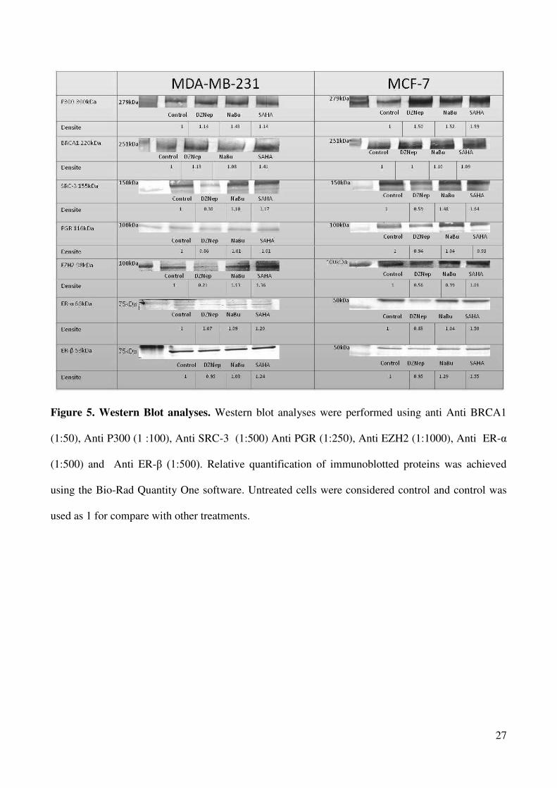
27
Figure 5. Western Blot analyses. Western blot analyses were performed using anti Anti BRCA1
(1:50), Anti P300 (1 :100), Anti SRC-3 (1:500) Anti PGR (1:250), Anti EZH2 (1:1000), Anti ER-
(1:500) and Anti ER- (1:500). Relative quantification of immunoblotted proteins was achieved
using the Bio-Rad Quantity One software. Untreated cells were considered control and control was
used as 1 for compare with other treatments.

28
Figure 6. Effects of DZNep, NaBu and SAHA treatments to m-RNA levels of selected genes. m-
RNA levels decreased in all cell lines with the treatment of DZNep, however with the treatment of
SAHA and NaBu m-RNA, levels decreased only in the MDA-MB-231 cell line. PGR does not have
a different level within itself for each treatment in MCF7 and MDA-MB-231 cell lines ,but MDA-
MB-231 and MCF7 levels are completely different. We think that it is due to the PGR status.
(DZNep: NaBu: SAHA: )

29
A
B
C
0
0.2
0.4
0.6
0.8
1 P300
MCF7
MDA MB 231
*
**
*
0
0.5
1
1.5
2
2.5
3
3.5
4
SRC3
MCF7
MDA MB 231
**
*
*
0
0.5
1
1.5
2
2.5
3
3.5
4
%(ChIP/TotalInput)
BRCA1
MCF7
MDA MB 231
*
* *
*
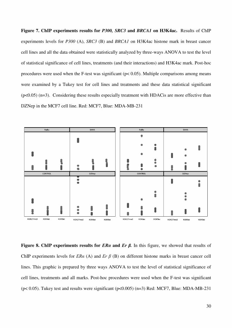
30
Figure 7. ChIP experiments results for P300, SRC3 and BRCA1 on H3K4ac. Results of ChIP
experiments levels for P300 (A), SRC3 (B) and BRCA1 on H3K4ac histone mark in breast cancer
cell lines and all the data obtained were statistically analyzed by three-ways ANOVA to test the level
of statistical significance of cell lines, treatments (and their interactions) and H3K4ac mark. Post-hoc
procedures were used when the F-test was significant (p< 0.05). Multiple comparisons among means
were examined by a Tukey test for cell lines and treatments and these data statistical significant
(p<0.05) (n=3). Considering these results especially treatment with HDACis are more effective than
DZNep in the MCF7 cell line. Red: MCF7, Blue: MDA-MB-231
Figure 8. ChIP experiments results for ER and Er . In this figure, we showed that results of
ChIP experiments levels for ER (A) and Er (B) on different histone marks in breast cancer cell
lines. This graphic is prepared by three ways ANOVA to test the level of statistical significance of
cell lines, treatments and all marks. Post-hoc procedures were used when the F-test was significant
(p< 0.05). Tukey test and results were significant (p<0.005) (n=3) Red: MCF7, Blue: MDA-MB-231

31
Table1: Primer and probe sequences
Genes Forward Reverse Probes
ER CCCTGACATTGGCTTAAACATCA TCTTTGGATCGCTCCAAAT 6FAM-TCCAGGCACAACTC-MGBNFQ
ERB GAGAGGCTTTGGGTTTGTCAAAT CCTCTAGTCCACGGCTTTGC 6FAM-CAGCAAACGTAACCTCGGGCCCTG-TAMRA
P300 CGATGGCACAGGTTAGTTTCG GCGCACCGAGTAGAAAAGATTAA 6FAM-CAGCCCCGGCCTTCCACGTT-TAMRA
SRC3 AAAATTAAGGGCAGGGCTAGGA GTGCGGCCGCTTTCG 6FAM-TCCGGATCCCGAGGGAGCTCC-TAMRA
EZH2 CCCTCCAGAAACACAATCAATAGA CCGCCTGGTCTGGCTTTAT 6FAM-CAGAGCAGCTCGACTCTTCCCTCAAACTT-TAMRA
BRCA1 CCCCGTCCAGGAAGTCTCA GCGCGGGAATTACAGATAAATT 6FAM-CGAGCTCACGCCGCGCAG-TAMRA
PGR GAGCCGCGTGTCACTAAATTG TCACAAGTCCGGCACTTGAG 6FAM-CGTCGCAGCCGCA-MGBNFQ
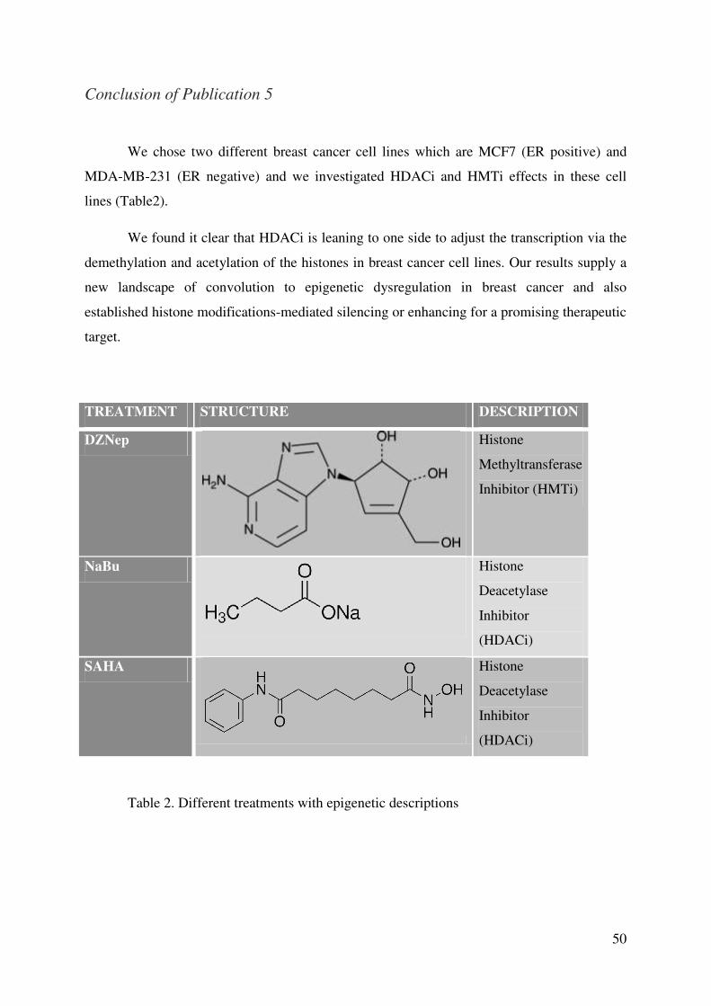
50
Conclusion of Publication 5
We chose two different breast cancer cell lines which are MCF7 (ER positive) and
MDA-MB-231 (ER negative) and we investigated HDACi and HMTi effects in these cell
lines (Table2).
We found it clear that HDACi is leaning to one side to adjust the transcription via the
demethylation and acetylation of the histones in breast cancer cell lines. Our results supply a
new landscape of convolution to epigenetic dysregulation in breast cancer and also
established histone modifications-mediated silencing or enhancing for a promising therapeutic
target.
TREATMENT STRUCTURE DESCRIPTION
DZNep
Histone
Methyltransferase
Inhibitor (HMTi)
NaBu
Histone
Deacetylase
Inhibitor
(HDACi)
SAHA
Histone
Deacetylase
Inhibitor
(HDACi)
Table 2. Different treatments with epigenetic descriptions

51
C. Epigenetic Modifications between Tumor and Normal Tissue in Sporadic
Breast Cancer According to Clinicopathological Parameters
Despite the fact that breast cancer progression is not well understood, it is likely that it
occurs due to a great amount of genetic mutations leading to widespread changes in gene
expression profiles and especially the expression of tumor suppressors and oncogenes. In
addition to the the genetic mutations, we must now further investigate epigenetic
modifications in breast cancer like histone marks. Many post-translational modifications of
histones, lysine acetylation, lysine methylation, and arginine methylation seem to be
implicated in gene regulation. So a combinatorial histone code is able to recognize these and
other modifications on chromatin regions and create transcription activation or repression of
genes. Though the “epigenetic” code is not completely elucidated, lysine acetylation
(H3K4ac, H3K9ac, and H4K8ac), and lysine trimethylation (H3K4me3) modifications are
able to generate transcriptionally active gene promoters, whereas other marks such as lysine
methylation (H3K9me3 and H3K27me3) are associated with repressed chromatin.
Therefore, in the team with Gaëlle Judes and Seher Karsli-Ceppioglu; we aimed to
assess chromatin immunoprecipitation (ChIP) of selected histone modifications in breast
tumors. The relative levels of three modified histones, including H3K27me3, H3K9ac, and
H3K4ac will be determined in breast tumors compared to normal tissue. In order to identify
the variations in global levels of histone marks in 200 tumors and normal tissues which are
classified by St Gallen, we proposed to follow ChIP with Q-PCR of 7 genes implicated in
breast cancer and for which the decrease or increase in their expression can result from bulk
histone modifications. We have chosen EZH2 and P300 genes that code respectively for a
histone methyl transferase (HMT) and a histone acetyl transferase (HAT) which are
chromatin-modifying enzymes implicated in gene regulation by catalyzing reversible post-
translational modifications of histones. SRC3, coded by the ncoa gene, is a steroid receptor
co-activator belonging to the p160 family (SRC1, SRC2 and SRC3), constituting a platform
for other co-activators such as P300 and BRCA1 tumor suppressor genes that are transcription
factors, Er genes that are nuclear receptors activated by 17 -estradiol that stimulate the
proliferation and ER that bind phytoestrogens and induce the cellular proliferation and
invasion. Also, we will study PGR gene which is important in triple negative breast cancer.
Likewise, we will follow the expression of the 7 selected genes with RTQ-PCR and western
blotting.

52
Until now, the function of H3K4c has received little attention. This acetylation mark is
particularly interesting, because it occurs on lysine residues; its methylation is associated with
transcriptional activation. The difference between methylation and acetylation of the same
residue results in a functional “switch” between transcriptional activation or silencing, as has
been described for H3K9 and H3K27 [79].
To perform this study, we used tumors and matched normal tissues of five different
classes belonging to the classification of breast cancer with Saint Gallen criteria (Table 3). We
have shown our results with boxplot graphics of gene expression levels. They are presented to
demonstrate the differences between tumor and matched-normal tissue samples. Expression
levels of ER , EZH2, BRCA1, SRC3, P300, ER and PGRgenes on H3K27me3, H3K9ac and
H3K4ac histone marks are shown in Figure 8.
Our results demonstrated that H3K27me3 histone marks have an important role in
controlling the gene expression. Gene expression levels of studied H3K27me3-enriched genes
were significantly low in breast cancer tumor samples when compared with matched-control
normal tissue (p<0.05). In addition, we have evaluated the effects of acetylated marks
H3K9ac and H3K4ac on gene expression, which are supposed to be transcriptionally active.
We found that expression levels of ER and PGR genes were decreased on H3K4ac mark-
enriched sites in tumor samples (p<0.05). ER and PGR genes play a crucial role in
determining breast cancer aggressiveness, thus the modified H3K4ac mark would have a
substantial transcriptional expression of these genes in breast cancer. However, we did not
find significant differences in gene expression levels related with H3K9ac mark modification.
Moreover, transcriptional effects of histone marks on gene expression levels based
upon different sub-types of breast cancer were investigated. The correlation between
transcriptional effects of histone marks on gene expression and breast cancer sub-types is
shown in Figure 9. Our results showed that over-expression of ER , ER and EZH2 genes
were found on H3K27me3-enriched sites in Luminal B-like (HER2 negative) breast tumors.
On the other hand, expression levels of ER and ER genes were decreased in basal-like
tumors. The expression level of the ER gene on the H3K9ac mark was increased in HER2
positive tumors. In addition, PGR was over-expressed on the H3K4ac mark in HER2 positive
tumors. As a result, we have found that modified histone marks effected transcriptional
expression of genes that code hormonal receptors.

53
CATEGORIES CHARACTERISTICS NUMBER of
PATIENTS
Luminal A
• ER and PgR positive
• HER2 negative
• Ki-67 low
• Recurrence risk ‘low’ based on
• Multi-gene-expression assay (if available)
40
Luminal B
HER2 negative
• ER positive
• HER2 negative
and at least one of:
• Ki-67 ‘high’
• PgR ‘negative or low’
HER2 positive
• ER positive
• HER2 over-expressed or amplified
• Any Ki-67
• Any PgR
40
40
HER2 Positive
Non-Luminal
• HER2 over-expressed or amplified
• ER and PgR absent
39
Basal like
• ER and PgR absent
• HER2 negative
40
Table 3. Check list of patients according to Saint Gallen classification.

54
The initiation and progression of breast cancer is apparently linked with epigenetic
alterations. In future studies, it is likely that the addition of multiple genes and the inclusion of
histone alterations to predictive panels may be determined as having an improving effect in
sensitivity and specificity. To increase the ability to remove all “pre-cancerous” tissues and
decrease local recurrences analysis of histologically normal tumor margins for epigenetic
alterations and field cancerisation can be used. Targeted therapies may someday be used as
preventive measures since epigenetic modifications can also be used as biomarkers.
Also we determined the protein levels of ER , EZH2, SRC3, P300, ER and PGR in
classified breast cancer tumors (Figure 10-11). According to our western blot results, ER
level in luminal-like tumors was higher than in control tissue samples. We found that EZH2
levels in basal-like tumors were increased when compared with other subtypes. We know that
EZH2 is a catalytic sub-unit of PRC2 and its over-expression is associated with various
cancers, especially breast cancer. Our findings are compatible with current knowledge. In
addition, mRNA levels of EZH2 were higher in tumor samples than in normal tissue samples.
Protein levels of P300, a histone acetyltransferase, were increased in tumor samples in all
subtypes of tumors. Function of P300 is critical for regulating gene expression. P300
undergoes a variety of covalent modifications like methylation, sumoylation and
phosphorylation; therefore it participates in different cancer types. Protein levels of PGR were
also increased in tumor tissues and these results were consisted with mRNA levels of PGR.
mRNA levels of ER , EZH2, BRCA1, SRC3, P300, ER and PGR were shown in Figure 12.

55
Figure 8. Gene expression levels of ER , EZH2, BRCA1, SRC3,P300, ER and PGR genes on H3K27me3, H3K9ac and H3K4ac histone marks (n=114).
ER- EZH2 BRCA1 SRC3
P300 ER- PGR

56
Figure 9. The correlation between transcriptional effects of histone marks on gene expression and breast cancer subtypes
H3K27me3 H3K9ac H3K4ac
Breast Cancer Subtypes
Luminal B-1 Luminal B-2
Luminal A
HER2+ Basal-like

57
Figure 10. Western blots ER , ER and PGR in classified breast cancer tumors.

58
Figure 11. Western blots P300, SRC3 and EZH2 in classified breast cancer tumors.

59
Figure 12. mRNA levels of ER , EZH2, BRCA1, SRC3, P300, ER and PGR (n=15)

60
D. Promoter Genome-Wide Analysis Tumor and Normal Tissue in Sporadic
Breast Cancer
Nowadays, ChIP has been coupled with promoter DNA microarrays to evaluate the
mechanisms of human gene regulation on a genome-wide scale. ChIP-on-chip technology
could be used to investigate the alterations of global gene expression in tumorigenesis. New
methodological improvements in epigenetic researches such as ChIP-on-chip methods, would
lead to a better understanding of underlying mechanisms of breast tumorigenesis and the
approaches to prevention and treatment with clarifying the role of additional mechanisms and
complex epigenetic regulations.
In this study, along with Seher Karsli-Ceppioglu, we aimed to investigate
differentially expressed genes associated with modified histones H3K27me3 and H3K9ac in
breast cancer tumors by ChIP-on-chip method. For this purpose, Agilent SurePrint G3 400k
microarrays containing approximately 21,000 of human transcripts were used to scan the
enriched region at each gene promoters in fifteen breast tumors with their matched normal
tissue samples.
For this study, we used 15 breast tumors with their matched normal tissue samples
and two histone marks where one had acetylation marks of H3K9ac and other one had
methylation marks of H3K27me3. It means that, we used three tumors and normal tissues for
each group belonging to the classification of Saint Gallen. So at the end 30 promoter
microarrays were used (Figure 13).

61
Figure13. Shedule of ChIP on chip
After the manipulation we sent all of the data to ViroScan 3D Profile expert for data
analysis. We have not received the results yet but we have added an image of the array and
some of the information generated with The Agilent Genomic Workbench software and each
Agilent microarray in the form of a QC (Quality Control) report and how to interpret in figure
14. Net signal statistics are an indication of the dynamic range of the signal on a microarray
for both non-control probes and spike-in probes. The QC report uses the range from the 1st
percentile to the 99th percentile as an indicator of dynamic range for that microarray. The
Negative Control Stats table includes the average and standard deviation of the net signals
(mean signal minus scanner offset) and the background-subtracted signals for both the red and
green channels in the negative controls. These statistics filter out saturated and feature non-
uniform and population outliers and give a rough estimate of the background noise on the
microarray.

62
A
B
Figure14. An example image and QC report of the Agilent Genomic Workbench software for TNBC group on H3K27me3(A) and H3K9ac (B) histone marks.
63
5. DISCUSSION
In 2012, 1.7 million of women were diagnosed with breast cancer and there were 6.3
million women alive who had been diagnosed with breast cancer in the previous five years.
Since the 2008 estimates, breast cancer incidence has increased by more than 20%, while
mortality has increased by 14%. Breast cancer is also the most common cause of cancer death
among women (522 000 deaths in 2012) and the most frequently diagnosed cancer among
women in 140 of 184 countries worldwide. It now represents one in four of all cancers in
women [80]. This study includes the epigenetic modifications with different treatments in
breast cancer cell lines and breast tumors.
In East and Southeast Asia, the average daily intake of phytoestrogens is estimated to
be between 20 and 50 mg [81]. In contrast, the typical diet of an adult in the United States
contains only 0.15–3 mg phytoestrogens per day, and in Europe the average daily
phytoestrogen consumption is estimated to be even lower, falling between 0.49 and 1 mg.
According to various epidemiologic studies, plasma isoflavone concentrations range from 2
M (Japanese men) to 5 nM (Finnish study subjects); however, local tissue phytoestrogen
concentrations are suggested to be 2–3 times higher than plasma levels [82, 83].
For this reason, our first study was about the effects of phytoestrogens on breast
cancer cell line with different histone marks. We chose three phytoestrogens which are
genistein, daidzein and equol; because these phytoestrogens are biologically active phenolic
compounds of plant origin that structurally mimic the principal mammalian estrogen 17 -
estradiol.
Epidemiologic evidence suggests that diet and nutrition can influence cancer
development and women living in Asia, where diets have traditionally included soybean
products, report fewer postmenopausal symptoms and experience fewer breast cancers than
women in Western countries. There are lot of mounting of evidence for interactions between
phytoestrogens and breast cancer. In recent years, investigations of their potential as
anticancer agents have increased dramatically.
Li et al. showed that histone modifications may play a more important role in
regulating genistein, modulate ER restoration as compared to DNA methylation. Histone
modifications affect the basic structure of the chromatin unit, the nucleosome, and histone

64
acetylation or deacetylation changes are considered to be the most prevalent mechanisms of
histone modifications. Histone acetylation results in an open chromatin structure leading to
active gene transcription. They found that treatment with genistein increased the histone
acetylation level in the ER promoter region, which could be considered as an important
contributor for ER reactivation. Although they did not find any methylation status changes in
the ER promoter region by genistein treatment, ER can be regulated by numerous cis-
regulatory elements located upstream of the coding sequence of ER and DNA methylation
may influence these elements leading to ER expression change. Hence Magee et al. have
demonstrated that daidzein and equol inhibit the invasive capacity of MDA-MB-231 human
breast cancer cells [84]. Also Charalambous et al. presented in their study related to equol and
MCF7; equol is significantly potent to induce gene expression [85].
No studies combining equol and histone modifications were found, so we thought that
our study would light the way for effects of equol on histone modifications in breast cancer
cell lines.
We studied the effects of phyto treatments on the variation of post-translational
modification of histones such as lysine methylation and acetylation of histone H3 and H4. We
provided evidence that phytoestrogens demethylated and acetylated histones of these selected
genes in MCF7 and MDA-MB 231 breast cancer cell lines, leads to unpack the chromatin and
enhance transcription.
When we looked our results of phytoestrogens; the fold enrichment of H3K9me3 and
H3K27me3 over control conditions decreased significatively such as 17 -estradiol treatment
in MCF7 and MDA-MB 231 cell lines. Also, when we controlled the results of acetylation
marks like H3Kac and H4K8ac, we saw that phytoestrogens and 17 -estradiol increased
acetylation significatively in selected genes.
These results suggested that phytoestrogens, histone methyltransferase inhibitors and
histone deacetylase inhibitors have beneficial effects on the genes which are up-regulated in
cancer. However, an adverse effect for down-regulated genes was observed. So the action did
not seem to be gene specific.

65
When we finished the study of phytoestrogens we start to work with other therapeutic
agents that are histone methylation inhibitors and histone deacetylase inhibitors on breast
cancer cell lines.
Previous studies have shown that, DZNep selectively inhibits H3K27me3 and
H4K20me3 in cancer cells [86]. This makes DZNep a possible candidate as an epigenetic
therapeutic for the treatment of cancer.
However, DZNep is a known S-adenosylhomocysteine (AdoHcy) hydrolase inhibitor,
which leads to the indirect inhibition of S-adenosyl-methionine (AdoMet)–dependent
reactions, including those carried out by many methyltransferases. In a study by Miranda et
al. they focused only on the methylation of H3K27, H3K9, and H4K20, then they expanded
the study to include other histone methylation modifications and to show that DZNep is not a
selective inhibitor of H3K27me3 and H4K20me3 as previously reported [86]. Instead DZNep
was found to globally inhibit both repressive and active histone methylation marks. In
addition, they tested other AdoHcy hydrolase (S-Adenosyl-L-homocysteine hydrolase which
is a potent feedback inhibitor of AdoMet-dependent methyltransferase enzymes) and global
methyltransferase inhibitors and found that EZH2 inhibition is not specific to DZNep.
However, DZNep may have more clinical potential than many of these other inhibitors due to
the known limitations of other AdoHcy hydrolase inhibitors [87]. Also they have shown that
although repressive histone marks can be reversed by DZNep treatment, a transcript cannot be
re-expressed if it contains a methylated CpG island. As a matter of fact there are many studies
accede to DZNep global methyltransferase inhibitors [87-89].
All these studies supported our results as we found that H3K27me3 level was reduced
on EZH2 in all cell line with DZNep treatment and also to strengthen our case, we did a
western blot analysis. The Western blot results have shown that there is a significant
difference between control and treatment with DZNep for the two studied cell lines.
Although it has clearly shown that DZNep acts globally and can inhibit both active
and repressive histone marks, DZNep may still be clinically useful. Therefore, we decided
that further investigations of the potential of DZNep as an epigenetic therapeutic were
warranted, in addition with using DZNep to further elucidate how chromatin structure affects
gene expression [90].

66
A previous study has shown that mutation in BRCA1 causes of increase EZH2
concentration particularly in the basal like breast carcinomas [91]. Generally EZH2 is
overexpressed in ER-negative breast cancer and inhibits BRCA1 phosphorylation. For this
reason, transition from G2 to M is accelerated in cell cycle and increased cell proliferation.
Hence, we found an EZH2 depletion by treatment with DZNep, indicating that an epigenetic
therapy that pharmacologically targets EZH2 via DZNep may constitute a novel approach to
treat breast cancer (Figure 15).
Figure15. Effects of DZNep in the cell.

67
We then applied another therapeutic, NaBu. Several research groups have reported
that NaBu halts DNA synthesis, arrests cell proliferation, alters cell morphology and increases
or decreases gene expression. As a potent HDACi in vivo, NaBu has been reported to regulate
a large number of genes in breast cell lines [92].
In a report by Davie et al., inhibition of HDAC activity with NaBu allows the HAT
activity of P300 to increase the histone acetylation levels at the promoter and nearby regions.
They also say that NaBu induces the expression of p21Waf1/Cip1 and thereby inhibits cyclin E–
Cdk2 activity and halts the subsequent events that are required for cells to enter S phase. The
cell cycle–arrested cells may differentiate or undergo cell death by apoptosis [93].
Furthermore Shin et al. utilized ChIP technology combined with next generation
sequencing technology (ChIP-seq) to analyze histone modification (acetylation) induced by
butyrate and to map the epigenomic landscape of normal histone H3 and acetylated H3K9 and
H3K27 on a large scale. Their data revealed that NaBu induced acetylation of H3K9 and
H3K27 played a role in gene expression regulation and in the complicated and dynamic
epigenomic landscape that is formed by a variety of histone modifications [94].
We have demonstrated that; NaBu treatment, H3K9ac and H3K4ac marks were more
effective than H3K27me3 mark in MCF7 cell line. This means that NaBu induced acetylation
of histone H3K9 and H3K4.
HDACs are enzymes that regulate histone acetylation on core nucleosomal histones by
catalyzing the removal of acetyl groups on their amino-terminal lysine residue. Decreased
histones H3 and H4 acetylation compact the chromatin structure resulting in disruption of the
access of transcriptional factors and repressing the transcription of certain genes [95]. To date,
the most convincing evidence that HDACs behave differently in cancer cells than in normal
cells is derived from the pharmacological manipulation of HDACs through HDAC inhibitors
[96]. However, the molecular mechanism of the tumor selective action of HDAC inhibitors is
unclear. Recent studies indicated that the HDAC inhibitors induced DNA damage in normal
and cancer cells, but cancer cells cannot repair themselves. Thus, the selectivity of HDAC
inhibitors in causing cancer cell death may be associated with impaired DNA repair
mechanism in cancer cells [97, 98].

68
Chiu et al. studied SAHA in MCF7 and MDA-MB-231. Their results are the first
demonstrating that SAHA enhances the radiation response. Furthermore, the combined
treatment induced stronger cytotoxicity in breast cancer cells. One of the mechanisms
whereby SAHA inhibited the cell capacity to repair IR-induced DNA damage by affecting the
DNA repair pathways could contribute to this combined effect. Induction of autophagy and
ER stress could also be involved in the underlying mechanisms. In addition, they further
found that SAHA inhibited the invasion and migration of breast cancer cells [99].
Moreover, Stark et al. reported that HDAC inhibitors can alter the gene expression
profile of mesenchimal-like triple negative breast cancer cells such as MDA-MB-231 to
become less aggressive and to have a more favorable prognostic profile [100]. Yi et al.
reported that SAHA effectively depletes ER in breast cancer MCF7 cells [101].
This result also supported our study because when we investigated the levels of ER
and ER they increased on the H3K27me3 mark especially with the SAHA treatment in
MDA-MB-231. Nevertheless, with the treatment of SAHA, rate of ER and ER increased on
the two activator marks H3K4ac and H3K9ac in MCF7 cell line.
These results suggested that phytoestrogens, histone methyltransferase inhibitors and
histone deacetylase inhibitors have beneficial effects on the genes which are up-regulated in
cancer. However, an adverse effect for down-regulated genes was observed. Thus, the action
did not seem to be gene specific.
Waby et al. assessed the effects of multiple members of the HDACi family on cell
cycle progression and on acetylation of Sp1. Their data indicated that a G2/M arrest is
consistently observed with several of the HDACi used and they though that Sp1 is acetylated
in response to multiple HDACi (Figure16) [102].

69
Figure16. Effects of HDACi in the cell
First ChIP experiments directed at the ER, focused on a limited number of known
binding sites and investigated binding of ER and cofactors. For example, Shang et al. found
that the ER and a number of coactivators are associated with chromatin at the c-Myc, pS2 and
CATD estrogen responsive promoters following estrogen treatment in a cyclic fashion [103].
More recently, a high throughput ChIP approach, ChIP cloning, was described by Laganiere
et al [104].
We chose the method of ChIP to understand the interactions between specific proteins
or modified forms of proteins and a genomic DNA region. ChIP can be used to monitor the
presence of histones with post-translational modifications at specific genomic locations and is
used to determine whether a transcription factor interacts with a candidate target gene. There
was a highly significant correlation between histone modification status, tumor biomarker
phenotype, and clinical outcome. High relative levels of global histone acetylation and
70
methylation were associated with a prognosis and detected almost exclusively in luminal-like
breast tumors (93%).
Elseikh et al. reported that basal like and HER2++ tumors were associated with low
levels of all seven histone marks. Low detection levels of H4R3me2, H3K9ac, and H4K16ac
were significantly associated with large tumor size and high detection levels of H4R3me2 and
H3K9ac were associated with low lymph node stage. These results may indicate that reduced
detection of selected histone modifications correlates with poor prognostic characteristics.
High-level detection of histone modifications was significantly correlated with steroid
receptor (ER, PR, AR)-positive tumors [105].
For our third project, we studied breast tumors and normal tissue comparing inside the
classification St Gallen. A number of genes have been investigated for changes in expression
level during progression of breast cancer. In all of the studies we used same selected genes
like EZH2, BRCA1, ER , ER , SRC3, P300 and PGR. This is because we know that ER ,
EZH2, SRC3 and P300 are overexpressed genes and promoted the activation of transcription
in breast cancer and also that BRCA1 is a human tumor suppressor gene. ER , ER and PGR
and their ligands play important roles in the development and function of the mammary gland
in order to identify gene expression changes that may be linked to clinical outcomes such as
relapse-free survival and overall survival.
Several experimental studies have established that elevated EZH2 levels in human
breast carcinomas are associated with the aggressive ER-negative basal-like phenotype
characterized by lack of ER expression, nuclear polymorphism and lack of BRCA1 protein
[106], [107].
Some studies have used a gene PRC2 target gene set and identified 391 (59.8%) and
336 (51.4%) genes that harbor epi-modification alterations in luminal and basal subtypes in
study of 2014. In particular, of these PRC2 targets like a EZH2, there were more genes having
reduced H3K27me3 signal in luminal subtype than that in basal subtype (228 and 146 genes
in luminal and basal, respectively). This finding is in line with the fact that basal breast cancer
has more undifferentiated features than luminal subtype [108].

71
When we investigated our results of EZH2 for each subtype of breast cancer, this data
supported our EZH2 results on H3K27me3 marks and inadditional, we demonstrated that
EZH2 reduced H3K27me3 in luminal B1 subtype than that in luminal A.
Hudelist et al. found that overexpression of SRC3 in breast tumors correlates with poor
disease-free survival. Higher SRC3 levels are also preferentially present in higher grade
invasive tumors. In addition Mc Ilroy et al. study results indicate an association between high
SRC3 levels and breast cancer [109, 110]. Furthermore Bouras et al. revealed that SRC3
overexpression also correlated with the absence of ER and PR in breast cancer [111]. Our
finding suggest that, overexpression of SRC3 accesible in Luminal A and Luminal B1 in
repressive marks in H3K27me3 besides subclass of HER2++ which is absence of ER and PR
groups, have a high level of SRC3 in activactrice marks like H3K4ac and H3K4ac.
Some studies indicate that P300 plays a role as tumor suppression, yet other studies
suggest it plays a role as tumor promotion. It has been reported that P300 behaves as a
classical tumor suppressor gene [112-114]. Xiao et al. reported that in 2012, high expression
of P300 was more frequently observed in breast cancer tissues when compared to that in the
non-malignant breast tissues. If we analyzed those results in comparison to our classification
of St Gallen; non-malignant breast tissues are combined with a class of Luminal. The
expression of P300 in non-malignant breast tissues was either absent or at low levels. In
contrast, high expression of P300 was frequently observed in large number of breast cancer
tissues [115].
We did not find any association between three histone marks H3K27me3, H3K9ac and
H3K4ac on expression level of P300. However, when we investigated the difference between
in subclass, we found that Luminal B1 has P300 high expression level for all modification of
histones.
Our results demonstrated that expression levels of ER and PGR genes were decreased
on H3K4ac mark-enriched sites in tumor samples. ER and PGR genes play a crucial role in
determining breast cancer aggressiveness, thus the modified H3K4ac mark would be
significant transcriptional expression of these genes in breast cancer. However, we did not
find significant differences in gene expression levels related with H3K9ac mark modification.
According to our results, over-expression of ER , ER and EZH2 genes were found on
H3K27me3-enriched sites in Luminal B-like (HER2 negative) breast tumors. On the other

72
hand, expression levels of ER and ER genes were decreased in basal-like tumors. The
expression level of ER gene on H3K9ac mark was increased in HER2 positive tumors. In
addition, PGR was over-expressed on H3K4ac mark in HER2 positive tumors. As a result, we
have found that modified histone marks affect transcriptional expression of genes that code
hormonal receptors.
After all these studies, we used promoter microarrays on breast cancer tumor and
matched normal tissue with previously selected histone marks to work with a wider spectrum
of genes. The overall goal of this project was to study breast cancer with reference to gene
expression analysis using clinical specimens and to advance the understanding of the
heterogeneity of breast cancer using gene expression analysis, and to identify gene expression
differences among the normal and cancer breast tissue by comparing various inside
classifications of St Gallen.
On the other hand, Li et al., compared the alterations of six types of histone
modifications H3K4me1, H3K4me3, H3K27ac, H3K36me3, H3K9me3 and H3K27me3 and
DNA methylation between two cell lines (MCF7, HCC1954) that had been shown to
represent two different breast cancer subtypes-luminal and basal. They used ChIP-seq and
microarray as in their previously study [116].
In all cell lines, they found housands of differential histone modification sites were
identified in both luminal and basal subtypes, compared with control. They also explained
that different patterns of alterations of epi-modifications between luminal and basal subtypes
were observed. In MCF7 cell line with the up-regulation of ER and PR in luminal subtype,
they found that H3K4me3 and H3K27ac were up-regulated and H3K27me3 was down-
regulated. However, the epigenetic patterns of ER and PR in this subtype were different from
those in basal subtype (HCC 1954 cell line).
Consistent with ERBB2 high expression in basal subtype, epigenetic alterations were
found within all the six types of histone modifications [117].
The advent of microarray technology has revolutionized the molecular profiling of
disease tissues and tumors. It is one of the most promising and powerful methodologies in
molecular oncology. Microarrays have a number of advantages over TaqMan assays,
especially when many clinical samples are to be screened. In addition, microarrays can
incorporate multiple positive and negative controls as well as multiples of the same samples at

73
different locations of the array to ensure reproducibility and statistical significance. Ideally,
initial screening of biological samples could be done by microarray analysis and individual
positive samples could be confirmed by additional tests using TaqMan assays.

74
6. CONCLUSION
This PhD research first focused on the biological effects of phytoestrogens and some
epidrugs in vitro and characterization of bioactive compounds from breast cancer.
These results suggested that phytoestrogens, histone methyltransferase inhibitors and
histone deacetylase inhibitors have beneficial effects on the genes which are up-regulated in
cancer. However, an adverse effect for down-regulated genes was observed. Thus, action did
not seem to be gene specific. Our results add a new outlook of complication to epigenetic
deregulation in breast cancer and also established histone modifications-mediated silencing or
enhancing for up-and-coming therapeutic target.
Subsequently, study with normal tissue and sporadic breast cancer have allowed us to
present effective differences on Saint Gallen classifications of breast tumors with ChIP. It also
allowed us to gain more insight into how epigenetic modification patterns are regulated in
detail especially how the patterns are established. However, future studies focusing on the
clinical relevance and mechanism of epigenetic modification of bio-active dietary factors are
needed to further assess the applicability of dietary factors as cancer preventive and chemo
preventive agents.
ChIP can be used to identify regions that are associated with proteins, or conversely, it
can be used to identify proteins that are associated with a particular region of the genome.
The combination of ChIP and microarray (ChIP on chip) allows genome-wide
identification of binding sites for the chromatin-associated proteins. Alternatively, high
throughput sequencing of the immunoprecipitated DNA (ChIP-Seq) offers a more precise
way of mapping protein-DNA interactions across the genome.
We have not yet received our results, but we hope that the transition from normal
tissue to tumor is an important aspect in understanding the biology of breast cancer. Gene
expression profiling can help identify the differences among the normal and tumor tissues and
can help better design drugs to target the disease. The normal and cancer specimens were
compared in order to identify genes and pathways that contribute to the transition from
normal breast tissue to a cancerous state.

75
7. REFERENCES
1. Jemal A, Bray F, Center MM, Ferlay J, Ward E, Forman D: Global cancer statistics. CA Cancer J Clin 2011, 61(2):69-90.
2. Clarke DB, Lloyd AS, Lawrence JM, Brown JE, Storey L, Raats M, Rainsbury RM, Culliford DJ, Bailey-Horne VA, Parry BM: Development of a food compositional
database for the estimation of dietary intake of phyto-oestrogens in a group of
postmenopausal women previously treated for breast cancer and validation with
urinary excretion. Br J Nutr 2013, 109(12):2261-2268. 3. Dumalaon-Canaria JA, Hutchinson AD, Prichard I, Wilson C: What causes breast
cancer? A systematic review of causal attributions among breast cancer survivors
and how these compare to expert-endorsed risk factors. Cancer Causes Control
2014, 25(7):771-785. 4. Lemaine V, Simmons PS: The adolescent female: Breast and reproductive
embryology and anatomy. Clin Anat 2013, 26(1):22-28. 5. Malvezzi M, Bertuccio P, Levi F, La Vecchia C, Negri E: European cancer
mortality predictions for the year 2013. Ann Oncol 2013, 24(3):792-800. 6. Ferlay J, Steliarova-Foucher E, Lortet-Tieulent J, Rosso S, Coebergh JW, Comber H,
Forman D, Bray F: Cancer incidence and mortality patterns in Europe: estimates
for 40 countries in 2012. Eur J Cancer 2013, 49(6):1374-1403. 7. Del Valle MO, Martin-Payo R, Lana A, Garcia JB, Folgueras MV, Lopez ML:
Behavioural cancer risk factors in women diagnosed with primary breast cancer. J Adv Nurs 2014.
8. Alsaker MD, Opdahl S, Romundstad PR, Vatten LJ: Association of time since last
birth, age at first birth and parity with breast cancer survival among parous
women: a register-based study from Norway. Int J Cancer 2013, 132(1):174-181. 9. Ma H, Bernstein L, Pike MC, Ursin G: Reproductive factors and breast cancer risk
according to joint estrogen and progesterone receptor status: a meta-analysis of
epidemiological studies. Breast Cancer Res 2006, 8(4):R43. 10. Setiawan VW, Monroe KR, Wilkens LR, Kolonel LN, Pike MC, Henderson BE:
Breast cancer risk factors defined by estrogen and progesterone receptor status:
the multiethnic cohort study. Am J Epidemiol 2009, 169(10):1251-1259. 11. Amin AL, Purdy AC, Mattingly JD, Kong AL, Termuhlen PM: Benign breast
disease. Surg Clin North Am 2013, 93(2):299-308. 12. Tice JA, O'Meara ES, Weaver DL, Vachon C, Ballard-Barbash R, Kerlikowske K:
Benign breast disease, mammographic breast density, and the risk of breast
cancer. J Natl Cancer Inst 2013, 105(14):1043-1049. 13. Bosviel R, Durif J, Dechelotte P, Bignon YJ, Bernard-Gallon D: Epigenetic
modulation of BRCA1 and BRCA2 gene expression by equol in breast cancer cell
lines. Br J Nutr 2012, 108(7):1187-1193. 14. Bagnardi V, Rota M, Botteri E, Tramacere I, Islami F, Fedirko V, Scotti L, Jenab M,
Turati F, Pasquali E et al: Light alcohol drinking and cancer: a meta-analysis. Ann
Oncol 2013, 24(2):301-308. 15. Gathani T, Ali R, Balkwill A, Green J, Reeves G, Beral V, Moser KA: Ethnic
differences in breast cancer incidence in England are due to differences in known
risk factors for the disease: prospective study. Br J Cancer 2014, 110(1):224-229. 16. O'Neill SC, Leventhal KG, Scarles M, Evans CN, Makariou E, Pien E, Willey S:
Mammographic breast density as a risk factor for breast cancer: awareness in a
recently screened clinical sample. Womens Health Issues 2014, 24(3):e321-326.

76
17. Banin Hirata BK, Oda JM, Losi Guembarovski R, Ariza CB, de Oliveira CE, Watanabe MA: Molecular markers for breast cancer: prediction on tumor
behavior. Dis Markers 2014, 2014:513158. 18. Rosner B, Glynn RJ, Tamimi RM, Chen WY, Colditz GA, Willett WC, Hankinson
SE: Breast cancer risk prediction with heterogeneous risk profiles according to
breast cancer tumor markers. Am J Epidemiol 2013, 178(2):296-308. 19. Hah N, Kraus WL: Hormone-regulated transcriptomes: lessons learned from
estrogen signaling pathways in breast cancer cells. Mol Cell Endocrinol 2014, 382(1):652-664.
20. Renoir JM, Marsaud V, Lazennec G: Estrogen receptor signaling as a target for
novel breast cancer therapeutics. Biochem Pharmacol 2013, 85(4):449-465. 21. Rizza P, Barone I, Zito D, Giordano F, Lanzino M, De Amicis F, Mauro L, Sisci D,
Catalano S, Wright KD et al: Estrogen receptor beta as a novel target of androgen
receptor action in breast cancer cell lines. Breast Cancer Res 2014, 16(1):R21. 22. Hagan CR, Lange CA: Molecular determinants of context-dependent progesterone
receptor action in breast cancer. BMC Med 2014, 12:32. 23. Gadkar-Sable S, Shah C, Rosario G, Sachdeva G, Puri C: Progesterone receptors:
various forms and functions in reproductive tissues. Front Biosci 2005, 10:2118-2130.
24. Dressing GE, Knutson TP, Schiewer MJ, Daniel AR, Hagan CR, Diep CH, Knudsen KE, Lange CA: Progesterone receptor-cyclin d1 complexes induce cell cycle-
dependent transcriptional programs in breast cancer cells. Mol Endocrinol 2014, 28(4):442-457.
25. Mavrova R, Radosa J, Schmitt K, Bohle RM, Rody A, Solomayer EF, Juhasz-Boess I: Estrogen, progesterone, and Her-2/neu receptor expression discrepancy in
primary tumors and in-breast relapse in patients with breast cancer. Breast J
2014, 20(3):322-324. 26. Chang CC, Tien CH, Tai SH, Chuang MT, Sze CI, Hung YC, Lee EJ: High-grade
glioma in a patient with breast cancer. Asian J Surg 2014, 37(3):162-166. 27. Teramoto A, Shimazu K, Naoi Y, Shimomura A, Shimoda M, Kagara N, Maruyama
N, Kim SJ, Yoshidome K, Tsujimoto M et al: One-step nucleic acid amplification
assay for intraoperative prediction of non-sentinel lymph node metastasis in
breast cancer patients with sentinel lymph node metastasis. Breast 2014. 28. Untch M, Gerber B, Harbeck N, Jackisch C, Marschner N, Mobus V, von Minckwitz
G, Loibl S, Beckmann MW, Blohmer JU et al: 13th st. Gallen international breast
cancer conference 2013: primary therapy of early breast cancer evidence,
controversies, consensus - opinion of a german team of experts (zurich 2013). Breast Care (Basel) 2013, 8(3):221-229.
29. Stathopoulos GP, Malamos NA, Markopoulos C, Polychronis A, Armakolas A, Rigatos S, Yannopoulou A, Kaparelou M, Antoniou P: The role of Ki-67 in the
proliferation and prognosis of breast cancer molecular classification subtypes. Anticancer Drugs 2014.
30. Eroles P, Bosch A, Perez-Fidalgo JA, Lluch A: Molecular biology in breast cancer:
intrinsic subtypes and signaling pathways. Cancer Treat Rev 2012, 38(6):698-707. 31. Vuong D, Simpson PT, Green B, Cummings MC, Lakhani SR: Molecular
classification of breast cancer. Virchows Arch 2014, 465(1):1-14. 32. Taherian-Fard A, Srihari S, Ragan MA: Breast cancer classification: linking
molecular mechanisms to disease prognosis. Brief Bioinform 2014. 33. Cornejo KM, Kandil D, Khan A, Cosar EF: Theranostic and molecular
classification of breast cancer. Arch Pathol Lab Med 2014, 138(1):44-56.
77
34. Lam SW, Jimenez CR, Boven E: Breast cancer classification by proteomic
technologies: current state of knowledge. Cancer Treat Rev 2014, 40(1):129-138. 35. Yang EJ, Shin EK, Shin HI, Lim JY: Psychometric properties of scale constructed
from the International Classification of Functioning, Disability and Health (ICF)
core set for breast cancer based on Rasch analysis. Support Care Cancer 2014. 36. Parise CA, Caggiano V: Breast Cancer Survival Defined by the ER/PR/HER2
Subtypes and a Surrogate Classification according to Tumor Grade and
Immunohistochemical Biomarkers. J Cancer Epidemiol 2014, 2014:469251. 37. Cardiff RD, Borowsky AD: At last: classification of human mammary cells
elucidates breast cancer origins. J Clin Invest 2014, 124(2):478-480. 38. Leidy J, Khan A, Kandil D: Basal-like breast cancer: update on clinicopathologic,
immunohistochemical, and molecular features. Arch Pathol Lab Med 2014, 138(1):37-43.
39. Wei W, Tweardy DJ, Zhang M, Zhang X, Landua J, Petrovic I, Bu W, Roarty K, Hilsenbeck SG, Rosen JM et al: STAT3 signaling is activated preferentially in
tumor-initiating cells in claudin-low models of human breast cancer. Stem Cells
2014. 40. Harrell JC, Pfefferle AD, Zalles N, Prat A, Fan C, Khramtsov A, Olopade OI, Troester
MA, Dudley AC, Perou CM: Endothelial-like properties of claudin-low breast
cancer cells promote tumor vascular permeability and metastasis. Clin Exp
Metastasis 2014, 31(1):33-45. 41. Dagdemir A, Durif J, Ngollo M, Bignon YJ, Bernard-Gallon D: Histone lysine
trimethylation or acetylation can be modulated by phytoestrogen, estrogen or
anti-HDAC in breast cancer cell lines. Epigenomics 2013, 5(1):51-63. 42. Mense SM, Hei TK, Ganju RK, Bhat HK: Phytoestrogens and breast cancer
prevention: possible mechanisms of action. Environ Health Perspect 2008, 116(4):426-433.
43. Pilsakova L, Riecansky I, Jagla F: The physiological actions of isoflavone
phytoestrogens. Physiol Res 2010, 59(5):651-664. 44. Bernard-Gallon DJ, Satih S, Chalabi N, Rabiau N, Bosviel R, Fontana L, Bignon YJ:
Phytoestrogens regulate the expression of genes involved in different biological
processes in BRCA2 knocked down MCF-7, MDA-MB-231 and MCF-10a cell
lines. Oncol Rep 2010, 23(3):647-653. 45. Hardy TM, Tollefsbol TO: Epigenetic diet: impact on the epigenome and cancer.
Epigenomics 2011, 3(4):503-518. 46. Ososki AL, Kennelly EJ: Phytoestrogens: a review of the present state of research.
Phytother Res 2003, 17(8):845-869. 47. Dagdemir A, Durif J, Ngollo M, Bignon YJ, Bernard-Gallon D: Breast cancer:
mechanisms involved in action of phytoestrogens and epigenetic changes. In Vivo
2013, 27(1):1-9. 48. Li Y, Meeran SM, Patel SN, Chen H, Hardy TM, Tollefsbol TO: Epigenetic
reactivation of estrogen receptor-alpha (ERalpha) by genistein enhances
hormonal therapy sensitivity in ERalpha-negative breast cancer. Mol Cancer
2013, 12:9. 49. Yuan B, Wang L, Jin Y, Zhen H, Xu P, Xu Y, Li C, Xu H: Role of metabolism in the
effects of genistein and its phase II conjugates on the growth of human breast cell
lines. AAPS J 2012, 14(2):329-344. 50. Yang Z, Kulkarni K, Zhu W, Hu M: Bioavailability and pharmacokinetics of
genistein: mechanistic studies on its ADME. Anticancer Agents Med Chem 2012, 12(10):1264-1280.

78
51. Gercel-Taylor C, Feitelson AK, Taylor DD: Inhibitory effect of genistein and
daidzein on ovarian cancer cell growth. Anticancer Res 2004, 24(2B):795-800. 52. Choi EJ, Kim GH: Daidzein causes cell cycle arrest at the G1 and G2/M phases in
human breast cancer MCF-7 and MDA-MB-453 cells. Phytomedicine 2008, 15(9):683-690.
53. Singh RP, Agarwal R: Natural flavonoids targeting deregulated cell cycle
progression in cancer cells. Curr Drug Targets 2006, 7(3):345-354. 54. Ju YH, Fultz J, Allred KF, Doerge DR, Helferich WG: Effects of dietary daidzein
and its metabolite, equol, at physiological concentrations on the growth of
estrogen-dependent human breast cancer (MCF-7) tumors implanted in
ovariectomized athymic mice. Carcinogenesis 2006, 27(4):856-863. 55. Verheus M, van Gils CH, Keinan-Boker L, Grace PB, Bingham SA, Peeters PH:
Plasma phytoestrogens and subsequent breast cancer risk. J Clin Oncol 2007, 25(6):648-655.
56. Atkinson C, Frankenfeld CL, Lampe JW: Gut bacterial metabolism of the soy
isoflavone daidzein: exploring the relevance to human health. Exp Biol Med
(Maywood) 2005, 230(3):155-170. 57. Rowland I, Faughnan M, Hoey L, Wahala K, Williamson G, Cassidy A:
Bioavailability of phyto-oestrogens. Br J Nutr 2003, 89 Suppl 1:S45-58. 58. Jackson RL, Greiwe JS, Schwen RJ: Emerging evidence of the health benefits of S-
equol, an estrogen receptor beta agonist. Nutr Rev 2011, 69(8):432-448. 59. Rufer CE, Glatt H, Kulling SE: Structural elucidation of hydroxylated metabolites
of the isoflavan equol by gas chromatography-mass spectrometry and high-
performance liquid chromatography-mass spectrometry. Drug Metab Dispos
2006, 34(1):51-60. 60. Magee PJ, Raschke M, Steiner C, Duffin JG, Pool-Zobel BL, Jokela T, Wahala K,
Rowland IR: Equol: a comparison of the effects of the racemic compound with
that of the purified S-enantiomer on the growth, invasion, and DNA integrity of
breast and prostate cells in vitro. Nutr Cancer 2006, 54(2):232-242. 61. Levin ER, Pietras RJ: Estrogen receptors outside the nucleus in breast cancer.
Breast Cancer Res Treat 2008, 108(3):351-361. 62. Castoria G, Migliaccio A, D'Amato L, Di Stasio R, Ciociola A, Lombardi M, Bilancio
A, Di Domenico M, de Falco A, Auricchio F: Integrating signals between cAMP
and MAPK pathways in breast cancer. Front Biosci 2008, 13:1318-1327. 63. Henson ES, Gibson SB: Surviving cell death through epidermal growth factor
(EGF) signal transduction pathways: implications for cancer therapy. Cell Signal
2006, 18(12):2089-2097. 64. Song RX, Fan P, Yue W, Chen Y, Santen RJ: Role of receptor complexes in the
extranuclear actions of estrogen receptor alpha in breast cancer. Endocr Relat
Cancer 2006, 13 Suppl 1:S3-13. 65. Cherdshewasart W, Panriansaen R, Picha P: Pretreatment with phytoestrogen-rich
plant decreases breast tumor incidence and exhibits lower profile of mammary
ERalpha and ERbeta. Maturitas 2007, 58(2):174-181. 66. Satih S, Savinel H, Rabiau N, Fontana L, Bignon YJ, Bernard-Gallon DJ: Expression
analyses of nuclear receptor genes in breast cancer cell lines exposed to soy
phytoestrogens after BRCA2 knockdown by TaqMan Low-Density Array
(TLDA). J Mol Signal 2009, 4:3. 67. Bosviel R, Dumollard E, Dechelotte P, Bignon YJ, Bernard-Gallon D: Can soy
phytoestrogens decrease DNA methylation in BRCA1 and BRCA2
oncosuppressor genes in breast cancer? OMICS 2012, 16(5):235-244.

79
68. Zhang G, Pradhan S: Mammalian epigenetic mechanisms. IUBMB Life 2014, 66(4):240-256.
69. Tung PY, Knoepfler PS: Epigenetic mechanisms of tumorigenicity manifesting in
stem cells. Oncogene 2014, 0. 70. Hackett JA, Surani MA: DNA methylation dynamics during the mammalian life
cycle. Philos Trans R Soc Lond B Biol Sci 2013, 368(1609):20110328. 71. Ziller MJ, Gu H, Muller F, Donaghey J, Tsai LT, Kohlbacher O, De Jager PL, Rosen
ED, Bennett DA, Bernstein BE et al: Charting a dynamic DNA methylation
landscape of the human genome. Nature 2013, 500(7463):477-481. 72. Jenuwein T, Allis CD: Translating the histone code. Science 2001, 293(5532):1074-
1080. 73. Connolly R, Stearns V: Epigenetics as a therapeutic target in breast cancer. J
Mammary Gland Biol Neoplasia 2012, 17(3-4):191-204. 74. Parrella P: Epigenetic Signatures in Breast Cancer: Clinical Perspective. Breast
Care (Basel) 2010, 5(2):66-73. 75. Nowsheen S, Aziz K, Tran PT, Gorgoulis VG, Yang ES, Georgakilas AG: Epigenetic
inactivation of DNA repair in breast cancer. Cancer Lett 2012. 76. Katz TA, Vasilatos SN, Harrington E, Oesterreich S, Davidson NE, Huang Y:
Inhibition of histone demethylase, LSD2 (KDM1B), attenuates DNA methylation
and increases sensitivity to DNMT inhibitor-induced apoptosis in breast cancer
cells. Breast Cancer Res Treat 2014, 146(1):99-108. 77. Lu S, Labhasetwar V: Drug Resistant Breast Cancer Cell Line Displays Cancer
Stem Cell Phenotype and Responds Sensitively to Epigenetic Drug SAHA. Drug
Deliv Transl Res 2013, 3(2):183-194. 78. Cazares Marinero Jde J, Lapierre M, Cavailles V, Saint-Fort R, Vessieres A, Top S,
Jaouen G: Efficient new constructs against triple negative breast cancer cells:
synthesis and preliminary biological study of ferrocifen-SAHA hybrids and
related species. Dalton Trans 2013, 42(43):15489-15501. 79. Garcia BA, Hake SB, Diaz RL, Kauer M, Morris SA, Recht J, Shabanowitz J, Mishra
N, Strahl BD, Allis CD et al: Organismal differences in post-translational
modifications in histones H3 and H4. J Biol Chem 2007, 282(10):7641-7655. 80. Kreiter E, Richardson A, Potter J, Yasui Y: Breast cancer: trends in international
incidence in men and women. Br J Cancer 2014, 110(7):1891-1897. 81. Sirtori CR, Arnoldi A, Johnson SK: Phytoestrogens: end of a tale? Ann Med 2005,
37(6):423-438. 82. Adlercreutz H, Markkanen H, Watanabe S: Plasma concentrations of phyto-
oestrogens in Japanese men. Lancet 1993, 342(8881):1209-1210. 83. Morton MS, Arisaka O, Miyake N, Morgan LD, Evans BA: Phytoestrogen
concentrations in serum from Japanese men and women over forty years of age. J
Nutr 2002, 132(10):3168-3171. 84. Magee PJ, Allsopp P, Samaletdin A, Rowland IR: Daidzein, R-(+)equol and S-(-
)equol inhibit the invasion of MDA-MB-231 breast cancer cells potentially via the
down-regulation of matrix metalloproteinase-2. Eur J Nutr 2014, 53(1):345-350. 85. Charalambous C, Pitta CA, Constantinou AI: Equol enhances tamoxifen's anti-
tumor activity by induction of caspase-mediated apoptosis in MCF-7 breast
cancer cells. BMC Cancer 2013, 13:238. 86. Tan J, Yang X, Zhuang L, Jiang X, Chen W, Lee PL, Karuturi RK, Tan PB, Liu ET,
Yu Q: Pharmacologic disruption of Polycomb-repressive complex 2-mediated
gene repression selectively induces apoptosis in cancer cells. Genes Dev 2007, 21(9):1050-1063.
80
87. Miranda TB, Cortez CC, Yoo CB, Liang G, Abe M, Kelly TK, Marquez VE, Jones PA: DZNep is a global histone methylation inhibitor that reactivates
developmental genes not silenced by DNA methylation. Mol Cancer Ther 2009, 8(6):1579-1588.
88. Girard N, Bazille C, Lhuissier E, Benateau H, Llombart-Bosch A, Boumediene K, Bauge C: 3-Deazaneplanocin A (DZNep), an inhibitor of the histone
methyltransferase EZH2, induces apoptosis and reduces cell migration in
chondrosarcoma cells. PLoS One 2014, 9(5):e98176. 89. Fujiwara T, Saitoh H, Inoue A, Kobayashi M, Okitsu Y, Katsuoka Y, Fukuhara N,
Onishi Y, Ishizawa K, Ichinohasama R et al: 3-Deazaneplanocin A (DZNep), an
inhibitor of S-adenosylmethionine-dependent methyltransferase, promotes
erythroid differentiation. J Biol Chem 2014, 289(12):8121-8134. 90. Su H, Altucci L, You Q: Competitive or noncompetitive, that's the question:
research toward histone deacetylase inhibitors. Mol Cancer Ther 2008, 7(5):1007-1012.
91. Puppe J, Drost R, Liu X, Joosse SA, Evers B, Cornelissen-Steijger P, Nederlof P, Yu Q, Jonkers J, van Lohuizen M et al: BRCA1-deficient mammary tumor cells are
dependent on EZH2 expression and sensitive to Polycomb Repressive Complex 2-
inhibitor 3-deazaneplanocin A. Breast Cancer Res 2009, 11(4):R63. 92. Hizel C, Maurizis JC, Rio P, Communal Y, Chassagne J, Favy D, Bignon YJ,
Bernard-Gallon DJ: Isolation, purification and quantification of BRCA1 protein
from tumour cells by affinity perfusion chromatography. J Chromatogr B Biomed
Sci Appl 1999, 721(2):163-170. 93. Davie JR: Inhibition of histone deacetylase activity by butyrate. J Nutr 2003, 133(7
Suppl):2485S-2493S. 94. Shin JH, Li RW, Gao Y, Baldwin Rt, Li CJ: Genome-wide ChIP-seq mapping and
analysis reveal butyrate-induced acetylation of H3K9 and H3K27 correlated with
transcription activity in bovine cells. Funct Integr Genomics 2012, 12(1):119-130. 95. Castro-Galache MD, Ferragut JA, Barbera VM, Martin-Orozco E, Gonzalez-Ros JM,
Garcia-Morales P, Saceda M: Susceptibility of multidrug resistance tumor cells to
apoptosis induction by histone deacetylase inhibitors. Int J Cancer 2003, 104(5):579-586.
96. Mai A, Massa S, Rotili D, Cerbara I, Valente S, Pezzi R, Simeoni S, Ragno R: Histone deacetylation in epigenetics: an attractive target for anticancer therapy. Med Res Rev 2005, 25(3):261-309.
97. Lee JH, Choy ML, Ngo L, Foster SS, Marks PA: Histone deacetylase inhibitor
induces DNA damage, which normal but not transformed cells can repair. Proc
Natl Acad Sci U S A 2010, 107(33):14639-14644. 98. Miller TA, Witter DJ, Belvedere S: Histone deacetylase inhibitors. J Med Chem
2003, 46(24):5097-5116. 99. Chiu HW, Yeh YL, Wang YC, Huang WJ, Chen YA, Chiou YS, Ho SY, Lin P, Wang
YJ: Suberoylanilide hydroxamic acid, an inhibitor of histone deacetylase,
enhances radiosensitivity and suppresses lung metastasis in breast cancer in vitro
and in vivo. PLoS One 2013, 8(10):e76340. 100. Stark A, Kleer CG, Martin I, Awuah B, Nsiah-Asare A, Takyi V, Braman M, Quayson
SE, Zarbo R, Wicha M et al: African ancestry and higher prevalence of triple-
negative breast cancer: findings from an international study. Cancer 2010, 116(21):4926-4932.
101. Yi X, Wei W, Wang SY, Du ZY, Xu YJ, Yu XD: Histone deacetylase inhibitor
SAHA induces ERalpha degradation in breast cancer MCF-7 cells by CHIP-

81
mediated ubiquitin pathway and inhibits survival signaling. Biochem Pharmacol
2008, 75(9):1697-1705. 102. Waby JS, Chirakkal H, Yu C, Griffiths GJ, Benson RS, Bingle CD, Corfe BM: Sp1
acetylation is associated with loss of DNA binding at promoters associated with
cell cycle arrest and cell death in a colon cell line. Mol Cancer 2010, 9:275. 103. Shang Y, Hu X, DiRenzo J, Lazar MA, Brown M: Cofactor dynamics and
sufficiency in estrogen receptor-regulated transcription. Cell 2000, 103(6):843-852.
104. Laganiere J, Deblois G, Giguere V: Functional genomics identifies a mechanism for
estrogen activation of the retinoic acid receptor alpha1 gene in breast cancer
cells. Mol Endocrinol 2005, 19(6):1584-1592. 105. Elsheikh SE, Green AR, Rakha EA, Powe DG, Ahmed RA, Collins HM, Soria D,
Garibaldi JM, Paish CE, Ammar AA et al: Global histone modifications in breast
cancer correlate with tumor phenotypes, prognostic factors, and patient outcome. Cancer Res 2009, 69(9):3802-3809.
106. Deb G, Thakur VS, Gupta S: Multifaceted role of EZH2 in breast and prostate
tumorigenesis: epigenetics and beyond. Epigenetics 2013, 8(5):464-476. 107. Gonzalez ME, Li X, Toy K, DuPrie M, Ventura AC, Banerjee M, Ljungman M,
Merajver SD, Kleer CG: Downregulation of EZH2 decreases growth of estrogen
receptor-negative invasive breast carcinoma and requires BRCA1. Oncogene
2009, 28(6):843-853. 108. Sparmann A, van Lohuizen M: Polycomb silencers control cell fate, development
and cancer. Nat Rev Cancer 2006, 6(11):846-856. 109. Hudelist G, Czerwenka K, Kubista E, Marton E, Pischinger K, Singer CF: Expression
of sex steroid receptors and their co-factors in normal and malignant breast
tissue: AIB1 is a carcinoma-specific co-activator. Breast Cancer Res Treat 2003, 78(2):193-204.
110. Mc Ilroy M, Fleming FJ, Buggy Y, Hill AD, Young LS: Tamoxifen-induced ER-
alpha-SRC-3 interaction in HER2 positive human breast cancer; a possible
mechanism for ER isoform specific recurrence. Endocr Relat Cancer 2006, 13(4):1135-1145.
111. Bouras T, Southey MC, Venter DJ: Overexpression of the steroid receptor
coactivator AIB1 in breast cancer correlates with the absence of estrogen and
progesterone receptors and positivity for p53 and HER2/neu. Cancer Res 2001, 61(3):903-907.
112. Gayther SA, Batley SJ, Linger L, Bannister A, Thorpe K, Chin SF, Daigo Y, Russell P, Wilson A, Sowter HM et al: Mutations truncating the EP300 acetylase in
human cancers. Nat Genet 2000, 24(3):300-303. 113. Bandyopadhyay D, Okan NA, Bales E, Nascimento L, Cole PA, Medrano EE: Down-
regulation of p300/CBP histone acetyltransferase activates a senescence
checkpoint in human melanocytes. Cancer Res 2002, 62(21):6231-6239. 114. Ait-Si-Ali S, Polesskaya A, Filleur S, Ferreira R, Duquet A, Robin P, Vervish A,
Trouche D, Cabon F, Harel-Bellan A: CBP/p300 histone acetyl-transferase activity
is important for the G1/S transition. Oncogene 2000, 19(20):2430-2437. 115. Xiao XS, Cai MY, Chen JW, Guan XY, Kung HF, Zeng YX, Xie D: High
Expression of p300 in Human Breast Cancer Correlates with Tumor Recurrence
and Predicts Adverse Prognosis. Chin J Cancer Res 2011, 23(3):201-207. 116. Xu H, Wei CL, Lin F, Sung WK: An HMM approach to genome-wide
identification of differential histone modification sites from ChIP-seq data. Bioinformatics 2008, 24(20):2344-2349.

82
117. Li Y, Li S, Chen J, Shao T, Jiang C, Wang Y, Chen H, Xu J, Li X: Comparative
epigenetic analyses reveal distinct patterns of oncogenic pathways activation in
breast cancer subtypes. Hum Mol Genet 2014.

83
8. REFERENCES OF WEB a. http://www.breastcancer.org/
b. http://www.nationalbreastcancer.org/
c. http://globocan.iarc.fr/Default.aspx
d. http://commons.wikimedia.org/wiki/File:Er_domains.svg
e. http://www.jpp.krakow.pl/journal/archive/12_08_s9/articles/07_article.html

ABSTRACT
Breast cancer is the leading cause of cancer mortality in women. Female death caused by
female breast cancers are consist of 6.1% of the total women deaths. Breast cancer is a significantly
heterogeneous disease in histology, genetics and prognosis, and it is clear that epigenetic alterations
have crucial role in breast cancer tumorigenesis.
Epigenetic alterations, which are heritable changes in gene expression that exist without
changes in DNA sequence, are important key factors for cancer development and prognosis. In recent
years, we have determined that epigenetic alterations are involved in the mechanisms of
carcinogenesis, especially DNA methylation or histone modifications. Epigenetic factors can regulate
the activation or repression of genes in the cell.
Therefore, we aimed to assess chromatin immunoprecipitation (ChIP) of selected histone
modifications (H3K27me3, H3K9me3, H3K4me3, H3K4ac, H3K9ac and H4K8ac) in breast cell lines
and their associations with phytoestrogens, histone methyltransferase inhibitors (HMTi) and histone
deacetylase inhibitors (HDACi). After that, we strived to understand the relative levels of three
modified histones, including H3K27me3, H3K9ac and H3K4ac that will be determined in breast
tumors compared with the matched normal tissue. In order to identify the presence of variations in
global levels of histone marks in tumors and the matched normal tissues, we proposed to follow ChIP
with Q-PCR of 7 genes implicated in breast cancer and for which the decrease or increase in their
expression can be resulted on histone modifications. Finally, we applied ChIP on chip, to investigate
differentially expressed genes associated with modified histones H3K27me3, H3K9ac and H3K4ac in
breast tumors.
Our results suggested that phytoestrogens, histone methyltransferase inhibitors and histone
deacetylase inhibitors have beneficial effects on the genes, which are up-regulated in cancer. However
an adverse effect for down-regulated genes was observed. So the action did not seem to be specific for
genes. Our results add a new outlook on the complication with epigenetic deregulation in breast cancer
and also established histone modifications-mediated silencing or enhancing as an up-and-coming
therapeutic target.