Embed Size (px)
Citation preview

Pbx1 restrains myeloid maturation while preserving lymphoid
potential in hematopoietic progenitors
Running title: Pbx1 hinders myeloid maturation
Francesca Ficara1,2, Laura Crisafulli1,2, Chenwei Lin3, Masayuki Iwasaki4, Kevin S. Smith4,
Luca Zammataro5 and Michael L. Cleary3,4,* 1 Milan Unit, Istituto di Ricerca Genetica e Biomedica, Consiglio Nazionale delle Ricerche,
Milan, Italy. 2 Humanitas Clinical and Research Center, via Manzoni 56, 20089 Rozzano (Mi) – Italy. 3 Department of Pediatrics - Cancer Biology; 4 Department of Pathology, Stanford University
School of Medicine, Stanford, CA 94305. 5 Computational Research Unit, Center for Genomics Science@SEMM, Istituto Italiano di
Tecnologia
*Correspondence: [email protected]
300 Pasteur Drive
Stanford, CA 94305-5324
Ph: 650-723-5471
Fax: 650-498-6222
© 2013. Published by The Company of Biologists Ltd.Jo
urna
l of C
ell S
cien
ceA
ccep
ted
man
uscr
ipt
JCS Advance Online Article. Posted on 9 May 2013

2
Summary
The capacity of the hematopoietic system to promptly respond to peripheral demands relies on
adequate pools of progenitors able to transiently proliferate and differentiate in a regulated
manner. However, little is known about factors that may restrain progenitor maturation to
maintain their reservoirs. Conditional knockout mice for the Pbx1 proto-oncogene have a
significant reduction in lineage-restricted progenitors in addition to a profound defect in
hematopoietic stem cell (HSC) self-renewal. Through analysis of purified progenitor
proliferation, differentiation capacity and transcriptional profiling, we demonstrate that Pbx1
regulates the lineage-specific output of multipotent and oligopotent progenitors. In the absence
of Pbx1 multipotent progenitor (MPP) and common myeloid progenitor (CMP) pools are
reduced due to aberrantly rapid myeloid maturation. This is associated with premature
expression of myeloid differentiation genes and decreased maintenance of proto-oncogene
transcriptional pathways including reduced expression of Meis1, a Pbx1 dimerization partner,
and its subordinate transcriptional program. Conversely, Pbx1 maintains lymphoid
differentiation potential of lymphoid-primed MPPs (LMPPs) and common lymphoid progenitors
(CLPs), whose reduction in the absence of Pbx1 is associated with a defect in lymphoid priming
that is also present in CMPs, which persistently express lymphoid and HSC genes underlying a
previously unappreciated lineage promiscuity that is maintained by Pbx1. These results
demonstrate a role for Pbx1 in restraining myeloid maturation while maintaining lymphoid
potential to appropriately regulate progenitor reservoirs.
Keywords: Pbx1, MPPs, CMPs, CLPs, myeloid differentiation Jour
nal o
f Cel
l Sci
ence
Acc
epte
d m
anus
crip
t

3
Introduction
Hematopoiesis is sustained by hematopoietic stem cells (HSCs), which have the capacity to self-
renew and differentiate into multiple blood cell lineages throughout the life of an individual. In
addition to HSCs, the hematopoietic hierarchy is critically dependent on various progenitors,
including transiently reconstituting multi-potent progenitors (MPPs), which in turn generate
downstream progenitors characterized by progressively reduced self-renewal potentials and
increasing lineage-restriction. Several transcription factors have been shown to have lineage-
instructive roles in specific progenitors (reviewed in Iwasaki and Akashi, 2007; Orkin and Zon,
2008; Rothenberg, 2007), however little is known about factors that may restrain progenitor
maturation, thus guaranteeing the constant presence of an adequate pool of undifferentiated cells
ready for transient proliferative expansion to facilitate prompt responses to peripheral stresses
such as infection or bleeding. This is especially important in the myeloid lineage, given the short
life span of granulocytes and monocytes, and the lack of “memory” cells in contrast to the
lymphoid lineage. Whether the same factors regulating HSC self-renewal are also implicated in
maintaining progenitor reservoirs has not been studied in detail. However, it is increasingly
evident that lineage-restricted progenitors can serve as targets for oncogenic mutations that
induce unlimited self-renewal and confer leukemia stem cell potential (Cano et al., 2008; Cleary,
2009; Krivtsov et al., 2006; Minami et al., 2008; Signer et al., 2010; Somervaille et al., 2009;
Tremblay et al., 2010; Wojiski et al., 2009).
Pbx1 is a homeodomain transcription factor that forms hetero-oligomeric complexes with
Hox, Meis and PKnox proteins to regulate developmental gene expression (Moens and Selleri,
2006). Postnatal hematopoiesis is profoundly perturbed in the absence of Pbx1, with severe
reductions of HSCs and progenitors (Ficara et al., 2008), and an extreme self-renewal defect
leading to non-functional stem cells. Pbx1 conditional ko mice also exhibit significant
reductions of common myeloid progenitors (CMPs) and common lymphoid progenitors (CLPs).
Although lymphocytes are reduced, mature myeloid cell numbers are unaffected despite
significant CMP reduction. This contrasts with several mouse models with mutations in other
genes implicated in maintaining HSC self-renewal and/or cell cycle properties that have no
phenotypes in the myeloid progenitor compartment (Galan-Caridad et al., 2007), or are
characterized by myeloproliferative-like disease (Metcalf et al., 2006; Santaguida et al., 2009;
Jour
nal o
f Cel
l Sci
ence
Acc
epte
d m
anus
crip
t

4
Tothova et al., 2007; Viatour et al., 2008; Yilmaz et al., 2006) as a consequence of the HSC
defect, strongly suggesting a specific role for Pbx1 in progenitors.
Here we demonstrate that Pbx1 restrains myeloid maturation in hematopoietic
progenitors. Multiple genes and pathways are aberrantly regulated in Pbx1-deficient CMPs, with
premature derepression of typical granulocyte and monocyte progenitor (GMP) transcripts, and
down-regulation of genes involved in malignant transformation. Pbx1 also sustains the
persistent expression of HSC and B-lymphoid genes in CMPs, highlighting a lineage promiscuity
that is maintained in committed progenitors by Pbx1.
Results
Multipotent progenitors differentially express Pbx1, which maintains their normal
frequencies. Pbx1 expression was measured by real-time PCR in different subsets of
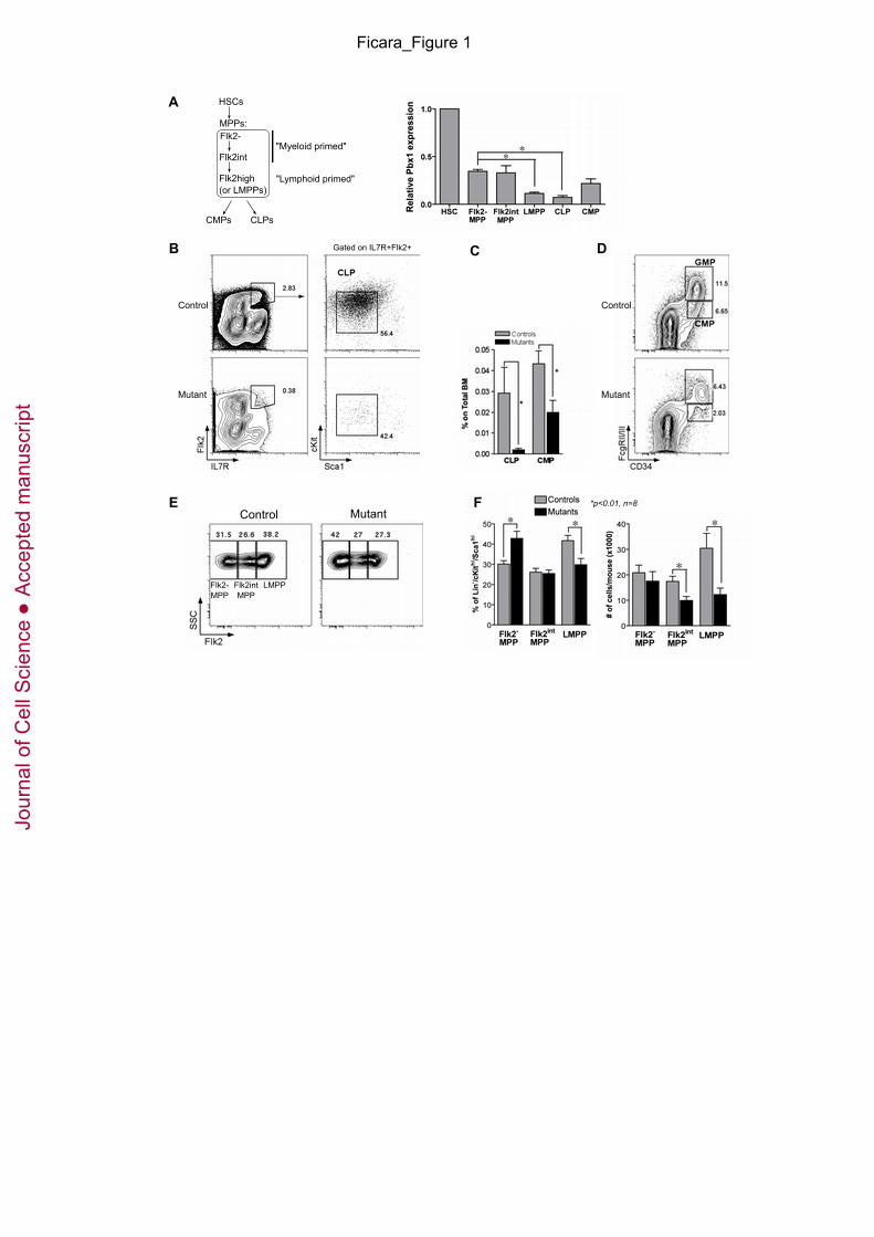
prospectively isolated wt stem and progenitor cells within the hematopoietic hierarchy (Fig. 1A).
For this purpose, MPPs were flow-sorted into three fractions according to Flk2 expression (Fig.
1A). Pbx1 transcripts were most abundant in HSCs, present at intermediate levels in MPPs with
robust myeloid maturation potential (Flk2- MPPs and Flk2int MPPs) and CMPs, and at lower
levels in lymphoid-primed MPPs (Flk2high, also known as LMPPs (Mansson et al., 2007) and
CLPs. This suggested that, in addition to HSCs, Pbx1 may serve important roles in MPPs and
downstream progenitors. Markedly lower Pbx1 expression in CLPs compared to MPPs raised
the possibility that the drastic reduction of CLPs observed in the absence of Pbx1 (Ficara et al.,
2008 and Fig. 1B, C) might be the consequence of up-stream defects. Consistent with these
observations, FACS analysis of MPP subsets revealed a significant decrease in the percentage
and absolute number of LMPPs in mutant (Tie2Cre+.Pbx1-/f) mice compared to control (Tie2Cre-
.Pbx1+/f) mice (Figs 1E, F), supporting a role for Pbx1 in their maintenance. In addition, a
consistent decrease in CMPs (Ficara et al., 2008 and Fig. 1C, D), and associated reduction of the
absolute number of their Flk2int progenitors (Fig. 1F), was observed.
Myeloid differentiation from Pbx1-deficient MPPs is accelerated. To determine if Pbx1
regulates the ability of multipotent progenitors to differentiate into down-stream progeny, Flk2+
Jour
nal o
f Cel
l Sci
ence
Acc
epte
d m
anus
crip
t
5
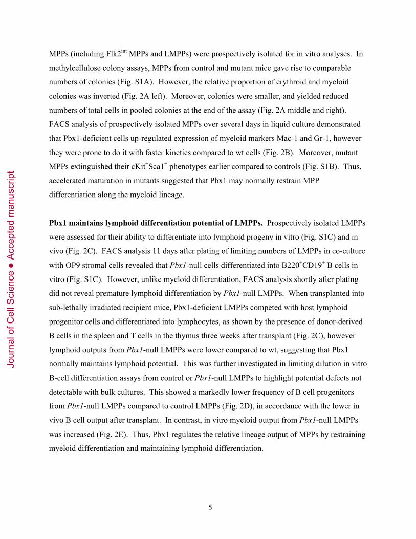
MPPs (including Flk2int MPPs and LMPPs) were prospectively isolated for in vitro analyses. In
methylcellulose colony assays, MPPs from control and mutant mice gave rise to comparable
numbers of colonies (Fig. S1A). However, the relative proportion of erythroid and myeloid
colonies was inverted (Fig. 2A left). Moreover, colonies were smaller, and yielded reduced
numbers of total cells in pooled colonies at the end of the assay (Fig. 2A middle and right).
FACS analysis of prospectively isolated MPPs over several days in liquid culture demonstrated
that Pbx1-deficient cells up-regulated expression of myeloid markers Mac-1 and Gr-1, however
they were prone to do it with faster kinetics compared to wt cells (Fig. 2B). Moreover, mutant
MPPs extinguished their cKit+Sca1+ phenotypes earlier compared to controls (Fig. S1B). Thus,
accelerated maturation in mutants suggested that Pbx1 may normally restrain MPP
differentiation along the myeloid lineage.
Pbx1 maintains lymphoid differentiation potential of LMPPs. Prospectively isolated LMPPs
were assessed for their ability to differentiate into lymphoid progeny in vitro (Fig. S1C) and in
vivo (Fig. 2C). FACS analysis 11 days after plating of limiting numbers of LMPPs in co-culture
with OP9 stromal cells revealed that Pbx1-null cells differentiated into B220+CD19+ B cells in
vitro (Fig. S1C). However, unlike myeloid differentiation, FACS analysis shortly after plating
did not reveal premature lymphoid differentiation by Pbx1-null LMPPs. When transplanted into
sub-lethally irradiated recipient mice, Pbx1-deficient LMPPs competed with host lymphoid
progenitor cells and differentiated into lymphocytes, as shown by the presence of donor-derived
B cells in the spleen and T cells in the thymus three weeks after transplant (Fig. 2C), however
lymphoid outputs from Pbx1-null LMPPs were lower compared to wt, suggesting that Pbx1
normally maintains lymphoid potential. This was further investigated in limiting dilution in vitro
B-cell differentiation assays from control or Pbx1-null LMPPs to highlight potential defects not
detectable with bulk cultures. This showed a markedly lower frequency of B cell progenitors
from Pbx1-null LMPPs compared to control LMPPs (Fig. 2D), in accordance with the lower in
vivo B cell output after transplant. In contrast, in vitro myeloid output from Pbx1-null LMPPs
was increased (Fig. 2E). Thus, Pbx1 regulates the relative lineage output of MPPs by restraining
myeloid differentiation and maintaining lymphoid differentiation.
Jour
nal o
f Cel
l Sci
ence
Acc
epte
d m
anus
crip
t

6
Pbx1-deficient CMPs display altered proliferation kinetics. To determine whether intrinsic
defects may contribute to the reduced number of CMPs and CLPs in the absence of Pbx1, we
quantified the in vivo steady-state cycling activity of lineage-restricted progenitors. Mice were
injected with BrdU and two hours later FACS analysis of CLP and CMP revealed no significant
decreases in BrdU incorporation indicating that the observed reductions in adult mutant mice
were not due to a decreased capacity to proliferate (Fig. 3A). Rather, there was a tendency
toward higher BrdU incorporation in mutant CMPs, although it did not reach statistical
significance (p=0.07). Furthermore, annexin V analysis excluded increased apoptosis as a major
cause underlying CMP and CLP reduction (not shown).
Proliferation of prospectively isolated CMPs and GMPs was then studied in liquid culture
in response to growth factor stimulation. Unexpectedly, Pbx1-null CMPs displayed a higher
proliferation rate compared to wt in the first few days of culture. However, their number
increased at a much lower rate compared to wt at later time points, so that the ratio of mutant CMPs
versus wt was inverted at day 8 (Fig. 3B). Of note, there was no evidence of increased cell death
as a major underlying cause of the observed phenotype at these time points (not shown). The in
vitro proliferation capacity of mutant GMPs, on the other hand, did not differ from normal
controls (not shown). The short-term higher proliferative response to cytokine stimulation in
mutant CMPs was confirmed by monitoring dilution of the CFSE dye after two days of liquid
culture (Fig. 3C). Thus, the reduced numbers of post-natal Pbx1-deficient committed
progenitors in vivo are not due to an intrinsic lack of proliferation, but rather to its aberrantly
rapid extinction in the myeloid lineage.
Pbx1-deficient CMPs differentiate prematurely. Prospectively isolated CMPs were placed in
liquid culture to assess whether the increased proliferation was associated with aberrant
differentiation capacity. Within 1 day of culture, a higher proportion of Pbx1-null CMPs had
acquired Mac-1 expression compared to controls (Fig. 4A). At day three, cultures initiated by
mutant CMPs contained a smaller proportion of immature c-Kit+Mac-1- cells, and a higher
fraction of c-Kit-Mac-1+ cells, demonstrating premature differentiation. Analysis of c-Kit+Mac-
1- gated cells showed that mutant CMPs lost their immature phenotype more rapidly than their
wt counterparts (Fig. 4A, B). Analysis of GMPs showed a similar but milder trend (Fig. S1D).
Concomitant FACS analysis for CFSE and Mac-1 after two days of culture showed that cells that
Jour
nal o
f Cel
l Sci
ence
Acc
epte
d m
anus
crip
t

7
had acquired Mac-1 had also diluted CFSE (Fig. S1E) indicating that the observed premature
differentiation by Pbx1-null CMPs was not due to an absence of proliferation.
In colony-forming-cell assays, CMPs from mutant and control mice gave rise to similar
numbers of colonies (Fig. S1A). Nevertheless, there was a significant reduction in the relative
proportion of erythroid colonies with a concomitant increase of myeloid colonies in cultures
initiated by mutant CMPs (Fig. 4C top left). Moreover, a lower proportion of undifferentiated,
“blast-like” cells in cytospin preparations from pooled Pbx1-null colonies was observed
compared to controls (Fig. 4C right). In addition, colonies from Pbx1-null CMPs tended to be
smaller compared to wt (not shown), resulting in a lower number of total cells (Fig. 4C bottom
left), in agreement with results in liquid culture at day 8 (Fig. 3B). Overall, these data indicate
that accelerated maturation to the next stage in the hematopoietic hierarchy in the absence of
sufficient self-renewal, with a preferential skewing towards the granulocytic-monocytic vs the
erythroid lineage, likely contributes to the observed CMP reduction in the absence of Pbx1.
To study differentiation capacity toward B cells by mutant lymphoid progenitors,
prospectively isolated CLPs from control or mutant mice were co-cultured with OP9 cells and
FACS analysis was performed at two time points (Fig. S1F). Pbx1-null CLPs differentiated as
well as control CLPs into B220+CD19+ cells in vitro. However, a lower frequency of functional
CLPs from Pbx1-null mice was observed in vitro in limiting dilution B-cell differentiation assays
(Fig. 4D). Prospectively isolated CLPs were also transplanted into sub-lethally irradiated wt
recipients. Transplanted mice were sacrificed one or three weeks later to monitor the presence of
donor-derived B cells in BM and spleen, and of T cells in the thymus. Pbx1-null CLPs
differentiated into mature lymphocytes in vivo, although the degree of chimerism in the B cell
lineage tended to be lower in mice transplanted with mutant CLPs compared to controls at both
time points (Fig. 4E and not shown). Thus, both myeloid and lymphoid progenitors differentiate
into mature progeny in the absence of Pbx1, but with a remarkably higher efficiency in the
myeloid lineage versus decreased efficiency in the lymphoid lineage.
GMP gene activation in Pbx1-deficient CMPs. Transcriptional profiling was performed to
define Pbx1-dependent genes and pathways responsible for the CMP premature maturation
phenotype. To provide a baseline for comparison, we initially interrogated the normal CMP-to-
GMP transition, which revealed 705 (592 non-redundant) differentially expressed transcripts
Jour
nal o
f Cel
l Sci
ence
Acc
epte
d m
anus
crip
t

8
(282 down-regulated in GMPs = typical CMP genes, including Pbx1, and 423 up-regulated in
GMPs = typical GMP genes) (Table S1; Fig. S2A). Their classification based on GO (Gene
Ontology, Fig. S3B) underscored a substantial increase in metabolic activity as CMPs progress
towards the GMP stage, with a concomitant reduction of transcripts coding for DNA-binding
proteins/transcription factors, as expected with an increase in specialization (Fig. S3A).
Comparison of Pbx1 mutant versus control CMPs revealed 329 (305 non-redundant)
differentially expressed transcripts (207 down-regulated, and 122 up-regulated in the absence of
Pbx1 –Tables S2, 3; Fig. S2A). Comparison of control versus mutant GMPs revealed fewer
(129) differentially regulated transcripts, in accordance with their milder phenotype, and with the
lower (although detectable) expression level of Pbx1 in wt GMPs relative to wt CMPs (Fig. 6A)
and to MPP1 (Fig. S2C), suggesting that Pbx1 does not play a crucial role in GMPs. Expression
of genes encoding surface markers used for isolation of CMPs and GMPs, respectively, were not
perturbed in mutants.
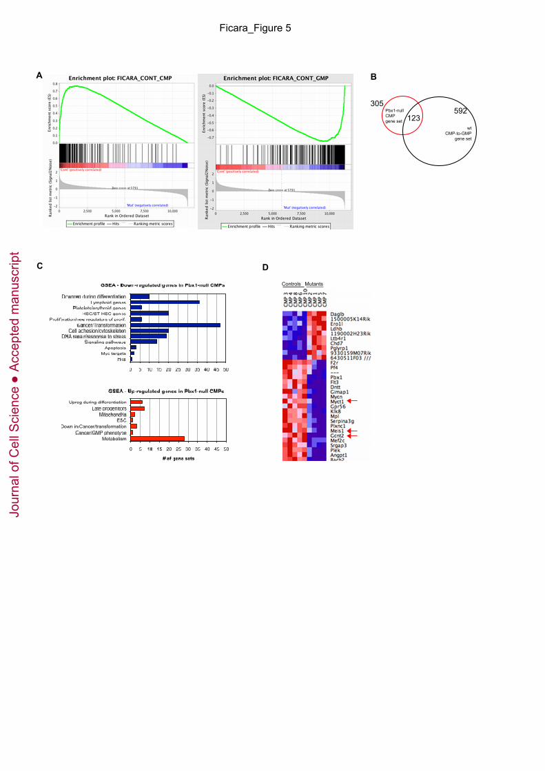
GSEA (Gene Set Enrichment Analysis (Mootha et al., 2003; Subramanian et al., 2005)
was employed to compare the rank-ordered dataset of mutant versus wt CMP transcripts against
the gene set defining normal CMP-to-GMP transition. Transcripts down-regulated in mutant
CMPs were highly enriched for normal CMP-specific genes (Fig. 5A), whereas transcripts up-
regulated in mutant CMPs correlated with normal GMP genes. Accordingly, 40% of the 305
non-redundant transcripts differentially expressed in mutant versus wt CMPs by SAM (Statistical
Analysis of Microarray) analysis were also differentially regulated in the normal CMP-to-GMP
transition (Fig. 5B). Principal Component Analysis (PCA) confirmed that Pbx1-null CMPs
tended to be located between normal GMPs and CMPs (Fig. S2B). In support of this notion,
classification of genes up-regulated in the absence of Pbx1 based on GO almost completely
overlapped with that of normal GMPs (Fig. S3D). Thus, in the absence of Pbx1, phenotypically
defined CMPs prematurely express a subset of genes typical of their downstream GMP progeny.
GSEA was then employed to compare the rank-ordered lists of mutant and normal CMP
and GMP transcripts against published gene sets. The transcripts down-regulated in Pbx1-null
CMPs were notably enriched for several gene sets (Table S4; Fig. 5C) down-regulated during
differentiation of myeloid or other cell types, providing further support to the notion that Pbx1
normally restrains myeloid differentiation. Unexpectedly, several lymphoid gene sets were
down-regulated in mutant CMPs, suggesting decreased lineage promiscuity and strong myeloid-
Jour
nal o
f Cel
l Sci
ence
Acc
epte
d m
anus
crip
t

9
commitment in Pbx1-null progenitors. Transcripts down-regulated in Pbx1-null CMPs were also
enriched for HSC genes, indicating that Pbx1 normally maintains at least part of the HSC
signature that is persistently expressed in committed progenitors.
Pbx1 maintains proto-oncogenic pathways in myeloid progenitors. The most numerous
group of annotated gene sets down-regulated in mutant CMPs was associated with various
malignancies, including myeloid leukemias, underscoring the fact that Pbx1 may normally
sustain pathways frequently perturbed in oncogenesis. In support of this hypothesis, a search of
the GEO and GSEA microarray databases revealed that Pbx1 is highly expressed in a subset of
human cancers (Fig. S4A-F; Table S4), and over-expressed compared to corresponding normal
tissues in mouse and human breast cancer (Fig. S4G-H), but down-regulated in response to
treatment (Figure S4I).
Thus, multiple genes and pathways are aberrantly regulated in Pbx1-null CMPs, with
premature derepression of typical GMP transcripts, and down-regulation of genes involved in
malignant transformation.
Meis1 is part of the Pbx1-dependent transcriptional program. Among the
cancer/transformation gene sets identified by GSEA, the two with the highest enrichment score
and the lowest FDR (0.00) and q value were comprised of Meis1 target genes in transformed cell
lines (Table S4). Meis1 forms a transcriptional complex with Pbx1 and is one of the few genes
down-regulated in Pbx1-null CMPs that are also down-regulated in Pbx1-null HSCs (Fig. 5D and
Ficara et al., 2008), suggesting that Pbx1 might directly or indirectly regulate expression of its
dimerization partner.
qPCR analysis on prospectively isolated wt CMPs and GMPs showed that Pbx1 and
Meis1 were down-regulated to similar extents during the CMP-to-GMP transition, as expected if
Pbx1 is transcriptionally up-stream of Meis1 (Fig. 6A). To further test the latter hypothesis,
cKit+ cells (enriched in myeloid progenitors) were transduced with a retroviral vector expressing
either GFP alone (MOCK) or Pbx1 and GFP (PBX1). Pbx1 and Meis1 over-expression was
measured by real-time PCR before (day 0, day 1) and after (day 3, in GFP+ cells) transduction
(Fig. 6B). In mock-transduced cells, both transcripts were down-regulated upon culture, as
expected since at day 3 most cells had differentiated (c-Kit-Mac-1+ phenotype). However, Meis1
Jour
nal o
f Cel
l Sci
ence
Acc
epte
d m
anus
crip
t

10
expression was up-regulated in Pbx1 transduced cells, in accordance with the hypothesis that
Meis1 is transcriptionally downstream of Pbx1, directly or indirectly. Flt3, a known Meis1
target gene, displayed a similar expression profile (Fig. S5). Nevertheless, forced expression of
Meis1 was unable to rescue the null phenotype in absence of Pbx1 (data not shown) consistent
with their function as a heterodimer. Thus, Meis1 is both a downstream effector as well as a
dimerization partner in the Pbx1-dependent transcriptional program of hematopoietic
progenitors.
Discussion
In this study we demonstrate a role for Pbx1 in sustaining expression of transcriptional programs
that restrain myeloid maturation in hematopoietic progenitors and are frequently perturbed in
human cancers. Pbx1 also sustains the persistent expression of HSC and B-lymphoid genes in
CMPs, highlighting a lineage promiscuity that is maintained by Pbx1. These data support the
conclusion that Pbx1 serves dual roles in hematopoietic progenitors to temporally restrict
myeloid differentiation, and maintain lymphoid potential, thus being crucial for homeostasis of
the hematopoietic system.
Our study provides new insight into the role of Pbx1 as a brake on cellular differentiation
and a regulator of the balance between self-renewal versus maturation. Previous studies have
attributed the hypoplasia or aplasia of multiple organ systems associated with embryonic
deficiency of Pbx1 to a progenitor proliferation defect (Brendolan et al., 2005; Manley et al.,
2004; Schnabel et al., 2003; Selleri et al., 2001). In contrast, our studies based on analyses of
hematopoietic progenitors at a single cell resolution showed increased short-term proliferation
that was prematurely extinguished as progenitors (MPP and CMP) rapidly progressed to the next
stage of differentiation. As a consequence, total cell numbers generated in culture and in vivo
were reduced but the proliferative index was not significantly perturbed. Accelerated myeloid
maturation was confirmed in vivo by gene expression profiles of freshly isolated Pbx1-deficient
CMPs, which displayed premature derepression of GMP genes and enrichment for differentiation
gene sets. Accelerated maturation has also been observed in embryonic chondrocytes (Selleri et
al., 2001), which undergo premature ossification in Pbx1-ko embryos. Taken together, these
Jour
nal o
f Cel
l Sci
ence
Acc
epte
d m
anus
crip
t

11
studies suggest that the primary defect associated with Pbx1 deficiency is not reduced
proliferation per se, but accelerated maturation that shortens the temporal phase of proliferative
expansion for progenitors resulting in hypoplasia or cytopenia.
In addition to functioning as a brake on myeloid differentiation, Pbx1 also maintains
lymphoid potential in hematopoietic progenitors. In the absence of Pbx1, the earliest progenitors
with skewed or restricted lymphoid differentiation capacity (LMPPs and CLPs, respectively) are
numerically reduced and functionally compromised with decreased lymphoid outputs in culture
and transplant experiments. This likely reflects a substantial role for Pbx1 in maintaining
lymphoid gene expression in progenitors consistent with previous studies showing that lymphoid
gene expression in HSCs (so-called lymphoid priming) is compromised in the absence of Pbx1
(Ficara et al., 2008). In the current studies, persistent expression of lymphoid genes in wt CMPs
enabled our demonstration that Pbx1 maintains their lineage inappropriate expression. The
functional significance for promiscuous expression of the lymphoid signature in wt CMPs is
unclear although it may underlie the well-documented lineage switching potential of myeloid
progenitors (Anderson et al., 2007; Boeckx et al., 2004; Chiang and Monroe, 1999; Yu et al.,
2003). Nevertheless, its perturbation in Pbx1 mutants provides evidence of a substantial role for
Pbx1 in maintaining lymphoid gene expression in a variety of progenitors, as well as HSCs,
which may account for the major reductions in LMPP, CLP and B cell progenitors of Pbx1-
deficient mice.
Our study provides a rationale to investigate whether Meis1 is a direct downstream target
of Pbx1, and as a possible mediator of Pbx1 function in CMPs. Meis1 is generally considered a
typical HSC gene based on its high expression in the most immature primitive hematopoietic
populations, and down-regulation upon differentiation (Argiropoulos and Humphries, 2007;
Pineault et al., 2002). In this study we confirm that the expression of Pbx1 and Meis1 is
decreased in myeloid oligopotent progenitors compared to HSCs, but nevertheless functions to
limit their otherwise rapid maturation. In accordance with our conclusion that Meis1 might be a
major mediator of Pbx1 in restraining myeloid differentiation, knockdown of Meis1 in murine
leukemia cells increases monocytic differentiation (Kumar et al., 2009). Meis1 is also a critical
downstream mediator of the MLL oncogenic program, functioning as an essential regulator of
leukemia stem cell (LSC) potential (Wong et al., 2007), and our data suggest that its reported
function in maintaining self-renewal of LSCs might partly be due to a role in preventing
Jour
nal o
f Cel
l Sci
ence
Acc
epte
d m
anus
crip
t

12
differentiation. Meis1 regulation by Pbx1 is unexpected considering that it is a Pbx1
dimerization partner (Chang et al., 1997), although is consistent with the recently reported
compensatory increase in Pbx1 levels in response to loss of Meis1 (Unnisa et al., 2012).
However, Pbx1 directly regulates Hox11 in spleen progenitor cells during spleen ontogeny, and
Hox11, in turn, regulates its own promoter in complex with Pbx1 (Brendolan et al., 2005).
Therefore, Pbx1 participates in complex cross-regulatory circuits with its DNA binding partners.
Our bioinformatics analyses indicated that Pbx1 maintains expression of transcriptional
pathways associated with oncogenesis suggesting a proto-oncogenic function for Pbx1 in various
lineages, including CMPs. This observation was confirmed upon removal of the annotated cell
cycle genes from the analysis (not shown), indicating that the proto-oncogenic signature is not a
consequence of the perturbation of CMP proliferation properties. The same observation was also
corroborated upon application of more stringent criteria for accepting gene sets as enriched for
(not shown). Genes associated with malignancies that are downstream of Pbx1 in CMPs include
Mpl, Pf4, Angpt1, Mef2c, Flt3, Fyn, Bcl2, Syk, and Abl1, among others. Despite these
hierarchical relationships, retroviral-mediated forced expression of wt Pbx1 was not sufficient to
induce leukemia in hematopoietic progenitors (data not shown) likely reflecting that its
oncogenic activation requires protein fusion as originally observed in B-cell precursor acute
lymphoblastic leukemia (Kamps et al., 1990; Nourse et al., 1990), which alters its transcriptional
properties. Nevertheless, wt Pbx1 is a component of crucial transcriptional cascades initiated by
oncoproteins in leukemia pathogenesis (Eklund, 2007; Wong et al., 2007), and our studies
demonstrate that Pbx1 in turn regulates transcriptional pathways that normally oppose
differentiation and are perturbed in a variety of malignancies.
Material and Methods
Mice. Tie2Cre+.Pbx1-/f mutant mice have been described previously (Ficara et al., 2008).
Congenic Ly5.1 mice, purchased from Jackson Laboratories (Bar Harbor, ME, USA), were
maintained in the Stanford animal facility and used as recipients in transplantation experiments.
All experiments were performed with the approval of and in accordance with Stanford’s and
Istituto Clinico Humanitas Administrative Panel on Laboratory Animal Care.
Jour
nal o
f Cel
l Sci
ence
Acc
epte
d m
anus
crip
t

13
Analysis and isolation of hematopoietic progenitor cells. BM cell suspensions were obtained
by crushing of multiple bones as described (Ficara et al., 2008). Progenitors were purified based
on the absence of lineage markers using a Lineage Cell Depletion kit (Miltenyi Biotec, Auburn,
CA, USA) and an automated cell separator (AutoMACS, Miltenyi Biotec), and then subjected to
cell surface staining prior to FACS analysis or sorting. Lineage-depleted BM was further
deprived of residual Lin+ cells after incubation with APC-Cy7 or PE-conjugated Streptavidin
(eBioscience, San Diego, CA, USA) and monoclonal antibodies (mAbs) to additional T cell
markers not present in the cocktail (CD3, 4, 8, BD Pharmingen, San Jose, CA
USA). In the present study the following subpopulations were analyzed: HSCs (Lin-
/cKithi/Scahi/CD34-/ Flk2-); MPPs at different stages of maturation (Lin-/CD127-
/cKithi/Scahi/CD34+/Flk2- or Flk2int or Flk2high); CLPs (Lin-/CD127+/Flk2high/cKitint/Scaint); CMPs
(Lin-/cKit+/Sca-/CD34+/FcgRII/IIIint); GMPs (Lin-/cKit+/Sca-/CD34+/FcgRII/IIIhigh). The
following fluorochrome-conjugated mAbs were purchased from eBioscience as conjugates to
APC, Cy5-PE, Cy7-PE, or PE: cKit (2B8), Sca1 (D7), CD127 (A7R34), CD16/32 (93), CD135
(AF2 10.1). FITC-conjugated CD34 (RAM34) was purchased from BD Pharmingen. CLPs were
isolated using procedures described previously (Karsunky et al., 2008). Cell sorts were
performed using a FACS Vantage (BD Biosciences, San Jose, CA, USA) equipped with Diva
software (BD, Franklin Lakes, New Jersey, USA), and data were analyzed using FlowJo (Tree
Star, Ashland, OR, USA). For in vitro B cell differentiation assays, lineage-depleted BM cells
were prepared with a lineage-depletion kit (Stem Cell Technologies, Vancouver, BC, Canada)
and the automated Robosep cell separator, prior to cell surface staining and sorting. Cell sorts
were performed using a FACS ARIA (BD Biosciences).
Real-time quantitative PCR. HSCs and progenitors were sorted from wt C57BL/6 mice or
from GFP+ cells prior to RNA extraction with Trizol (Invitrogen, Carlsbad, CA, USA). cDNA
was prepared using Superscript First-Strand Synthesis System for RT-PCR (Invitrogen) and then
subjected to real-time PCR using Taqman probes (Applied Biosystems, Foster City, CA, USA).
Cell culture. For colony-forming unit assays, sorted hematopoietic progenitors were seeded into
methylcellulose-containing medium (methoCult 3234; Stem Cell Technologies) in the presence
Jour
nal o
f Cel
l Sci
ence
Acc
epte
d m
anus
crip
t

14
of SCF, Flt3 ligand (20 ng/ml each), IL-6, IL-3, GM-CSF, TPO (10 ng/ml each) and Epo (3
U/ml), and colonies were scored at day 7. MPPs, CMPs and GMPs were cultured in IMDM
supplemented with 10% FBS (Hyclone, Logan, UT, USA) and SCF (20 ng/ml), IL-6, IL-3 (10
ng/ml each), in U-bottomed 96 well plates, and analyzed by FACS 3 or 6 days later upon
staining with Abs to Mac1/CD11b (M1/70, BD Pharmingen), Gr1 (RB6-8C5, eBioscience),
and/or Abs defined above. LMPPs and CLPs were co-cultured with OP9 stromal cells in Mem-
alpha supplemented with heat-inactivated 10% FBS, 50 mM b-Mercaptoethanol, and SCF, Flt3L
(20 ng/ml each), and IL-7 (10 ng/ml), and analyzed by FACS 4 or 10-11 days later upon staining
with Abs to B220 (RA3-6B2, eBioscience) and CD19 (1D3, BD Pharmingen). For limiting
dilution assays, OP9 cells were seeded in flat-bottomed 96-well plates, and different amounts of
sorted LMPPs or CLPs from pools of three mice were added to each well (10 to 96
wells/condition). Medium was half-replaced every third day, and at day 10 cells from each well
were analyzed by FACS to assess the presence of B220+CD19+CD11b-Gr1- B cells.
Transplantation assays. Transplants of LMPPs or CLPs (2,000-4,500 cells) from mutant or
control mice (Ly5.2) were performed by retro-orbital injection of sublethally irradiated Ly5.1
mice (450 cGy). Spleens and thymuses were dissected from transplanted animals one or three
weeks later as described (Karsunky et al., 2008). Donor and recipients were distinguished using
mAbs to Ly5.2/CD45.2 (104) and Ly5.1/CD45.1 (A20).
Cell cycle analysis. Mice received a single intraperitoneal injection of 5-bromodeoxyuridine
(BrdU) 2 hrs prior to sacrifice. Analysis of BrdU incorporation in BM progenitors was
performed using the FITC BrdU Flow Kit (BD Pharmingen) according to the manufacturer’s
protocol. Concomitant use of the anti-BrdU mAb and Abs to cell surface markers allowed
detection of proliferating cells within the different progenitor subsets after lineage-depletion
(LMPPs and CLPs) or cKit positive-selection (CMP, GMP, MEP). CFSE labeling (Invitrogen)
was performed according to manufacturer’s instructions.
Statistical analysis. Significance of differences was determined by two-tailed Student’s t-test.
Error bars in bar graphs indicate ± s.e.m.
Jour
nal o
f Cel
l Sci
ence
Acc
epte
d m
anus
crip
t
15
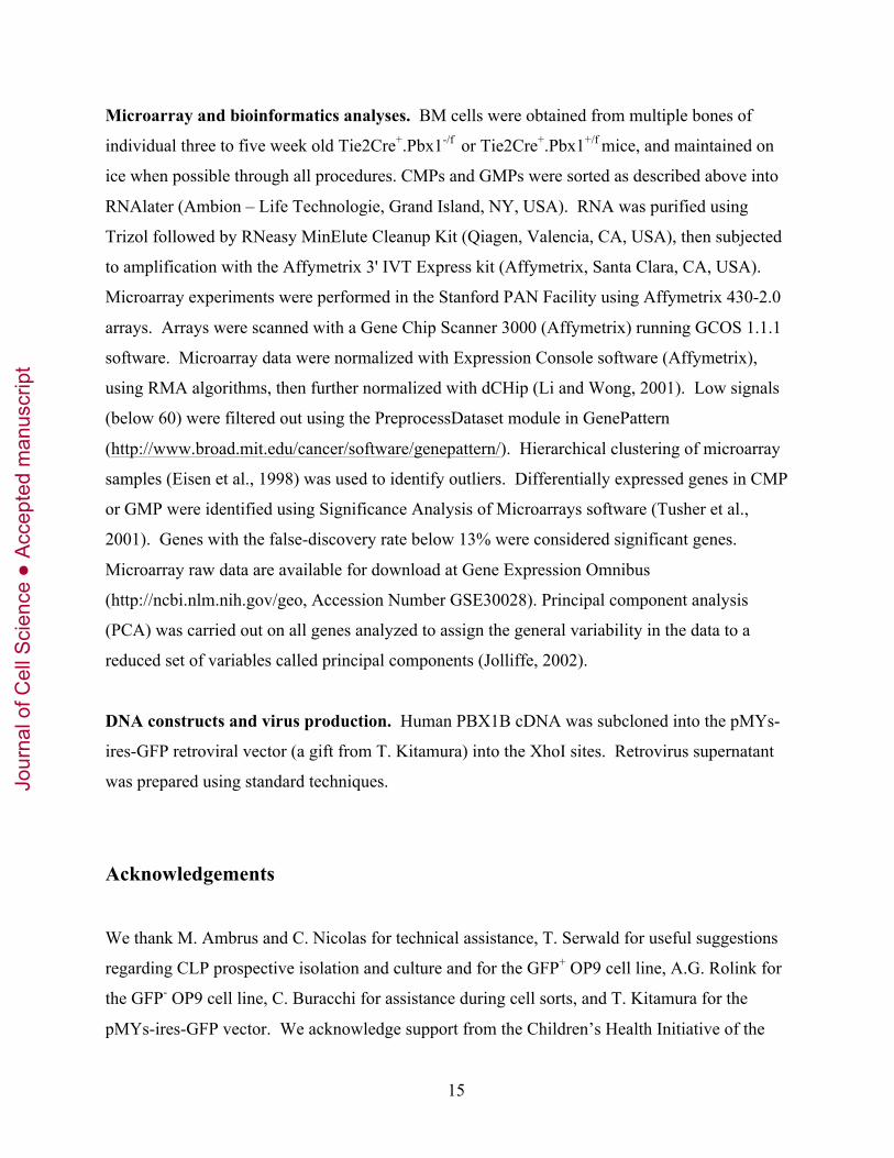
Microarray and bioinformatics analyses. BM cells were obtained from multiple bones of
individual three to five week old Tie2Cre+.Pbx1-/f or Tie2Cre+.Pbx1+/f mice, and maintained on
ice when possible through all procedures. CMPs and GMPs were sorted as described above into
RNAlater (Ambion – Life Technologie, Grand Island, NY, USA). RNA was purified using
Trizol followed by RNeasy MinElute Cleanup Kit (Qiagen, Valencia, CA, USA), then subjected
to amplification with the Affymetrix 3' IVT Express kit (Affymetrix, Santa Clara, CA, USA).
Microarray experiments were performed in the Stanford PAN Facility using Affymetrix 430-2.0
arrays. Arrays were scanned with a Gene Chip Scanner 3000 (Affymetrix) running GCOS 1.1.1
software. Microarray data were normalized with Expression Console software (Affymetrix),
using RMA algorithms, then further normalized with dCHip (Li and Wong, 2001). Low signals
(below 60) were filtered out using the PreprocessDataset module in GenePattern
(http://www.broad.mit.edu/cancer/software/genepattern/). Hierarchical clustering of microarray
samples (Eisen et al., 1998) was used to identify outliers. Differentially expressed genes in CMP
or GMP were identified using Significance Analysis of Microarrays software (Tusher et al.,
2001). Genes with the false-discovery rate below 13% were considered significant genes.
Microarray raw data are available for download at Gene Expression Omnibus
(http://ncbi.nlm.nih.gov/geo, Accession Number GSE30028). Principal component analysis
(PCA) was carried out on all genes analyzed to assign the general variability in the data to a
reduced set of variables called principal components (Jolliffe, 2002).
DNA constructs and virus production. Human PBX1B cDNA was subcloned into the pMYs-
ires-GFP retroviral vector (a gift from T. Kitamura) into the XhoI sites. Retrovirus supernatant
was prepared using standard techniques.
Acknowledgements
We thank M. Ambrus and C. Nicolas for technical assistance, T. Serwald for useful suggestions
regarding CLP prospective isolation and culture and for the GFP+ OP9 cell line, A.G. Rolink for
the GFP- OP9 cell line, C. Buracchi for assistance during cell sorts, and T. Kitamura for the
pMYs-ires-GFP vector. We acknowledge support from the Children’s Health Initiative of the
Jour
nal o
f Cel
l Sci
ence
Acc
epte
d m
anus
crip
t

16
Packard Foundation and PHS grant CA90735. F.F. was supported by an ASH Scholar Award
from the American Society of Hematology, and by a Marie Curie International Reintegration
Grant.
References
Anderson, K., Rusterholz, C., Mansson, R., Jensen, C. T., Bacos, K., Zandi, S., Sasaki, Y.,
Nerlov, C., Sigvardsson, M. and Jacobsen, S. E. (2007). Ectopic expression of PAX5 promotes
maintenance of biphenotypic myeloid progenitors coexpressing myeloid and B-cell lineage-
associated genes. Blood 109, 3697-705.
Argiropoulos, B. and Humphries, R. K. (2007). Hox genes in hematopoiesis and
leukemogenesis. Oncogene 26, 6766-76.
Boeckx, N., van der Velden, V. H., Boogaerts, M., Hagemeijer, A., Vandenberghe, P. and
van Dongen, J. J. (2004). An inv(16)(p13q22) positive acute myeloid leukaemia relapsing as
acute precursor B-cell lymphoblastic leukaemia. Haematologica 89, 96-98.
Brendolan, A., Ferretti, E., Salsi, V., Moses, K., Quaggin, S., Blasi, F., Cleary, M. L. and
Selleri, L. (2005). A Pbx1-dependent genetic and transcriptional network regulates spleen
ontogeny. Development 132, 3113-26.
Cano, F., Drynan, L. F., Pannell, R. and Rabbitts, T. H. (2008). Leukaemia lineage
specification caused by cell-specific Mll-Enl translocations. Oncogene 27, 1945-50.
Chang, C. P., Jacobs, Y., Nakamura, T., Jenkins, N. A., Copeland, N. G. and Cleary, M. L.
(1997). Meis proteins are major in vivo DNA binding partners for wild-type but not chimeric
Pbx proteins. Mol Cell Biol 17, 5679-87.
Chiang, M. Y. and Monroe, J. G. (1999). BSAP/Pax5A expression blocks survival and
Jour
nal o
f Cel
l Sci
ence
Acc
epte
d m
anus
crip
t

17
expansion of early myeloid cells implicating its involvement in maintaining commitment to the
B-lymphocyte lineage. Blood 94, 3621-32.
Cleary, M. L. (2009). Regulating the leukaemia stem cell. Best Pract Res Clin Haematol 22,
483-7.
Eisen, M. B., Spellman, P. T., Brown, P. O. and Botstein, D. (1998). Cluster analysis and
display of genome-wide expression patterns. Proc Natl Acad Sci U S A 95, 14863-8.
Eklund, E. A. (2007). The role of HOX genes in malignant myeloid disease. Curr Opin Hematol
14, 85-9.
Ficara, F., Murphy, M. J., Lin, M. and Cleary, M. L. (2008). Pbx1 regulates self-renewal of
long-term hematopoietic stem cells by maintaining their quiescence. Cell Stem Cell 2, 484-96.
Galan-Caridad, J. M., Harel, S., Arenzana, T. L., Hou, Z. E., Doetsch, F. K., Mirny, L. A.
and Reizis, B. (2007). Zfx controls the self-renewal of embryonic and hematopoietic stem cells.
Cell 129, 345-57.
Iwasaki, H. and Akashi, K. (2007). Myeloid lineage commitment from the hematopoietic stem
cell. Immunity 26, 726-40.
Jolliffe, I. T. (2002). Principal Component Analysis. New York: Springer-Verlag.
Kamps, M. P., Murre, C., Sun, X. H. and Baltimore, D. (1990). A new homeobox gene
contributes the DNA binding domain of the t(1;19) translocation protein in pre-B ALL. Cell 60,
547-55.
Karsunky, H., Inlay, M. A., Serwold, T., Bhattacharya, D. and Weissman, I. L. (2008).
Flk2+ common lymphoid progenitors possess equivalent differentiation potential for the B and T
lineages. Blood 111, 5562-70.
Jour
nal o
f Cel
l Sci
ence
Acc
epte
d m
anus
crip
t

18
Krivtsov, A. V., Twomey, D., Feng, Z., Stubbs, M. C., Wang, Y., Faber, J., Levine, J. E.,
Wang, J., Hahn, W. C., Gilliland, D. G. et al. (2006). Transformation from committed
progenitor to leukaemia stem cell initiated by MLL-AF9. Nature 442, 818-22.
Kumar, A. R., Li, Q., Hudson, W. A., Chen, W., Sam, T., Yao, Q., Lund, E. A., Wu, B.,
Kowal, B. J. and Kersey, J. H. (2009). A role for MEIS1 in MLL-fusion gene leukemia. Blood
113, 1756-8.
Li, C. and Wong, W. H. (2001). Model-based analysis of oligonucleotide arrays: expression
index computation and outlier detection. Proc Natl Acad Sci U S A 98, 31-6.
Manley, N. R., Selleri, L., Brendolan, A., Gordon, J. and Cleary, M. L. (2004).
Abnormalities of caudal pharyngeal pouch development in Pbx1 knockout mice mimic loss of
Hox3 paralogs. Dev Biol 276, 301-12.
Mansson, R., Hultquist, A., Luc, S., Yang, L., Anderson, K., Kharazi, S., Al-Hashmi, S.,
Liuba, K., Thoren, L., Adolfsson, J. et al. (2007). Molecular evidence for hierarchical
transcriptional lineage priming in fetal and adult stem cells and multipotent progenitors.
Immunity 26, 407-19.
Metcalf, D., Dakic, A., Mifsud, S., Di Rago, L., Wu, L. and Nutt, S. (2006). Inactivation of
PU.1 in adult mice leads to the development of myeloid leukemia. Proc Natl Acad Sci U S A
103, 1486-91.
Minami, Y., Stuart, S. A., Ikawa, T., Jiang, Y., Banno, A., Hunton, I. C., Young, D. J.,
Naoe, T., Murre, C., Jamieson, C. H. et al. (2008). BCR-ABL-transformed GMP as myeloid
leukemic stem cells. Proc Natl Acad Sci U S A 105, 17967-72.
Moens, C. B. and Selleri, L. (2006). Hox cofactors in vertebrate development. Dev Biol 291,
193-206.
Jour
nal o
f Cel
l Sci
ence
Acc
epte
d m
anus
crip
t

19
Mootha, V. K., Lindgren, C. M., Eriksson, K. F., Subramanian, A., Sihag, S., Lehar, J.,
Puigserver, P., Carlsson, E., Ridderstrale, M., Laurila, E. et al. (2003). PGC-1alpha-
responsive genes involved in oxidative phosphorylation are coordinately downregulated in
human diabetes. Nat Genet 34, 267-73.
Nourse, J., Mellentin, J. D., Galili, N., Wilkinson, J., Stanbridge, E., Smith, S. D. and
Cleary, M. L. (1990). Chromosomal translocation t(1;19) results in synthesis of a homeobox
fusion mRNA that codes for a potential chimeric transcription factor. Cell 60, 535-45.
Orkin, S. H. and Zon, L. I. (2008). Hematopoiesis: an evolving paradigm for stem cell biology.
Cell 132, 631-44.
Pineault, N., Helgason, C. D., Lawrence, H. J. and Humphries, R. K. (2002). Differential
expression of Hox, Meis1, and Pbx1 genes in primitive cells throughout murine hematopoietic
ontogeny. Exp Hematol 30, 49-57.
Rothenberg, E. V. (2007). Negotiation of the T lineage fate decision by transcription-factor
interplay and microenvironmental signals. Immunity 26, 690-702.
Santaguida, M., Schepers, K., King, B., Sabnis, A. J., Forsberg, E. C., Attema, J. L., Braun,
B. S. and Passegue, E. (2009). JunB protects against myeloid malignancies by limiting
hematopoietic stem cell proliferation and differentiation without affecting self-renewal. Cancer
Cell 15, 341-52.
Schnabel, C. A., Selleri, L. and Cleary, M. L. (2003). Pbx1 is essential for adrenal
development and urogenital differentiation. Genesis 37, 123-30.
Selleri, L., Depew, M. J., Jacobs, Y., Chanda, S. K., Tsang, K. Y., Cheah, K. S., Rubenstein,
J. L., O'Gorman, S. and Cleary, M. L. (2001). Requirement for Pbx1 in skeletal patterning and
programming chondrocyte proliferation and differentiation. Development 128, 3543-57.
Jour
nal o
f Cel
l Sci
ence
Acc
epte
d m
anus
crip
t

20
Signer, R. A., Montecino-Rodriguez, E., Witte, O. N. and Dorshkind, K. (2010). Immature B
cell progenitors survive oncogenic stress and efficiently initiate Ph+ B-acute lymphoblastic
leukemia. Blood 116, 2522-30.
Somervaille, T. C., Matheny, C. J., Spencer, G. J., Iwasaki, M., Rinn, J. L., Witten, D. M.,
Chang, H. Y., Shurtleff, S. A., Downing, J. R. and Cleary, M. L. (2009). Hierarchical
maintenance of MLL myeloid leukemia stem cells employs a transcriptional program shared
with embryonic rather than adult stem cells. Cell Stem Cell 4, 129-40.
Subramanian, A., Tamayo, P., Mootha, V. K., Mukherjee, S., Ebert, B. L., Gillette, M. A.,
Paulovich, A., Pomeroy, S. L., Golub, T. R., Lander, E. S. et al. (2005). Gene set enrichment
analysis: a knowledge-based approach for interpreting genome-wide expression profiles. Proc
Natl Acad Sci U S A 102, 15545-50.
Tothova, Z., Kollipara, R., Huntly, B. J., Lee, B. H., Castrillon, D. H., Cullen, D. E.,
McDowell, E. P., Lazo-Kallanian, S., Williams, I. R., Sears, C. et al. (2007). FoxOs are
critical mediators of hematopoietic stem cell resistance to physiologic oxidative stress. Cell 128,
325-39.
Tremblay, M., Tremblay, C. S., Herblot, S., Aplan, P. D., Hebert, J., Perreault, C. and
Hoang, T. (2010). Modeling T-cell acute lymphoblastic leukemia induced by the SCL and
LMO1 oncogenes. Genes Dev 24, 1093-105.
Tusher, V. G., Tibshirani, R. and Chu, G. (2001). Significance analysis of microarrays applied
to the ionizing radiation response. Proc Natl Acad Sci U S A 98, 5116-21.
Unnisa, Z., Clark, J. P., Roychoudhury, J., Thomas, E., Tessarollo, L., Copeland, N. G.,
Jenkins, N. A., Grimes, H. L. and Kumar, A. R. (2012). Meis1 preserves hematopoietic stem
cells in mice by limiting oxidative stress. Blood Oct 22. [Epub ahead of print].
Viatour, P., Somervaille, T. C., Venkatasubrahmanyam, S., Kogan, S., McLaughlin, M. E.,
Jour
nal o
f Cel
l Sci
ence
Acc
epte
d m
anus
crip
t

21
Weissman, I. L., Butte, A. J., Passegue, E. and Sage, J. (2008). Hematopoietic stem cell
quiescence is maintained by compound contributions of the retinoblastoma gene family. Cell
Stem Cell 3, 416-28.
Wojiski, S., Guibal, F. C., Kindler, T., Lee, B. H., Jesneck, J. L., Fabian, A., Tenen, D. G.
and Gilliland, D. G. (2009). PML-RARalpha initiates leukemia by conferring properties of self-
renewal to committed promyelocytic progenitors. Leukemia 23, 1462-71.
Wong, P., Iwasaki, M., Somervaille, T. C., So, C. W. and Cleary, M. L. (2007). Meis1 is an
essential and rate-limiting regulator of MLL leukemia stem cell potential. Genes Dev 21, 2762-
74.
Yilmaz, O. H., Valdez, R., Theisen, B. K., Guo, W., Ferguson, D. O., Wu, H. and Morrison,
S. J. (2006). Pten dependence distinguishes haematopoietic stem cells from leukaemia-initiating
cells. Nature 441, 475-82.
Yu, D., Allman, D., Goldschmidt, M. H., Atchison, M. L., Monroe, J. G. and Thomas-
Tikhonenko, A. (2003). Oscillation between B-lymphoid and myeloid lineages in Myc-induced
hematopoietic tumors following spontaneous silencing/reactivation of the EBF/Pax5 pathway.
Blood 101, 1950-5.
Jour
nal o
f Cel
l Sci
ence
Acc
epte
d m
anus
crip
t

22
Figure Legends
Fig. 1. Evaluation of multi-potent progenitors in Pbx1-conditional knock-out mice. (A)
HSCs and progenitors indicated on the left were prospectively isolated by FACS from pooled
BM of adult wt mice prior to RNA purification. The histogram shows Pbx1 transcript levels
relative to HSCs as measured by real-time PCR (3 to 4 biological replicates, each performed in
duplicate; *p<0.01). Expression levels of all progenitor subsets are significantly different from
HSC’s (p≤0.03). MPPs are defined as Lin-/cKithi/Scahi CD34+, divided into three fractions
according to Flk2 expression: Flk2- MPPs, previously called short-term HSCs, Flk2-intermediate
MPPs (Flk2int), Flk2high MPPs or LMPPs. (B) Representative FACS analysis of CLPs from
control (Tie2Cre-.Pbx1+/f) or mutant (Tie2Cre+.Pbx1-/f) mice. Contour plots on the left are
referred to the Lin- gate. Percentages are relative to the parent gate. Quantitative data are shown
in panel C. (C) Histograms represent the average CLP and CMP percentage within the BM of
control or mutant mice (n=14 and 15, respectively; *p<0.05). (D) Representative FACS analysis
of CMPs and GMPs from control or mutant mice. Contour plots are referred to the Lin-
cKit+Sca1- gate. Percentages are relative to the parent gate. Quantitative data are shown in panel
C. (E) Representative FACS analysis is shown for MPPs from control or mutant mice, relative
to the Lin-/cKithi/Scahi gate. (F) Histograms show the percentage of Flk2-negative, Flk2-
intermediate and Flk2-high MPPs within the Lin-/cKithi/Scahi gate (left; qualitative plots are
shown in E), or their absolute numbers per mouse (right). *p=0.01, n=8. Note that the mean
fluorescence intensity was similar in mutants and controls, indicating that the reduction in
progenitor subpopulations was not due to altered expression of the markers used to define them.
Fig. 2. Differentiation ability of Pbx1-null multi-potent progenitors. (A) Methylcellulose
colony assay of prospectively isolated Flk2+ MPPs. Erythroid and myeloid colonies were
counted nine days after plating. The histogram on the left represents the average of four
independent assays, each performed in duplicate (*p≤0.04). Representative myeloid colonies
(CFU-GM) are shown in the middle panels. The histogram on the right represents pooled
colonies from three independent experiments. (B) Time-course FACS analysis (representative of
four) was performed on prospectively isolated MPPs from control or mutant mice, cultured in the
Jour
nal o
f Cel
l Sci
ence
Acc
epte
d m
anus
crip
t

23
presence of serum and cytokines. Contour plots show events within the live/forward and side
light scatter gate, and the average percentage of Mac-1+ cells is represented in the histograms on
the right. (C) Prospectively isolated LMPPs from control or mutant mice were transplanted into
wt recipients. FACS plots show representative analysis of spleen (left, gated on CD19+ cells)
and thymus (right, gated on CD45+ cells) from a mouse transplanted three weeks earlier with
Pbx1-null cells. The histogram depicts the average of four experiments (4 to 7 independent
donors/group, *p=0.05). (D) Limiting dilution analysis (representative of two) of the frequency
of control or mutant LMPPs able to develop into CD19+ B cells in co-culture with GFP-OP9
stromal cells (R-squared = 0.91 and 0.98, respectively). (E) Frequency of wells negative for the
presence of myeloid cells when 10 LMPPs/well were seeded in the OP9 B-cell differentiation
assay.
Fig. 3. Altered progenitor proliferation. (A) Bar graph shows the percentage of BrdU+ cells
in vivo within the indicated progenitor compartments (two independent experiments; average of
three mutants and four controls, *p<0.02). (B) Prospectively isolated CMPs (2,000 to 10,000
cells) were placed in liquid culture in the presence of cytokines. The graph shows the ratio of
Pbx1-null cell number versus control at different time points (n=3, *p=0.003). (C) Prospectively
isolated CMPs were labeled with CFSE immediately after sorting and cultured for two or four
days. FACS analysis was performed immediately after labeling (d0), or at the indicated time
points after plating. Histograms are representative of two independent experiments (n=3).
Fig. 4. Premature differentiation of Pbx1-null lineage-restricted progenitors. (A) FACS
analysis (representative of five) is shown for the progeny of CMPs prospectively isolated from
control or mutant mice. Dot plots on the right are relative to the c-Kit+Mac-1- gate at day 3. (B)
Histograms summarize data shown in (A), day 3 (n=5, *p<0.05). (C) Methylcellulose colony
assay from prospectively isolated CMPs. The top and bottom histograms represent the average
of four and two independent assays, respectively, each performed in duplicate (*p≤0.02). Once
scored, colonies were pooled, counted (bottom histogram), cytospun and stained with May-
Grunwald and Giemsa. Arrowheads on cytospin images indicate blast-like, not terminally
differentiated cells (number of blast-like cells/field: 11.5±1.25 and 1.2±0.57 in control and
mutant, respectively; n=10, p<0.0001). Pictures were taken using a Nikon ECLIPSE E1000M
Jour
nal o
f Cel
l Sci
ence
Acc
epte
d m
anus
crip
t

24
microscope equipped with SPOT advanced software, using a 20X magnification. (D) Limiting
dilution analysis (representative of two) of the frequency of control or mutant CLPs able to
develop into CD19+ B cells as in Figure 2D (R-squared = 0.92 and 0.96, respectively). (E) Bar
graph shows average % of donor-derived cells within B lymphocytes in bone marrow (BM) or
spleen (SP), and within T cells in the thymus (TH), one week after transplant of control or
mutant CLPs (3 recipient mice analyzed/group, 2 different donors).
Fig. 5. Pbx1 deficiency leads to derepression of GMP genes in CMPs. (A) GSEA plots show
enrichment of the gene set characteristic of the normal CMP-to-GMP transition in the Pbx1-null
CMP transcriptome. Transcripts down-regulated in Pbx1-null CMPs positively correlate with
typical CMP genes (left panel, FDR q-value 0), and negatively correlate with typical GMP genes
(right panel, FDR q-value 0). (B) Venn diagram shows overlapping gene expression among
Pbx1-null CMPs and the normal CMP-to-GMP transition. (C) Bar graphs shows the number of
gene sets belonging to different functional groups that are enriched in Pbx1-null CMP down-
regulated (in blue) and up-regulated (in red) transcripts. Gene sets with FDR q-value <0.2 and
Nominal p-value ≤0.05 were considered significantly enriched. (D) Heat map shows the
expression of the top 31 differentially regulated transcripts between control and mutant CMPs,
whose level of expression changed at least three-fold between the two groups. Red arrows
indicate genes that were also down-regulated in Pbx1-null HSCs (Ficara et al. 2008).
Fig. 6. Meis1 is transcriptionally downstream of Pbx1. (A) Bar graph shows Pbx1 and Meis1
transcript levels in GMPs relative to CMPs measured by real-time PCR on progenitors freshly
isolated from wt BM cells (*p≤0.002). (B) Bar graph shows Pbx1 and Meis1 transcript levels
measured by real-time PCR either on freshly isolated wt c-Kit+ cells (day 0), or from cultured
cells before (day 1) or after (day3) transduction with a retroviral vector encoding Pbx1
(*p≤0.0007).
Jour
nal o
f Cel
l Sci
ence
Acc
epte
d m
anus
crip
t
A
CB
HSCs
Flk2-
Flk2int
Flk2high(or LMPPs)
"Myeloid primed"
"Lymphoid primed"
CMPs CLPs
MPPs:
E
Control
Mutant
Control Mutant
Flk2-MPP
Flk2intMPP
LMPP
Control
Mutant
Gated on IL7R+Flk2+
Ficara_Figure 1
C D
F *p<0.01, n=8
Jour
nal o
f Cel
l Sci
ence
Acc
epte
d m
anus
crip
t

B
d3
d6
Total cells
Ficara_Figure 2
C
Spleen Thymus
A
D
Control Mutant
EFraction of wellsneg for myeloid cells
Control Mutant
0.42 0.15
Cells frompooled colonies
Jour
nal o
f Cel
l Sci
ence
Acc
epte
d m
anus
crip
t

Ficara_Figure3
a b
c
(representative of three)
Jour
nal o
f Cel
l Sci
ence
Acc
epte
d m
anus
crip
t

Ficara_Figure 4
C
Ad3
Control
Bd1 d3, cKit+Mac1- gate
D
Mutant
E
Cells frompooled colonies
Jour
nal o
f Cel
l Sci
ence
Acc
epte
d m
anus
crip
t
Ficara_Figure 5
A
C
B
D
Controls Mutants
305
123592
wtCMP-to-GMP
gene set
Pbx1-nullCMPgene set
Jour
nal o
f Cel
l Sci
ence
Acc
epte
d m
anus
crip
t

Ficara_Figure 6
DAY 3
A
B
Jour
nal o
f Cel
l Sci
ence
Acc
epte
d m
anus
crip
t