Embed Size (px)
Citation preview
Protein Data Bank in Europe http://pdbe.org PISA Tutorial
PDBe PDBePISA (Protein Interfaces, Surfaces and
Assemblies)
http://pdbe.org/pisa/
This tutorial introduces the PDBePISA (PISA for short) service, which is a web-‐
based interactive tool offered by the PDBe to investigate stability of formation of
macromolecular complexes (protein, DNA/RNA and ligand). In addtion to a
detailed analysis of surfaces, interfaces and assemblies for all entries in the
Protein Data Bank (PDB), the service also allows upload and analysis of one’s
own PDB or mmCIF format coordinate files.
Background
The stability of a macromolecular complex in a biological system is essentially
governed by the following physicochemical properties:
1) free energy of formation.
2) solvation energy gain.
3) interface area.
4) hydrogen bonds.
5) saltbridges across the interface.
6) hydrophobic specificity.
These interactions are also likely prevalent in crystal systems where protein
molecules interact with one another in the crystallization solution and arrange
themselves in orderly lattices to form crystals. Therefore, given that some
crystallization conditions may mimic actual biological interactions in solution,
the analysis of crystal interfaces and the prediction of higher order structures
may actually reflect those present in a biological system. The PISA service uses
all the criteria listed above in order to analyse a given structure and make a
prediction of the possible stable complex. However, it is important to emphasize
that PISA results are predictions and any conclusions drawn from the same must
Protein Data Bank in Europe http://pdbe.org PISA Tutorial
be in light of other biological and experimental proof. The results from PISA may
be used for validation of independent biochemical data if the structure of the
protein under study is available.
Tutorial
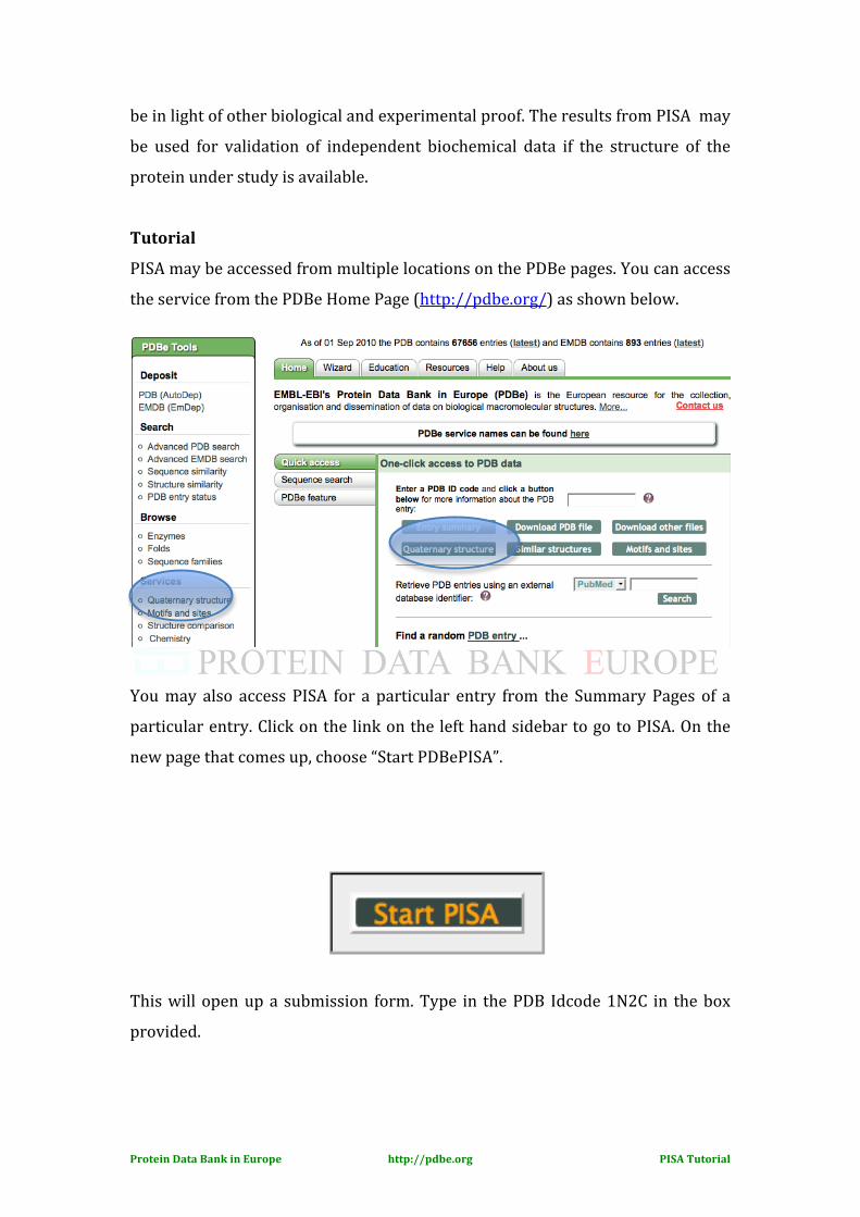
PISA may be accessed from multiple locations on the PDBe pages. You can access
the service from the PDBe Home Page (http://pdbe.org/) as shown below.
You may also access PISA for a particular entry from the Summary Pages of a
particular entry. Click on the link on the left hand sidebar to go to PISA. On the
new page that comes up, choose “Start PDBePISA”.
This will open up a submission form. Type in the PDB Idcode 1N2C in the box
provided.

Protein Data Bank in Europe http://pdbe.org PISA Tutorial
As soon as the file gets uploaded to the server, it will give you preliminary information regarding the PDB entry (number of proteins chains and bound ligands). The entry 1n2c has 8 protein chains and 22 ligands. The most probable assembly is stated as an 8-‐mer. [To know more about this PDB entry (e.g. Name of the protein, origin etc.), then go to the summary pages for this entry http://www.pdbe.org/1n2c/). The atlas page for this entry gives us information that it is a nitrogenase complex structure stabilized by ADP-‐ tetrafluoroaluminate. There are three different proteins NIFD (Chains A and B), NIFK (Chains C and D) and NIFH1 (Chains E, F, G, H).] You may click on the View button (shown above) in blue to view the loaded PDB entry.
Protein Data Bank in Europe http://pdbe.org PISA Tutorial
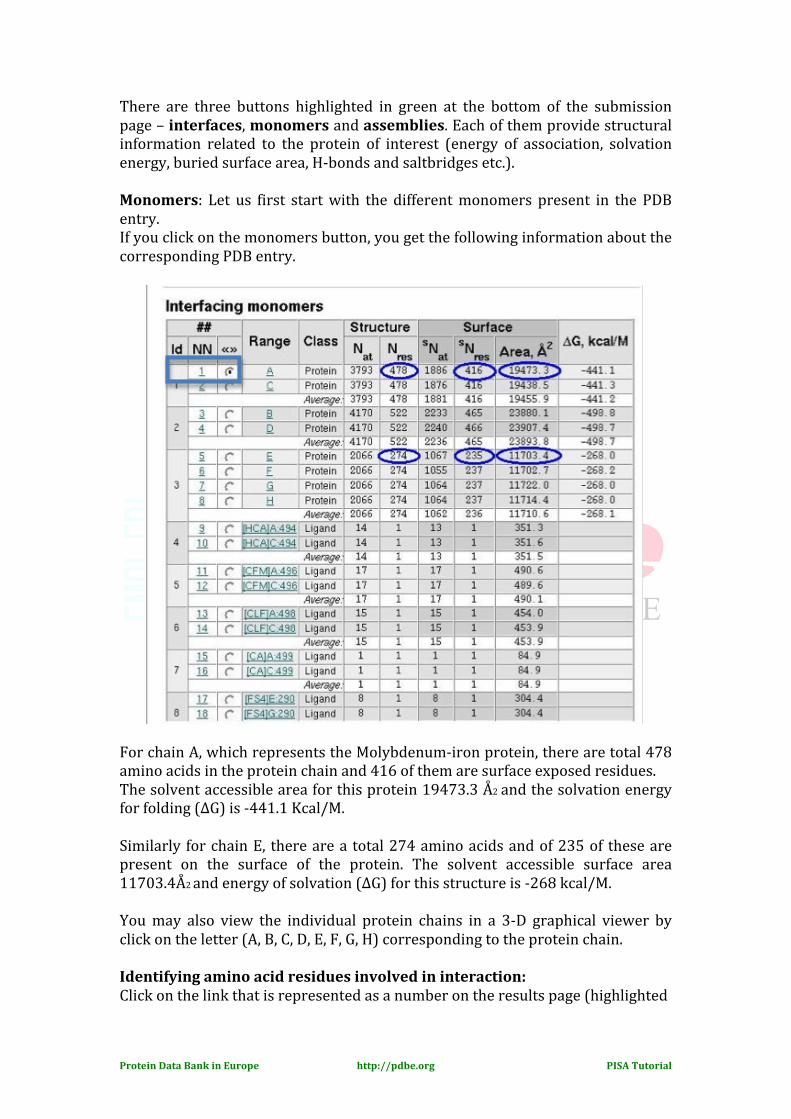
There are three buttons highlighted in green at the bottom of the submission page – interfaces, monomers and assemblies. Each of them provide structural information related to the protein of interest (energy of association, solvation energy, buried surface area, H-‐bonds and saltbridges etc.). Monomers: Let us first start with the different monomers present in the PDB entry. If you click on the monomers button, you get the following information about the corresponding PDB entry. For chain A, which represents the Molybdenum-‐iron protein, there are total 478 amino acids in the protein chain and 416 of them are surface exposed residues. The solvent accessible area for this protein 19473.3 Å2 and the solvation energy for folding (∆G) is -‐441.1 Kcal/M. Similarly for chain E, there are a total 274 amino acids and of 235 of these are present on the surface of the protein. The solvent accessible surface area 11703.4Å2 and energy of solvation (∆G) for this structure is -‐268 kcal/M. You may also view the individual protein chains in a 3-‐D graphical viewer by click on the letter (A, B, C, D, E, F, G, H) corresponding to the protein chain. Identifying amino acid residues involved in interaction: Click on the link that is represented as a number on the results page (highlighted
Protein Data Bank in Europe http://pdbe.org PISA Tutorial
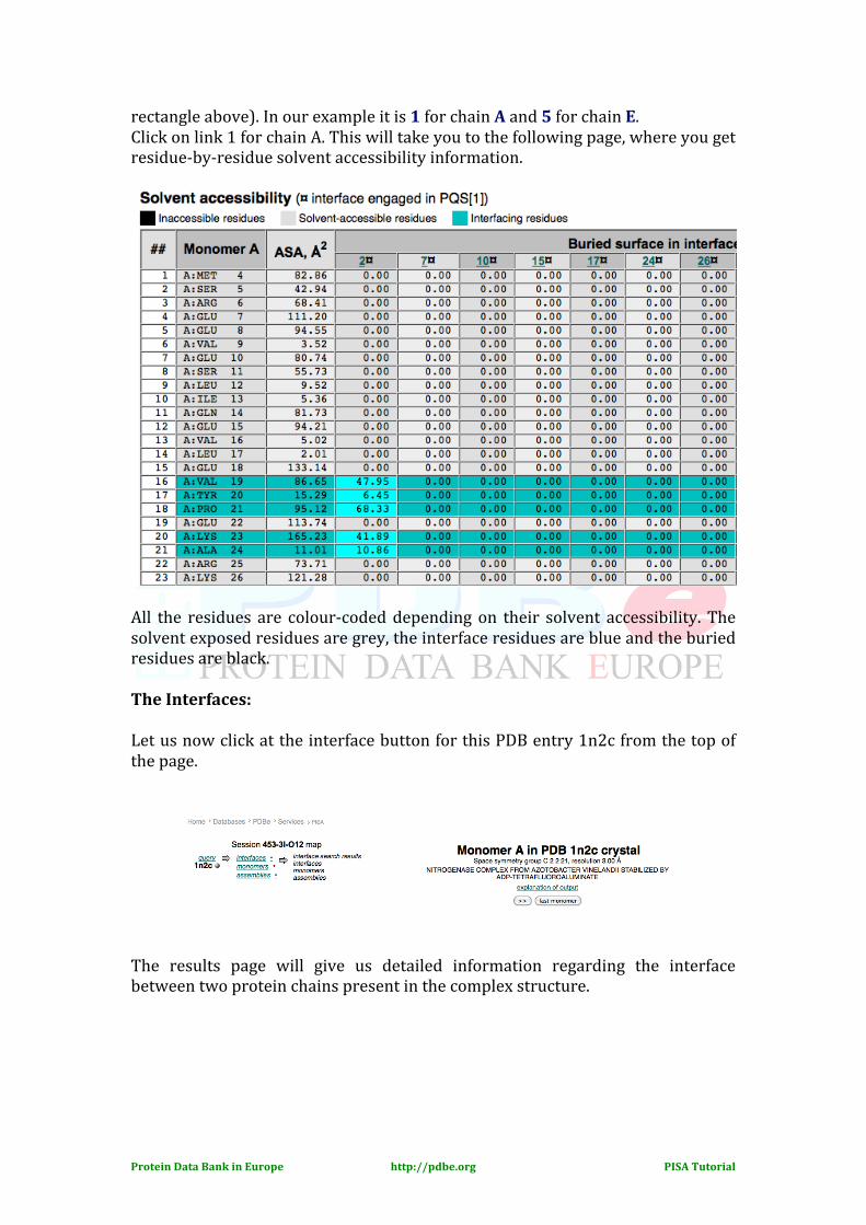
rectangle above). In our example it is 1 for chain A and 5 for chain E. Click on link 1 for chain A. This will take you to the following page, where you get residue-‐by-‐residue solvent accessibility information.
All the residues are colour-‐coded depending on their solvent accessibility. The solvent exposed residues are grey, the interface residues are blue and the buried residues are black. The Interfaces: Let us now click at the interface button for this PDB entry 1n2c from the top of the page. The results page will give us detailed information regarding the interface between two protein chains present in the complex structure.
Protein Data Bank in Europe http://pdbe.org PISA Tutorial
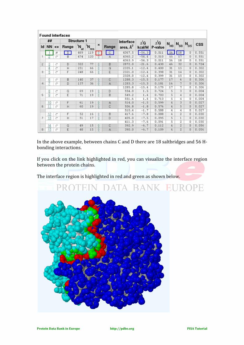
In the above example, between chains C and D there are 18 saltbridges and 56 H-‐ bonding interactions. If you click on the link highlighted in red, you can visualize the interface region between the protein chains. The interface region is highlighted in red and green as shown below.
Protein Data Bank in Europe http://pdbe.org PISA Tutorial
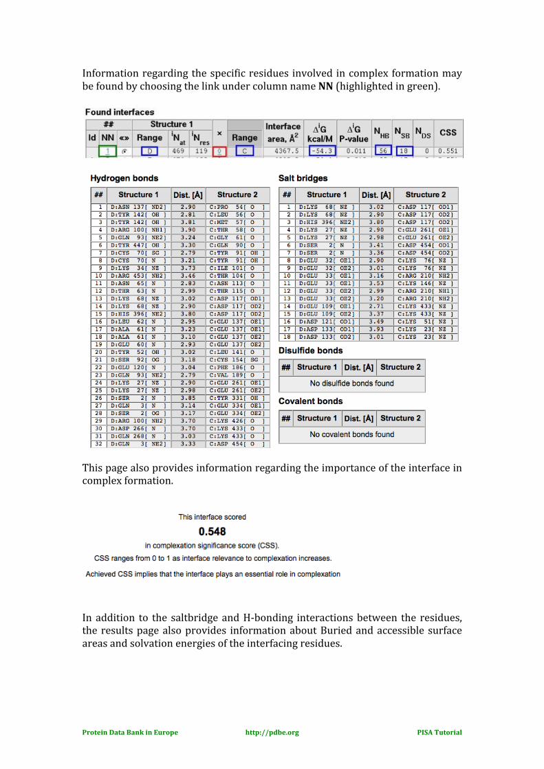
Information regarding the specific residues involved in complex formation may be found by choosing the link under column name NN (highlighted in green).
This page also provides information regarding the importance of the interface in complex formation.
In addition to the saltbridge and H-‐bonding interactions between the residues, the results page also provides information about Buried and accessible surface areas and solvation energies of the interfacing residues.
Protein Data Bank in Europe http://pdbe.org PISA Tutorial
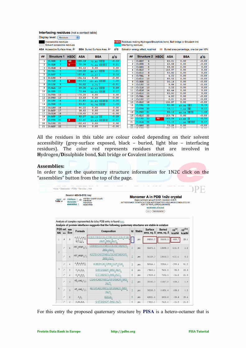
All the residues in this table are colour coded depending on their solvent accessibility (grey-‐surface exposed, black – buried, light blue – interfacing residues). The color red represents residues that are involved in Hydrogen/Disulphide bond, Salt bridge or Covalent interactions. Assemblies: In order to get the quaternary structure information for 1N2C click on the “assemblies” button from the top of the page. For this entry the proposed quaternary structure by PISA is a hetero-octamer that is
Protein Data Bank in Europe http://pdbe.org PISA Tutorial
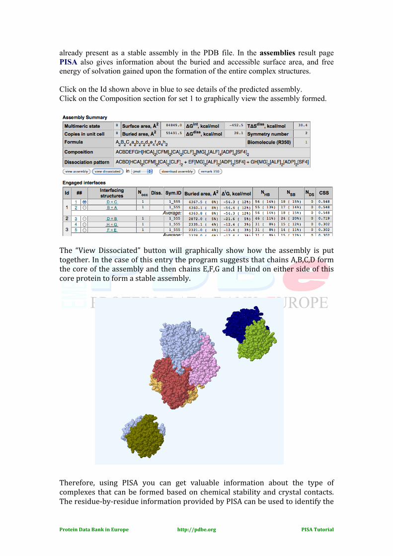
already present as a stable assembly in the PDB file. In the assemblies result page PISA also gives information about the buried and accessible surface area, and free energy of solvation gained upon the formation of the entire complex structures. Click on the Id shown above in blue to see details of the predicted assembly. Click on the Composition section for set 1 to graphically view the assembly formed.
The “View Dissociated” button will graphically show how the assembly is put together. In the case of this entry the program suggests that chains A,B,C,D form the core of the assembly and then chains E,F,G and H bind on either side of this core protein to form a stable assembly. Therefore, using PISA you can get valuable information about the type of complexes that can be formed based on chemical stability and crystal contacts. The residue-‐by-‐residue information provided by PISA can be used to identify the
Protein Data Bank in Europe http://pdbe.org PISA Tutorial
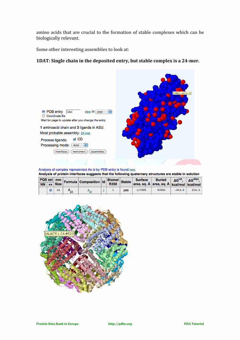
amino acids that are crucial to the formation of stable complexes which can be biologically relevant. Some other interesting assemblies to look at: 1DAT: Single chain in the deposited entry, but stable complex is a 24-mer.
Protein Data Bank in Europe http://pdbe.org PISA Tutorial
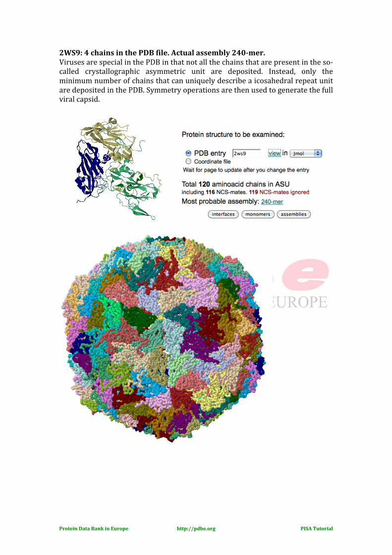
2WS9: 4 chains in the PDB file. Actual assembly 240-mer. Viruses are special in the PDB in that not all the chains that are present in the so-‐called crystallographic asymmetric unit are deposited. Instead, only the minimum number of chains that can uniquely describe a icosahedral repeat unit are deposited in the PDB. Symmetry operations are then used to generate the full viral capsid.
Protein Data Bank in Europe http://pdbe.org PISA Tutorial
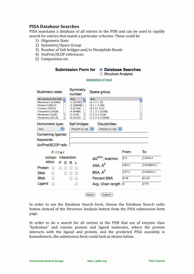
PISA Database Searches PISA maintains a database of all entries in the PDB and can be used to rapidly search for entries that match a particular criterion. These could be
1) Oligomeric State 2) Symmetry/Space Group 3) Number of Salt bridges and/or Disulphide Bonds 4) UniProt/SCOP references 5) Composition etc.
In order to see the Database Search form, choose the Database Search radio button instead of the Structure Analysis button from the PISA submission form page. In order to do a search for all entries in the PDB that are of enzyme class “hydrolase” and contain protein and ligand molecules, where the protein interacts with the ligand and protein, and the predicted PISA assembly is homodimeric, the submission form could look as shown below.
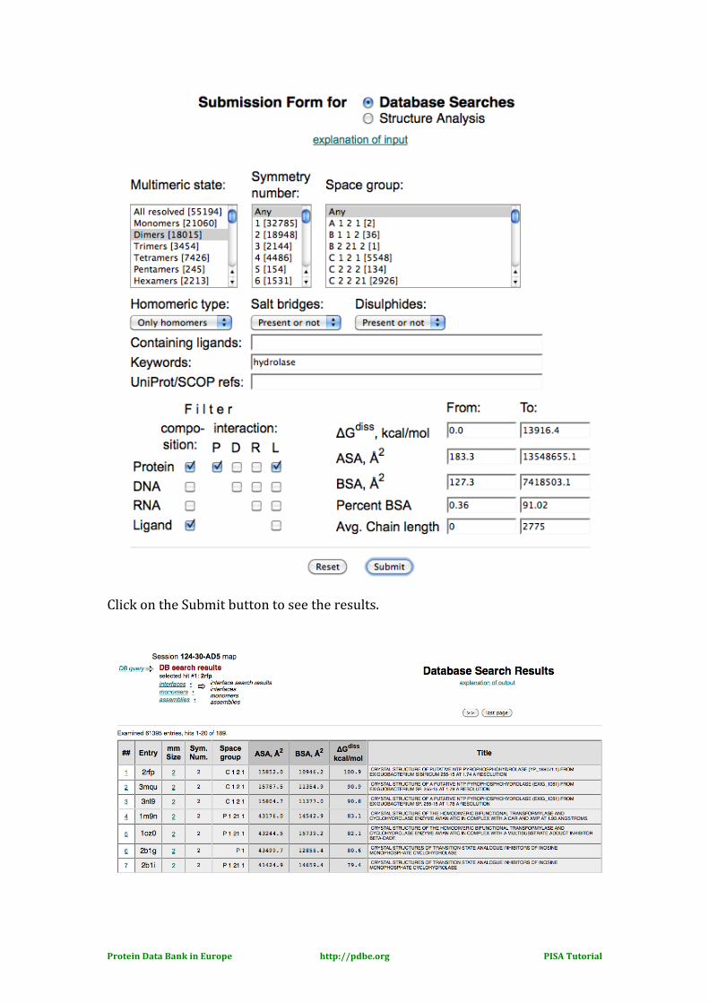
Protein Data Bank in Europe http://pdbe.org PISA Tutorial
Click on the Submit button to see the results.
Protein Data Bank in Europe http://pdbe.org PISA Tutorial
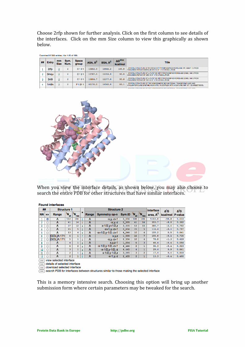
Choose 2rfp shown for further analysis. Click on the first column to see details of the interfaces. Click on the mm Size column to view this graphically as shown below.
When you view the interface details, as shown below, you may also choose to search the entire PDB for other structures that have similar interfaces.
This is a memory intensive search. Choosing this option will bring up another submission form where certain parameters may be tweaked for the search.
Protein Data Bank in Europe http://pdbe.org PISA Tutorial
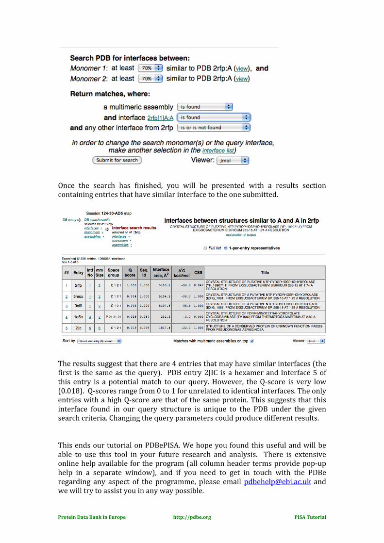
Once the search has finished, you will be presented with a results section containing entries that have similar interface to the one submitted.
The results suggest that there are 4 entries that may have similar interfaces (the first is the same as the query). PDB entry 2JIC is a hexamer and interface 5 of this entry is a potential match to our query. However, the Q-‐score is very low (0.018). Q-‐scores range from 0 to 1 for unrelated to identical interfaces. The only entries with a high Q-‐score are that of the same protein. This suggests that this interface found in our query structure is unique to the PDB under the given search criteria. Changing the query parameters could produce different results. This ends our tutorial on PDBePISA. We hope you found this useful and will be able to use this tool in your future research and analysis. There is extensive online help available for the program (all column header terms provide pop-‐up help in a separate window), and if you need to get in touch with the PDBe regarding any aspect of the programme, please email [email protected] and we will try to assist you in any way possible.