Embed Size (px)
Citation preview
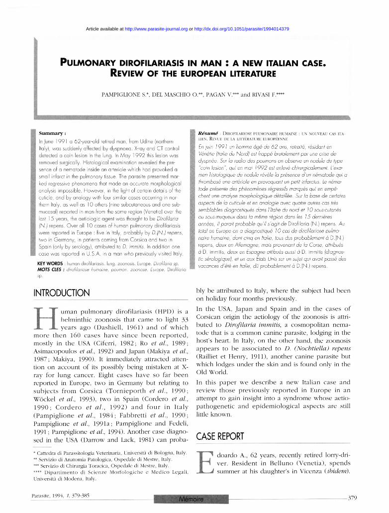
PULMONARY DIROFILARIASIS I N M A N : A NEW ITALIAN CASE. REVIEW OF THE EUROPEAN LITERATURE
PAMPIGLIONE S.*, DEL MASCHIO O.**. PAGAN V.*** and RIVASI F.****
Summary : Injure 1991 a 62-year-old retired man, from Udine (northern Italy), was suddenly affected by dyspnoea. X-ray and CT control detected a coin lesion in the lung. In May 1992 this lesion was removed surgically. Histological examination revealed the presence of a nematode inside an arteriole which had provoked a small infarct in the pulmonary tissue. The parasite presented marked regressive phenomena that made an accurate morphological analysis impossible. However, in the light of certain details of the cuticle, and by analogy with four similar cases occurring in northern Italy, as well as 10 others (nine subcutaneous and one submucosal) reported in man from the same region (Venetia) over the last 1 5 years, the aetiologic agent was thought to be Dirofilaria (N.) repens. Over all 1 0 cases of human pulmonary dirofilariasis were reported in Europe : five in Italy, probably by D. (N.) repens, two in Germany, in patients coming from Corsica and two in Spain (only by serology), attributed to D. immitis. In addition one case was reported in U.S.A. in a man who previously visited Italy.
KEYWORDS : human dirofilariasis. lung, zoonosis. Europe. Dirofilaria sp. MOTS CLES : dirofilariose humaine. poumon. zoonose. Europe. Dirofilaria sp.
Résumé : DIROFILARIOSE PULMONAIRE HUMAINE : UN NOUVEAU CAS ITA-LIEN. REVUE DE LA LITTÉRATURE EUROPÉENNE
En juin 1991 un homme âgé de 62 ans, retraité, résidant en Vénétie (Italie du Nord) est frappé brutalement par une crise de dyspnée. Sur la radio des poumons on observe un nodule du type "coin lésion", qui en mai 1992 est enlevé chirurgicalement. L'examen histologique du nodule révèle la présence d'un nématode qui a thrombosé une artériole en provoquant un petit infarctus. Le nématode présente des phénomènes régressifs marqués qui en empêchent une analyse morphologique détaillée. Sur la base de certains aspects de la cuticule et en analogie avec quatre autres cas très semblables diagnostiqués dans l'Italie du nord et 10 sous-cutanés ou sous-muqueux dans la même région dans les 15 dernières années, il parait probable qu'il s'agit de Dirofilaria (N.) repens. Au total en Europe on a diagnostiqué 10 cas de dirofilariose pulmonaire humaine, dont cinq en Italie, tous dus probablement à D.(N.) repens, deux en Allemagne, mais provenant de la Corse, attribués à D. immitis, deux en Espagne attibués aussi à D. immitis (diagnostic sérologique), et un aux États Unis sur un sujet qui avait passé des vacances d'été en Italie, dû probablement à D.(N.) repens.
INTRODUCTION
H uman pulmonary dirofilariasis ( H P D ) is a
helminthic zoonos is that came to light 33
years a g o (Dashie l l , 1 9 6 1 ) and o f w h i c h
m o r e t h e n 1 6 0 c a s e s h a v e s i n c e b e e n r e p o r t e d ,
mostly in the USA (Ciferri, 1 9 8 2 ; Ro et al., 1 9 8 9 ;
Asimacopoulos et al., 1992) and Japan (Makiya et al.,
1987 ; Makiya, 1990) . It immediately attracted atten
tion on account of its possibly being mistaken at X-
ray for lung cancer . Eight cases have so far b e e n
reported in Europe, two in Germany but relating to
s u b j e c t s f r o m C o r s i c a ( T o r n i e p o r t h et al., 1 9 9 0 ;
W ö c k e l et al., 1993) , two in Spain (Cordero et al.,
1 9 9 0 ; C o r d e r o et al., 1 9 9 2 ) a n d f o u r in I ta ly
( P a m p i g l i o n e et al., 1 9 8 4 ; Fabbret t i et al., 1 9 9 0 ;
Pampiglione et al., 1991a ; Pampiglione and Fedeli,
1991 ; Pampiglione et al., 1994) . Another case diagno
sed in the USA (Darrow and Lack, 1981) can proba-
* Cattedra di Parassitologia Veterinaria, Università di Bologna, Italy. ** Servizio di Anatomia Patologica, Ospedale di Mestre, Italy. *** Servizio di Chirurgia Toracica, Ospedale di Mestre, Italy. **** Dipartimento di Scienze Morfologiche e Medico Legali. Università di Modena, Italy.
bly b e attributed to Italy, where the subject had been
on holiday four months previously.
In the USA, Japan and Spain and in the cases o f
Corsican origin the aetiology of the zoonosis is attri
buted to Dirofilaria immitis, a cosmopolitan nema
tode that is a c o m m o n canine parasite, lodging in the
host's heart. In Italy, on the other hand, the zoonosis
appears to b e associated to D. (Nochtiella) repens
(Railliet et Henry, 1911), another canine parasite but
which lodges under the skin and is found only in the
Old World.
In this paper w e descr ibe a n e w Italian case and
review those previously reported in Europe in an
attempt to gain insight into a syndrome whose aetio-
pa thogenet i c and epidemiologica l aspects are still
little known.
CASE REPORT
E doardo A., 62 years, recently retired lorry-dri
ver . Resident in B e l l u n o ( V e n e t i a ) , s p e n d s
summer at his daughter's in Vicenza (ibidem).
Parasite, 1994, 1. 379-385 Mémoire 379
Article available at http://www.parasite-journal.org or http://dx.doi.org/10.1051/parasite/1994014379
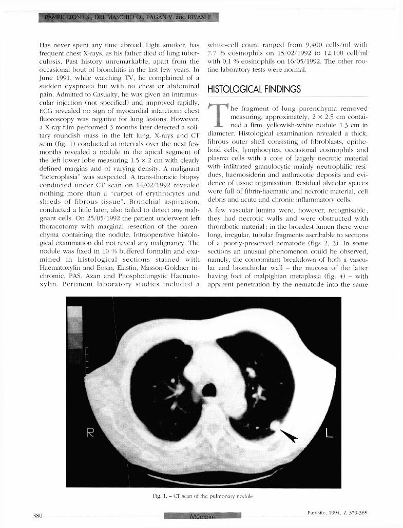
Has never spent any time abroad. Light smoker, has frequent chest X-rays, as his father died of lung tuberculosis . Past history unremarkable , apart from the occasional bout of bronchitis in the last few years. In J u n e 1991, while watching TV, he complained of a sudden dyspnoea but with no chest or abdominal pain. Admitted to Casualty, he was given an intramuscular injection (not specified) and improved rapidly. ECG revealed no sign of myocardial infarction; chest f luoroscopy was negative for lung lesions. However, a X-ray film performed 3 months later detected a solitary roundish mass in the left lung. X-rays and CT scan (fig. 1) conducted at intervals over the next few months revealed a nodule in the apical segment of the left lower lobe measuring 1.5 x 2 cm with clearly defined margins and of varying density. A malignant "heteroplasia" was suspected. A trans-thoracic biopsy c o n d u c t e d under CT scan on 14/02/1992 revealed nothing m o r e than a "carpet o f e ry throcytes and s h r e d s o f f i b r o u s t i s s u e " . B r o n c h i a l a s p i r a t i o n , conducted a little later, also failed to detect any malignant cells. On 25/05/1992 the patient underwent left thoracotomy with marginal resection of the parenchyma containing the nodule. Intraoperative histological examination did not reveal any malignancy. The nodule was fixed in 10 % buffered formalin and exam i n e d in h i s t o l o g i c a l s e c t i o n s s t a i n e d w i t h Haematoxylin and Eosin, Elastin, Masson-Goldner tri-chromic, PAS, Azan and Phosphotungstic Haematox y l i n . P e r t i n e n t laboratory s t u d i e s i n c l u d e d a
whi te -ce l l c o u n t ranged from 9 , 4 0 0 cells/ml with 7.7 % eosinophi ls on 15/02/1992 to 12 ,100 cell/ml with 0.1 % eosinophils on 16/05/1992. The other routine laboratory tests were normal.
HISTOLOGICAL FINDINGS
T he fragment o f lung p a r e n c h y m a r e m o v e d measuring, approximately, 2 x 2.5 cm contained a firm, yellowish-white nodule 1.3 cm in
diameter. Histological examination revealed a thick, fibrous outer shell consisting o f fibroblasts, epithelioid cells, lymphocytes, occasional eosinophils and plasma cells with a core of largely necrotic material with infiltrated granulocytic mainly neutrophilic residues, haemosiderin and anthracotic deposits and evidence of tissue organisation. Residual alveolar spaces were full o f fibrin-haematic and necrotic material, cell debris and acute and chronic inflammatory cells.
A few vascular lumina were, however, recognisable; they had necrot ic walls and w e r e obstructed with thrombotic material; in the broadest lumen there were long, irregular, tubular fragments ascribable to sections of a poorly-preserved nematode (figs 2, 3 ) . In some sections an unusual phenomenon could be observed, namely, the concomitant breakdown of both a vascular and bronchiolar wall - the mucosa of the latter having foci of malpighian metaplasia (fig. 4 ) - with apparent penetration by the nematode into the same
Fig. 1. - CT scan of the pulmonary nodule.
380 Parasite, 1994, 7, 379-385
PAMPIGLIONE S., D E L MASCHIO O., PAGAN V. and RIVASI F.
Mémoire
HUMAN PULMONARY DIROFILARIASIS IN ITALY
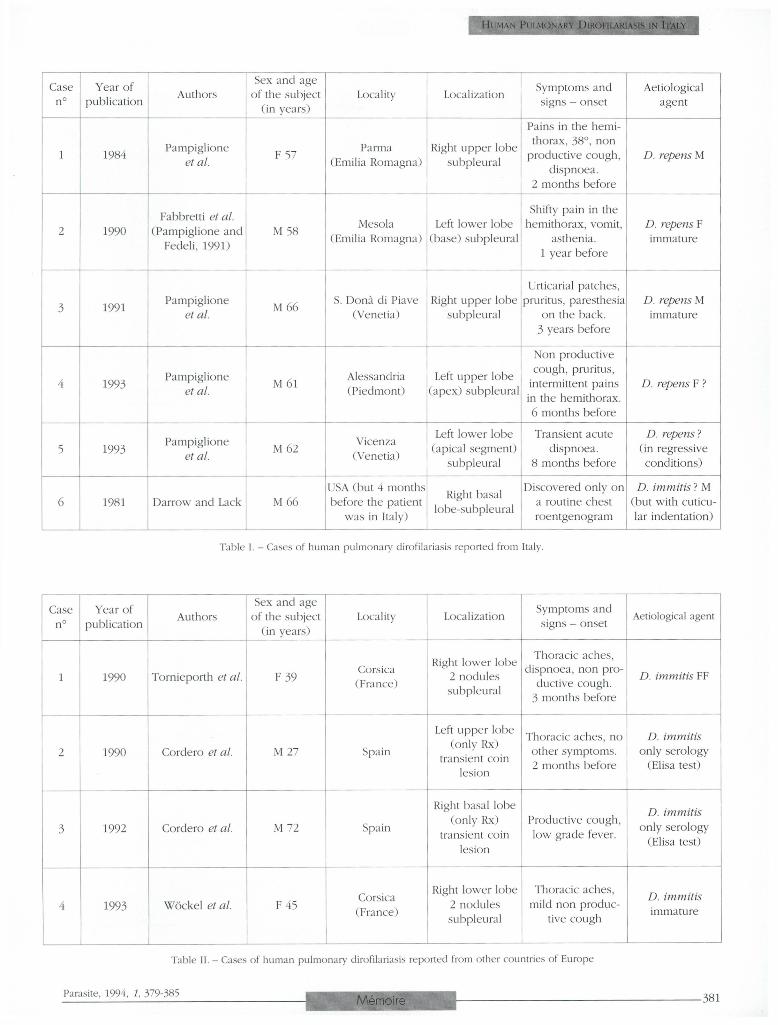
Table I. - Cases of human pulmonary dirofilariasis reported from Italy.
Table II. - Cases of human pulmonary dirofilariasis reported from other countries of Europe
Parasite, 1994, 1, 379-385 Mémoire 381
PAMPIGLIONE S., DEL MASCHIO O., PAGAN V. and RIVASI F.
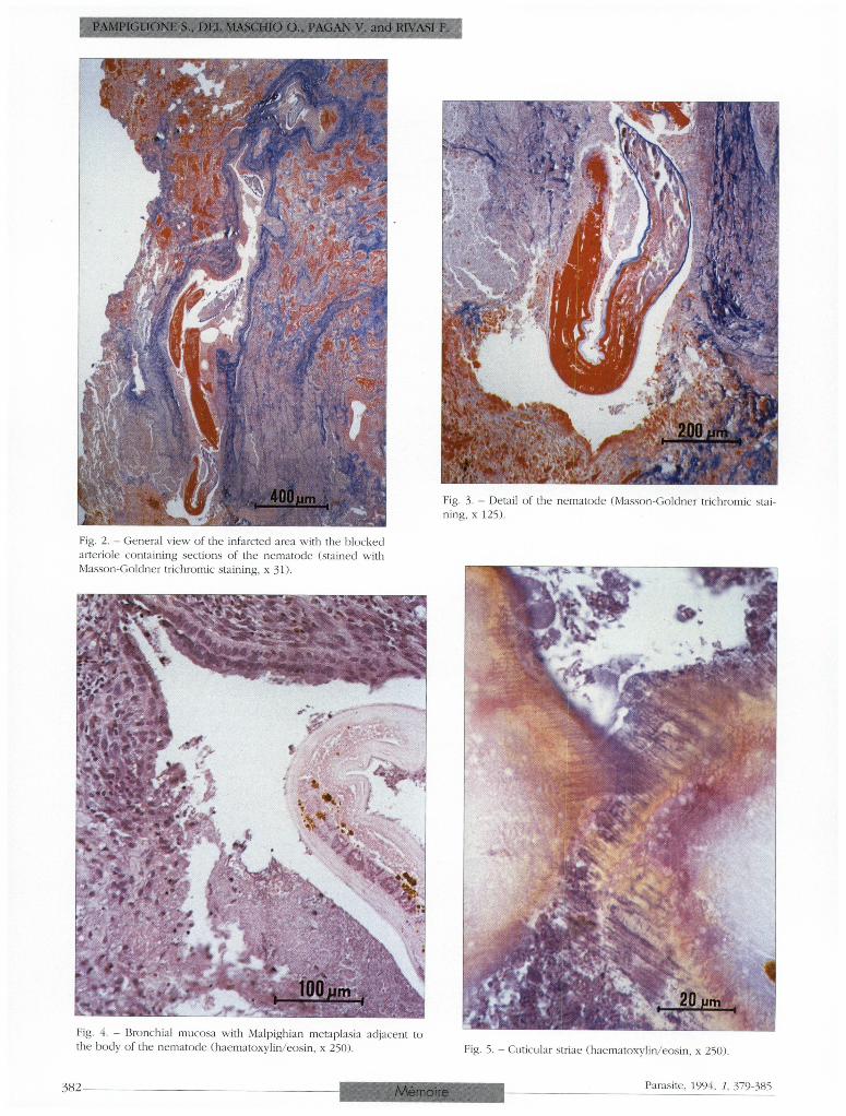
Fig. 2. - General view of the infarcted area with the blocked arteriole containing sections of the nematode (stained with Masson-Goldner trichromie staining, x 3D.
Fig. 3. - Detail of the nematode (Masson-Goldner trichromie staining, x 125).
Fig. 4. - Bronchial mucosa with Malpighian metaplasia adjacent to the body of the nematode (haematoxylin/eosin, x 250). Fig. 5. - Cuticular striae (haematoxylin/eosin, x 250).
382 Parasite, 1994, 7, 379-385 Mémoire
bronchiolar lumen. In the surrounding lung tissue an aspecific inflammatory reaction involving eosinophils and focal fibrosis was evident.
PARASITOLOGICAL FINDINGS
T he histological e x a m i n a t i o n revealed s o m e nematode sect ions ( from three to eight per slide, depending on the level of the section),
mostly in a state of advanced regression, surrounded by infarctual tissue inside a small arterial vessel. Some sections of the nematode were completely necrotic and had partially disintegrated; its shape could only b e deduced from the remains of cutícula, which had worn very thin and had partly broken down. The sections of the parasite were mainly paralongitudinal or oblique, in a few cases paratransversal. The diameter of the nematode varied from 82 to 190 µm. The cutícula was often the only morphological feature still d i s c e r n i b l e ; it w a s in p o o r condi t ion , s o m e t i m e s exfoliated, sometimes swollen. Where more intact, it varied in thickness be tween 7 and 13 pm and appeared to consist o f three layers. Along the edge of s o m e not ent ire ly n e c r o s e d s e c t i o n s longi tudinal striae were present (fig. 5) , 6-14 urn apart, corresponding to the cutícula; however, it was impossible to determine their thickness, nor could they be said to be actual ridges on the outer cuticular surface, given their regressive state. Other thinner striae about 1 pm apart and transversal to the longitudinal axis of the n e m a t o d e were also occas ional ly discernible . In a few sections, a thin hypoderma (2-3 pm) and some muscular fibers were observed. Inside the nematode, only disparate necrotic tissue with no distinguishing features w a s present , a l though, in s o m e sec t ions , two longitudinal tubular structures, 22-30 pm in diam e t e r a n d p r e s u m a b l y b e l o n g i n g to t h e g e n i t a l and/or intestinal apparatus, were visible.
The morphological characteristics described above do not permit of any more than a very generic parasito-logical diagnosis of Dirofilaria sp. However, by analogy with the four Italian cases of HPD previously observed, and in view of the similarities both in the subject's clinical picture and in certain morphological features of the nematode (longitudinal striae in some sections on the surface of the cutícula), we can postulate an immature individual of D. (N.) repens.
DISCUSSION
T his case conforms to the well-defined picture of HPD. The clinical signs are modest pulmon a r y i n v o l v e m e n t t o g e t h e r w i t h a " c o i n
lesion" X-ray picture, corresponding to a small infarctual nodule on the periphery of a lung. This nodule is caused by a thrombosis in the arteriole in which the nematode is lodging. The sudden bout of dyspnoea in J u n e 1991, almost a year before the operation, is likely to have co inc ided with the occlusion of the arteriole; this is borne out by the appearance of the infarctual tissue, which was already partly structured at the t ime o f the h i s t o l o g i c a l e x a m i n a t i o n . T h e patient was probably bitten the previous year by an in fec ted m o s q u i t o in Vicenza , w h e r e he is in the habit of spending the summer. Mosquitoes abound in summer in this region and D. (N.) repens has been reported here in dogs (Canestri-Trotti et al., 1986). Furthermore, another case of HPD (Fabbretti et al, 1990) as well as 10 cases of subcutaneous or submu-cosa involvement (Pampiglione et al, 1991b) , all o f them due to D. (N.) repens, have b e e n reported in the Venet ia region. C o m p a r i n g our f indings with those previously reported in Italy (table I) and elsewhere in Europe (table II), we note that all 10 cases emerged in the last 13 years. This may b e due not so much to a hypothetical spread of zoonosis as to greater importance being attached to solitary pulmonary nodules and to more frequent thoracic surgery thanks to an improvement in the facilities. The patients vary in age between 27 and 66 years, the elderly being predominant, in line with the findings in the USA (Gershwin et al, 1 9 7 4 ; Dayal and Neafie, 1975 ; Ciferri, 1982 ; Ro et al, 1989). Males are more c o m m o n l y affected than females ( seven out of 10 cases), again in line with American statistics. The parasite always lodges in the subpleural region, six in the right lung and four in the left. The nodules are single in eight cases ; in the two cases from Corsica they are double. The diameter of the nodules does not exceed 3 cm and they appear in X-rays as "coin lesions". The symptomatology was acute, with sudden dyspnoea, in two cases, while in the remaining ones it was unobtrusive. In one case, discovered by chance during a routine radiological check-up. there were no symptoms whatsoever. Symptoms generally consisted of more or less localised chest pain and nonproductive c o u g h ; two patients ran a temperature, two experienced pruritus, one came out in a rash and one suffered vomiting. The time elapsing between the appearance of the first symptoms and removal of the nodule ranged from two months to three years.
W h e r e a s in many subcutaneous cases in Italy, the nematode was well preserved or even alive, in cases o f HPD the n e m a t o d e was usually in a markedly regressive state. In three cases, it was male, in two female, while in five cases it was impossible to determine the sex. Correct diagnosis was possible in eight cases only after histological examination and disclo-
HUMAN PULMONARY DIROFILARIASIS IN ITALY
Parasite, 1994, 7, 379-385 Mémoire 383
sure of the parasite. In the Italian cases the parasite was identified as D. (N.) repens with stated reservations in the present case . Those of Corsican origin were thought to be D. immitis, while those reported in Spain and tentatively ascribed to D. immitis were diagnosed only radiologically and immunologically thus casting a slight doubt on the reliability of the diagnosis. In the eight cases subjected to histological examination the clinical diagnosis was consistently one of "malignant tumour", this justifying the importance that the American authors attach to the syndrome in the differential diagnosis of primary or metastatic lung cancer (Ro et al., 1989). In these cases surgery involved the removal of that portion of the lung containing the nodule. The histological finding was invariably of an infarct ion around a thrombot i c arteriole in w h o s e l u m e n the n e m a t o d e was lodging s u r r o u n d e d by necrotic tissue. The infarction was always roundish, whereas the classical t h r o m b o e m b o l i c infarction is generally pyramidal in shape (Kochar, 1985).
In none of the American or European cases o f HPD subjected to histological examination - and here we include our Italian cases - does it seem plausible to attribute infarction to mechanical obstruction of arterial f low by the nematode , for the vessel is never complete ly b l o c k e d by the parasite. T h e thrombus a r o u n d the paras i te a p p e a r s to have f o r m e d gradually, and may be due either to a direct action on the intima of the vessel by heterogeneous substances released by the parasite,which was dead on arrival (Przyjemski, 1 9 8 1 ; Bloch et al., 1990) , or to a local antigen-antibody reaction or to both (Navarrete-Reyna and Noon, 1 9 6 8 ; Neafie and Piggott, 1971) . It is likely that, in time, the nematode would be broken down completely by inflammatory reaction and disappear; tissue-repair processes would then take place, as the findings o f Cordero et al. ( 1 9 9 0 , 1992 ) relating to transient lung nodules, monitored radiologically and immunological ly , would appear to demonstrate . In our case the formation of a passage between the pulmonary artery and the bronchus could be explained by the ropture o f the necrotic vascular wall.
Analysis of the present case and of the others taken into account raises questions that are hard to answer for now. The five Italian cases all occurred in the same geographical locality, the Po Valley, and in a re la t ive ly r e s t r i c t e d a r e a . T h e a e t i o l o g i c a l a g e n t a p p e a r s to b e D.(N.) repens and not D. immitis, although it might seem more logical to suspect the latter species, given its affinity for the vascular system, its abundance in dogs in the area, often greater than that of D. (N.) repens (Canestri-Trotti et al, 1986, 1988) , and its potent ia l pa thogenic i ty for man, as amply demonstrated in other continents and in neighbouring
Corsica. Is it a question of different vectors ? Or of local strains with a differentiated propensity for thriving in man ? Or of different immunological resistance in populations exposed for different periods of time to the parasite ? In order to find the answer to these questions, further entomological and immunological research is needed. Another question, already raised by some American authors (Beaver and Orihel, 1965) relates to the age of the subjects affected : why should the elderly appear to be more at risk ? One could suspect that it has to do with the lowering of the immune d e f e n c e s at that t ime o f l i fe . Ciferr i , ( 1 9 8 2 ) , for example, noted that 11 % of American cases of HPD involved subjects with impaired immunity to disease.
T o conclude, autochthonous HPD can b e said to exist in Europe, even if rarely reported. The syndrome is endemic to certain restricted areas o f the Po Valley (northern Italy), to Corsica and perhaps to Spain. The presence of D. (N) repens and/or D. immitis in dogs in vast areas of southern Europe leads one to suspect its occurrence in other regions. It is likely that undiag n o s e d c a s e s e s c a p e t h e a t t e n t i o n o f the h e a l t h authorities not only because there may b e few or no symptoms but also because , if the microscopic examination is not meticulously performed, the nematode can escape detection and so remain unidentified (Ro etal, 1989) .
ACKNOWLEDGEMENTS
This research received financial support from MURST (60 % ) .
REFERENCES
ASIMACOPOTJLOS P.J., KOTRAS A. and CHRISTIE B. Pulmonary dirofilariasis. The largest single-hospital experience. Chest, 1 9 9 2 , 102, 8 5 1 - 8 5 4 .
BEAVER P.C. and ORIHEL T.C. Human infection with filariae of animáis in the United States. American Journal of Tropical Medicine and Hygiene, 1 9 6 5 , 14, 1 0 1 0 - 1 0 2 9 .
BLOCH T., GLYNN T. and HINSHAW M. Human pulmonary
dirofilariasis. Indiana Medicine, 1 9 9 0 , 83, 2 4 - 2 7 .
CANESTRI-TROTTI G . , GOVERNATORI M., RIVASI F., TAMPIERI A.
and TARTONI P.L. Indagine sulla dirofilariosi canina in provincia di Modena. Modello epidemiologico statistico previsionale. Tecnica Sanitaria, 1 9 8 8 , 2 1 , 2 8 3 - 2 9 7 .
CANESTRI-TROTTI G . , PAMPIGLIONE S. and VISCONTI S. Ricerche sulla diffusione delle filariasi canine in alcune provincie della pianura padana. Annali Istituto Superiore di Sanità, 1 9 8 6 , 2 2 , 4 4 9 - 4 5 2
CIFERRI F. Human pulmonary dirofilariasis in the United States : a criticai review. American Journal of Tropical Medicine and Hygiene, 1 9 8 2 , 3 1 , 3 0 2 - 3 0 8 .
3 8 4 Parasite, 1994, 1, 379-385
PAMPIGLIONE S., DEL MASCHIO O., PAGAN V. and RIVASI F.
Mémoire
HUMAN PULMONARY DIROFILARIASIS IN ITALY
CORDERO M . , MUNOZ M.R. , MURO A. and SIMON F. Transient
solitary pulmonary nodule caused by Dirofilaria immitis. European Respiratory Journal 1990, 3, 1070-1071.
CORDERO M , MURO A., SIMON F., TAPIA J.I. and ESPINOZA E.
Are transient pulmonary solitary nodules a common event in human dirofilariosis ? Clinical Investigator, 1992, 70, 437-440.
DARROW J . C . and LACK E.E. Solitary lung nodule due by Dirofilaria immitis (clog "heartworm"). Journal of Surgical Oncology, 1981, 16, 219-224.
DASHIELI. C F . A case of dirofilariasis involving the lung. American Journal of Tropical Medicine and Hygiene, 1961, 10. 37-38.
DAYAL Y. and NEAFIF. R.C. Human pulmonary dirofilariasis : a case report and review of the literature. American Review of'Respiratory Diseases, 1975, 112, 437-443.
FAHHRETTI G . . FEDELI F., ALESSI A., BOARON M . , SALPIETRO V .
and BRISIGOTTI M . Human pulmonary dirofilariasis : report of a new European case . Histology and Histopathology, 1990, 5, 311-313.
GERSHWIN L.J.. GERSHWIN M.E . and KRITZMAN J . Human pul
monary dirofilariasis. Chest, 1974, 66, 92-96.
KOCHAR A.S. Human pulmonary dirofilariasis. Report of three cases and brief review of the literature. American Journal of Clinical Pathology, 1985, 84, 19-23.
MAKIYA K . Dirofilariasis a parasitic disease possible acquired in the quotidian life environment. Medico, 1990, 21, 14-17, (in Japanese).
MAKIYA K . , TSUKAMOTO M . and KAGEI N . Fifty-six cases of
human dirofilariasis reported from Japan - a compiled table. Journal of the University of Occupational and Environmental Health, 1987, 9, 233-242. (in Japanese)
NAVARRETE-REYNA A. and NOON G . Pulmonary dirofilariasis manifested as a coin lesion. Archives of Pathology, 1968, 85. 266-271.
NEAFIE R.C. and PIGGOTT J . Human pulmonary dirofilariasis. Archives of Pathology, 1971, 92, 342-349.
PAMPIGLIONE S., BOSI F . , MACONT A.G., MERIGGI F., REMOTTI G . and SCAGLIA M . Pulmonary dirofilasiasis : clinical and parasitologic findings of a new human case. Giornale Italiano Malattie del Torace, 1994, 48, 1-4.
PAMPIGLIONE S., CANDIANI G . , DEL MASCHIO O . and PAGAN V .
Dirofilariasi polmonare nell'uomo : un terzo caso in Italia. Pathologica. 1991a, 83, 21-27.
PAMPIGLIONE S., CANESTRI-TROTTI G . and RIVASI F. La dirofila-
riose humaine en I t a l i c Annales de Parasitologic Humaine et Comparée. 1991b, 66, 195-203.
PAMPIGLIONE S. and FEDELI F. Dirofilariasi polmonare umana : aspetti parassitologici del secondo caso segnalato in Italia. Parassitologia, 1991, 33, 153-157.
PAMPIGLIONE S., RIVASI F. and CANESTRI-TROTTI G . Human
Pulmonary dirofilariasis in Italy. The Lancet, 1984, February' 11, 333.
PRZYJEMSKI C . J . Dirofilaria immitis lung nodules. Journal of the American Medical Association, 1981, 245, 30-31.
Ro J.Y., TSAKALAKIS P.J., WHITE V.A., LUNA M.A., CHANG-TUNG
E . G . , GREEN L. , CRIBBETT L. and ALAYA A.G. Pulmonary
dirofilariasis : the great imitator of primary or metastatic lung tumor. Human Pathology, 1989, 20, 69-76.
TORNIEPORTH N . . BRANDIS A., V O G E L В. and D I S K O R. Autochthone pulmonale Dirofi lariose in Europe. Deutsche Medizinische Wochenschrift, 1990, 115, 15-19.
WOCKEL W . , ECKERT J LOSCHER Т.Н., HAUSSINGER К. und MORRESI A. Autochtone europäische lungen-Dirofilariose. Pneumologie, 1993, 47, 227-231.
Accepté le 8 juillet 1994
Parasite, 1994, 1, 379-385 385 Mémoire