Embed Size (px)
Citation preview
Panda et al. Parasites & Vectors 2013, 6:203http://www.parasitesandvectors.com/content/6/1/203
RESEARCH Open Access
Decreased prevalence of sepsis but not mild orsevere P. falciparum malaria is associated withpre-existing filarial infectionMadhumita Panda1†, Prakash K Sahoo2†, Alok Das Mohapatra1, Soumya kanti Dutta3, Pravat K Thatoi3,Rina Tripathy4, Bidyut K Das3, Ashok K Satpathy2 and Balachandran Ravindran1*
Abstract
Background: Enhanced inflammatory host responses have been attributed as the cellular basis for development ofsevere malaria as well as sepsis. In contrast to this, filarial infections have been consistently reported to beassociated with an immunological hypo-responsive phenotype. This suggests that successful control of filariasis byemploying mass drug administration, could potentially contribute to an increase in incidence of sepsis and cerebralmalaria in human communities. A case control study was undertaken to address this critical and urgent issue.
Methods: Eighty-nine patients with sepsis and one hundred and ninety-six patients with P. falciparum malaria alloriginating from Odisha, were tested for prevalence of circulating filarial antigens - a quantitative marker of activefilarial infection. Antibodies to four stage specific malarial recombinant proteins were measured by solid phaseimmunoassays and circulating CD4+CD25high T-cells were quantified by flow cytometry with an objective to study ifpre-existing filarial infections influence antibody responses to malarial antigens or the levels of circulating T-regulatory cells in P. falciparum infected patients.
Results: Prevalence of filarial antigenemia was significantly less in sepsis patients as compared to controlssuggesting that pre-existing filariasis could influence development of sepsis. On the other hand, levels of circulatingfilarial antigen were comparable in severe malaria cases and healthy controls suggesting that development ofsevere malaria is independent of pre-existing W. bancrofti infections. Plasma TNF-a, RANTES and antibodies torecombinant malarial proteins as well as levels of circulating CD4+ CD25high cells were comparable in malariapatients with or without filarial infections.
Conclusions: These observations imply that successful control of filariasis could have adverse consequences onpublic health by increasing the incidence of sepsis, while the incidence of severe malaria may not adverselyincrease as a consequence of elimination of filariasis.
Keywords: Coinfection, Filariasis, Severe malaria, Sepsis, P. falciparum, Regulatory T cells
BackgroundLymphatic dwelling filarial parasites cause severe mor-bidity in human hosts and persist for long durations ininfected hosts. The Global Programme to EliminateLymphatic Filariasis (GPELF) is currently targetingelimination of the disease through annual mass drug
* Correspondence: [email protected]†Equal contributors1Infectious Disease Biology group, Institute of Life Sciences, Bhubaneswar,Odisha, IndiaFull list of author information is available at the end of the article
© 2013 Panda et al.; licensee BioMed Central LCommons Attribution License (http://creativecreproduction in any medium, provided the or
administration (MDA) of albendazole with either DECor ivermectin. This has been widely acclaimed to be oneof the successful public health programmes and isexpected to block transmission of filariasis in endemiccountries by 2020 [1].Sepsis is one of the major causes of mortality around
the world and malaria is considered to be one of themost severe infectious diseases afflicting the world’smost impoverished populations. Severe malaria presentsitself with a range of biological dysfunctions, i.e. anemia, re-spiratory complications, acidosis, renal failure, pulmonary
td. This is an Open Access article distributed under the terms of the Creativeommons.org/licenses/by/2.0), which permits unrestricted use, distribution, andiginal work is properly cited.
Panda et al. Parasites & Vectors 2013, 6:203 Page 2 of 9http://www.parasitesandvectors.com/content/6/1/203
edema, multi-organ failure and cerebral malaria etc. [2].Development of sepsis and severe malaria share a commonbiology with activation of uncontrolled inflammatory hostresponses being the cellular and molecular basis for clinicalmanifestation. Both diseases are associated with elevatedplasma levels of TNF-a, IL-6, IL-1β, etc. [3,4]. TNF-a is pro-duced and released by host cells following exposure to vari-ous malarial antigens. The increase of TNF-a release isresponsible for the overexpression of adhesion molecules,hence influencing sequestration of parasitized RBCs [5].Similarly RANTES (Regulated on Activation Normal T-Cell Expressed and Secreted) is a chemokine involved inthe generation of inflammatory infiltrates. Recent studiesindicate that degradation of cell-cell junctions, blood–brainbarrier dysfunction, recruitment of leukocytes and Plasmo-dium-infected erythrocytes and occlusion of microvesselsare associated with RANTES expression. Additionally, acti-vated lymphocytes, platelets and endothelial cells releaselarge quantities of RANTES, suggesting a unique role forRANTES in generation and maintenance of the malaria-induced inflammatory response [6]. Low levels of RANTEScorrelate with disease severity and mortality in individualswith sepsis [7]. However, data on the role of RANTES inmalaria appears to be contradictory. Decreased plasmalevels of RANTES were documented in children with se-vere malarial anemia [8], but in another study, increasedmRNA expression of RANTES was found in the brains ofchildren who died of CM [9]. One of the probable reasonsfor low levels of RANTES in severe malaria may be thethrombocytopenia commonly associated with this condi-tion, as platelets are major reservoirs of RANTES in per-ipheral circulation [10]. Systemic nematodes on the otherhand are known to down regulate such responses ininfected hosts [11]. This later phenomenon has been attrib-uted to production of an array of immunomodulatory mol-ecules released by helminthes that skew host responsesaway from a pro-inflammatory phenotype [12]. These ob-servations suggest that pre-existing helminth infectionscould influence development of sepsis or malaria in a givenhost. Prevalence of cerebral malaria has been reported tobe low in children with Ascaris lumbricoides infection,suggesting that endemic subjects harboring helminthic in-fections could become protected against development ofcerebral malaria [13]. Animal models of sepsis and cerebralmalaria have been used to address the issue, although suchmodels do not truly represent the human disease. Con-comitant infection with S. mansoni and P. berghei ANKAinfection has been reported to lead to reduced cerebralmanifestations [14]. More recently, it has been demon-strated that filarial parasite induced secretion of IL-10 is re-sponsible for developing resistance to murine cerebralmalaria [15], although this does not appear to be a consist-ent feature since observations to the contrary have alsobeen reported [16]. For example, in a study on co-infection
of mice with Litomosoides sigmodontis and P. chabaudi,mice free of circulating microfilaria were shown to developmore severe forms of malaria than animals free of microfil-aria [16].Here we report a case control study in two cohorts of
patients, one with clinically proven sepsis and the otherwith severe P. falciparum malaria and quantified circu-lating filarial antigen (CFA), to test the hypothesiswhether pre-existing filarial infections could influencedevelopment of severe malaria or sepsis. Insights intothis aspect are of critical public health importance inpredicting possible outcomes of the ongoing successfulfilariasis control programme on the incidence of sepsisor severe malaria in human populations.
MethodsStudy area & subject recruitmentPatients with symptoms of sepsis admitted to the De-partment of Medicine at S.C.B. Medical College wereclassified into three categories. 1) Sepsis (n=36): Infec-tion, documented or suspected, with signs and symp-toms of an inflammatory response, viz., leukocytosis orleucopenia, increased C-reactive protein, increasedprocalcitonin levels. 2) Severe sepsis (n=24): Sepsis com-plicated by multi-organ dysfunction. 3) Septic shock(n=29): Severe sepsis with acute circulatory failure char-acterized by persistent arterial hypotension despiteadequate volume administration. Details of sepsispatients are shown in Table 1. For classifying patientsand determining outcomes, the Acute Physiology andChronic Health Evaluation II (APACHE II) scoringsystem was used [4].Patients reporting at the out-patient department and/
or admitted to the Department of Internal Medicine atS.C.B. Medical College (Cuttack, India), with a short his-tory of fever associated with unarousable coma or multiorgan dysfunction were clinically assessed. Patients withmicroscopically demonstrable P. falciparum in thickblood smears were recruited for the study. Diagnosisby microcopy was further confirmed by immuno-chromatographic card test. Details of malaria patientsare shown in Table 2. Non-complicated malaria (NCM)was defined as patients reporting to the outpatient de-partment with fever and evidence of P. falciparum infec-tion. Patients classified with severe malaria belonged toone of the following three groups:1) Cerebral malaria(CM, n=48), 2) Non cerebral severe malaria (NCSM,n=13) and 3) Multi-organ-dysfuction (MOD, n=64) [17].Thirty-eight normal subjects of comparable ethnicityand originating from the same areas as that of patientsand free of demonstrable malarial infections were takenas healthy controls. The current studies on sepsis andmalaria were approved by the Ethics Committee of SCBMedical College and blood samples were collected after
Table 1 Prevalence of sepsis in filariasis infected subjects
Subjects Sepsis (n=89) Healthy control (n=38) P value
CFA+ve CFA-ve CFA+ve CFA-ve
Number (%) *6 (6.7) 83 (93.3) *16 (42.2) 22 (57.8) CFA+ve in sepsis vs HC (<0.0001), CFA-ve vs insepsis vs HC(<0.0001)
Sex(M/F) 6/0 55/28 9/7 10/12 -
Mean Age in years(range)
42.83(24–73) 46.24(17–85) 31.22(20–75) 29.85(12–69) -
APACHE II score 14.25 ± 4.15 12.63 ± 0.58 - - CFA+ve vs CFA-ve, NS
Mechanical ventillaion 2 (33.34) 7 (8.43) - - CFA+ve vs CFA-ve, NS
Vasopressors 2 (33.34) 18 (21.6) - - CFA+ve vs CFA-ve, NS
Total leukocyte count(103/mm3)
13.4-30 7.2-150 ND ND CFA+ve vs CFA-ve, NS
CRP (mean±SD) mg/ml(Range)
86.92 ± 34.10(16.33-146.6)
97.17±5.18(10.42-150.8)
ND ND CFA+ve vs CFA-ve, NS
NOTE: CFA; circulating filarial antigen.*p value<0.0001, 95% Confidence Interval, Odds ratio = 0.099. Data are no. (%) of participants unless otherwise specified.
Panda et al. Parasites & Vectors 2013, 6:203 Page 3 of 9http://www.parasitesandvectors.com/content/6/1/203
obtaining written consent from patients or accompany-ing persons.
Flow cytometryAbout 5ml of the venous blood was collected in heparinfrom patients, plasma was separated and frozen at −20°Cuntil further use. 100 ul of whole blood was used fortwo color staining with PE-cy5 labelled anti-CD4 andFITC labelled anti-CD25 (BD Biosciences), along with ap-propriate isotype controls. Stained cells were then acquiredon a 2-laser/4 channel BD FACS Calibur Flow Cytometerand analysed using CellQuest Pro Software.
Enzyme-linked immunosorbent assay (ELISA)Plasma concentrations of TNF-a and RANTES wereestimated using commercial sandwich ELISA kits(Sanquin, Amsterdam) according to the manufacturer’sinstructions. Circulating Filarial Antigens (CFA) weremeasured by Trop Bio ELISA test kit (Trop Bio Pvt Ltd,Townsville, Australia) as described earlier by us [18].Antibodies to malarial recombinant proteins, by solidphase assay using sporozoite surface protein (SSP-2),circum-sporozoite protein (CSP), exported antigen AG5.1(Exp-1) and liver stage antigen-1 (LSA-1) of P. falcipaarum.Sera were tested after 200 fold dilutions and bound anti-bodies were detected using 1000-fold-diluted HRP-labeledanti-human IgG (P0216; Dako) and enzyme activity wasmeasured using OPD and absorbance read at 492 nm. The
Table 2 Details of study participants
Subjects SM NCM HC
Total number (n) 125 71 38
Sex(M/F) 96/29 47/24 19/19
cp 34(15–72) 30(14–70) 30(12–75)
NOTE: SM Severe malaria1, NCM Non- complicated malaria, HCHealthy controls.
results were expressed as arbitrary ELISA units, using in-ternal laboratory standards.
StatisticsStatistical analyses were performed by using GraphPadPrism (version 5.01). For analysis of Figure 1(D and E)and Figure 2 (A, B, C and D) unpaired student’s t testwas applied to determine differences between groups.For analysis of Figure 1A, B and Table 1, Fisher-exacttest was employed to calculate Odds ratios (ORs) at 95%confidence intervals. For analysis of Figure 1C and Figure 3(B, C, D and E) the association within the groups wereanalysed by one way analysis of variance (ANOVA)followed by Tukey’s post-hoc test. P-values<0.05 were con-sidered to be statistically significant.
ResultsPrevalence of filariasis in sepsis and malaria patientsPrevalence of circulating filarial antigen (CFA) was sig-nificantly less in patients with sepsis in comparison tohealthy endemic controls (Table 1). While 42.2% of con-trols were found to harbor CFA, only 6.7% of patientswith sepsis were positive for antigenemia with relativelylow levels of antigenemia (CFA levels of the six CFA+vepatients were 154, 398, 625, 154, 354 & 542 respect-ively). These findings suggest that pre-existing filarialinfections could be preventing development of clinicalsepsis (Table 1). On the other hand prevalence offilarial antigenemia was comparable in severe malariapatients, non complicated malaria and healthy controls(Figure 1A). Similarly, the prevalence of filarialantigenemia was also comparable in three subgroups ofsevere malaria i.e. cerebral malaria, non-cerebral severemalaria and multiorgan dysfunction (Figure 1B). About45-50% of patients in all the groups were found to har-bor filarial antigenemia and no statistically significant
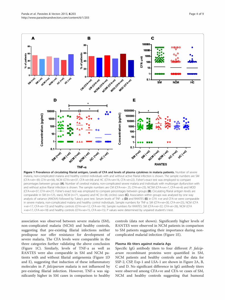
Figure 1 Prevalence of circulating filarial antigen, Levels of CFA and levels of plasma cytokines in malaria patients. Number of severemalaria, non-complicated malaria and healthy control individuals with and without active filarial infection is shown. The sample numbers are SM(CFA+ve= 69, CFA-ve=56), NCM (CFA+ve=37, CFA-ve=34) and HC (CFA+ve=16, CFA-ve=22). Fisher’s-exact test was employed to comparepercentages between groups (A). Number of cerebral malaria, non-complicated severe malaria and individuals with multiorgan dysfunction withand without active filarial infection is shown. The sample numbers are CM (CFA+ve= 25, CFA-ve=23), NCSM (CFA+ve=7, CFA-ve=6) and MOD(CFA+ve=37, CFA-ve=27). Fisher’s-exact test was employed to compare percentages between groups (B). Circulating filarial antigen levels arecomparable in SM (n=125, stars), NCM (n=71, squares) and HC (n=38, circles) cases (C). Association within groups was analysed by one wayanalysis of variance (ANOVA) followed by Tukey’s post test. Serum levels of TNF- a (D) and RANTES (E) in CFA +ve and CFA-ve were comparablein severe malaria, non-complicated malaria and healthy control individuals. Sample numbers for TNF-a: SM (CFA+ve=30, CFA-ve=25), NCM (CFA+ve=17, CFA-ve=13) and healthy controls (CFA+ve=12, CFA-ve=16). Sample numbers for RANTES: SM (CFA+ve=32, CFA-ve=26), NCM (CFA+ve=17, CFA-ve=18) and healthy controls (CFA+ve=15, CFA-ve=15). P values were determined by unpaired student’s t-test.
Panda et al. Parasites & Vectors 2013, 6:203 Page 4 of 9http://www.parasitesandvectors.com/content/6/1/203
association was observed between severe malaria (SM),non-complicated malaria (NCM) and healthy controls,suggesting that pre-existing filarial infections neitherpredispose nor offer resistance for development ofsevere malaria. The CFA levels were comparable in thethree categories further validating the above conclusion(Figure 1C). Similarly, levels of TNF-a as well asRANTES were also comparable in SM and NCM pa-tients with and without filarial antigenemia (Figure 1Dand E), suggesting that induction of these inflammatorymolecules in P. falciparum malaria is not influenced bypre-existing filarial infection. However, TNF-a was sig-nificantly higher in SM cases in comparison to healthy
controls (data not shown). Significantly higher levels ofRANTES were observed in NCM patients in comparisonto SM patients suggesting their importance during non-complicated malarial infection (Figure 1E).
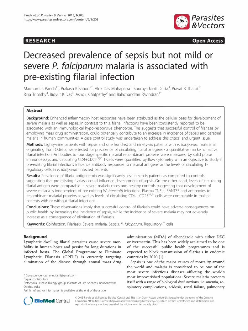
Plasma Ab titers against malaria AgsSpecific IgG antibody titers to four different P. falcip-arum recombinant proteins were quantified in SM,NCM patients and healthy controls and the data forSSP-2, CSP, Exp-1 and LSA-1 are shown in Figure 2A, B,C and D. No significant difference in IgG antibody titerswere observed among CFA+ve and CFA-ve cases of SM,NCM and healthy controls suggesting that humoral
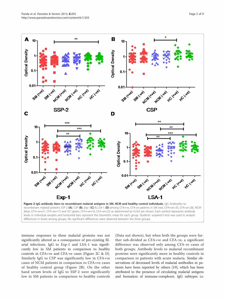
Figure 2 IgG antibody titers to recombinant malarial antigens in SM, NCM and healthy control individuals. IgG Antibodies torecombinant malarial proteins SSP-2 (A), CSP (B), Exp-1(C) & LSA-1 (D) among CFA+ve, CFA-ve patients in SM (red, CFA+ve=35, CFA-ve=26), NCM(blue, CFA+ve=21, CFA-ve=17) and HC (green, CFA+ve=16, CFA-ve=22) as determined by ELISA are shown. Each symbol represents antibodylevels in individual samples and horizontal bars represent the Geometric mean for each group. Students’ unpaired t-test was used to analyzedifferences in levels among groups. No significant differences were observed between the three groups.
Panda et al. Parasites & Vectors 2013, 6:203 Page 5 of 9http://www.parasitesandvectors.com/content/6/1/203
immune responses to these malarial proteins was notsignificantly altered as a consequence of pre-existing fil-arial infections. IgG to Exp-1 and LSA-1 was signifi-cantly low in SM patients in comparison to healthycontrols in CFA+ve and CFA-ve cases (Figure 2C & D).Similarly IgG to CSP was significantly low in CFA+vecases of NCM patients in comparison to CFA+ve casesof healthy control group (Figure 2B). On the otherhand serum levels of IgG to SSP-2 were significantlylow in SM patients in comparison to healthy controls
(Data not shown), but when both the groups were fur-ther sub-divided as CFA+ve and CFA-ve, a significantdifference was observed only among CFA-ve cases ofboth groups. Antibody levels to malarial recombinantproteins were significantly more in healthy controls incomparison to patients with acute malaria. Similar ob-servations of decreased levels of malarial antibodies in pa-tients have been reported by others [19], which has beenattributed to the presence of circulating malarial antigensand formation of immune-complexes. IgG subtypes i.e.
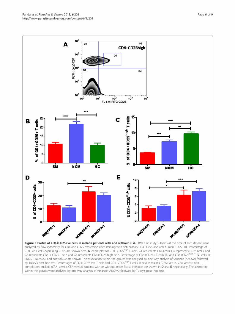
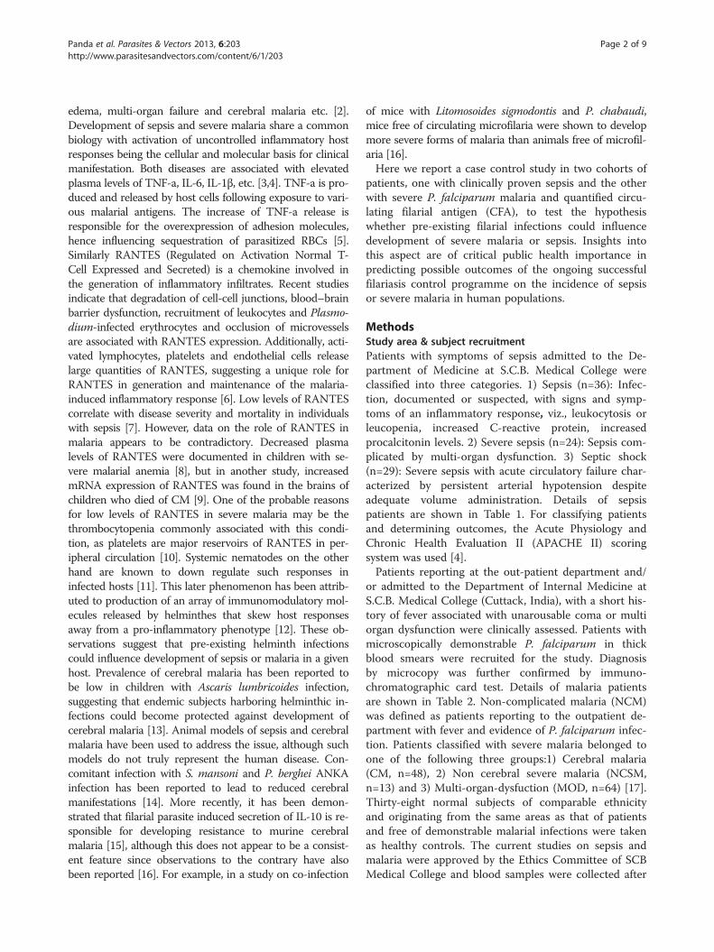
Figure 3 Profile of CD4+CD25+ve cells in malaria patients with and without CFA. PBMCs of study subjects at the time of recruitment wereanalyzed by flow-cytometry for CD4 and CD25 expression after staining with anti-human CD4-PE.cy5 and anti-human CD25-FITC. Percentage ofCD4+ve T cells expressing CD25 are shown here, A: Zebra plot for CD4+CD25high T–cells, G1 represents CD4+cells, G4 represents CD25+cells, andG3 represents CD4 + CD25+ cells and G5 represents CD4+CD25 high cells. Percentage of CD4+CD25+ T cells (B) and CD4+CD25high T (C) cells inSM=91, NCM=58 and control=22 are shown. The association within the groups was analysed by one way analysis of variance (ANOVA) followedby Tukey’s post-hoc test. Percentages of CD4+CD25+ve T cells and CD4+CD25high T cells in severe malaria (CFA+ve=14, CFA-ve=64), non-complicated malaria (CFA+ve=13, CFA-ve=34) patients with or without active filarial infection are shown in D and E respectively. The associationwithin the groups were analysed by one way analysis of variance (ANOVA) followed by Tukey’s post- hoc test.
Panda et al. Parasites & Vectors 2013, 6:203 Page 6 of 9http://www.parasitesandvectors.com/content/6/1/203
Panda et al. Parasites & Vectors 2013, 6:203 Page 7 of 9http://www.parasitesandvectors.com/content/6/1/203
IgG1, IgG2, IgG3 or IgG4 specific to malaria recombinantsmay have revealed differences between CFA+ve and –vesubjects. However, such a possibility could not be testeddue to shortage of malarial recombinant proteins.
CD4+CD25high T cells in the study population with orwithout active filarial infectionFilariasis and chronic malaria have been reported toinfluence the normal balance of immune-regulatory Tlymphocytes. For this preliminary study CD4+CD25high
cells were regarded as T-regulatory cells and the gatingstrategy to score them is shown in Figure 3A [20]. Whilescoring CD4+CD25high populations, we also scored CD4+CD25+ T cells and significantly higher levels of boththe cell populations were observed in NCM cases com-pared to SM patients (Figure 3B and C). However, whenSM, NCM and healthy controls are analyzed in the con-text of filarial antigenemia, no significant difference inlevels of this T cell phenotype was observed between thegroups indicating that levels of circulating CD4+CD25+and CD4+CD25highT cells in malaria patients are notinfluenced as a consequence of pre-existing filarial infec-tion (Figure 3D and E).
DiscussionThe primary objective of this case–control study was toaddress a critical public health issue viz., what will bethe consequences of an ongoing successful Filariasiscontrol programme on the incidence of severe malariaor sepsis in human communities? The study wasconducted in a tertiary hospital in eastern India that re-ports high incidence of all three diseases. The observa-tions on incidence of severe malaria vis-à-vis filarialantigenemia should come as a relief to public healthprofessionals since the data suggests that incidence ofsevere manifestations of P.falciparum malaria such ascerebral malaria, multi-organ dysfunction etc., may notadversely increase as a consequence of control/elimin-ation of filariasis in human communities. These resultsdo not appear to be in consonance with several co-infection studies conducted in experimental models ofsevere malaria and nematode infections such as L.sigmodontis [16], H. polygyrus [21] or B. pahangi [15].Co-infection experiments conducted in animals, how-ever, need to be interpreted with caution since a) animalmodels of cerebral malaria are not considered as trulyrepresentative of severe malaria in humans [22], b) useof genetically defined strains of mice do not reflect gen-etic heterogeneity observed in human populations andmore importantly c) such studies do not factor-in epi-genetic parameters and other common variables ob-served in human communities.Our finding on significantly low prevalence of sepsis
in subjects with filarial antigenemia suggests that
development of clinical sepsis (a hyperinflammationstate) is prevented by pre-existing chronic filarial infec-tions. This is analogous to another report in the litera-ture in which prevalence of chronic inflammatorydiseases such as type1 diabetes was less frequent in sub-jects who were positive for filarial antigenemia [23]. Re-cently we also reported absence of filarial antigenemia inrheumatoid arthritis patients living in filarial endemicareas [24]. These observations give credence to our earl-ier proposal that circulating filarial antigens bind toTLR-4 and block activation of human monocytes byendotoxin. We have also previously demonstrated that afilarial glycoprotein activates murine macrophages andhuman monocytes in vitro in a non-inflammatory path-way and also blocks development of endotoxemia inmice. LPS failed to activate PBMCs of subjects with ac-tive filarial infection as shown by significantly low levelsof synthesis and release of inflammatory cytokinesin vitro [25]. A somewhat similar scenario has beenreported in human Schistosomiasis also furthersuggesting that systemic nematode infections could beoffering resistance to hosts from developing clinical sep-sis [26]. The above studies are further supported by apaper published by Hubner et al. in Litomosoidessigmodontis, which suggests that adult worms suppressLPS induced endotoxemia [27]. However, since thepopulation in the studied region is covered by the MDAprogramme of DEC and albendazole, it is possible thatthe infected patients are positive only for antigenemia(CFA) and not for microfilaraemia and thus areprotected from sepsis. It has been widely proposed thatas a consequence of significant decrease or eliminationof infectious diseases in economically developed coun-tries there has been an increase in incidence of allergyand autoimmune diseases over the last century [28].Low prevalence of filarial antigenemia in sepsis patientsobserved in this study tends to support the notion thatincreased incidence of sepsis in developed nations dur-ing the last 100 years could be due to elimination ofmetazoan pathogens in these countries. If this is true theresults of the current study suggest that success of theGlobal drive for filariasis elimination could have adverseconsequences by way of increasing incidence of sepsis.Validation of this conclusion is an urgent requirementand emphasizes the need to undertake similar investiga-tions on filarial infections in sepsis patients in other geo-graphical locations and to arrive at a clearer and moreconclusive portrait of the consequences of elimination offilariasis. The current study, however, has two minorlimitations; a) prevalence of geo-helminthic infection,which might influence severity of malaria, was notscored due to non-availability of stool samples from co-matose patients or from sepsis patients in ICUs. How-ever, the prevalence of soil transmitted helminthes in
Panda et al. Parasites & Vectors 2013, 6:203 Page 8 of 9http://www.parasitesandvectors.com/content/6/1/203
India is available in (Report of the Informal Consultationon Scaling up Treatment of Soil Transmitted Helminthiasisin the South-East Asia Region, 2011(26) a WHO report ona survey of soil transmitted helminthes in India, whichshows the prevalence rates from 0.5% to 42% between 1999to 2003. b) circulating microfilaria could not be quantified.Both these limitations were due to ethical as well as prac-tical considerations since patients in ICUs cannot besubjected to nocturnal blood or stool sample collection.However, the second issue is not very critical since circulat-ing microfilaria are rapidly eliminated by single annualMDA which does not influence antigenemia, taken as abasis of filariasis in the current study.
ConclusionsThe above study suggests that incidence of severe mal-aria may not adversely increase as a consequence offilariasis elimination programme in human communities.Furthermore a significantly low prevalence of sepsis insubjects with filarial antigenemia was observed whichsuggests that development of clinical sepsis (ahyperinflammation state) is prevented by pre-existingchronic filarial infections.
Competing interestsThe authors declare that they have no competing interests.
Authors’ contributionsMP, ADM and PS carried out the experiments. MP, ADM wrote and draftedthe manuscript. BKD supplied the blood samples and performed clinicalcategorization of patients. AKS contributed to analysis of data. BR conceivedthe idea, designed the experiment and finalized the manuscript. All authorsread and approved the manuscript.
AcknowledgementsThe authors are grateful to the patients and healthy controls whoparticipated in this study. Senior Research fellowship to MP and ADM wasprovided by The Indian Council of Medical Research. The authors arethankful to Dr.Sanjai Kumar, FDA for providing recombinant malaria antigens.The authors are also thankful to Mr.Paritosh Nath and Mr. Subrat Mohanty fordrawing blood from patients. Institute of Life Sciences is funded by TheDepartment of Biotechnology, Government of India.
Author details1Infectious Disease Biology group, Institute of Life Sciences, Bhubaneswar,Odisha, India. 2Division of Immunology, Regional Medical Research Centre,Bhubaneswar, Odisha, India. 3Department of Internal Medicine, SCB MedicalCollege, Cuttack, Odisha, India. 4Department of Biochemistry, SCB MedicalCollege, Cuttack, Odisha, India.
Received: 2 March 2013 Accepted: 3 July 2013Published: 10 July 2013
References1. World Health Organization: Global Programme to Eliminate Lymphatic
Filariasis: Progress report on mass drug administrations in 2005. WklyEpidemiol Rec 2006, 22:221–232.
2. Jallow M, Casals-Pascual C, Ackerman H, Walther B, Walther M, Pinder M,Sisay-Joof F, Usen S, Abubakar I, Olaosebikan R, et al: Clinical features ofsevere malaria associated with death: a 13-year observational study inthe Gambia. PLoS One 2012, 7(9):e45645.
3. Clark IA, Alleva LM, Mills AC, Cowden WB: Pathogenesis of malaria andclinically similar conditions. Clin Microbiol Rev 2004, 17(3):509–539.
4. Bozza FA, Salluh JI, Japiassu AM, Soares M, Assis EF, Gomes RN, Bozza MT,Castro-Faria-Neto HC, Bozza PT: Cytokine profiles as markers of diseaseseverity in sepsis: a multiplex analysis. Crit Care 2007, 11(2):R49.
5. Gimenez F, de Lagerie Barraud S, Fernandez C, Pino P, Mazier D: Tumornecrosis factor alpha in the pathogenesis of cerebral malaria. Cell Mol LifeSci 2003, 60(8):1623–1635.
6. Sarfo BY, Armah HB, Irune I, Adjei AA, Olver CS, Singh S, Lillard JW Jr, StilesJK: Plasmodium yoelii 17XL infection up-regulates RANTES, CCR1, CCR3and CCR5 expression, and induces ultrastructural changes in thecerebellum. Malar J 2005, 4:63.
7. Cavaillon JM, Adib-Conquy M, Fitting C, Adrie C, Payen D: Cytokine cascadein sepsis. Scand J Infect Dis 2003, 35(9):535–544.
8. Ochiel DO, Awandare GA, Keller CC, Hittner JB, Kremsner PG, Weinberg JB,Perkins DJ: Differential regulation of beta-chemokines in children withcpmalaria. Infect Immun 2005, 73(7):4190–4197.
9. Sarfo BY, Singh S, Lillard JW Jr, Quarshie A, Gyasi RK, Armah H, Adjei AA,Jolly P, Stiles JK: The cerebral-malaria-associated expression of RANTES,CCR3 and CCR5 in post-mortem tissue samples. Ann Trop Med Parasitol2004, 98(3):297–303.
10. Appay V, Rowland-Jones SL: RANTES: a versatile and controversialchemokine. Trends Immunol 2001, 22(2):83–87.
11. Metenou S, Dembele B, Konate S, Dolo H, Coulibaly YI, Diallo AA, SoumaoroL, Coulibaly ME, Coulibaly SY, Sanogo D, et al: Filarial infection suppressesmalaria-specific multifunctional Th1 and Th17 responses in malaria andfilarial coinfections. J Immunol 2011, 186(8):4725–4733.
12. Hewitson JP, Grainger JR, Maizels RM: Helminth immunoregulation: therole of parasite secreted proteins in modulating host immunity. MolBiochem Parasitol 2009, 167(1):1–11.
13. Nacher M, Gay F, Singhasivanon P, Krudsood S, Treeprasertsuk S, Mazier D,Vouldoukis I, Looareesuwan S: Ascaris lumbricoides infection is associatedwith protection from cerebral malaria. Parasite Immunol 2000, 22(3):107–113.
14. Waknine-Grinberg JH, Gold D, Ohayon A, Flescher E, Heyfets A, DoenhoffMJ, Schramm G, Haas H, Golenser J: Schistosoma mansoni infectionreduces the incidence of murine cerebral malaria. Malaria J 2010, 9:5.
15. Specht S, Ruiz DF, Dubben B, Deininger S, Hoerauf A: Filaria-inducedIL-10 suppresses murine cerebral malaria. Microbes Infect 2010,12(8–9):635–642.
16. Graham AL, Lamb TJ, Read AF, Allen JE: Malaria-filaria coinfection in micemakes malarial disease more severe unless filarial infection achievespatency. J Infect Dis 2005, 191(3):410–421.
17. Panda AK, Panda SK, Sahu AN, Tripathy R, Ravindran B, Das BK: Associationof ABO blood group with severe falciparum malaria in adults: casecontrol study and meta-analysis. Malaria J 2011, 10:309.
18. Sahu BR, Mohanty MC, Sahoo PK, Satapathy AK, Ravindran B: Protectiveimmunity in human filariasis: a role for parasite-specific IgA responses.J Infect Dis 2008, 198(3):434–443.
19. Bostrom S, Giusti P, Arama C, Persson JO, Dara V, Traore B, Dolo A, DoumboO, Troye-Blomberg M: Changes in the levels of cytokines, chemokinesand malaria-specific antibodies in response to Plasmodium falciparuminfection in children living in sympatry in Mali. Malaria J 2012, 11:109.
20. Baecher-Allan C, Brown JA, Freeman GJ, Hafler DA: CD4+CD25high regulatorycells in human peripheral blood. J Immunol 2001, 167(3):1245–1253.
21. de Souza B, Helmby H: Concurrent gastro-intestinal nematode infection doesnot alter the development of experimental cerebral malaria. Microbes Infect2008, 10(8):916–921.
22. White NJ, Turner GD, Medana IM, Dondorp AM, Day NP: The murinecerebral malaria phenomenon. Trends Parasitol 2010, 26(1):11–15.
23. Aravindhan V, Mohan V, Surendar J, Rao MM, Ranjani H, Kumaraswami V,Nutman TB, Babu S: Decreased prevalence of lymphatic filariasisamong subjects with type-1 diabetes. Am J Trop Med Hyg 2010,83(6):1336–1339.
24. Panda AK, Ravindran B, Das BK: Rheumatoid Arthritis Patients Are Free ofFilarial Infection in an Area Where Filariasis is Endemic: Comment on theArticle by Pineda et al. Arthritis Rheum 2013, 65(5):1402–1403.
25. Panda SK, Kumar S, Tupperwar NC, Vaidya T, George A, Rath S, Bal V,Ravindran B: Chitohexaose activates macrophages by alternatepathway through TLR4 and blocks endotoxemia. PLoS Pathog 2012,8(5):e1002717.
26. Onguru D, Liang Y, Griffith Q, Nikolajczyk B, Mwinzi P, Ganley-Leal L: Humanschistosomiasis is associated with endotoxemia and Toll-like receptor 2-and 4-bearing B cells. Am J Trop Med Hyg 2011, 84(2):321–324.
Panda et al. Parasites & Vectors 2013, 6:203 Page 9 of 9http://www.parasitesandvectors.com/content/6/1/203
27. Hubner MP, Pasche B, Kalaydjiev S, Soboslay PT, Lengeling A, Schulz-Key H,Mitre E, Hoffmann WH: Microfilariae of the filarial nematode Litomosoidessigmodontis exacerbate the course of lipopolysaccharide-induced sepsisin mice. Infect Immun 2008, 76(4):1668–1677.
28. Okada H, Kuhn C, Feillet H, Bach JF: The 'hygiene hypothesis' for autoimmuneand allergic diseases: an update. Clin Exp Immunol 2010, 160(1):1–9.
doi:10.1186/1756-3305-6-203Cite this article as: Panda et al.: Decreased prevalence of sepsis but notmild or severe P. falciparum malaria is associated with pre-existingfilarial infection. Parasites & Vectors 2013 6:203.
Submit your next manuscript to BioMed Centraland take full advantage of:
• Convenient online submission
• Thorough peer review
• No space constraints or color figure charges
• Immediate publication on acceptance
• Inclusion in PubMed, CAS, Scopus and Google Scholar
• Research which is freely available for redistribution
Submit your manuscript at www.biomedcentral.com/submit