Embed Size (px)
Citation preview
1/2016
Review Packet
EKG Competency
2016
This packet is a review of the information you will need to know for the proctored EKG
competency test.
1/2016
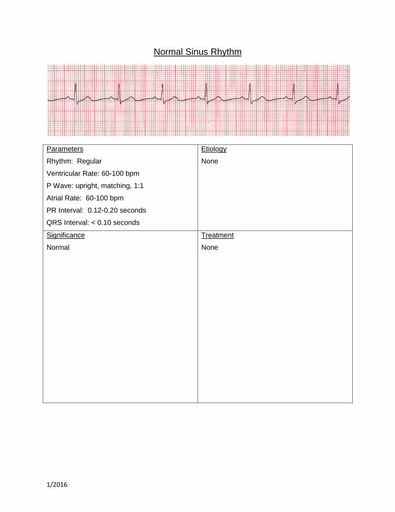
Normal Sinus Rhythm
Parameters
Rhythm: Regular
Ventricular Rate: 60-100 bpm
P Wave: upright, matching, 1:1
Atrial Rate: 60-100 bpm
PR Interval: 0.12-0.20 seconds
QRS Interval: < 0.10 seconds
Etiology
None
Significance
Normal
Treatment
None
1/2016
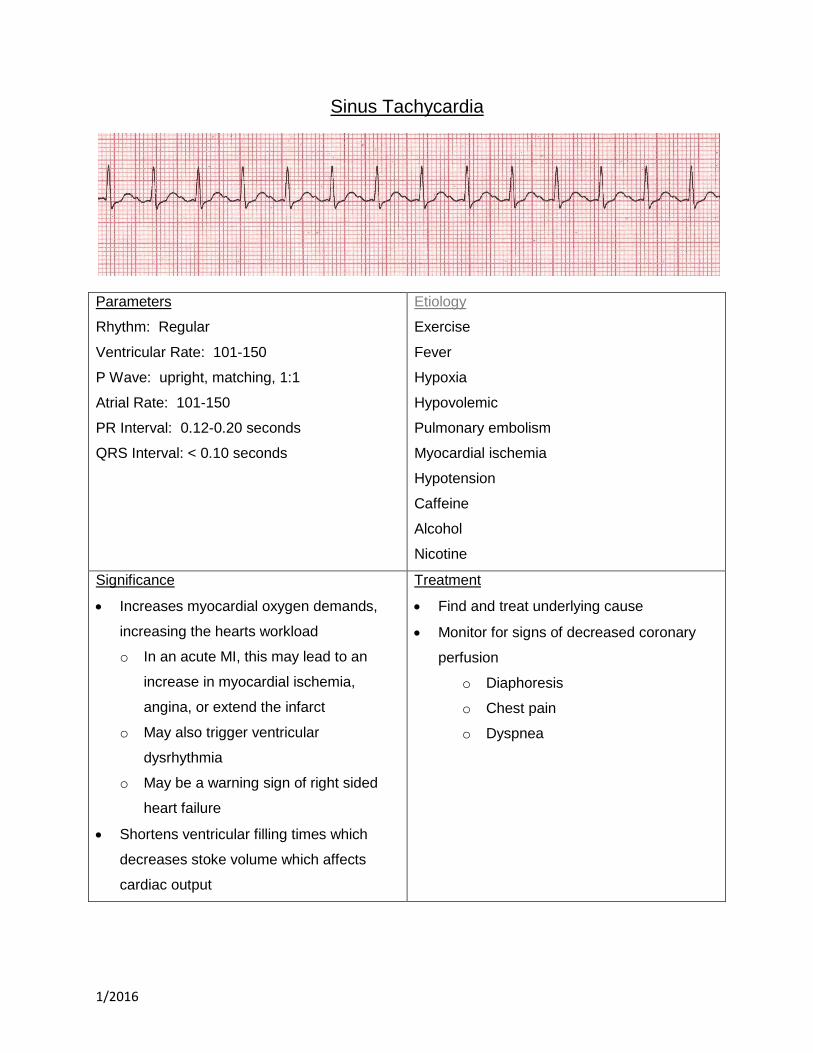
Sinus Tachycardia
Parameters
Rhythm: Regular
Ventricular Rate: 101-150
P Wave: upright, matching, 1:1
Atrial Rate: 101-150
PR Interval: 0.12-0.20 seconds
QRS Interval: < 0.10 seconds
Etiology
Exercise
Fever
Hypoxia
Hypovolemic
Pulmonary embolism
Myocardial ischemia
Hypotension
Caffeine
Alcohol
Nicotine
Significance
Increases myocardial oxygen demands,
increasing the hearts workload
o In an acute MI, this may lead to an
increase in myocardial ischemia,
angina, or extend the infarct
o May also trigger ventricular
dysrhythmia
o May be a warning sign of right sided
heart failure
Shortens ventricular filling times which
decreases stoke volume which affects
cardiac output
Treatment
Find and treat underlying cause
Monitor for signs of decreased coronary
perfusion
o Diaphoresis
o Chest pain
o Dyspnea
1/2016
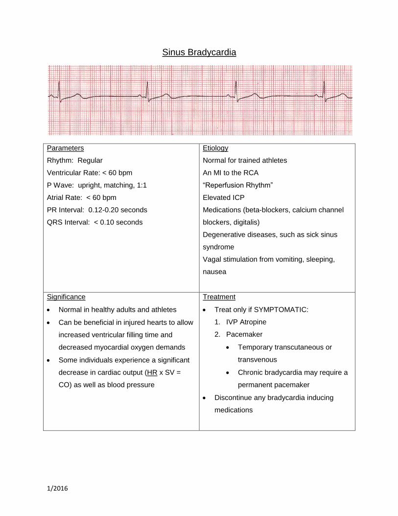
Sinus Bradycardia
Parameters
Rhythm: Regular
Ventricular Rate: < 60 bpm
P Wave: upright, matching, 1:1
Atrial Rate: < 60 bpm
PR Interval: 0.12-0.20 seconds
QRS Interval: < 0.10 seconds
Etiology
Normal for trained athletes
An MI to the RCA
“Reperfusion Rhythm”
Elevated ICP
Medications (beta-blockers, calcium channel
blockers, digitalis)
Degenerative diseases, such as sick sinus
syndrome
Vagal stimulation from vomiting, sleeping,
nausea
Significance
Normal in healthy adults and athletes
Can be beneficial in injured hearts to allow
increased ventricular filling time and
decreased myocardial oxygen demands
Some individuals experience a significant
decrease in cardiac output (HR x SV =
CO) as well as blood pressure
Treatment
Treat only if SYMPTOMATIC:
1. IVP Atropine
2. Pacemaker
Temporary transcutaneous or
transvenous
Chronic bradycardia may require a
permanent pacemaker
Discontinue any bradycardia inducing
medications
1/2016
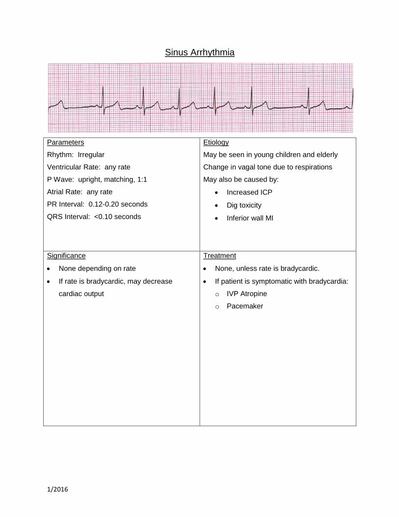
Sinus Arrhythmia
Parameters
Rhythm: Irregular
Ventricular Rate: any rate
P Wave: upright, matching, 1:1
Atrial Rate: any rate
PR Interval: 0.12-0.20 seconds
QRS Interval: <0.10 seconds
Etiology
May be seen in young children and elderly
Change in vagal tone due to respirations
May also be caused by:
Increased ICP
Dig toxicity
Inferior wall MI
Significance
None depending on rate
If rate is bradycardic, may decrease
cardiac output
Treatment
None, unless rate is bradycardic.
If patient is symptomatic with bradycardia:
o IVP Atropine
o Pacemaker

1/2016
Premature Atrial Contraction(PAC)
Parameters
Rhythm: that of underlying rhythm
Ventricular Rate: that of underlying
P Wave: upright, abnormal in size and shape,
p wave may be in T wave
Atrial Rate: that of underlying rhythm
PR Interval: 0.12-0.20 seconds
QRS Interval: <0.10 seconds
Etiology
Can occur in normal hearts
o Can be seen with emotional distress
Heart disease
Ingestion of alcohol, caffeine, or nicotine
Hypoxia
Myocardial ischemia
Chronic lung disease
Medications
Significance
Usually common and do not require
treatment
Frequent PAC’s may warn of or intiate:
o PAT
o Atrial Fibrillation
o Atrial Flutter
Treatment
Usually no treatment
Remove underlying cause:
o Nicotine
o Alcohol
o Caffeine
1/2016
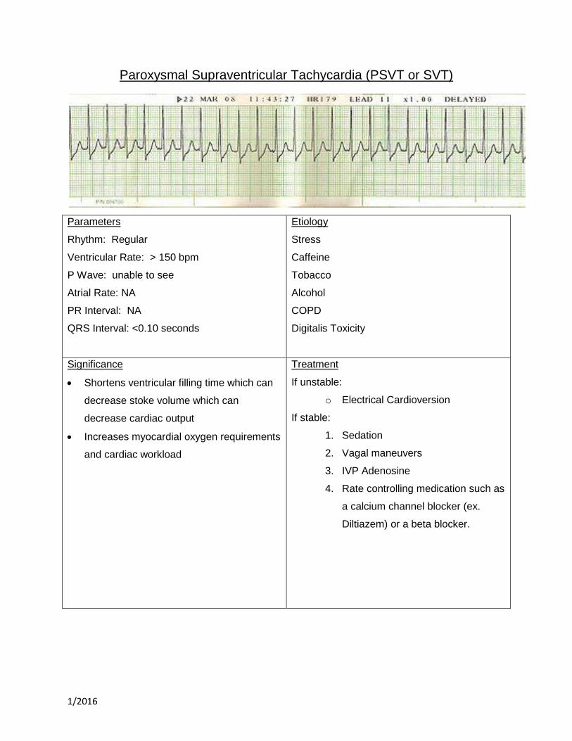
Paroxysmal Supraventricular Tachycardia (PSVT or SVT)
Parameters
Rhythm: Regular
Ventricular Rate: > 150 bpm
P Wave: unable to see
Atrial Rate: NA
PR Interval: NA
QRS Interval: <0.10 seconds
Etiology
Stress
Caffeine
Tobacco
Alcohol
COPD
Digitalis Toxicity
Significance
Shortens ventricular filling time which can
decrease stoke volume which can
decrease cardiac output
Increases myocardial oxygen requirements
and cardiac workload
Treatment
If unstable:
o Electrical Cardioversion
If stable:
1. Sedation
2. Vagal maneuvers
3. IVP Adenosine
4. Rate controlling medication such as
a calcium channel blocker (ex.
Diltiazem) or a beta blocker.
1/2016
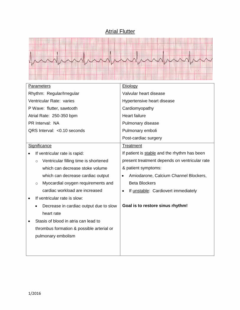
Atrial Flutter
Parameters
Rhythm: Regular/Irregular
Ventricular Rate: varies
P Wave: flutter, sawtooth
Atrial Rate: 250-350 bpm
PR Interval: NA
QRS Interval: <0.10 seconds
Etiology
Valvular heart disease
Hypertensive heart disease
Cardiomyopathy
Heart failure
Pulmonary disease
Pulmonary emboli
Post-cardiac surgery
Significance
If ventricular rate is rapid:
o Ventricular filling time is shortened
which can decrease stoke volume
which can decrease cardiac output
o Myocardial oxygen requirements and
cardiac workload are increased
If ventricular rate is slow:
Decrease in cardiac output due to slow
heart rate
Stasis of blood in atria can lead to
thrombus formation & possible arterial or
pulmonary embolism
Treatment
If patient is stable and the rhythm has been
present treatment depends on ventricular rate
& patient symptoms:
Amiodarone, Calcium Channel Blockers,
Beta Blockers
If unstable: Cardiovert immediately
Goal is to restore sinus rhythm!
1/2016
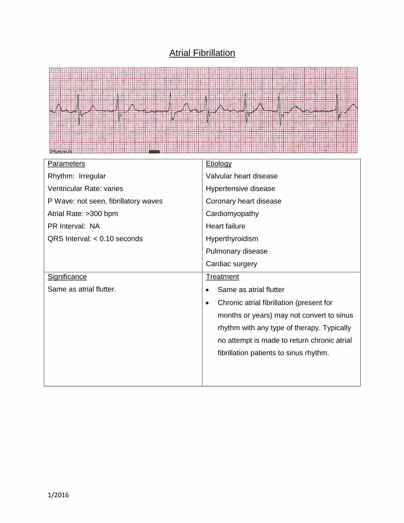
Atrial Fibrillation
Parameters
Rhythm: Irregular
Ventricular Rate: varies
P Wave: not seen, fibrillatory waves
Atrial Rate: >300 bpm
PR Interval: NA
QRS Interval: < 0.10 seconds
Etiology
Valvular heart disease
Hypertensive disease
Coronary heart disease
Cardiomyopathy
Heart failure
Hyperthyroidism
Pulmonary disease
Cardiac surgery
Significance
Same as atrial flutter.
Treatment
Same as atrial flutter
Chronic atrial fibrillation (present for
months or years) may not convert to sinus
rhythm with any type of therapy. Typically
no attempt is made to return chronic atrial
fibrillation patients to sinus rhythm.
1/2016
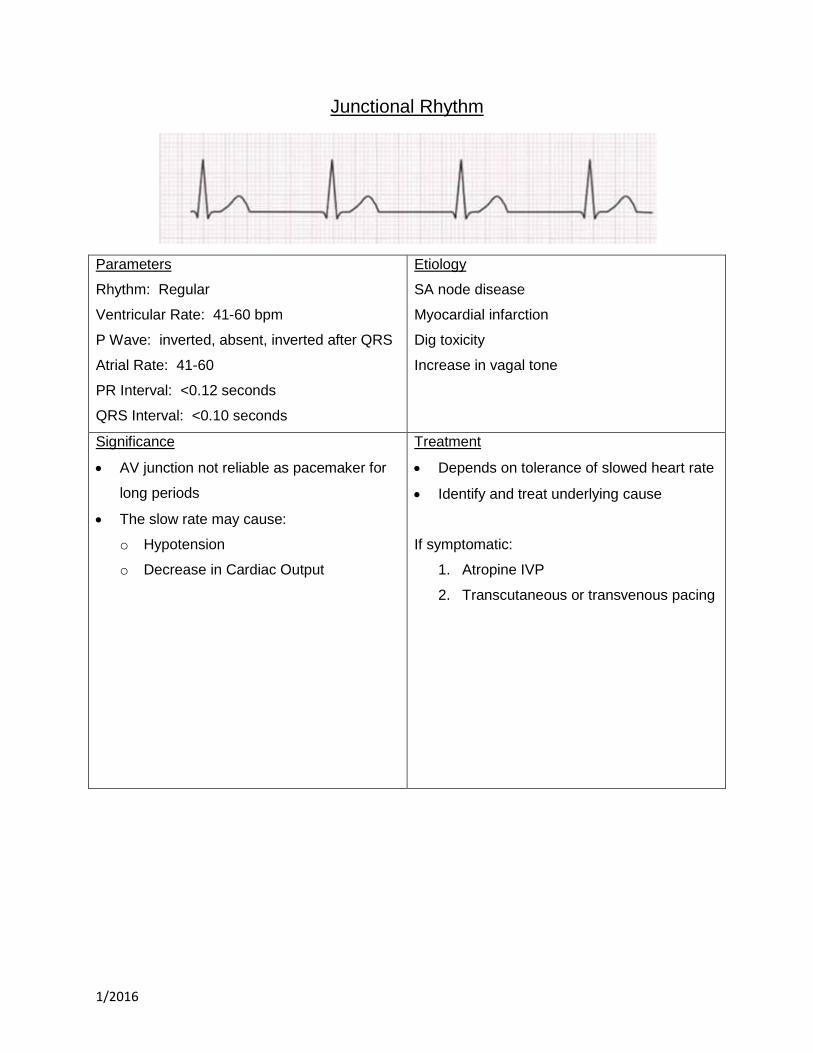
Junctional Rhythm
Parameters
Rhythm: Regular
Ventricular Rate: 41-60 bpm
P Wave: inverted, absent, inverted after QRS
Atrial Rate: 41-60
PR Interval: <0.12 seconds
QRS Interval: <0.10 seconds
Etiology
SA node disease
Myocardial infarction
Dig toxicity
Increase in vagal tone
Significance
AV junction not reliable as pacemaker for
long periods
The slow rate may cause:
o Hypotension
o Decrease in Cardiac Output
Treatment
Depends on tolerance of slowed heart rate
Identify and treat underlying cause
If symptomatic:
1. Atropine IVP
2. Transcutaneous or transvenous pacing
1/2016
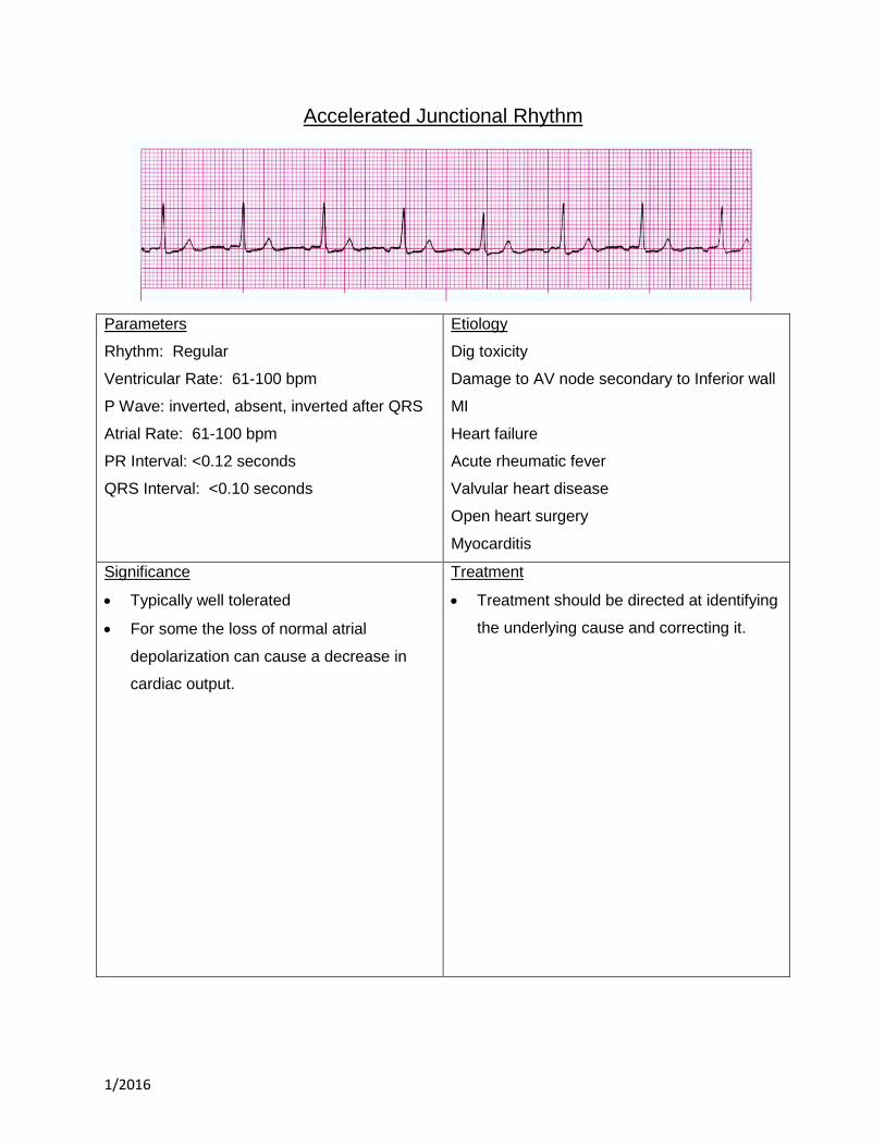
Accelerated Junctional Rhythm
Parameters
Rhythm: Regular
Ventricular Rate: 61-100 bpm
P Wave: inverted, absent, inverted after QRS
Atrial Rate: 61-100 bpm
PR Interval: <0.12 seconds
QRS Interval: <0.10 seconds
Etiology
Dig toxicity
Damage to AV node secondary to Inferior wall
MI
Heart failure
Acute rheumatic fever
Valvular heart disease
Open heart surgery
Myocarditis
Significance
Typically well tolerated
For some the loss of normal atrial
depolarization can cause a decrease in
cardiac output.
Treatment
Treatment should be directed at identifying
the underlying cause and correcting it.
1/2016
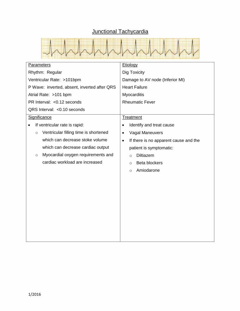
Junctional Tachycardia
Parameters
Rhythm: Regular
Ventricular Rate: >101bpm
P Wave: inverted, absent, inverted after QRS
Atrial Rate: >101 bpm
PR Interval: <0.12 seconds
QRS Interval: <0.10 seconds
Etiology
Dig Toxicity
Damage to AV node (Inferior MI)
Heart Failure
Myocarditis
Rheumatic Fever
Significance
If ventricular rate is rapid:
o Ventricular filling time is shortened
which can decrease stoke volume
which can decrease cardiac output
o Myocardial oxygen requirements and
cardiac workload are increased
Treatment
Identify and treat cause
Vagal Maneuvers
If there is no apparent cause and the
patient is symptomatic:
o Diltiazem
o Beta blockers
o Amiodarone
1/2016
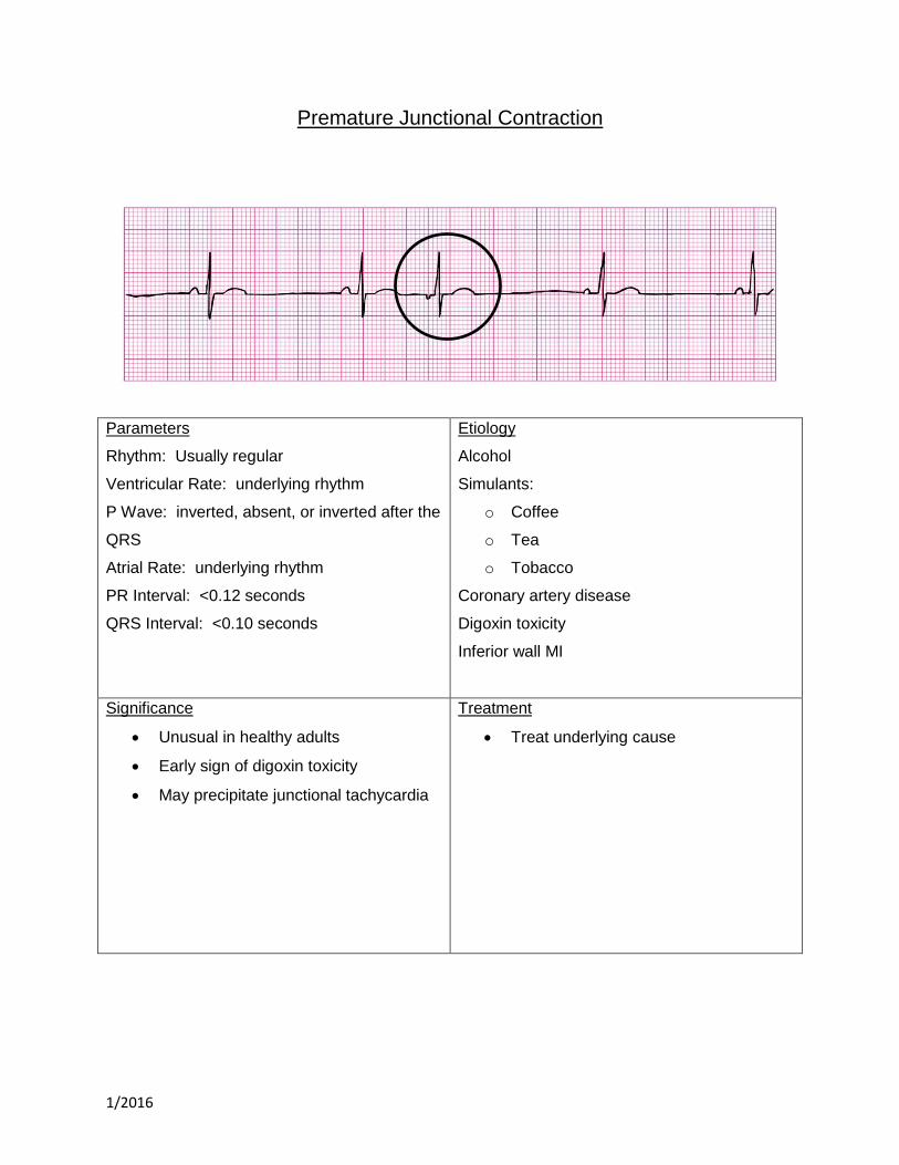
Premature Junctional Contraction
Parameters
Rhythm: Usually regular
Ventricular Rate: underlying rhythm
P Wave: inverted, absent, or inverted after the
QRS
Atrial Rate: underlying rhythm
PR Interval: <0.12 seconds
QRS Interval: <0.10 seconds
Etiology
Alcohol
Simulants:
o Coffee
o Tea
o Tobacco
Coronary artery disease
Digoxin toxicity
Inferior wall MI
Significance
Unusual in healthy adults
Early sign of digoxin toxicity
May precipitate junctional tachycardia
Treatment
Treat underlying cause
1/2016
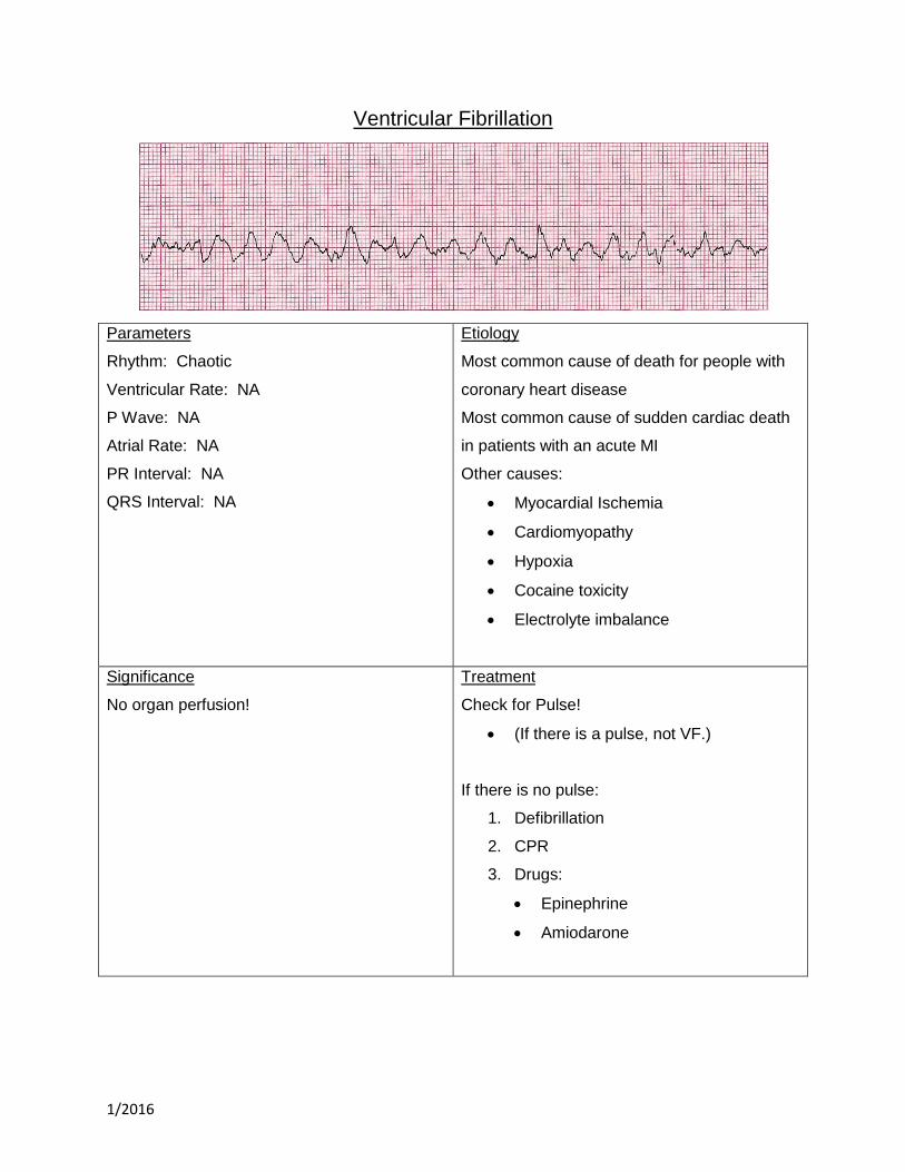
Ventricular Fibrillation
Parameters
Rhythm: Chaotic
Ventricular Rate: NA
P Wave: NA
Atrial Rate: NA
PR Interval: NA
QRS Interval: NA
Etiology
Most common cause of death for people with
coronary heart disease
Most common cause of sudden cardiac death
in patients with an acute MI
Other causes:
Myocardial Ischemia
Cardiomyopathy
Hypoxia
Cocaine toxicity
Electrolyte imbalance
Significance
No organ perfusion!
Treatment
Check for Pulse!
(If there is a pulse, not VF.)
If there is no pulse:
1. Defibrillation
2. CPR
3. Drugs:
Epinephrine
Amiodarone
1/2016
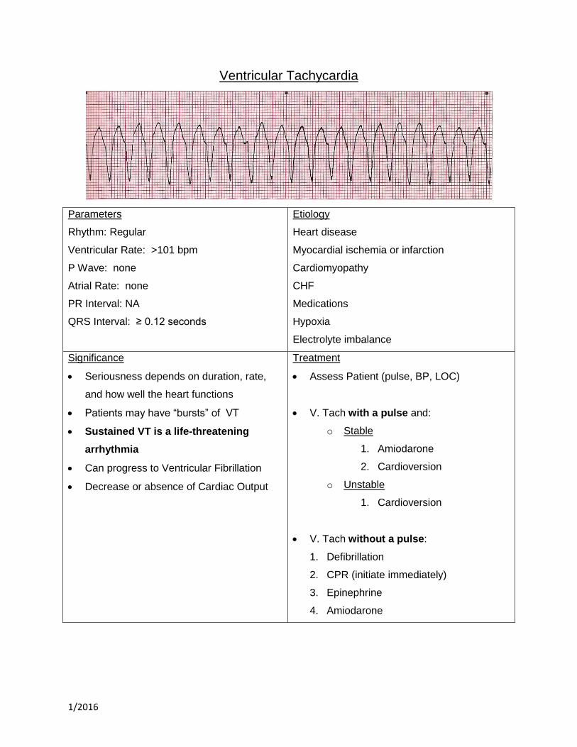
Ventricular Tachycardia
Parameters
Rhythm: Regular
Ventricular Rate: >101 bpm
P Wave: none
Atrial Rate: none
PR Interval: NA
QRS Interval: ≥ 0.12 seconds
Etiology
Heart disease
Myocardial ischemia or infarction
Cardiomyopathy
CHF
Medications
Hypoxia
Electrolyte imbalance
Significance
Seriousness depends on duration, rate,
and how well the heart functions
Patients may have “bursts” of VT
Sustained VT is a life-threatening
arrhythmia
Can progress to Ventricular Fibrillation
Decrease or absence of Cardiac Output
Treatment
Assess Patient (pulse, BP, LOC)
V. Tach with a pulse and:
o Stable
1. Amiodarone
2. Cardioversion
o Unstable
1. Cardioversion
V. Tach without a pulse:
1. Defibrillation
2. CPR (initiate immediately)
3. Epinephrine
4. Amiodarone

1/2016
Ventricular Standstill
Parameters
Rhythm: Atrial Regular
Ventricular Rate: NA
P Wave: upright, matching
Atrial Rate: varies
PR Interval: NA
QRS Interval: NA
Etiology
Acidosis
Hypoxia
Hyperkalemia
Hypothermia
Drug Overdose
1/2016
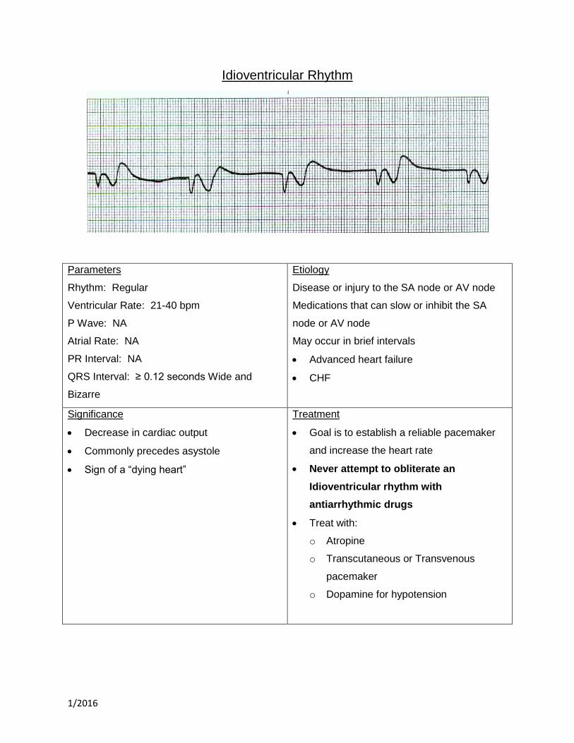
Idioventricular Rhythm
Parameters
Rhythm: Regular
Ventricular Rate: 21-40 bpm
P Wave: NA
Atrial Rate: NA
PR Interval: NA
QRS Interval: ≥ 0.12 seconds Wide and
Bizarre
Etiology
Disease or injury to the SA node or AV node
Medications that can slow or inhibit the SA
node or AV node
May occur in brief intervals
Advanced heart failure
CHF
Significance
Decrease in cardiac output
Commonly precedes asystole
Sign of a “dying heart”
Treatment
Goal is to establish a reliable pacemaker
and increase the heart rate
Never attempt to obliterate an
Idioventricular rhythm with
antiarrhythmic drugs
Treat with:
o Atropine
o Transcutaneous or Transvenous
pacemaker
o Dopamine for hypotension
1/2016
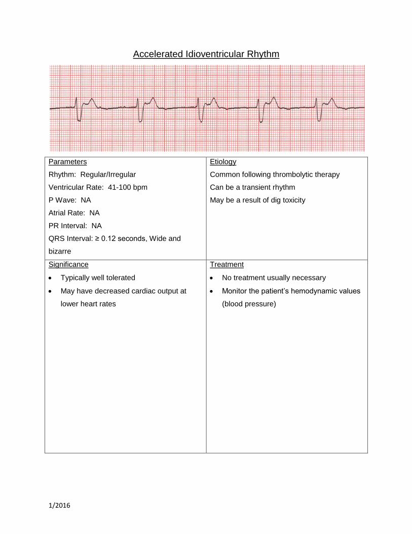
Accelerated Idioventricular Rhythm
Parameters
Rhythm: Regular/Irregular
Ventricular Rate: 41-100 bpm
P Wave: NA
Atrial Rate: NA
PR Interval: NA
QRS Interval: ≥ 0.12 seconds, Wide and
bizarre
Etiology
Common following thrombolytic therapy
Can be a transient rhythm
May be a result of dig toxicity
Significance
Typically well tolerated
May have decreased cardiac output at
lower heart rates
Treatment
No treatment usually necessary
Monitor the patient’s hemodynamic values
(blood pressure)
1/2016
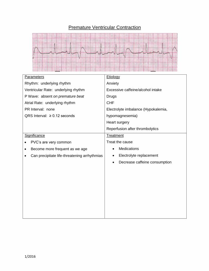
Premature Ventricular Contraction
Parameters
Rhythm: underlying rhythm
Ventricular Rate: underlying rhythm
P Wave: absent on premature beat
Atrial Rate: underlying rhythm
PR Interval: none
QRS Interval: ≥ 0.12 seconds
Etiology
Anxiety
Excessive caffeine/alcohol intake
Drugs
CHF
Electrolyte imbalance (Hypokalemia,
hypomagnesemia)
Heart surgery
Reperfusion after thrombolytics
Significance
PVC’s are very common
Become more frequent as we age
Can precipitate life-threatening arrhythmias
Treatment
Treat the cause
Medications
Electrolyte replacement
Decrease caffeine consumption
1/2016
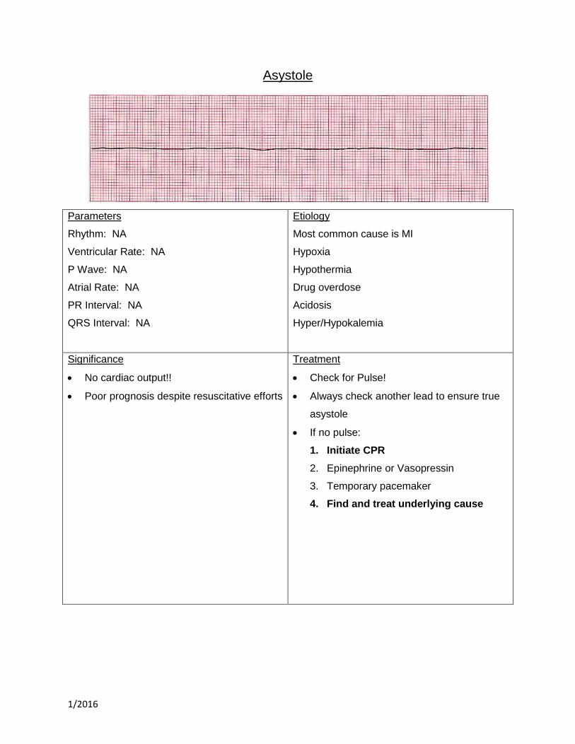
Asystole
Parameters
Rhythm: NA
Ventricular Rate: NA
P Wave: NA
Atrial Rate: NA
PR Interval: NA
QRS Interval: NA
Etiology
Most common cause is MI
Hypoxia
Hypothermia
Drug overdose
Acidosis
Hyper/Hypokalemia
Significance
No cardiac output!!
Poor prognosis despite resuscitative efforts
Treatment
Check for Pulse!
Always check another lead to ensure true
asystole
If no pulse:
1. Initiate CPR
2. Epinephrine or Vasopressin
3. Temporary pacemaker
4. Find and treat underlying cause
1/2016
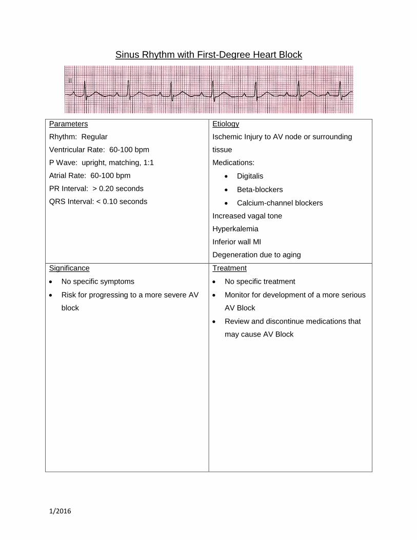
Sinus Rhythm with First-Degree Heart Block
Parameters
Rhythm: Regular
Ventricular Rate: 60-100 bpm
P Wave: upright, matching, 1:1
Atrial Rate: 60-100 bpm
PR Interval: > 0.20 seconds
QRS Interval: < 0.10 seconds
Etiology
Ischemic Injury to AV node or surrounding
tissue
Medications:
Digitalis
Beta-blockers
Calcium-channel blockers
Increased vagal tone
Hyperkalemia
Inferior wall MI
Degeneration due to aging
Significance
No specific symptoms
Risk for progressing to a more severe AV
block
Treatment
No specific treatment
Monitor for development of a more serious
AV Block
Review and discontinue medications that
may cause AV Block

1/2016
Second-Degree Heart Block Type 1 (Wenkebach)
Parameters
Rhythm: Ventricular-Irregular/Regular
Atrial-Regular
Ventricular Rate: varies
P Wave: upright, matching
Atrial Rate: varies
PR Interval: progressively lengthens
QRS Interval: < 0.10 seconds
Etiology
Ischemia of the AV due to an inferior MI
Same medications as First Degree
Acute Infections
May be normal in athletes
Significance
Seldom a serious form of heart block
Rarely progresses to higher degree of
block
Treatment
Monitor for development of a more serious
AV Block
Review and discontinue medications that
may cause AV Block
If symptomatic due to bradycardia:
o Atropine
o Pacemaker, not usually necessary
1/2016
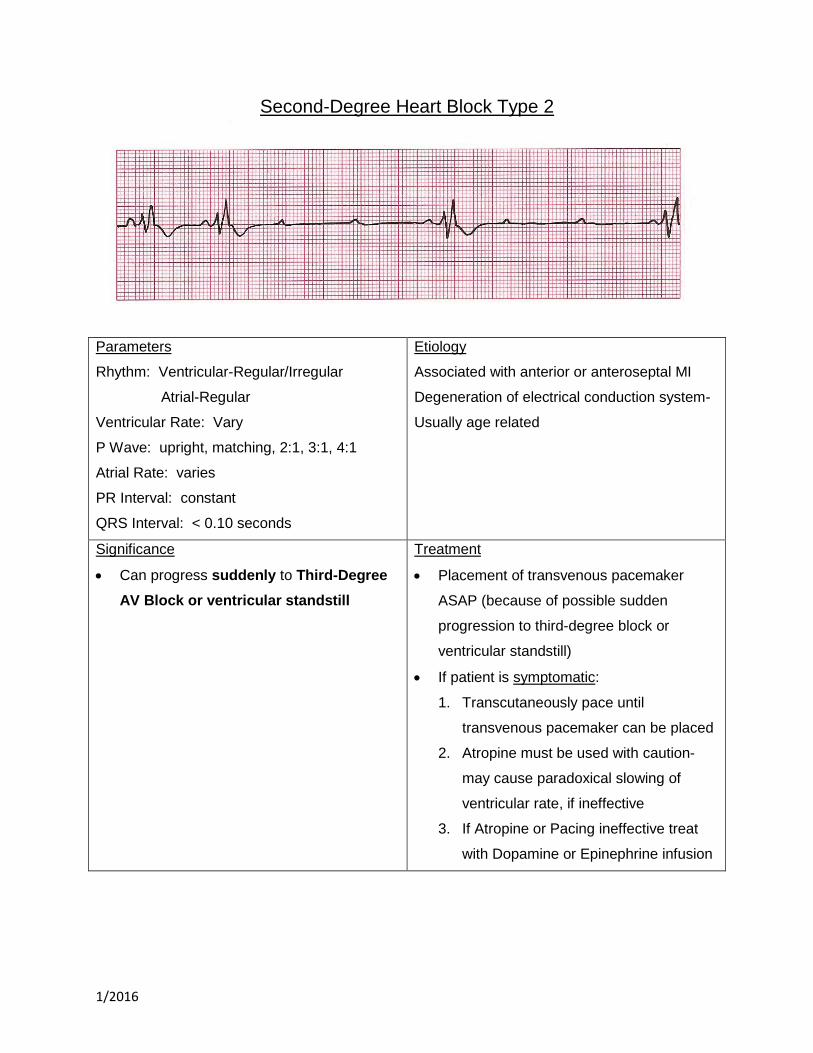
Second-Degree Heart Block Type 2
Parameters
Rhythm: Ventricular-Regular/Irregular
Atrial-Regular
Ventricular Rate: Vary
P Wave: upright, matching, 2:1, 3:1, 4:1
Atrial Rate: varies
PR Interval: constant
QRS Interval: < 0.10 seconds
Etiology
Associated with anterior or anteroseptal MI
Degeneration of electrical conduction system-
Usually age related
Significance
Can progress suddenly to Third-Degree
AV Block or ventricular standstill
Treatment
Placement of transvenous pacemaker
ASAP (because of possible sudden
progression to third-degree block or
ventricular standstill)
If patient is symptomatic:
1. Transcutaneously pace until
transvenous pacemaker can be placed
2. Atropine must be used with caution-
may cause paradoxical slowing of
ventricular rate, if ineffective
3. If Atropine or Pacing ineffective treat
with Dopamine or Epinephrine infusion
1/2016
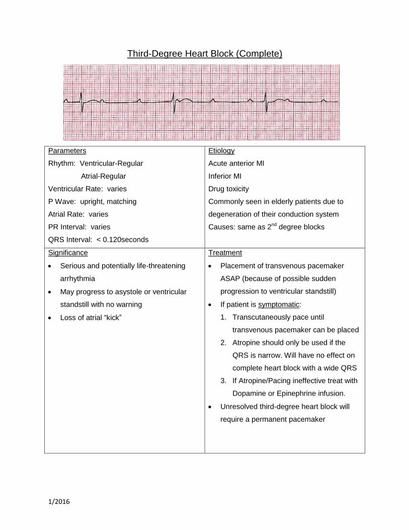
Third-Degree Heart Block (Complete)
Parameters
Rhythm: Ventricular-Regular
Atrial-Regular
Ventricular Rate: varies
P Wave: upright, matching
Atrial Rate: varies
PR Interval: varies
QRS Interval: < 0.120seconds
Etiology
Acute anterior MI
Inferior MI
Drug toxicity
Commonly seen in elderly patients due to
degeneration of their conduction system
Causes: same as 2nd degree blocks
Significance
Serious and potentially life-threatening
arrhythmia
May progress to asystole or ventricular
standstill with no warning
Loss of atrial “kick”
Treatment
Placement of transvenous pacemaker
ASAP (because of possible sudden
progression to ventricular standstill)
If patient is symptomatic:
1. Transcutaneously pace until
transvenous pacemaker can be placed
2. Atropine should only be used if the
QRS is narrow. Will have no effect on
complete heart block with a wide QRS
3. If Atropine/Pacing ineffective treat with
Dopamine or Epinephrine infusion.
Unresolved third-degree heart block will
require a permanent pacemaker
1/2016
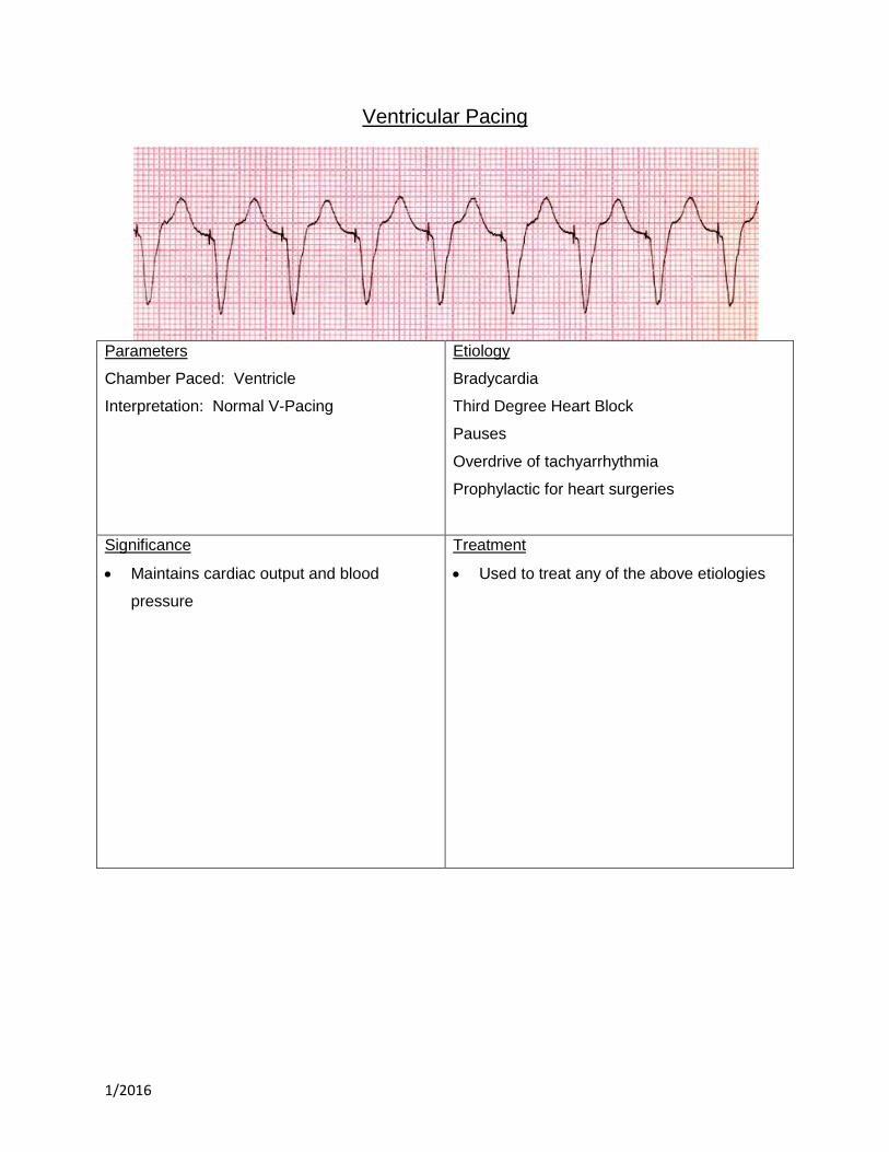
Ventricular Pacing
Parameters
Chamber Paced: Ventricle
Interpretation: Normal V-Pacing
Etiology
Bradycardia
Third Degree Heart Block
Pauses
Overdrive of tachyarrhythmia
Prophylactic for heart surgeries
Significance
Maintains cardiac output and blood
pressure
Treatment
Used to treat any of the above etiologies
1/2016
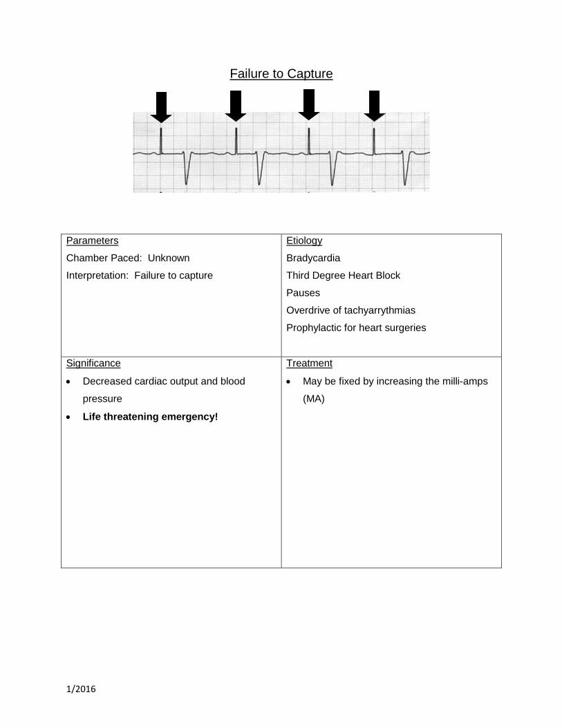
Failure to Capture
Parameters
Chamber Paced: Unknown
Interpretation: Failure to capture
Etiology
Bradycardia
Third Degree Heart Block
Pauses
Overdrive of tachyarrythmias
Prophylactic for heart surgeries
Significance
Decreased cardiac output and blood
pressure
Life threatening emergency!
Treatment
May be fixed by increasing the milli-amps
(MA)

1/2016
Undersensing
Parameters
Chamber Paced: Ventricular
Interpretation: V-Paced with undersensing
Etiology
Bradycardia
Third Degree Heart Block
Pauses
Overdrive of tachyarrythmias
Prophylactic for heart surgeries
Significance
Decreased cardiac output and blood
pressure
May cause PVC’s or V. Tach if spikes land
on t-wave during relative refractory period
Treatment
May be fixed by increasing the milli-volts
(MV)
1/2016
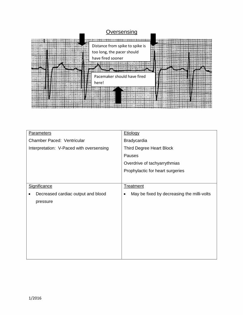
Oversensing
Parameters
Chamber Paced: Ventricular
Interpretation: V-Paced with oversensing
Etiology
Bradycardia
Third Degree Heart Block
Pauses
Overdrive of tachyarrythmias
Prophylactic for heart surgeries
Significance
Decreased cardiac output and blood
pressure
Treatment
May be fixed by decreasing the milli-volts
Distance from spike to spike is
too long, the pacer should
have fired sooner
Pacemaker should have fired
here!
1/2016
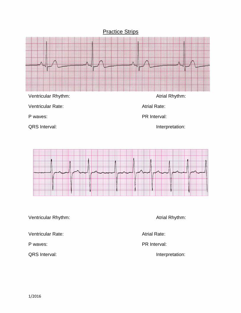
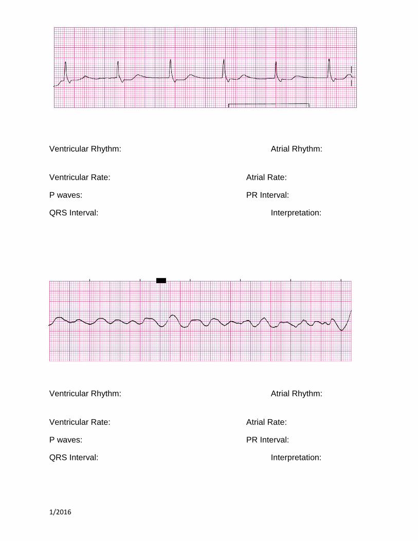
Practice Strips
Ventricular Rhythm: Atrial Rhythm:
Ventricular Rate: Atrial Rate:
P waves: PR Interval:
QRS Interval: Interpretation:
Ventricular Rhythm: Atrial Rhythm:
Ventricular Rate: Atrial Rate:
P waves: PR Interval:
QRS Interval: Interpretation:
1/2016
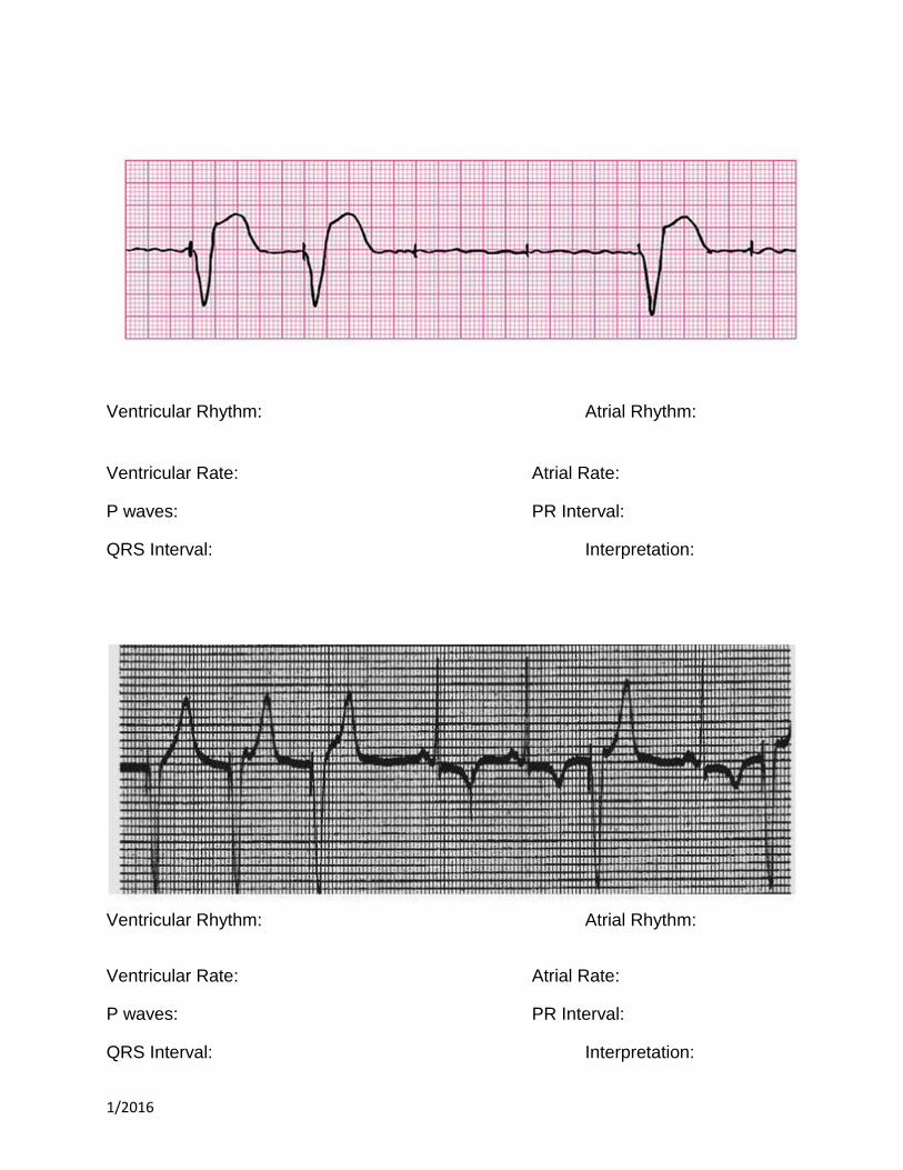
Ventricular Rhythm: Atrial Rhythm:
Ventricular Rate: Atrial Rate:
P waves: PR Interval:
QRS Interval: Interpretation:
Ventricular Rhythm: Atrial Rhythm:
Ventricular Rate: Atrial Rate:
P waves: PR Interval:
QRS Interval: Interpretation:

1/2016
Ventricular Rhythm: Atrial Rhythm:
Ventricular Rate: Atrial Rate:
P waves: PR Interval:
QRS Interval: Interpretation:
Ventricular Rhythm: Atrial Rhythm:
Ventricular Rate: Atrial Rate:
P waves: PR Interval:
QRS Interval: Interpretation:
1/2016
Ventricular Rhythm: Atrial Rhythm:
Ventricular Rate: Atrial Rate:
P waves: PR Interval:
QRS Interval: Interpretation:
Ventricular Rhythm: Atrial Rhythm:
Ventricular Rate: Atrial Rate:
P waves: PR Interval:
QRS Interval: Interpretation:
1/2016
Answers to Practice Strips
Strip 1:
Ventricular Rhythm: Regular Atrial Rhythm: Regular Ventricular Rate: 42 Atrial Rate: 42 P waves: Upright, Matching, 1:1 PR Interval: 0.16 seconds QRS: 0.12 seconds Interpretation: Sinus Bradycardia with IVCD Strip 2: Ventricular Rhythm: Irregular Atrial Rhythm: NA Ventricular Rate: 80-90 Atrial Rate: NA P waves: Fibrillatory PR Interval: NA QRS: 0.12 seconds Interpretation: Atrial Fibrillation with IVCD Strip 3: Ventricular Rhythm: Regular Atrial Rhythm: NA Ventricular Rate: 58-60 Atrial Rate: NA P waves: Inverted after QRS complex PR Interval: NA QRS: 0.06 seconds Interpretation: Junctional Rhythm Strip 4: Ventricular Rhythm: NA Atrial Rhythm: NA Ventricular Rate: NA Atrial Rate: NA P waves: NA PR Interval: NA QRS: NA Interpretation: Ventricular Fibrillation
1/2016
Strip 5: Ventricular Rhythm: Regular Atrial Rhythm: NA Ventricular Rate: 60-63 Atrial Rate: NA P waves: NA PR Interval: NA QRS: 0.16-0.18 seconds Interpretation: AIVR Strip 6: Ventricular Rhythm: Regular Atrial Rhythm: Regular Ventricular Rate: 35-36 Atrial Rate: 94 P waves: Upright, Matching PR Interval: 0.36, 0.18, 0.50 seconds QRS: 0.24 seconds Interpretation: Third Degree Heart Block or Complete Heart Block Strip 7: Chamber Paced: Ventricle Paced Rate: 60 Interpretation: Venticular paced rhythm with non capture Strip 8: Chamber Paced: Ventricle Paced Rate: 75 Interpretation: V-Paced with Undersensing