Embed Size (px)
Citation preview
EditionWissenschaftForschungsgemeinschaft Funk e. V. . G 14515 . Issue No. 21 . December 2005
Roland Glaser
Wis
sens
chaf
tEd
itio
n Are thermoreceptorsresponsible for„non-thermal“ effectsof RF fields?
2 Edition Wissenschaft December 2005
Editorial
as “Edition Wissenschaft Nr. 21“ the
Forschungsgemeinschaft Funk likes
to present you at the end of the year
2005 an very interesting article of
Prof. Dr. Roland Glaser, Humboldt
University, Berlin about the question:
“Are thermoreceptors responsible
for “non-thermal” effects of RF
fields?”. This should be a valuable
contribution to the still open discus-
sion, whether there are reliable
existing evidences for “non-thermal”
effects far under the limits of the
ICNIRP recommendations.
During the last years Prof. Roland
Glaser had taken several times the
opportunity to aim on clarification
of the burning question, which
biophysical mechanism can contrib-
ute to “non-thermal” or “athermal”
effects. This question is not new.
Neither there was a reliable com-
monly accepted reproducible effect,
nor an unanimously accepted
definition for the usage of the terms
“non-thermal” or “athermal”. Very
often in discussions the scientific
community even could not be
brought on the same level of
understanding of underlying mecha-
nism or processes in cells. On
different occasions this study
question was hotly discussed. We
have reported about several FGF-
and COST 281- Workshops in the
”FGF Newsletter”. During the last
four years a special group of scien-
tists with the support of MMF and
FGF have donated much time to look
for concepts to understand the
issue. We are eager to look at the
output of this task force and we will
inform you immediately after the
presentations of the results.
Till today it can be stated, that there
is a lack of knowledge concerning
research on cellular and molecular
mechanisms. And that lack of
knowledge has to be filled under the
consideration, that without solid
basic research of that study issue the
question will be still remain open.
This will underline the necessity of
that kind of research: “to look for
mechanism under the condition of
very small energy input in biological
systems like it is with EMF energy
emission of modern radio communi-
cation systems”
If one is speculating that may be
one of the results of further epide-
miological research will be: „no clear
cancer evidence“, the results of
human and animal studies will
getting more and more important.
Sometimes the result of studies is
like that following statement: „there
are some effects, but probably non
dangerous and possibly subtle-
thermal“. Then the old stereotype
questions arise: „but what will be
with other conditions or other
frequencies or pulsations?...“ These
questions can only be answered on
the basis of a known mechanism.
That exactly underlines the main
points of further research needs.
It is absolutely correct, that: „The
only established mechanisms that
relate to health consequences are
caused by temperature elevation ...“
but: excitation of molecular vibra-
tions and protein conformations can
be the result of this, not only by
radical pair mechanism (which, as
already known, is not relevant for
weak RF-fields!). This is in accor-
dance with the statement in the
WHO research agenda that: „Micro-
dosimetry research (i.e., at the
cellular or subcellular levels) that
may yield new insights concerning
biologically relevant targets of RF“.
This exactly should be the start point
for high priority research. Including
the recent results of thermosensible
molecules (TRP-receptors, GrpE, as
„riboswitches“ for HSP-expression
etc.) one really could understand the
so called „non-thermal“ effects of
weak RF-fields. The papers of Ken
Foster and Earl Prohofsky are
important steps to find the answer
of this question, but they did not
include these new results of ther-
moreception. FGF recently have
organized a workshop “Subtle
Thermal Effects of RF-Fields in vitro
and in vivo”, which exactly tackle
this point (Stuttgart, Nov. 2005).
Only knowing the theoretical
background of possible molecular
absorption and heat-dissipation of
RF-energy at molecular and su-
pramolecular level, predictions could
be made on frequency dependence,
pulsation etc. Furthermore, these
results can indicate, what really does
the term „dose“ mean, and what
kind of “dosimetry” we really need
(SAR, SAR* Time, W*m-2 ...).
To realize this kind of research,
experiments should be performed in
cooperation with specialists of
thermoreception on RF-field effects
on established models of molecular
and cellular thermoreceptors. These
sorts of experiments are much faster
and cheaper than animal studies.
They easily can realize conditions of
frequency dependence or pulsations
and can produce data, which leads
to new insights in to molecular
mechanisms. So, let us try to set this
kind of research in force. Glaser´s
contribution in this edition is just
one step. Other studies have to
follow.
Kindest regards
Gerd Friedrich
Dear readers,
3Edition Wissenschaft December 2005
Contents
Introduction: What does „non-thermal“ mean?Introduction: What does „non-thermal“ mean?Introduction: What does „non-thermal“ mean?Introduction: What does „non-thermal“ mean?Introduction: What does „non-thermal“ mean? 44444
Thermoregulatory response to IR versus RF energy absorptionThermoregulatory response to IR versus RF energy absorptionThermoregulatory response to IR versus RF energy absorptionThermoregulatory response to IR versus RF energy absorptionThermoregulatory response to IR versus RF energy absorption 55555
Limitations of behavioural experiments performedLimitations of behavioural experiments performedLimitations of behavioural experiments performedLimitations of behavioural experiments performedLimitations of behavioural experiments performed
in animals and human volunteersin animals and human volunteersin animals and human volunteersin animals and human volunteersin animals and human volunteers 66666
What do we know about the physiology of specializedWhat do we know about the physiology of specializedWhat do we know about the physiology of specializedWhat do we know about the physiology of specializedWhat do we know about the physiology of specialized
thermoreceptors in animals?thermoreceptors in animals?thermoreceptors in animals?thermoreceptors in animals?thermoreceptors in animals? 66666
Molecular mechanisms of thermoreceptionMolecular mechanisms of thermoreceptionMolecular mechanisms of thermoreceptionMolecular mechanisms of thermoreceptionMolecular mechanisms of thermoreception 88888
Conclusions: The system of thermoreception and its side reactionsConclusions: The system of thermoreception and its side reactionsConclusions: The system of thermoreception and its side reactionsConclusions: The system of thermoreception and its side reactionsConclusions: The system of thermoreception and its side reactions
– a possible reason for „non-thermal“ reactions of RF-fields– a possible reason for „non-thermal“ reactions of RF-fields– a possible reason for „non-thermal“ reactions of RF-fields– a possible reason for „non-thermal“ reactions of RF-fields– a possible reason for „non-thermal“ reactions of RF-fields 1010101010
ReferencesReferencesReferencesReferencesReferences 1111111111
AbstractAbstractAbstractAbstractAbstract 1313131313
4 Edition Wissenschaft December 2005
Introduction
Roland Glaser
Are thermoreceptors responsible for„non-thermal“ effects of RF fields?
Introduction:What does „non-thermal“ mean?In contrast to ELF, the fields in the
frequency range of mobile phones
do not excite nerve and muscle cells.
Therefore, the only obvious effect of
RF and MW fields on biological
systems is heating potentially
bringing about the consequences of
temperature increases. Although
interactions of high frequency fields
were investigated already in the
thirties of the last century [Rajewsky,
1938], the question whether there
exist additional “non-thermal“
effects that would have to be
considered in defining safety limits,
still remains controversial. The
reason for this are publications
claiming to have found such “non-
thermal“ effects in in vitro experi-
ments with cells, as well as in
experiments with animals or human
volunteers.
What do the terms “thermal“ and
“non-thermal“ (or “athermal“) really
mean? This question can be an-
swered either from a biophysical
perspective, considering possible
mechanisms of interaction, or just
empirically, based on experimental
setups. From the empirical point of
view, effects usually are called “non-
thermal“:
• if irradiation intensity in the
experiment is so low that changes
in temperature are unlikely to
occur physically [Geletyuk et al.,
1995; Kwee et al., 2001; Phillips
et al., 1998; Preece et al., 1999;
Weisbrot et al., 2003];
• if, during irradiation, no signifi-
cant change in temperature has
been measured in the body or in
the experimental vessel, or if
water jacketing, etc., has been
part of the experimental setup
warranting a constant tempera-
ture during exposure [Bohr and
Bohr, 2000; Byus et al., 1988;
Leszczynski et al., 2002; Mark-
kanen et al., 2004];
• if, in comparison, a temperature
increase caused by conventional
heating does not show effects
similar to those induced by RF
exposure [Cao et al., 1995; de
Pomerai et al., 2000; Peinnequin
et al., 2000; Velizarov et al.,
1999], or if, due to a normal
temperature increase, an opposite
effect is expected related to RF
exposure [Allis et al., 1987].
In general, in these papers an effect
is considered “non-thermal“ if it is
not accompanied by a predictable or
measurable temperature increase, or
if it does not correspond to effects
occurring after conventional heating
of the same amount.
Contrary to this, the biophysical
definition is based on the types of
mechanisms of field interaction.
Concomitant heating is not consid-
ered in this approach. Consequently,
a mechanism is seen as non-thermal
if the interaction of the electrical (or
magnetic) vector of the RF field with
charges or dipoles of molecules in
the living system directly leads to
specific effects other than heating.
Fröhlich [1982] already pointed out
that: “An effect is non-thermal
when, under the influence of a field,
the system changes its properties in
a way that can not be achieved by
heating, i.e. when its response is
non-linear“. His calculations made
clear that extremely strong fields
would be necessary to move molecu-
lar dipoles.
Non-thermal effects in this sense
actually are well known, as for
example in dielectrophoreses or
electrorotation of cells [Fuhr and
Hagedorn, 1996; Fuhr et al., 1996;
Gimsa, 1991; Glaser and Fuhr, 1987;
Glaser, 2000]. To induce them,
however, electric field strengths are
required which are many magni-
tudes above those which are used by
normal telecommunications systems.
Consequently, dielectrophoresis and
electrorotation are accompanied by
considerable heat production.
According to the „empirical“
definition, they should therefore be
classified as “thermal“. They are of
particular interest regarding biotech-
nological applications, but irrelevant
to the issue of safety limits as
discussed in this paper.
5Edition Wissenschaft December 2005
Thus, the use of the empirical versus
the biophysical definition of the
term “non-thermal“ sometimes leads
to rather contradictory evaluations.
It would probably make sense to
introduce the terms “quasi-non-
thermal“ or “subtle thermal“ to
make the empirical definition of
“non-thermal“ more precise. For
simplifying matters, however, in the
following we will enclose the term
“non-thermal“ in quotation marks
when referring to the empirical
definition in the sense of “so-called
non-thermal“ effects.
In this context, the question arises:
Are effects that are called “non-
thermal“ in the above mentioned
empirical sense of the word “subtle
thermal“ effects, or do non-thermal
effects, according to the biophysical
definition, really occur also at weak
RF field exposure? At first glance,
this question may seem sophistical,
but in fact it is crucial where safety
aspects, i.e. the setting of exposure
limits, are concerned. If such
responses to weak RF fields found in
some experiments are just that:
“subtle temperature“ effects –
meaning that they simply activate
common reactions of the biological
system to a very low and probably
only locally detectable degree of
heating – they are harmless and can
be neglected. Such effects can be
treated similarly to those caused by
temperature influences to which the
organism is exposed on a daily basis
and under various environmental
conditions. If, on the other hand,
particular interaction mechanisms of
weak RF or MW fields do exist that
are not identical with those of
normal heating, they should be
carefully investigated considering
potential health consequences, and
should also taken into account when
defining exposure limits.
This paper is an attempt to verify
hypotheses claiming that “non-
thermal“ effects of RF fields, at least
those found in experiments per-
formed under well-controlled
conditions, ultimately are the result
of thermoreceptor activation.
Possibly only single molecules or
thermosensitive cells were stimulat-
ed that triggered various reactions
of the highly complicated system of
thermoregulation without involving
conscious perception of test volun-
teers, or behavioural consequences
in animals. Temperature elevation
gains and the size of the heated
volume may be so small that mea-
surement is technically impossible
(this aspect was already discussed in
detail by Stern et al. [1979]).
Thermoregulatoryresponse to IR versusRF energy absorption
The concept of radiofrequency (RF)
and microwave (MW) fields activat-
ing the thermoregulatory system is
not new. Recently, Eleanor R. Adair
and D. R. Black summarized related
investigations in a diligent review
[Adair and Black, 2003]. First
detailed experiments investigating
the activation of human thermore-
ceptors by GHz fields in comparison
to infrared (IR) irradiation were
already performed by Hendler and
Hardy [1960]. In the seventies, a
number of related studies followed,
summarized in the book “Micro-
waves and Thermoregulation“ that
was edited by Eleanor A. Adair
[1983]. Blick at al. [1997] repeated
and extended investigations using
experiments with advanced dosime-
try, and at an extended frequency
range. Thresholds for microwave-
evoked skin sensations of warmth at
frequencies of 2.45, 7.5, 10, 35,
and 94 GHz were measured and
compared to warmth evoked by
infrared radiation (IR). These find-
ings are generally consistent with
those reported by earlier investiga-
tors indicating dependency on
frequency which corresponds to the
depth of tissue penetration of
radiation. Sensitivity monotonically
increased with increasing frequency
throughout the range of microwave
frequencies tested. When irridiating
an area of 327 cm2 for 10 seconds,
the threshold at 94 GHz (4.5 ±
0.6 mW/cm2) was more than an
order of magnitude below that at
2.45 GHz (63.1 ± 6.7 mW/cm2), and
was comparable to the threshold for
IR (5.34 ± 1.07 mW/cm2) (Tab. 1).
These differences were due to the
effectivity of the absorbed radiation
in attaining a defined temperature
elevation across the regions where
thermoreceptors are located. The
energy absorbed by other regions is
dissipated without any effect on the
thermoregulatory system. Thermore-
ceptors of the skin located in the
outer 0.6 mm skin layer are primarily
activated [Adair and Black, 2003].
Riu et al. [1997] calculated the
temperature profiles generated by
these frequencies after 3 and 10
seconds of skin exposure and found
corresponding thresholds of temper-
ature sensation of about 0.07°C.
The thresholds found in these
experiments therefore clearly exceed
the exposure limits recommended by
the ICNIRP and prescribed by official
regulations. Furthermore, they are
above the level of exposure at which
“non-thermal“ effects have some-
times been found. Does this mean
that thermosensors can be ignored
in such low level experiments?
6 Edition Wissenschaft December 2005
Limitations of behav-ioural experimentsperformed in animalsand human volunteers
The above mentioned experiments
testing the warmth sensation of
human volunteers or the behaviour
of animals are of relevance when
investigating the basic problem of RF
field influence on the system of
thermoregulation, but cannot
exclude the possibility of thermosen-
sory activation in general. This is
already made evident by two
circumstances which were observed
in these experiments: (I) The thresh-
old of activation depends not only
on what region of the body is
exposed to the field, but also on
what area of the skin is irradiated.
This means that a response was
detected only if a minimum number
of singular thermoreceptors were
activated. The observed intensity
does therefore not reflect the
threshold of a single thermoreceptor
cell. (II) The time constant of the
sensation is >1 s. The sensitivity to
thermal stimulation was found to
increase with exposure time ranging
from 3 s up to 10 s [Riu et al.,
1997]. This indicates that the
reaction was not determined by the
primary response of thermosensors
but by the time constant of the
heating of the tissue surrounding
the thermoreceptors and by subse-
quent neuronal processes.
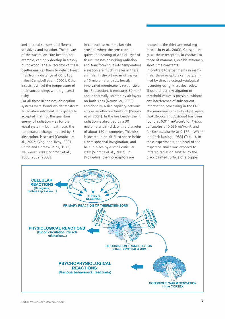
In fact, warmth sensation does not
simply reflect the activation of
thermoreceptors in a specific region
of the body, but is a result of the
subsequent information processing
in the hypothalamus and cortex (see:
Fig. 1). Thermoreceptors of warm-
blooded animals are located on the
surface as well as in many other
parts of the body, including the
brain and the spinal cord. Whilst
thermoreceptors in the skin measure
the environmental temperature,
internal thermoreceptors are respon-
sible for controlling blood tempera-
ture. They are all part of a
complicated network transmitting
information to the center of temper-
ature control: the preoptic area of
the anterior hypothalamus. Con-
scious warmth sensation, therefore,
is the result of processing the
information provided by all these
sensors, including a lot of other
physiological conditions.
In the last years our knowledge on
molecular and cellular mechanisms
of thermoreception has remarkably
increased. So it was shown that not
only nerve endings and dendrids are
responsible for thermoreception, but
thermosensitive ion channels are
also found in keratinocytes, ovary
cells [Peier et al., 2002 a, b] and
aorta endothelial cells [Watanabe et
al., 2002]. These investigations must
be taken into consideration to
understand the results of experi-
ments on “non-thermal“ reactions of
RF-fields.
What do we knowabout the physiology ofspecialized thermore-ceptors in animals?
Provided that many processes in
living nature are similar across
organisms of different organization-
al levels, it makes sense firstly to
take a look at the highly specialized
thermoreceptors of various animals
and insects, aiding them in hunting,
feeding and overall survival. Boas,
pythons and pit vipers are known to
use IR radiation emanated by warm-
blooded animals. Their pit organs
are radiant heat detectors helping
them to locate an IR source with an
angle resolution of few degrees
[Campbell et al., 2002; de Cock
Buning, 1983; Neuweiler, 2003]. A
similarly functioning pit organ is
found in vampire bats, as for
example in Desmodus rotundus
[Kürten and Schmidt, 1982]. Many
insects also are equipped with IR
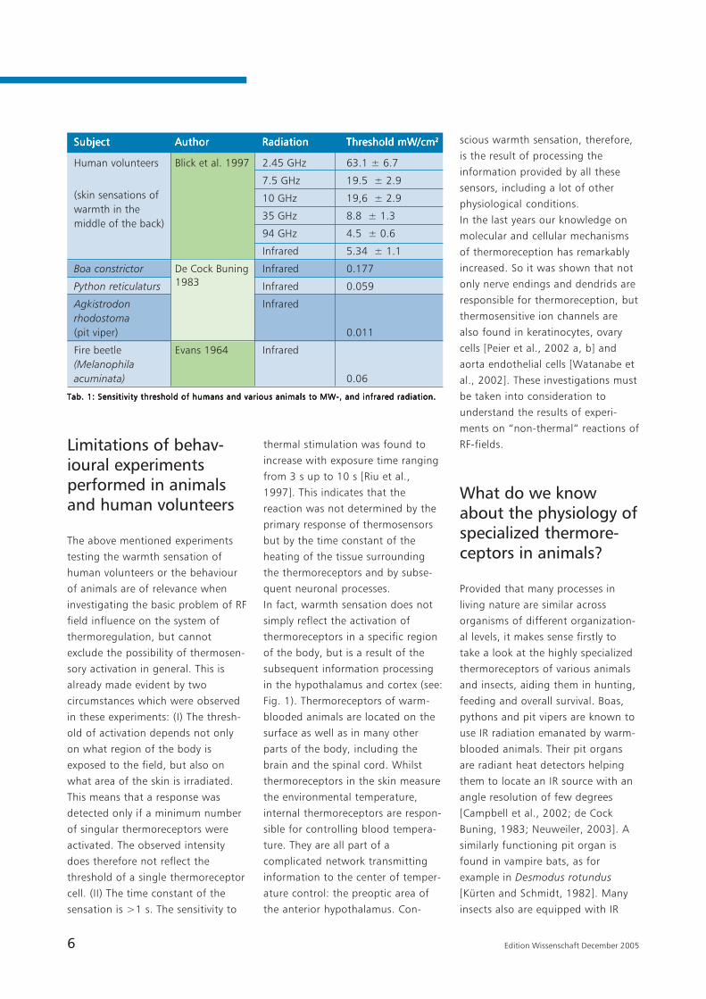
Tab. 1: Sensitivity threshold of humans and various animals to MW-, and infrared radiation.Tab. 1: Sensitivity threshold of humans and various animals to MW-, and infrared radiation.Tab. 1: Sensitivity threshold of humans and various animals to MW-, and infrared radiation.Tab. 1: Sensitivity threshold of humans and various animals to MW-, and infrared radiation.Tab. 1: Sensitivity threshold of humans and various animals to MW-, and infrared radiation.
(skin sensations ofwarmth in themiddle of the back)
SubjectSubjectSubjectSubjectSubject AuthorAuthorAuthorAuthorAuthor RadiationRadiationRadiationRadiationRadiation Threshold mW/cmThreshold mW/cmThreshold mW/cmThreshold mW/cmThreshold mW/cm22222
Human volunteers Blick et al. 1997 2.45 GHz 63.1 ± 6.7
7.5 GHz 19.5 ± 2.9
10 GHz 19,6 ± 2.9
35 GHz 8.8 ± 1.3
94 GHz 4.5 ± 0.6
Infrared 5.34 ± 1.1
Boa constrictor De Cock Buning Infrared 0.177
Python reticulaturs Infrared 0.059
Agkistrodon Infraredrhodostoma(pit viper) 0.011
Fire beetle Evans 1964 Infrared(Melanophilaacuminata) 0.06
1983
7Edition Wissenschaft December 2005
and thermal sensors of different
sensitivity and function. The larvae
of the Australian “fire beetle“, for
example, can only develop in freshly
burnt wood. The IR receptor of these
beetles enables them to detect forest
fires from a distance of 60 to100
miles [Campbell et al., 2002]. Other
insects just feel the temperature of
their surroundings with high sensi-
tivity.
For all these IR sensors, absorption
systems were found which transform
IR radiation into heat. It is generally
accepted that not the quantum
energy of radiation – as for the
visual system – but heat, resp. the
temperature change induced by IR
absorption, is sensed [Campbell et
al., 2002; Gingl and Tichy, 2001;
Harris and Gamow 1971, 1972;
Neuweiler, 2003; Schmitz et al.,
2000, 2002, 2003].
In contrast to mammalian skin
sensors, where the sensation re-
quires the heating of a thick layer of
tissue, masses absorbing radiation
and transforming it into temperature
elevation are much smaller in these
animals. In the pit organ of snakes,
a 15 micrometer thick, heavily
innervated membrane is responsible
for IR reception. It measures 30 mm2
and is thermally isolated by air layers
on both sides [Neuweiler, 2003];
additionally, a rich capillary network
acts as an effective heat sink [Pappas
et al. 2004]. In the fire beetle, the IR
radiation is absorbed by a 30
micrometer thin disk with a diameter
of about 120 micrometer. This disk
is located in an air-filled space inside
a hemispherical invagination, and
held in place by a small cuticular
stalk [Schmitz et al., 2002]. In
Drosophila, thermoreceptors are
located at the third antennal seg-
ment [Liu et al., 2003]. Consequent-
ly, all these receptors, in contrast to
those of mammals, exhibit extremely
short time constants.
In contrast to experiments in mam-
mals, these receptors can be exam-
ined by direct electrophysiological
recording using microelectrodes.
Thus, a direct investigation of
threshold values is possible, without
any interference of subsequent
information processing in the CNS.
The maximum sensitivity of pit vipers
(Agkistrodon rhodostoma) has been
found at 0.011 mW/cm2, for Python
reticulatus at 0.059 mW/cm2, and
for Boa constrictor at 0.177 mW/cm2
[de Cock Buning, 1983] (Tab. 1). In
these experiments, the head of the
respective snake was exposed to
infrared radiation emitted by the
black painted surface of a copper
8 Edition Wissenschaft December 2005
Molecular mechanismsof thermoreception
Over the last years a number of
detailed investigations of the
thermoregulation processes of
procaryotic and eucaryotic cells have
been published. These investigations
provided new insight into the
molecular mechanisms of ther-
mosensation of various cells, and
reveal the ambivalence of their
functions. The implication of cells in
the neuronal system of thermosensa-
tion just reflects one special case in
this diversity. Thermosensitive
molecules are responsible for various
cellular functions, such as the
protection against physiologically
adverse temperatures, or even for
the activation of cells in the case of
favorable temperatures [Chowdhury
et al., 2003]. Some of them are
temperature dependent RNA mole-
cules, a special kind of the so-called
„riboswitches“ [Lai, 2003]. Bacteria
and cells of many other organisms
regulate their phase transition of
membrane lipids in response to
temperature by controlling the fatty
acid composition of lipids. In B.
subtilis, for example, this is done by
a temperature dependent expression
of oxygen-dependent saturase, in E.
coli activating enzymes for synthesiz-
ing new phospholipids [Mansilla et
al., 2004]. The expression of heat-
shock proteins in procaryotes such
as E. coli is controlled by tempera-
ture dependent structural alterations
of the nucleotide exchange factor
GrpE exhibiting “non-Arrhenius“
behaviour. This means there is an
increasing activity in one, and a
decreasing activity in another
temperature region [Gelinas et al.,
2003; Grimshaw et al., 2003].
Although these kinds of cellular
thermoregulation seem to be
important too, sine they explain
some other “non-thermal“ cellular
effects, we will focuse here on
neuronal thermosensors. A growing
number of so called TRPV-transport
proteins in membranes of various
cells has been found over the last
years, some of them showing a high
degree of temperature dependence
of their function (TRP stands for
“transient receptor potential“, V
indicates a vallinoid sensitive sub-
family) [Tominaga et al., 2004]. In
mammals, TRPV3 and TRPV4 are the
most important channels responding
at temperatures in the physiological
range [Benham et al., 2003].
Notably, they do not only occur in
neurons but are also found in a
number of other cells. TRPV4
channels, for example, occur in a
HEK293 cell expression system and
in native mouse aorta endothelial
cells [Watanabe et al., 2002].
Gating mechanisms of these chan-
nels, at a specific temperature
interval, show a much larger sensi-
tivity to temperature elevation than
standard biochemical reactions. At
block with a temperature warmer by
10°C than the environment, at
distances of 16.4 to 66.6 cm.
Recordings were conducted at heat-
sensitive neurons in the brain. The
Australian fire beetle Melanophila
acuminata shows a maximum
sensitivity at 0.06 mW/cm2 [Evans
1964].
The real temperature increase of the
receptor sides in these experiments
are not measurable. Calculations of
the corresponding temperature
elevations, however, led to estimat-
ed values between 0.003 and
0.01 K. This is below the sensitivity
of conventional technical ther-
mosensors. By the way – from the
use of the empirical definition it
would follow that the infrared
sensation of these animals is based
on “non-thermal“ effects!
The time constants of these ther-
moreceptors were limited not so
much by the heating of the absorber
– as is the case in humans, monkeys
and rats – as by the time constants
and the firing frequency of nerves.
So the response to a stepwise
increased irradiation was below 20
ms [Schmitz et al., 2000; Gingl and
Tichy, 2001].
Unfortunately, there are no detailed
experiments on the sensibility of
these thermoreceptors to RF-fields.
Only Harris and Gamow [1972]
tested the response of the pit organs
of boas to 10.7 GHz microwave
radiation. They found a response to
pulses of about 11.1 mW/cm2, at a
distance of 4 cm from the horn
antenna “making an accurate
measurement of the power density
nearly impossible“. The aim of this
paper was just to demonstrate that
it is not a specific infrared effect
that is responsible for this reaction,
but an unspecific temperature
elevation.
By exceeding the scope of biological
objects and including those special-
ized for thermosensation to better
focus on the primary reactions as
occur in mammals, thermoreceptors
with extremely low thresholds and
short time constants were found.
Even such neurophysiological
experiments, however, fail to reflect
the real time constant of the primary
reaction, because they nevertheless
include a time delay for nerve
reactions. A closer look at the
molecular mechanisms is necessary
to obtain information about the real
data of this reaction, and also on
the probable sensitivity of single
thermosensitive cells in mammals.
9Edition Wissenschaft December 2005
times, this property is expressed by a
Q10-value, a change in the reaction
rate resulting from a 10 °C tempera-
ture rise. For normal biochemical
reactions as well as for normal ion
channels, this value is a factor of 2,
approximately. In the thermosensi-
tive temperature range of these
channels, however, it can be much
larger. Between 24 and 36 °C, TRPV4
channels, for example, indicate a
current increased by a Q10 of 19.1
[Watanabe et al., 2002]. Exposed in
Chinese hamster ovary cells, TRPV3
channels showed a Q10 of 1.9±0.3
for lower temperatures, and a Q10
=17.3±3.0, after the temperature
rose above 32 °C. Cells exposed to
rapid heating typically exhibited a
steep initial activation phase with
Q10 =21.6±4.2 [Xu et al., 2002]. In
keratinocytes, a TRPV3 channel was
found that was activated at temper-
atures above 35 °C, showing a mean
Q10 = 6.62 [Peier et al., 2002]. The
reason for this extremely sensitive
reactions in a narrow, specific
temperature range are structural
alterations of the molecule, a kind of
molecular phase transition. As a
result, for example, the TRPV4
channels transform temperature
stimuli into electrophysiological
responses of the cell connected by a
calcium influx from outside of the
cell [Güler et al., 2002].
Although TRPV1 to TRPV 4 channels
have not been found yet in inverte-
brates, similar channels were
detected in Drosophila and in the
roundworm Caenorhabditis elegans.
Another channel of the TRP family,
the ANKTM1 protein is found in
Drosophila as well as in vertebrates,
and possibly is also involved in
thermosensation [Viswanath et al.,
2003]. Unfortunately, the channels
which are responsible for the
enormous sensibility of the pit
organs of snakes have not yet been
isolated. However the functional
similarity of the corresponding
membrane proteins with the TRPV1-
channel has already been demon-
strated [Pappas et al. 2004].
It seems important to note that the
reaction of these temperature
sensitive molecules depends on
several conditions. The response of
the TRPV3 channel, for example,
increases with repeated stimulation
at suprathreshold temperatures,
indicating pronounced sensitization
of this receptor by heat. Its respons-
es increased at each subsequent
stimulus so that current responses
were enhanced about 10-fold over
the course of the experiment
[Benham et al., 2003]. This may be
one molecular reason for the above
mentioned dependence of the
thermal reaction of humans and
animals on the duration of IR
irradiation, besides other reactions
such as heating of the tissue etc.
Furthermore, the sensitivity of the
corresponding cells depends on the
osmolarity of the medium, the lipid
composition of the membrane and
other conditions [Güler et al., 2002].
Consequently, these channels exhibit
a surprising temperature depen-
dence of their function, particularly
at narrow temperature ranges. A
single cell usually contains many
copies of different types of such
channels, each being responsible for
a specific temperature region. The
general response of this cell, i.e. the
signal given by this cell to the
thermoreceptor, therefore is the
resulting average of the behavior of
of a large number of proteins. Since
thermoreceptors moreover represent
the response of many cells, this is to
be understood as a system of
substantial noise suppression.
Unfortunately, at present we know
only the temperature dependence of
these molecules, but not the thresh-
old of their response. A full analysis
of the temperature threshold of
these primary reactions cannot be
given yet.
Conclusions:The system ofthermoreceptionand its side reactions– a possible reasonfor „non-thermal“reactions of RF-fields
In Fig. 1 an attempt was made to
roughly illustrate the system of
thermosensation, from the primary
reaction of the membrane proteins,
the response of the cells, the
transduction of nervous information
to the hypothalamus up to the
conscious warm sensation as a
reaction of the cortex. Considering
the properties of this system and its
elements, as summarized in this
paper, new insights into the issue of
“non-thermal“ RF field interaction
become possible.
The extremely high sensitivity of
thermoreceptors of several animals
and insects, as listed in Tab. 1,
shows that in general this system is
able to react to temperature eleva-
tions below those measurable in
experiments investigating RF- and
microwave effects, as well as below
calculated temperature elevations
which usually occur in the skin and
surface of the brain during the use
of a mobile phone. Of course, the
sensitivity of the specialized ther-
mosensors in snakes or insects
possibly is higher than those of
mammals. As can be seen in many
other cases, however, similar
molecular systems are used conser-
Concclusions
10 Edition Wissenschaft December 2005
vatively in nature, at different levels
of evolution. There is a similarity, for
example, with the molecules of the
TRP-transporters, or thermosensitive
RNA-systems. Therefore, a response
of other cells at this level of temper-
ature elevation cannot be excluded.
The signal-to-noise ratio of ther-
moreception obviously is optimized
by averaging and filtering at all
information processing steps.
Thermoreceptor cells average the
information from many proteins; the
thermosensors at the nerve endings
use the information from many
thermosensitive cells; in the hypo-
thalamus, information from various
thermosensors in the body is
evaluated. The physiological evalua-
tion of signals sent by thermorecep-
tors obviously depends on the
number of activated thermorecep-
tors (i.e. the area of exposed skin),
the duration of activation, but also
on a number of general psychophys-
iological parameters.
The conscious warm sensation is
therefore the endpoint of the
system, the result of various neu-
ronal processes, signaling the body
there is a need of behavioral reac-
tions, like moving away from a
source etc. The absence of this
sensation, however, does not mean
that several less dramatic regulatory
processes were initiated. EEG
modifications as a result of weak RF-
field exposure, for example, are
understandable as being caused by
locally modified circulation in the
brain, even without a conscious
warm sensation. The dependence of
this reaction on a number of other
physiological factors certainly
explain why replications of these
results in human volunteers some-
times were impossible [e.g.: Freude
et al., 2000; Wagner et al., 2000].
Consequently, if „non-thermal“
Conclusions
effects – at least those found in
experiments using accurate exposure
systems and exact dosimetry - really
are based on the activation of the
molecular or cellular system of
thermosensors, they must be
classified as ‘everyday’ responses
without real consequences to health.
Acknowledgement
This paper was supported by the
Forschungsgemeinschaft Funk e.V.
Bonn.
11Edition Wissenschaft December 2005
Adair ER, Black DR 2003. Thermoregulatoryresponses to RF energy absorption. Bioelectro-magnetics Supp. 6: S17-S38.
Adair ER (Edit.) 1983. Microwaves andThermoregulations. Academic Press, New York.
Allis JW, Sinha-Robinson BL. 1987. Tempera-tur-specific inhibition of human red cell Na+/K+ATPase by 2,450-MHz microwave radiation.Bioelectromagnetics 8: 203-212.
Benham CD, Gunthorpe M J, Davis J B. 2003.TRPV channels as temperature sensors. CellCalcium 33: 479-487.
Blick DW, Adair ER, Hurt WD, Sherry CJ,Walters TJ, Merritt JH. 1997. Thresholds ofmicrowave-evoked warmth sensation in humanskin. Bioelectromagnetics 18: 403-409.
Bohr H, Bohr J. 2000. Microwave enhancedkinetics observed in ORD studies of a protein.Bioelectromagnetics 21: 68-72.
Byus CV, Dartun K, Pieper S, Adey WR. 1988.Increased ornithine decarboxylase activity incultured cells exposed to low energy modulat-ed microwave fields and phorbol ester tumorpromoters. Cancer Res. 48: 4222-4226.
Campbell A L, Naik R R, Sowards L, Stone MO.2002. Biological infrared imaging and sensing.Micron 33: 211-225.
Cao G, Liu L-M, Cleary SF. 1995. Cell cyclealterations induced by isothermal 27 MHzradio-frequency radiation exposure. Bioelectro-chem. Bioenerg. 37: 131-140.
Chowdhury S, Ragaz C, Kreuger, E, Narber-haus, F. 2003. Temperature-controlledstructural alterations of an RNA thermometer.J. Biol. Chem. 278: 47915-47921.
de Cock Buning TJ. 1983. Thresholds ofinfrared sensitive tectal neurons in Pytonreticulatus, Boa constrictor and Agkistrodonrhodostoma. J. Comp. Physiol. 151: 461-467.
de Pomerai D, Daniells C, David H, et al. 2000.Non-thermal heat-shock response to micro-waves. Nature 405: 417-418.
Evans, W. G. 1964. Infrared receptors inMelanophila acuminata De Geer.Nature; 202, 211
Freude G, Ullsperger P, Eggert S, Ruppe I.2000. Microwaves emitted by cellulartelephones affect human slow brain potentials.Eur. J. Appl. Physiol. 81: 18-27.
Fröhlich H. 1982. What are non-thermalelectric biological effects? Bioelectromagnetics3: 45-46.
Fuhr G, Hagedorn R. 1996. Cell Electrorota-tion. In: Lynch, P. T., M. R. Davey (Edrs.):Electrical Manipulation of Cells. Chapman &Hall, New York 38-70.
Fuhr G, Zimmermann U, Shirley SG. 1996. Cellmotion in time-varying fields: principles andpotential. in: U. Zimmermann, G. Neil (Edit.)Electromanipulation of cells. CRC Press INC,Boca Raton 259-328.
Geletyuk VI, Kazachenko VN, Chemeris NK,Fesenko EE. 1995. Dual effects of microwaveson single Ca2+-activated K+ channels incultured kidney cells vero. FEBS Letters 359:85-88.Gelinas AD, Toth J, Bethoney KA, Langsetmo K,Stafford WF, Harrison CJ. 2003. Thermodynam-ic linkage in the GrpE nucleotide exchangefactor, a molecular thermosensor. Biochemistry42: 9050-9059.
Gimsa J, Marszalek P, Loewe U, Tsong TY.1991. Dielectrophoresis and electrorotation ofneurospora slime and murine myeloma cells.Biophys. J. 60: 749-760.
Gingl E, Tichy H. 2001. Infrared sensitivity ofthermoreceptors. J. Comp. Physiol. 187: 467-475.
Glaser R. 2000. Biophysics. Springer, BerlinHeidelberg New York.
Glaser R, Fuhr G. 1987. Electrorotation – Thespin of cells in rotating high frequency electricfields. In: M. Blank and E. Findl (eds.);Mechanistic Approaches to Interactions ofElectric and Electromagnetic Fields with LivingSystems; Plenum Press, New York 271-289.
Grimshaw JPA, Jelesarov I, Siegenthaler RK,Christen P. 2003. Thermosensor action of GrpE- The DnaK chaperone system at heat shocktemperatures. J.Biol. Chem. 278: 19048-19053.
Güler AD, Lee HS, Iida T, Shimizu I, TominagaM, Caterina M. 2002. Heat-evoked activationof the ion channel TRPV4. J. Neuroscience 22:6408-6414.
Harris JF, Gamow RI. 1971. Snake infraredreceptors. Thermal or photochemical mecha-nisms? Science 172: 1252-1253.
Harris JF, Gamow RI. 1972. An analysis of heatreceptors by means of microwave radiation.Biol. Med. Sci. Instrument. 9: 187-190.
Hendler E, Hardy JD. 1960. Infrared andmicrowave effects on skin heating andtemperature sensation. IRE Trans. Med.Electronics 7: 143-152.
Kürten L, Schmidt U. 1982. Thermoreception inthe common vampire bat (Desmodusrotundus). J. Comp. Physiol. A 146: 223-228.
Kwee, S., Raskmark, P., Velizarov, S. 2001.Changes in cellular proteins due to environ-mental non-ionizing radiation. I. Heat-shockproteins. Electro and Magnetobiology 20: 141-152.
Lai, EC. 2003. RNA sensors and riboswitches.Self-regulating messages. Current Biology2003; 13: R285-R291.
Leszczynski D, Joenvaara S, Reivinen J, KuokkaR. 2002. Non-thermal activation of the hsp27/p38MAPK stress pathway by mobile phoneradiation in human endothelial cells: Molecularmechanism for cancer- and blood-brain barrier-related effects. Differentiation 70: 120-129.
Liu L, Yermolaieva O, Johnson WA, AbboudFM, Welsh M J. 2003. Identification andfunction of thermosensory neurons in Droso-phila larvae. Nature Neuroscience 6: 267-273.
Mansilla, MC, Cybulski, LE, Albanesi, D.,deMendoza, D. 2004. Control of membranelipid fluidity by molecular thermosensors. J.Bacteriol. 186: 6681-6688
Markkanen A, Penttinen P, Naarala J, PelkonenJ, Sihvonen AP, and Juutilainen J. 2004.Apoptosis induced by ultraviolet radiation isenhanced by amplitude modulated radiofre-quency radiation in mutant yeast cells.Bioelectromagnetics 25: 127-133.
Neuweiler G. 2003. Neuro- und Sinnesphysiolo-gie - 3. Temperaturempfindlichkeit. In: G. Held-maier u. G. Neuweiler: Vergleichende Tier-physiologie, Springer-Verlag Berlin 1: 89-96.
Pappas, TC, Motamedi, M., and Christensen,BN 2004. Unique temperature-activatedneurons from pit viper thermosensors. Am. J.Physiol. Cell Physiology 287: C1219-C1228.
Peier AM, Moqrich A, Hergarden AC, et al.2002. A TRP channel that senses cold stimuliand menthol. Cell 108: 705-715.
Peier AM, Reeve AJ, Andersson EA, et al. 2002.A heat-sensitive TRP channel expressed inkeratinocytes. Science 296: 2046-2049.
Peinnequin A, Piriou A, Mathieu J, Dabouis V,Sebbah C, Malabiau R, Debouzy JC. 2000.Non-thermal effects of continuous 2.45 GHzmicrowaves on Fas-induced apoptosis inhuman Jurkat T-cell line. Bioelectrochem.Bioenerg. 51: 157-161.
Phillips JL, Ivaschuk O, IshidaJones T, Jones RA,CampbellBeachler M, Haggren W. 1998. DNAdamage in Molt-4 T-lymphoblastoid cellsexposed to cellular telephone radiofrequencyfields in vitro. Bioelectrochem. Bioenerg. 45:103-110.
Preece AW, Iwi G, DaviesSmith A, Wesnes K,Butler S, Lim E, Varey A. 1999. Effect of a 915-MHz simulated mobile phone signal oncognitive function in man. Int. J. Radiat. Biol.75: 447-456.
Rajewsky B. 1938. Biophysikalische Grundlagender Ultrakurzwellen-Wirkung im lebendenGewebe. in: B. Rajewsky (Hrsg.) Ultra-kurzwellen in ihren medizinischen-biologischenAnwendungen, G. Thieme, Leipzig.
Riu PJ, Foster KR, Blick DW, Adair ERA. 1997.Thermal model for human thresholds ofmicrowave-evoked warmth sensations.Bioelectromagnetics 18: 578-583.
Schmitz H, Schmitz A, Bleckmann H. 2000.A new type of infrared organ in the Australian„fire-beetle“ Merimna atrata (Coleoptera: Bupres-tidae). Naturwissenschaften 87: 542-545.
Schmitz H, Schmitz A, Trenner S, Bleckmann H.2002. A new type of insect infrared organ oflow thermal mass. Naturwissenschaften 89:226-229.
Schmitz H, Trenner S. 2003. Electrophysiologi-cal characterization of the multipolarthermoreceptors in the ‘’fire-beetle’’ Merimnaatrata and comparison with the infraredsensilla of Melanophila acuminata (BothColeoptera, Buprestidae). J. Compar. Physiolo-gy A Neuroethology Sensory Neural andBehavioral Physiology 189: 715-722.
References
12 Edition Wissenschaft December 2005
Stern S, Margolin L, Weiss B, Lu ST, MichaelsonSM. 1979. Microwaves: Effect on thermoregula-tory behavior in rats. Science 206: 1198-1201.
Tominaga, M., Caterina, M J. 2004. Ther-mosensation and pain. J. Neurobiology 6:3-11
Velizarov S. 1999. Electric and magnetic fieldsin microbial biotechnology: Possibilities,limitations, and perspectives. Electro andMagnetobiology 18: 185-212.
Viswanath V, Story GM, Peier AM, Petrus MJ,Hwang SW, Patapoutian A, Jegla T. 2003. Ionchannels – Opposite thermosensor in fruitflyand mouse. Nature 423: 822-823.
Wagner P, Röschke J, Mann K, Fell J, Hiller W,Frank C, Grozinger M. 2000. Human sleep EEGunder the influence of pulsed radio frequencyelectromagnetic fields – Results frompolysomnographies using submaximal highpower flux densities. Neuropsychobiology 42:207-212.
Watanabe H, Vriens SH, Suh SH, Benham CD,Droogmans G, Nilius B. 2002. Heat-evokedactivation of TRPV4 channels in a HEK293 cellexpression system and in native mouse aortaendothelial cells. J. Biol. Chem. 277: 47044-47051.
Weisbrot D, Lin H, Ye L, Blank M., andGoodman R.2003. Effects of mobile phoneradiation on reproduction and development inDrosophila melanogaster. J. Cell. Biochem.;89: 48-55
Xu HX, Ramsey IS, Kotecha SA, et al. 2002.TRPV3 is a calcium permeable temperature-sensitive cation channel. Nature 418: 181-186.
13Edition Wissenschaft December 2005
Author:
Roland Glaser
Inst. of Biology/Experimental
Biophysics, Humboldt University-
Berlin /Germany
Abstract
The existence of “non-thermal“
effects of weak RF-fields has been
discussed again and again. In these
papers the expression “non-thermal“
is mostly not related to the biophysi-
cal mechanism, but used in a
empirical sense. Usually, an effect is
considered “non-thermal“ if it is not
accompanied by a predictable or
measurable temperature increase, or
if it does not correspond to effects
occurring after conventional heating
of a similar degree. This approach
does not take into consideration the
real system of thermoregulation, its
ambivalence, and its high sensitivity.
Recently, a specialized class of
transport proteins have been found
functioning as thermosensors in cell
membranes not only in cells of
specialized organs, but also in
normal keratinocytes and other cells.
The signal-to-noise ratio in this
system of thermoreception is
optimized by averaging the response
of many proteins and cells and many
steps of information processing with
various time constants from below
microseconds for the primary
reactions of the membrane proteins,
milliseconds for nerve excitations,
and eventually, tenths of seconds or
even minutes for behavioral conse-
quences. The threshold of this
system can be lower than the
sensitivity of our technical devices
for measuring or controlling temper-
ature in experiments. Considering
this, the effects found in experi-
ments with weak RF-fields in fact
could be “quasi-thermal“ or “subtle
thermal“ reactions of the biological
system of thermosensation and
thermoregulation. A number of
reactions is conceivable, occurring at
temperature elevations that are not
high enough to be registered by the
central nervous system. Protein
expression, influences on local blood
circulation, or other effects could be
the the result of thermal stimulation,
even if there is no measurable
temperature increase and no behav-
ioral consequences or conscious
“warm“ sensations. If “non-thermal“
effects, at least those found in
experiments using accurate exposure
systems and exact dosimetry, really
are based on the activation of the
molecular system of thermosensors,
they consequently must be classified
as ‘everyday’ responses without real
significance to health.
Abstract

14 Edition Wissenschaft December 2005Wis
sens
chaf
tEd
itio
n
ImprintEdition Wissenschaft der FGF e. V.
Publisher: Forschungsgemeinschaft Funk e. V., Rathausgasse 11a,
D-53111 Bonn, Phone: +49(0)228 / 72 62 2-0, Fax: +49(0)228 / 72 62 21 1
Email: [email protected], http://www.fgf.de
Editors: Gerd Friedrich (responsible), Regina Reichardt
Layout: setz it. Richert GmbH, Sankt Augustin
This study in hand was carried out on behalf of the Forschungsgemeinschaft
Funk e.V. (Research Association for Radio Applications). The report repre-
sents the authors’ opinions, not implicitly the opinion of FGF.