Embed Size (px)
Citation preview

PEARL MILLET AND GHRELIN IN POULTRY
By
JOSHUA DEAN HAMBURG
(under the direction of Adam J. Davis)
ABSTRACT
Poultry production allows the efficient production of animal protein in the form of
meat and eggs. In developing countries where affordable animal protein sources for
human consumption are deficient, the consumer price of poultry products and the
expansion of the commercial poultry industry are negatively impacted by the utilization of
costly imported feed ingredients to make poultry diets. Identifying and utilizing locally
grown feed ingredients would be beneficial in these locations. Pearl millet is a drought
resistant plant that produces a nutritious grain. Its cultivation in present-day Mali spans
thousands of years. It is still widely cultivated in this country where poultry production is
limited. The nutrient composition of different varieties of pearl millet grown in Mali was
assessed and then experiments were completed which indicated that whole pearl millet
grain grown in this region is a suitable replacement for corn in broiler and laying hen diets.
INDEX WORDS: pearl millet, broilers, laying hens

i
PEARL MILLET AND GHRELIN IN POULTRY
By
JOSHUA DEAN HAMBURG
B.S.A., The University of Georgia, 2009
A Thesis submitted to the Graduate Faculty of The University of Georgia in Partial
Fulfillment of the Requirements for the Degree
MASTERS OF SCIENCE
ATHENS, GEORGIA
2013

ii
2013
Joshua Dean Hamburg
All Rights Reserved

iii
PEARL MILLET AND GHRELIN IN POULTRY
By
JOSHUA DEAN HAMBURG
Major Professor: Adam J. Davis
Committee: Michael J. Azain Kristen J. Navara
Electronic Version Approved: Maureen Grasso Dean of the Graduate School The University of Georgia May 2013

iv
DEDICATION
In loving memory of my grandfather Dean Bural Hamburg. Thank you for all of your
advice and encouragement, I miss you every day.

v
ACKNOWLEDGMENTS
To my family, thank you for all of your support and encouragement throughout the
years. Without you I would not have become the person that I am today.
To my advisor, Dr. Adam Davis, thank you for keeping me busy and on my toes. You
guidance is as unmatched as your wit and humor. Thank you for allowing me this
opportunity and for all of your motivation.
To Dr. Mike Lacy, thank you for all of your kind words and for always being willing
to lend an open ear and a helping hand. You have a way of making our department feel like
a family, welcoming and supportive, despite the many different personalities and opinions
that are present—You are the glue that keep us all together.
To Martha Elizabeth Freeman, thank you for being the best teacher a student could
as for. Your patience and supportiveness are unrivaled. I will take all of the little “-isms”
that you have taught me in the lab and use them in my future endeavors.
To my fellow graduate students, past and present, thank you for being shining
examples of success. Your positive words of encouragement have helped guide me. You
have been the backbone and given me strength when I needed it most.
To Carey Drake, Erin Anderson, Erika Elmore, and Tamara Loeffler, you have all
pushed me and supported me: physically, mentally, and emotionally. I thank you for that.
To Lindsey Racket and the farm crew, thank you for the helping hands and for
making our facilities a highly conducive environment for research.
vi
TABLE OF CONTENTS
Page
ACKNOWLEDGEMENTS………………………………………………………………………………………..v
CHAPTER
1 MILLET…………………………………………………………………..................................................1
General overview……………………………………………………………………………1 Pearl millet……..………………………………………………………………………………1 Nutrient composition …………………………………..…………………………………3
Pearl millet grain in poultry production…………………………………………...4 Laying hens ………………………………………………………………………………..…5 Broilers………………………………………………………………………………………..…6 Whole pearl millet in poultry diets………………………….……………………….7 Pearl millet as an alternative grain in poultry diets……………………..……8
Summary………………………………………………………………………………………..9 2 GHRELIN AND THE AVIAN OVARY…………….………………………………………...…...10 The avian ovary…………………………………………………………………………….10 Avian follicular tissues and follicular maturation………….…………..……11 Feed restriction and ovarian function………………………………………...….11 Ghrelin overview……..…………………………………………………………………...14
Ghrelin synthesis…………..………………………………………………………………15 Ghrelin secretion and tissue distribution…………………….…………………16
vii
Ghrelin receptor………….………………………………………………………………..17 Physiological effects of ghrelin.……………………………..……………………….18 Regulation of ghrelin secretion………………..…………………………………….18 Ghrelin and reproduction……………………………………………………….……..19 Avian ghrelin…………………………………………………...…………………………...22 Avian GOAT……………………………….……………………………………………….…23
Avian ghrelin receptor………………………………………………………………..…23 Avian ghrelin synthesis and functions……………...…………………………….24 Summary……………………………………………………………………………………...25 3 STATEMENT OF PURPOSE…………………………………………………………………….…28 4 MATERIALS AND METHODS…………………………………………………………………….30 Pearl millet experiments……………………………………………………………….30 Experiment 1: Whole pearl millet in laying hen diets ……………………..31 Experiment 2: Whole pearl millet in broilers ……………………………..…..33 Experiment 3: Ground pearl millet in broilers………………………………..39
Ghrelin experiments……………………………………………………………………..39
Experiment 4: Ghrelin expression in the hen ovary…………………….…..39
Experiment 5: Ghrelin in fed and fasted broiler breeder hens…………40
RNA extraction……………………………………………………………………………..42
Real time RT-PCR………………………………………………………………………….42
Statistics……………………………………………………………………………………….45
viii
5 RESULTS AND CONCLUSIONS…..………………………………………………………………46 Experiment 1………………………………………………………………………………..46 Experiment 2………………………………………………………………………………..52 Experiment 3………………………………………………………………………………..57 Experiment 4………………………………………………………………………………..66 Experiment 5………………………………………………………………………………..66
6 DISCUSSION……………………………………………………………………………………….……76 Pearl millet…………………………………………………………………………………...76 Protein and energy content of Malian pearl millet…………………………..76 Malian pearl millet in poultry production………………………………………77 Whole pearl millet………………………………………………………………………...80 Ghrelin…………………………………………………………………………………………81 7 REFERENCES……………………………………………………………………………….………….85

1
CHAPTER 1
MILLET
General overview
Millets are a collection of divergent small seeded grasses that are grouped together
based on their agronomic characteristics rather than close genetic relatedness. Thus,
millet grains vary widely in color, size and shape. Millets are known for their short
growing season, drought resistance and tolerance of high temperatures. They are
important crops in Southeast Asia, particularly India and in Sub-Sahara Africa in countries
such as Mali, Niger and Nigeria. Millet grain is an important human food staple in
developing countries and the grass serves as important forage for livestock. In the United
States, millet is often planted in southern areas of the country such as Texas, where it
serves as pasture forage for livestock. In addition, proso millet is grown for feeding wild
and domesticated birds.
Pearl millet
Although cultivated around the world, millet likely evolved in western Africa and
many varieties of wild and cultivated millet are still found there. Worldwide, pearl millet
(Pennisetum glaucum) is the most widely grown millet and accounts for approximately
50% of millet production. Pearl millet domestication can be traced back to the Lower
Tilemsi Valley in northeastern Mali to the period of 2500 to 2600 BC (Manning et al.,
2011). From this location domesticated pearl millet spread eastward across the Sahel

2
region and reached India by sea contact within less than 100 years of its domestication
(Manning et al., 2011). Its rapid spread was likely by mobile pastoralist societies that
valued pearl millets drought resistance and suitability as a pasture forage.
Today pearl millet is still a favored crop in the semiarid, low soil fertility regions of
Southeast Asia (especially India) and in the Sahel and tropical savanna regions south of the
Sahara Desert in Africa. Pearl millet is a tall, fast growing, deep-rooted forage and cereal
grain that is utilized for both human and livestock consumption (Baltensperger, 2002) and
can be grown successfully under environmental conditions where corn and wheat fail to
survive. Pearl millet grain out-yields other cereal grains under poor environmental
conditions such as: drought, heat stress, infertile soil quality, and limited growing season
(Freeman and Bocan, 1973). There are several genetic strains of pearl millet which means
the color of millet grain can vary from white, pale yellow, brown, grey, slate blue or purple
and the kernel shape can be classified as obovate, hexagonal, lanceolate, globular and
elliptical. Pearl millet grains are typically about 2 to 4 mm long (Lee et al., 2004). In the
Sahel region of Africa, pearl millet grain can account up to 60% of total cereal food
consumption. It is often eaten as porridge, used as a flour to make flat bread or processed
into a beverage component.
The agronomic characteristics that allow pearl millet to grow in the poor soils of
semi-arid Africa and southeast Asia makes it suitable to be grown in the coastal plain
region of the southeast United States, where soils tend to be acidic, arid, and have poor
fertility. Pearl millet also matures quickly under proper environmental conditions
(maturing in 75-80 days) allowing for double-cropping in the long summers of the
southeast as well as rotational cropping systems. Pearl millet can be planted as early as

3
May 1st, when temperatures are ideal; around 70°F. and as late at August 10th (Lee et al.,
2004). However, widespread use and cultivation of pearl millet has been limited in the
U.S. because of its susceptibility to rust disease. This fungal disease can cause significant
losses of yield and grain weight (Wilson et al., 1995). More recently the development of
rust resistant pearl millet varieties has renewed interest in cultivating pearl millet in the
southeastern United States (Davis et al., 2003; Lee et al., 2004). These rust resistant
varieties have reliably produced 70 or more bushels/acre with proper management and
environmental conditions (Lee et al., 2004).
Nutrient composition
The nutrient composition of pearl millet is similar to or better than corn. Although
variable, the crude protein level of pearl millet typically ranges from 10-16% (Burton et
al., 1972; Sullivan et al., 1990; Adeola and Rogler, 1994; Amato and Forrester, 1995; Davis
et al., 2003; Singh et al., 2005; Vasan et al., 2008a, b). Protein levels vary from harvest to
harvest and are different depending on the variety of pearl millet and the agronomic
conditions, particularly the nitrogen content in the soil from which it was harvested. The
recommended amount of nitrogen fertilizer is 80 to 100lbs per acre in shallow soils and up
to 140lbs per acre in deep soil (Lee et al., 2004).
On a weight-to-weight comparison, millet has up to 60% more protein, 40% more
lysine and methionine, and up to 30% more threonine than corn (Singh and Perez-
Maldonado, 2003). Other research also indicates that pearl millet has higher lysine
concentrations than corn (Adeola and Rogler, 1994; Amato and Forrester, 1995; Davis et
al., 2003; Singh, 2004; Vasan et al., 2008a, b). Singh et al., (2005) reported that newly
4
developed pearl millet varieties in Australia had about 50% more lysine, methionine,
threonine, and tryptophan than sorghum and that the digestibility of the essential amino
acids determined in poultry was superior (cystine, lysine, and threonine) or equivalent to
sorghum. The apparent digestibilities of the essential amino acids of pearl millet are
similar to corn (Vasan et al., 2008a, b).
Pearl millet is rich in oil, typically having a fat content above 5%, slightly higher than
corn (3.8%) and other common cereal grains (Rooney, 1978; Hill and Hanna 1990;
Sullivan, et al., 1990; Adeola and Rogler, 1994; Hidalgo, et al., 2004). The nitrogen
corrected true metabolizable energy (TMEn) of corn is 3,350 kcal/kg compared to 3,300 to
3450 kcal/kg for pearl millet (Adeola, et al., 1994; Collins, et al., 1997; Davis, et al., 2003;
Hidalgo, et al., 2004). Pearl millet has a high proportion of linolenic acid (LNA; C18:3n-3),
about 4% of its fatty acid content, giving it a much higher content of n-3 fatty acids than
many other cereal grains (Rooney, 1978). Pearl millet has a lower content of omega-6
fatty acids, specifically linoleic acid (LA; C18:2n-6).
Pearl millet grain in poultry production
The nutritional profile of millet would allow it to replace corn as an energy source,
as well as a portion of the soybean meal as a protein source in poultry diets. Thus, a
growing body of research has been conducted to determine the feasibility of including
pearl millet in both laying hen and broiler diets.
5
Laying hens
Collins et al. (1995) reported that replacing 100% of the dietary corn (68% of the
formulated diet) with millet caused no reduction in egg production for a 6-week trial
period. Similar results were reported by Purushothaman and Thirumalai (1997) and
Collins et al. (1997). In contrast, Mehri et al. (2010) conducted a 12-week experiment and
found that 100% replacement of corn (58% of the formulated diet) with millet decreased
egg production (P<0.05). However, replacing 25, 50, or 75% of the corn content of the
laying diet with millet did not affect egg production. The authors attributed the decrease
in egg production when corn was completely replaced by millet in the diet to the high fiber
content of the local Iranian variety of millet which had a crude fiber content of 64 g/kg
while the fiber content of the corn was 22 g/kg. Similarly, Rama Rao et al. (2000)
suggested that the higher crude fiber level of their millet (44.4 g/kg versus corn 19.3 g/kg)
might have increased passage rate, therefore reduced nutrient utilization in hens.
Egg weights and egg quality are typically unaffected by replacing dietary corn with
millet, even when the dietary corn is completely replaced by millet (Collins et al., 1997;
Amini and Ruiz-Feria, 2007). However, yolk pigmentation is reduced when dietary corn is
replaced by millet (Collins et al., 1997; Amini and Ruiz-Feria, 2007). Kumar et al. (1991)
reported an increase in egg size when dietary corn was completely replaced by millet. As
with egg production, Mehri et al. (2010) reported replacing the corn content of a laying
diet with 25, 50 or 75% millet did not affect egg weight, but a 100 replacement of dietary
corn with millet reduced egg weight. Similar reductions in egg weight were reported by
Purushothaman and Thirumalai (1997) in laying hens and by Rama Rao et al. (2000).
6
In corn omega-3 fatty acids only comprise 0.9% of the total fatty acids while in pearl
millet 4% of the fatty acids are n-3 fatty acids (Rooney, 1978). The benefit of having high
n-3 fatty acids in feed ingredients for hens is that more of these fatty acids can then be
deposited into eggs. Through marketing strategies, omega-3 enriched eggs are sold for
higher prices to humans for consumption. There are potential benefits of elevated n-3
fatty acid intake from the eggs, which include: increased immune function, prevention of
cardiovascular disease, diabetes, some cancers and a significant role in neonatal growth
(Kinsella et al., 1990; Simopoulos, 2000). Traditionally omega-3 enriched eggs have been
produced by incorporating flaxseed and menhaden fish oil but both of these ingredients
lead to the production of eggs with off-flavors and that are more susceptible to oxidative
rancidity (Scheideler et al., 1994). The utilization of pearl millet as a replacement for corn
in laying hen diets allows the levels of flaxseed or menhaden fish oil to be reduced while
yielding omega-3 enriched eggs that are of better consumer quality.
Broilers
Total replacement of corn with pearl millet improved body weight and feed
conversion in male Ross 508 broilers during a 42-day grow-out experiment conducted in
cages (Baurhoo et al., 2011). Additionally, digesta viscosity, jejunum villus, height, width
and surface area were equal between broilers fed the control corn containing diet or diets
in which the corn was replaced by millet (Baurhoo et al., 2011). In a 42 day floor pen
study, male and female Ross broilers were fed diets containing 0, 25 or 50% pearl millet
and live performance and carcass yields were equivalent or better for the broilers fed the
pearl millet diets relative to the control (0% millet) corn and soybean meal diet (Davis, et
7
al., 2003). Similarly, pearl millet was reported to be equivalent or superior to corn as a
grain source for broiler rations in prior research (Sharma et al., 1979; Smith et al., 1989;
Collins et al., 1994; Amato and Forrester, 1995).
Whole pearl millet in poultry diets
Often with the introduction of new cultivars of pearl millet there has been limited
supply of the grain for poultry research and for incorporation into commercial diets.
Additionally, many commercial poultry feed mills lack the post-grinding storage capacity
for minor grains such as pearl millet. Therefore, the majority of research that has been
conducted was to determine the feasibility of incorporating whole millet into poultry diets.
Incorporation of up to 10% whole pearl millet in diets fed throughout the entire broiler
production period of 42 days did not adversely affect broiler performance, carcass yields,
or pellet quality (Hidalgo et al., 2004). Disappearance of whole millet determined by the
absence of millet seed in the excreta was greater than 95% for broilers that were fed diets
containing 5, 10, 15 or 20% whole millet from 1 to 15 days of age (Hidalgo et al., 2004) or
in laying hens fed for 7 days diets containing 5, 10, 20, 30 or 40%whole millet (Garcia and
Dale, 2006). Laying hens fed a diet containing 10% whole millet for 4-weeks had no effect
on feed consumption or egg production (Garcia and Dale, 2006).
Laying hens fed diets containing 15% whole millet versus 15% ground millet had
significantly higher starch digestibility of the diet than the hens fed the diet with ground
millet. Previous studies have also indicated that broilers fed diets containing whole wheat
or barley had increased dietary starch digestibility (Hetland et al., 2002; Plavnik et al.,
2002; Svihus et al., 2004). The increased starch digestibility may be due to increased
8
enzyme accessibility to starch granules, which may result from enhanced gizzard function
in birds fed whole grains. Hidalgo et al., (2004) reported that broilers fed diets containing
10, 15 or 20% whole millet had increased relative gizzard weights to body size than birds
fed diet with no whole millet. Similar increases in gizzard weight have been seen in
studies using whole grains, such as wheat, triticale, and barley as reported by Kiiskinen
(1996), Svihus et al. (1997) and Jones and Taylor (2001).
Pearl millet as an alternative grain in poultry diets
The positive performance results obtained in poultry when using millet in poultry
research along with its favorable agronomic characteristics has stimulated interest in
developing pearl millet as an alternative feed grain for poultry diets. With the increase in
corn prices due to ethanol production, pearl millet becomes increasingly more attractive
as a feed ingredient since its nutritive value is typically at least as good as, if not better
than corn. Alternative feed ingredients are often locally produced crops or crop by-
products , which also can make them more attractive as it is more economical to use a
product that is harvested locally to save on shipping and handling costs. Thus, millet,
which could be easily grown in the southern U. S., where poultry production is
concentrated, has tremendous potential as a crop. In the Sahel Region of Africa where
millet production is abundant and corn does not thrive, millet has been used as a human
food staple for centuries. Poultry production in this region of Africa has not developed
greatly in part because commercial producers use imported corn and soybean meal to feed
their birds, which results in very elevated and inflated consumer prices. However, the use
of locally grown pearl millet in poultry diets, as a corn replacement, could greatly reduce
9
poultry production costs, stimulate commercial development, and offer an affordable
alternative protein source.
Summary
Pearl millet is the leading millet variety produced in the world. As a native of the
western and southern edges of the Sahara desert, it grows well under conditions
characterized by sporadic irregular rainfall, high temperatures and poor soil quality. Pearl
millet is a fast growing crop that produces a grain that has energy and protein values that
are variable based on variety and growing conditions, but that are typically similar to or
better than the values for corn. Research indicates that pearl millet can easily replace a
portion or all of the corn in poultry diets without negatively effecting growth and egg
production.

10
CHAPTER 2
GHRELIN AND THE AVIAN OVARY
The avian ovary
The functional left ovary of the sexually mature laying hen typically contains a
visually evident hierarchy of follicles based on size and time until ovulation. In the laying
hen, there are commonly four to six large yellow yolk-filled follicles, referred to as
hierarchical follicles that range from approximately 12 to 40 mm in diameter. There are
several additional follicles, called small yellow follicles (SYF), measuring 5 to 12 mm in
diameter. In addition to the SYF, there are a large number of white follicles that are less
than 5 mm in diameter. The SYF and white follicles are referred to as pre-hierarchical
follicles.
The large yellow hierarchical follicles are named according to size and expected
time of ovulation. The largest follicle, designated F1, will ovulate within 24 hours. The next
largest follicle is called the F2 follicle and will ovulate approximately 24-26 hours after the
ovulation of the F1 follicle. The remaining large yolk filled hierarchical follicles are named
accordingly (F3-Fn). With the ovulation of each F1 follicle, the next follicle in position
advances one position forward in the naming hierarchy while a new follicle is recruited
into the hierarchy from the pool of SYF. Meanwhile, some of the larger white follicles begin
the uptake of yellow yolk and advance to the pool of SYF. The vast majority of small yellow
and white follicles will never advance into the hierarchy (Gilbert et al., 1983) and instead
will undergo atresia by apoptosis (Johnson et al., 1996).

11
Avian follicular tissues and follicular maturation
Each preovulatory follicle consists of distinct tissue layers that surround the yolk-
filled oocyte. In each hierarchical follicle, the yolk-filled oocyte is surrounded by its plasma
membrane, then the inner perivitelline layer, followed by the granulosa cell layer, the
basement membrane, and theca tissue layers. The theca tissue is highly vascularized, in
contrast to the avascular granulosa cell layer, and facilitates the transfer of yolk precursors
from plasma to the developing follicles in the ovary (Etches and Cheng, 1981).
In general terms, follicular maturation can be described by the accumulation of yolk
and the development of endocrine capabilities within the follicular tissues. The theca cells
of the small yellow and white follicles are steroidogenically competent and are the primary
source of plasma estrogen (Lee and Bahr, 1989; Senior and Furr, 1975). However, the
granulosa cells of these small follicles are steroidogenically incompetent because they lack
P450 side chain cleavage (P450 SCC) enzyme activity which catalyses the initial step in the
metabolic pathway that converts cholesterol to steroid hormones (Li and Johnson, 1993).
Once selected into the hierarchy, the granulosa cells become increasingly steroidogenically
competent and luteinizing hormone (LH) sensitive (Li and Johnson, 1993; Johnson and
Bridgham, 2001). This LH sensitivity promotes progesterone production (Calvo and Bahr,
1983; Robinson et al., 1988; Davis et al., 1999; Davis et al., 2001; Johnson et al., 2004)
which allows the F1 follicle to ovulate.
Feed restriction and ovarian function
Through genetic selection, better nutrition and better bird management today’s
broilers reach a market weight of about 2.5 kilograms in 5 to 6 weeks after hatching. To

12
support this rapid growth rate, broilers have been bred to possess voracious appetites.
These appetites and rapid growth rates are problematic for optimal reproductive
performance in the genetically similar parent stocks of broilers. Optimum reproductive
efficiency in broilers is dependent in large part on attaining and maintaining an ideal body
weight to support reproduction, consuming a nutritionally adequate diet, and being
photostimulated. Although the ideal body weight for reproduction is a little less than
market size, the optimum sensitivity to photostimulation, for reproduction. in broilers does
not occur until about 20 weeks of age.
To prevent broiler breeder pullets from growing too quickly and becoming too large
and fat by the photosensitivity-based sexual maturity that occurs at 20 weeks of age, their
dietary intake is restricted. Typically, feed allocations are 60-80 percent less during the
rearing period and 25-50 percent less during the laying period than what breeder hens
would consume ad libitum (Renema and Robinson, 2004). Feed restriction of broiler
breeder hens is a successful management tool in increasing the reproductive efficiency of
these birds. Feed restricting broiler breeder hens delays sexual maturation (Robbins et al.,
1986; Yu et al., 1992a; Heck et al., 2004; Bruggeman et al., 2005; Hocking and Robertson,
2005; Onagbesan et al., 2006) and decreases mortality (Robbins et al., 1986; Katanbaf et al.,
1989; Heck et al., 2004; Bruggeman et al., 2005). Additionally, feed restriction during the
rearing and the laying period reduces the development of an abnormally high number of
large follicles on the ovary of broiler breeder hens (Hocking, 1987; Hocking et al., 1989;
Heck et al., 2004; Hocking and Robertson, 2005). More importantly, broiler breeder hens
that have been feed-restricted produce more eggs (Yu et al., 1992a; Heck et al., 2004;
Bruggeman et al., 2005; Onagbesan et al., 2006) because they lay longer sequences

13
(Robinson et al., 1991), persist in lay longer (Fattori et al., 1991), lay fewer abnormal eggs
and have fewer multiple ovulations in a single day (Fattori et al., 1991; Yu et al., 1992a;
Heck et al., 2004) compared to full-fed broiler breeder hens. Overweight broiler breeders
also have compromised fertility due to reduced locomotion and their physical difficulty in
successfully copulating (Duff and Hocking, 1986). Fertility is reduced in overweight hens
even when artificial insemination is used (Brake and McDaniel, 1981), this may be due to
the fact that the excess body fat may actually make the insemination more difficult, possibly
block the sperm storage tubules or inhibit sperm movement (Hocking, 1987).
The implementation of feed restriction in the United States typically results in
broiler breeder pullets being fed once every other day during rearing and once a day
sometime between photostimulation and when total egg production reaches about 5% for
the flock. This feed is quickly consumed by the birds resulting in significant fasting periods
for the birds between feedings. Morris and Nalbandov (1961) suggested that the lack of
gonadotropin secretion from the pituitary was responsible for the loss of egg production in
fasted birds. Subsequently, Scanes et al. (1976) reported that plasma LH concentrations
were significantly depressed in 6 week old male chicks fasted for 12 hours compared to
control-fed cockerels. In addition, fasted laying hens have lower plasma concentrations of
LH after 48 hours of fasting and lower estradiol and progesterone concentrations after 24
hours of fasting compared to ad libitum fed control hens (Tanabe et al., 1981).
Research supports the idea that the fasting periods created as a result of current
poultry industry feed restriction practices depress total egg production in broiler breeder
hens. Egg production through peak lay was reported to be improved by reducing the
fasting duration between meals in broiler breeder hens by feeding them twice a day rather

14
than once a day (Spradley, 2007). In addition, reducing the fasting period by feeding once a
day instead of every other day during the period after photostimulation for reproduction
until the flock reached 8% egg production significantly enhanced total egg production by
19 eggs per hen (Gibson, 2006).
The research of Gibson (2006) and Spradley (2007) reinforces the hypothesis that
nutritional status and caloric intake are both intricately connected to reproductive function
in birds. Furthermore, despite the success of feed restriction in broiler breeder hens, these
hens still produce annually over 100 eggs less than their Leghorn laying hen counterparts,
and follicular maturation and ovulation are still plagued by an unacceptable incidence of
atresia of large yellow follicles and internal ovulations. The complex hormonal interactions
defining how caloric intake affects reproduction have yet to be clarified, but a key
component of this interaction may be the hormone ghrelin.
Ghrelin overview
Ghrelin is a 28-amino acid peptide hormone involved in the regulation of energy
homeostasis and growth hormone (GH) secretion. In 1999, Kojima et al., isolated ghrelin
from rat gastric tissue and identified it as the endogenous ligand for the growth hormone
secretagogue receptor (GHSR). Since its discovery, ghrelin has been found to be a potent
orexigenic compound that centrally and peripherally effects metabolism. In addition to
modulating a variety of metabolic processes, ghrelin has been found to influence
reproduction, both centrally and peripherally, in mammalian species (Van der Lely et al.
2004). There are two identified isoforms of GHSR, 1a and 1b, however only GHSR1a
activation results in GH secretion in the pituitary (Howard et al., 1996). Ghrelin also has

15
two identified forms; des-acyl ghrelin (DAG) and acylated ghrelin (ghrelin), both of which
are found in circulation. Only acylated ghrelin binds to the ghrelin receptor.
Ghrelin synthesis
In humans, the ghrelin gene (GHRL) encodes for a 117-residue peptide,
preproghrelin, which undergoes proteolysis of a signal peptide comprised of 23 amino
acids to generate a 94 amino acid peptide known as proghrelin (amino acids 24-117).
While this immature peptide form of ghrelin is still within the leaflets of the endoplasmic
reticulum, acylation by ghrelin O-acyltransferase (GOAT) may occur (Yang et al., 2008b).
Thus, both the des-acyl proghrelin and acylated proghrelin may be transported from the
endoplasmic reticulum to the golgi body for further processing (Kojima and Kangawa,
2010). Once within the golgi body, prohormone convertases (PC1/3) cleaves proghrelin
after amino acid 51 (Zhu et al., 2006). The result is the 28 amino acid ghrelin (amino acids
24-51) and a 66 amino acid C-terminal propeptide named C-ghrelin (amino acids 52-117)
(Hosoda et al., 2003).
The acylated form of ghrelin has a characteristic N-octanoic acid functional group on
Ser3 (Thr3 in frogs), which is added to DAG by GOAT. In 2008, two separate groups
identified GOAT as the membrane bound O-acyltransferase (MBOAT4) responsible for
modifying ghrelin. Currently, ghrelin is the only known peptide hormone in which a fatty
acid modification is crucial for biological function, as only the acylated ghrelin binds to and
activates GHSR1 (Kojima et al. 1999; Gutierrez et al., 2008; Yang et al., 2008a).
The acylated form of ghrelin was originally believed to be the only biologically
active form, however, recent studies indicate that DAG is also involved in cell signaling

16
pathways independent of the GHSR system (Inoue et al., 2010). It has now become
generally accepted that DAG is not a biologically inert hormone (Baldanzi et al., 2002;
Murata et al., 2002; Gauna et al., 2005; Toshinai et al., 2006; Barazzoni et al., 2007; Zhang
et al., 2008), but the mechanisms by which it causes its effects are unknown. DAG is
involved in a wide range of regulatory pathways including insulin homeostasis,
orexigenesis, cell proliferation, and adipogenesis (Cassoni et al., 2004; Muccioli et al.,
2004; Thompson et al., 2004; Asakawa et al., 2005; Granata et al., 2006; Sato et al., 2006;
Granata et al., 2007).
Ghrelin secretion and tissue distribution
In mammals, approximately two thirds of plasma total ghrelin (DAG + ghrelin) is
produced in the stomach by cells within the gastric fundus mucosa while the remaining
source of plasma total ghrelin is the small intestine (Date et al., 2000; Ariyasu et al., 2001;
Gualillo et al., 2003). Other tissues that express ghrelin mRNA include the pancreas, heart,
adipose tissue, adrenals, thyroid, pituitary, hypothalamus, placenta, ovary, and testes
(Gnanapavan et al., 2002; Volante et al., 2002; Gronberg et al., 2008). Of the total plasma
ghrelin content, the concentration of DAG is approximately 50 times higher than that of
ghrelin (Hosoda et al., 2000; Murakami et al., 2002).
Initially, many researchers measured plasma total ghrelin levels based on the
assumption that DAG was inactive and that increases in total ghrelin reflected increases in
ghrelin. While the ratio between total ghrelin and ghrelin secretion have been reported to
remain constant under a variety of conditions (Ariyasu et al., 2002; Murikami et al., 2002),
the scientifically preferred method for determining plasma ghrelin concentration is to

17
measure it directly. Furthermore, since the N-octanoic acid modification of ghrelin is
relatively unstable, collected plasma samples should be acidified immediately to prevent
hydrolysis of ghrelin to DAG (Hosoda et al., 2004). Thus, research reports dealing with
plasma ghrelin concentrations should be reviewed carefully to determine if total ghrelin or
acylated ghrelin was being measured and if the samples were processed properly to
prevent degradation of acylated ghrelin.
Ghrelin receptor
Discovery of GHSR preceded the successful isolation of its natural ligand, ghrelin
(Howard et al., 1996). Because binding of synthetic substrates to the G-coupled protein
receptor stimulated the release of growth hormone (GH), the receptor was reasonably
named Growth Hormone Secretagogue Receptor. Currently, there are two identified
isoforms, GHSR1a and GHSR1b; however, only GHSR1a is activated by ghrelin and can
initiate downstream signal transduction of secondary messengers to stimulate GH
secretion. The protein structure of GHSR1a is predicted to consist of an extracellular N-
terminal domain, 7 transmembrane domains, and an intracellular C-terminal domain
(Howard et al., 1996). GHSR1b is composed of 289 amino acids and lacks the first 77
amino acids encoded by the beginning of the second exon. It is predicted that this
truncated form of GHSR has only the first 5 of the 7 transmembrane domains. Though the
latter GHSR form cannot bind ghrelin, it may serve to down regulate availability of GHSR1a
for binding. The highest expression levels of GHSR1a mRNA are found in somatotrophs,
cells responsible for GH secretion within the pituitary, and in the hypothalamus in the
arcuate nucleus, an area crucial for neuroendocrine regulation of appetite stimulation

18
(Guan et al., 1997; Kojima and Kangawa, 2005). However, GHSR1a mRNA is expressed at
lower levels in a variety of other tissues such as heart, lung, liver, pancreas, intestine,
adipocytes, thyroid, spleen, adrenal, ovarian and testicular tissue (Guan et al., 1997;
Kojima et al., 2001; Gnanapavan et al., 2002; Barreiro et al., 2003; Gaytan et al., 2003).
GHSR1b mRNA has been detected within the same tissues in which GSHR1a has been
identified (Gnanapavan et al., 2002), but the mRNA expression of GHSR1b tends to be less
than the expression of GHSR1a (Korbonits et al., 2001; Gauna et al., 2005).
Physiological effects of ghrelin
Ghrelin was originally discovered as the natural ligand for a growth hormone
secretagogue receptor within the pituitary. Therefore, it is not surprising that ghrelin
causes the potent release of growth hormone in somatotrophs both in vitro and in vivo in a
dose dependant manner (Kojima et al., 1999; Peino et al., 2000; Hataya et al. 2001).
However, the most striking physiological effect of ghrelin, independent of growth hormone
releasing activity, is the stimulation of appetite and feeding behavior. Infusion of ghrelin,
either intracerebroventricularly or peripherally, into mice or rats stimulates feeding
behavior and if the injections are given long enough the animals will gain weight (Tschop
et al., 2000; Wren et al., 2000; Kamegai et al., 2001).
Regulation of ghrelin secretion
Plasma ghrelin concentrations and mRNA expression within the stomach directly
reflect energy balance within mammals. Rats that have been fasted for 24 or 48 hours, and
consequently have a negative energy balance, have higher levels of ghrelin mRNA
19
expression in their stomachs, as well as increased plasma levels of total ghrelin, when
compared to rats that have been fed (Toshinai et al., 2001). Six hours after refeeding the
fasted rats, there was a decrease in stomach ghrelin mRNA expression and plasma levels of
total ghrelin (Toshinai et al., 2001). Mechanical distention of the stomach with non-
nutritive substances, such as water, will not suppress plasma ghrelin levels in mice or rats
(Tschop et al., 2000; Williams et al., 2003), but providing total parenteral nutrition will
reduce plasma ghrelin levels (Qader et al., 2005).
Ghrelin and reproduction
Hyperghrelinemia delays puberty in male rats, but has no effect on puberty onset in
females (Fernandez-Fernandez et al., 2005a; Martini et al., 2006). Prepubertal males
receiving subcutaneous injections of ghrelin have lower levels of plasma LH than normal
controls, as well as decreased plasma testosterone levels. However, females undergoing
the same treatment had no changes in plasma LH, FSH, or estradiol concentrations
(Fernandez-Fernandez et al., 2005a). These results suggest that male reproduction may
be more sensitive to the influence of ghrelin than female reproduction.
In female and male rats, female monkeys, ewes, and human males, intracerebro-
ventricular or peripheral ghrelin injections will decrease the pulse frequency of LH release
from the pituitary and plasma LH levels, regardless of whether or not the animal is
gonadally intact (Furata et al., 2001; Tena-Sempere et al., 2002; Fernandez-Fernandez et
al., 2004; Vulliemoz et al., 2004; Iqbal et al., 2006; Kluge et al., 2007). The effect on LH
secretion appears to be mediated by gonadotropin releasing hormone (GnRH), since GnRH
secretion by hypothalamic fragments from ovariectomized female rats is inhibited by

20
ghrelin, as is pituitary cell responsiveness to GnRH, as measured by LH production
(Fernandez-Fernandez et al., 2005b). Interestingly, ghrelin will stimulate LH and FSH
secretion in a dose dependant manner when it is added to isolated pituitary cells collected
from ovariectomized female rats (Fernandez-Fernandez et al., 2004; Fernandez-Fernandez
et al., 2005b; Lebrethon et al., 2006). The biological basis for this unique effect in vitro of
ghrelin on gonadotropin secretion is not known, but it has not affected the overall
conclusion that ghrelin inhibits LH production in vivo and provides a mechanism that
when there is caloric insufficiency the further energy demands of reproduction are
prevented (Barreiro and Tena-Sempere, 2004; Tena-Sempere, 2005).
Peripheral injection of ghrelin into human males lowers plasma testosterone levels
(Kluge et al., 2007). This is likely due to ghrelin inhibiting LH production, since LH is the
primary stimulus for leydig cells to produce testosterone (Moyle and Ramachandran,
1973). However, ghrelin produced by leydig cells, possibly in response to LH stimulation
(Barreiro, et al., 2002) may have a role in fine-tuning testosterone production by the leydig
cells (Barreiro and Tena-Sempere, 2004). Neither ghrelin or GHSR1a (mRNA. or protein)
is expressed in leydig cells before they are steriodogenically competent (Barreiro et al.,
2002; Barreiro et al., 2003). Once the testes are capable of producing steroids, rat
testicular tissue incubated with ghrelin produces less testosterone than untreated control
tissue (Tena-Sempere et al., 2002). This direct effect of ghrelin on testosterone production
is associated with a decrease in mRNA expression of steriodogenic enzymes, such as
steriodogenic acute regulatory protein (StAR) and cholesterol side chain cleavage enzyme
required for testosterone synthesis (Tena-Sempere et al., 2002) and would diminish the
stimulatory effect of LH on testosterone production. Ghrelin also decreases proliferative

21
activity of immature rat leydig cells differentiating into adult leydig cells (Barreiro et al.,
2004).
Although there is evidence of ovarian production of ghrelin and for the presence of
GHSR1a in most ovarian tissue, the role of ghrelin in follicular development has not been
widely researched and remains unclear. Ghrelin and GHSR1a (mRNA and protein) are
expressed in mature human and rat ovaries (Caminos et al., 2003; Gaytan et al., 2003).
Ghrelin mRNA expression within the rat ovary is cyclic in nature, with the lowest
expression found during proestrus and highest expression level occurring during the
luteal phase (Caminos et al., 2003). In humans, ghrelin protein is present in interstitial
cells and granulosa cells, but not detectable in oocytes or theca cells (Gaytan et al., 2003).
In addition, ghrelin protein is not detectable in newly formed or regressing corpus luteum,
but is detected in the young and mature corpus luteum (Gaytan et al., 2003).
Ghrelin mRNA and protein are expressed by rodent endometrial and placental cells
(Gualillo et al., 2001a) and are secreted by endometrial cells into uterine fluid (Kawamura
et al., 2003). The injection of exogenous ghrelin during early pregnancy significantly
decreased litter size in rats, most likely due to an inhibition in the development of
preimplantation embryos (Fernandez-Fernandez et al., 2005a). However, placental
ghrelin production may be crucial to some aspects of fetal development. Ghrelin mRNA
and protein expression in human and rat placentas sharply peaks during the last half of
gestation (Gualillo et al., 2001a). Furthermore, exogenous ghrelin given to pregnant rats
late in gestation increases fetal body weight and immunization against ghrelin will
decrease fetal body weight (Hayashida et al., 2002; Nakahara et al., 2006). Thus, during
implantation and early pregnancy, ghrelin may serve to link nutrient status with the
22
demands of pregnancy by preventing pregnancy or limiting litter size in females with
energy insufficiency. However, during the later stages of pregnancy ghrelin from
placental production may actually promote fetal growth due to the stimulatory effect of
growth hormone secretion.
Avian ghrelin
Avian ghrelin was cloned in 2002 by Kaiya et al. Avian preproghrelin is composed
of 116 amino acids and shares very little amino acid sequence homology with mammalian
preproghrelin except in the core DAG sequence (Yuan et al., 2007). Similar to the
processing of human ghrelin, the first 23 amino acids of chicken preproghrelin are cleaved
to yield proghrelin. Although the chicken proghrelin sequence has no amino acid deletions
in the amino acid DAG core sequence that follows the signal sequence, it is processed
differently than human ghrelin. N-terminal sequencing of isolated chicken ghrelin reveals
that it consists of only 26 amino acids (Kaiya et al., 2002). When DAG is cleaved from
proghrelin the amino acid residues in position 27 and 28 are left with the proghrelin
portion of the protein (Kaiya et al., 2002; Yuan et al., 2007). The same as mammalian
species, it is the third amino acid residue, a serine, which is acylated (Kaiya et al., 2002).
The tissue distribution pattern of avian ghrelin mRNA and protein expression is
similar to that of mammalian species as well. The highest expression levels are found in
the proventriculus, the glandular portion of the avian stomach, followed by the small
intestines (Kaiya et al., 2002; Wada et al., 2003; Richards et al., 2005). Avian ghrelin
mRNA is also expressed in the pancreas, adipose tissue, lung, spleen, and brain, but at
levels lower than what is detected in the digestive tract (Kaiya et al., 2002; Wada et al.,

23
2003; Richards et al., 2005; Kaiya et al., 2007). It is assumed that the proventriculus is the
main source of circulating ghrelin in avian species (Richards et al., 2005; Kaiya et al.,
2007).
Avian GOAT
Avian GOAT was cloned and the mRNA expression of GOAT is detected in the
proventriculus of broilers and broiler breeders, but is not detected in most tissues
(Dimova, 2012). However, GOAT mRNA is detected in granulosa cells, but not theca cells
of preovulatory follicles, and its expression decreases with follicular maturity. Fasting
broiler breeder hens increases GOAT mRNA expression in the proventriculus, but not in
granulosa cells isolated from preovulatory follicles of any size.
Avian ghrelin receptor
Avian GHSR was characterized in 2003 by two separate research groups (Geelissen
et al., 2003; Tanaka et al., 2003). Both groups reported that chickens have a GHSR gene
structure analogous to that seen in humans with the avian GHSR gene being composed of
two exons. The GHSR1a mRNA sequence in chickens codes for a protein of 347 amino
acids. Alternate splicing of the GHSR transcript yields two other forms of the chicken
ghrelin receptor, GHSR1aV and GHSRtv (Tanaka et al., 2003; Sirotkin et al., 2006).
GHSR1aV lacks the first 16 amino acids coded by exon 2 and is predicted to lack
transmembrane region 6 (Tanaka et al., 2003) and has its C-terminal region located on the
extracellular side of the cell membrane. The GHSR1tv transcript forms from a premature
splicing from exon 1, retention of a 126bp fragment from intron 1, and premature

24
initiation of exon 2, all of which results in a shift in the open reading frame of the message
that results in a new stop codon at amino acid 221. It is unclear if both of these truncated
forms of the chicken GHSR receptor are even translated from their altered mRNA
sequences (Sirotkin et al., 2006).
GHSR1a mRNA expression in avian species is very high in the hypothalamus and
pituitary. Lower levels of expression are found in the proventriculus, duodenum, adrenals,
ovary, testes, liver, muscles, heart, and skin (Geelissen et al., 2003; Richards et al., 2006).
The mRNA for the ghrelin receptor has been detected in the theca and granulosa cells from
hierarchical and nonhierarchical follicles and the mRNA expression of the ghrelin receptor
is down regulated by FSH and LH in cultured granulosa cells (Freeman, 2008).
Avian ghrelin synthesis and functions
In 2006, Richards, et al. reported that broilers fasted for 48 hours had increased
ghrelin mRNA levels in the proventriculus, but that plasma concentrations of total ghrelin
remained unchanged. In addition, ghrelin mRNA expression remained high in the
proventriculus 12 hours after the birds had been re-fed. Rodents ,on the other hand, have
an increase in both ghrelin mRNA levels in the stomach and plasma levels of total ghrelin
when they are fasted and both levels decrease within 6 hours after re-feeding (Toshinai et
al., 2001). Research reports in quail and male Leghorn chicks, in which acylated ghrelin
was specifically measured, indicate that plasma ghrelin levels increased and subsequently
decreased upon re-feeding (Shousha et al., 2005; Kaiya et al., 2007). However, it was again
noted by Kaiya et al. (2007) that the level of ghrelin mRNA in the proventriculus remained
high 24 hours after the Leghorn chicks had been re-fed. Plasma acylated ghrelin levels

25
also increase in broiler breeder hens that are fasted (Freeman, 2008). Similar to
mammalian species, ghrelin has been found to be a potent in vivo and in vitro stimulator of
growth hormone release in Leghorn chickens (Ahmed and Harvey, 2002; Baudet and
Harvey, 2003). However, the effects of ghrelin upon feeding behavior are not as clearly
defined in avian species as they are in rodents and humans. Since the discovery of ghrelin
in avian species, there has been only one report of peripherally injected ghrelin
stimulating feeding behavior in birds and that occurred in adult quail (Shousha et al.,
2005). Intracerebro-ventricular injection of ghrelin into broiler chicks or peripheral
injection of ghrelin into Leghorn chicks inhibited (Furuse et al., 2001; Saito et al., 2002;
Saito et al., 2005) or had no effect on feed intake (Kaiya et al., 2007).
Very little research has been performed on ghrelin and reproduction in avian
species. In 2005, Yoshimura et al. reported that ghrelin mRNA and protein was expressed
in the mucosal epithelium layers in the infundibulum and magnum of mature quail, yet
there was no ghrelin expression found in the oviducts of immature quail. Sirotkin et al.
(2006) found ghrelin and GHSR mRNA to be present in follicular wall fragments of
developing hierarchal follicles, but Freeman (2008) was unable to replicate these results
or detect ghrelin in theca or granulosa tissue from preovulatory follicles. However,
Dimova (2012) reported that GOAT mRNA was detectable in granulosa tissue from
preovulatory follicles.
Summary
Ghrelin plays a key role in signaling a negative energy balance and stimulating
appetite in mammalian species. In addition, ghrelin has a negative impact on reproduction

26
by decreasing LH secretion in both males and females when caloric intake is not sufficient
or during fasting. In avian species, ghrelin is also prominently produced by the glandular
stomach and implicated in regulation of energy homeostasis. The broiler breeder hen
provides a unique opportunity to explore the role of ghrelin in ovarian development.
Restricting caloric intake is essential in broiler breeder hens to prevent excessive weight
gain that is detrimental to reproductive efficiency. At the same time, management
practices in implementing this caloric restriction may negatively impact reproduction.
The negative impact of fasting on reproduction could be mediated in part by ghrelin and
the histological structure of the avian preovulatory follicles, as well as the arrangement of
the preovulatory follicles in a size hierarchy relative to ovulation make it an ideal model to
determine the role of ghrelin in ovarian development.

27
Preproghrelin (117 aa)
signal peptide (1-23)
Proghrelin (94 aa)
Unacylated ghrelin (24-57) Obestatin (76-98)
L
Y
IK
S
GAQ
QQHR
GA
L
F N A PE D V G
O = C - (CH2)6 - CH3
O
H
G
E
FL S P E H Q
KA
RK
SKKP
PA K L Q P R
S S
H
O
+
G
E
FL S P E H Q
KA
RK
SKKP
PA K L Q P R
S S
O = C - (CH2)6 - CH3
O
Unacylated ghrelin (28 aa)
N-Octanoic Acid
Obestatin (23 aa)
Acylated ghrelin (28 aa)
1 117
24 117
Figure 1.1. Post-translational processing of human preproghrelin into unacylated ghrelin
(UAG), acylated ghrelin, and obestatin.
28
CHAPTER 3
STATEMENT OF PURPOSE
Pearl millet was domesticated in Mali over 4,500 years ago and Mali is still one of
the top 5 countries in the world for pearl millet production. Pearl millet is well suited to
growing in the arid environmental conditions of Mali. Pearl millet produced in other parts
of the world typically yields grain that is comparable to, or slightly better than, corn with
regards to nutritional characteristics. Pearl millet grain has been successfully used as a
corn replacement in poultry diets without compromising growth or feed efficiency in
broilers or egg production and feed efficiency in laying hens. Commercial poultry
production in Mali is not widespread and utilizes imported corn and soybean meal as
dietary ingredients that make poultry production expensive and consumer prices for
poultry products high. Despite being a leading producer of pearl millet, the grain from
pearl millet is not utilized in poultry production in Mali. This is based on following the
lead of commercial poultry production in developed countries such as the U. S. where corn
and soybean meal are used in commercial poultry production. Ghrelin is a hormone that
regulates the effects of nutrient status on reproductive function in mammalian species.
Broiler breeders have to be severely feed restricted not only during rearing, but also
during production, to prevent them from over eating and becoming too large and
reproductively unfit with poor livability. Previous research indicates the presence of
ghrelin receptor mRNA in the developing follicles of the hen ovary. For ghrelin to bind to
its receptor it needs to be acylated and this is accomplished by the enzyme GOAT, which is
29
expressed in the developing follicles of the hen ovary. Further characterization of the
ghrelin system in the hen ovary, in particular the broiler breeder hen ovary, by detecting
ghrelin itself could be important for determining why follicular development is abnormal
in these hens relative to laying hens. Therefore, the goals of the present research are 1) to
characterize the nutrient composition of pearl millet produced in Mali, 2) to determine if
whole pearl millet produced in Mali could be incorporated in its whole unground form into
laying hen and broiler diets without compromising performance, 3) to characterize the
mRNA expression of ghrelin in developing hen follicles and 4) to determine whether
fasting changes ghrelin mRNA expression in the developing follicles of the ovary of the
broiler breeder hen.
30
CHAPTER 4
MATERIALS AND METHODS
Pearl millet experiments
Seven different widely available varieties of pearl millet, grown in different regions
of Mali, were obtained. In addition, an eighth sample was obtained, which was a
commercial blend of the most widely available varieties of millet in Mali. The obtained
millet samples were ground and then proximate composition (AOAC, 2006), amino acid
content (AOAC, 2006), digestible amino acid content and nitrogen corrected true
metabolizable energy (TMEN) (Sibbald, 1976; Dale and Fuller, 1984) were determined for
the different pearl millet samples. For the digestible amino acid content determinations of
each pearl millet sample, 8 cecectomized White Leghorn roosters (60 weeks of age)
underwent a feed withdrawal of 30 hours to clear the digestive tract, followed by each
rooster being fed 35 grams of the given pearl millet variety, using the precision fed rooster
assay describe by Sibbald (1976). An additional 8 un-fed roosters were used as
endogenous controls. All of the roosters were placed in individual wire cages suspended
over an aluminum pan. Feces were collected for 48 hours after feeding for each individual
rooster. Fecal samples were dried and analyzed for amino acid content (AOAC, 2006) at
Ajinomoto Heartland Laboratories, Chicago, Illinois.
The TMEN procedure mirrored the digestible amino acid procedure except non-
cecectomized roosters were utilized and the dried feces was analyzed at The University of

31
Georgia Ag. Services, Athen, GA. as previously described (Sibbald, 1976; Dale and Fuller,
1984) for the determination of TMEN.
Experiment 1: Whole pearl millet in laying hen diets
Based on nutrient analyses and its continual and widespread availability, the
commercial blend of pearl millet from Mali was chosen for the live bird research. A few
thousand kilograms of this pearl millet was purchased and imported from Mali via Senegal.
In the first experiment, whole pearl millet was tested as a feed ingredient in laying hen
diets. The decision to use whole pearl millet was based on the potential savings for Malian
poultry farmers from not having to grind pearl millet, if it was proven that whole pearl
millet could replace a large portion of the corn in a typical laying hen diet.
For this experiment, 300 individually caged, 33 week old, Hy-line W-36 White
Leghorn laying hens, were selected from a larger farm stock flock, in which egg production
and hen body weight were monitored for 3 weeks, prior to the start of the experiment.
Each cage had a sloped floor for egg displacement from the interior of the cage. The height
of the cage was 41cm in the rear and 46 cm in the front. The width of the cage was 25 cm
and the depth was 41 cm. Each cage was equipped with a nipple drinker and access to a
galvanized trough feeder. The birds were housed in an environmentally controlled
building, had free access to a standard laying hen diet and were maintained on daily
lighting schedule of 16 hours light and 8 hours dark.
At the end of the 3-week period, the 300 birds with the best egg production and
body weights were selected and divided into 4 dietary treatments; each treatment
consisted of 3 blocks of 25 individually caged hens. The hens were distributed such that

32
Table 4.1. Ingredient composition of layer diets (Experiment 1)1. Ingredient Diet 1 Diet 2 Diet 3 Diet 4 ----------------------(%) -------------------- Corn 56.357 45.122 33.795 22.381 Whole pearl millet 0.000 14.193 28.387 42.580 Soybean meal 27.754 26.036 24.372 22.719 Limestone 9.785 9.794 9.802 9.810 Dicalcium phosphate 1.360 1.343 1.325 1.307 Salt 0.239 0.107 0.043 0.185 S-carbonate 0.088 0.181 0.220 0.098 L-Lysine, 78.8% 0.016 0.060 0.102 0.144 DL-Methionine, 99.0% 0.249 0.255 0.260 0.266 L-Threonine, 98.0% 0.023 0.033 0.043 0.053 L-Tryptophan, 98.0% 0.008 0.001 0.000 0.000 Choline chloride, 60% 0.020 0.059 0.097 0.136 Soybean oil 3.780 2.497 1.232 0.000 Phytase2 0.020 0.020 0.020 0.020 Trace mineral mix3 0.075 0.075 0.075 0.075 Vitamin mix4 0.227 0.227 0.227 0.227
Calculated analyses CP, % 17.36 17.32 17.30 17.27 ME, kcal/kg 2856 2856 2856 2856 Calcium, % 4.20 4.20 4.20 4.20 Available phosphorus, % 0.480 0.480 0.480 0.480 Digestible lysine, % 0.850 0.850 0.850 0.850 Digestible threonine, % 0.595 0.595 0.595 0.595 Digestible methionine, % 0.488 0.491 0.494 0.497
1Pelleted diets were fed for 16 weeks (hen age at start 36 weeks). 2Quantum 2500 XT (AB Vista, NC) 3Trace mineral premix provides the following in milligrams per kilogram of diet: manganese, 176; zinc, 176; iron, 64; copper, 8.8; iodine, 2.8; selenium, 0.5. 4Vitamin premix provides the following per kilogram of diet: vitamin A, 7,000 IU; vitamin D3, 2,500 IU; vitamin E, 19 IU; vitamin K, 1.3 mg; vitamin B1, 1.6 mg; vitamin B2, 6.3 mg; vitamin B6, 2.4 mg; vitamin B12, 0.01 mg; niacin, 40 mg; pantothenic acid, 11 mg; folic acid, 0.7 mg; biotin, 0.08 mg.

33
the 12 blocks of hens did not differ in egg production or body weight profile at the start of
the experiment, when the birds were 36 weeks of age.
The hens in dietary treatment 1 were fed a standard corn/soybean meal laying hen
diet (Table 4.1) while the hens in dietary treatments 2, 3 or 4 were fed a diet in which 25,
50 or 75 percent of the corn in dietary treatment 1 was replaced with whole pearl millet,
respectively (Table 4.1). The hens were fed these diets for 16 weeks. The diets were
formulated on digestible amino acid basis.
During the 16-week experimental period, all hens were individually weighed every
4 weeks and total feed consumption per block of 25 hens was also determined every 4
weeks. Egg production was recorded daily for each bird and hen-housed and hen-day egg
production was calculated weekly from daily egg counts. Every other week, the eggs from
two days worth of production were weighed for each replicate block of hens. Specific
gravities were determined by using the saline flotation method (Phillips and Williams,
1944) on 2 days of egg production from each replicate block starting at week 4 of the
experiment (hen age 40 weeks) and continuing every two weeks thereafter until the
conclusion of the experiment when the birds were 52 weeks of age. Finally, egg yolk color
was assessed during the last week of the experiment on eggs collected from 2 days worth of
production using a Minolta colorimeter to measure L* (lightness), a* (redness), and b*
(yellowness).
Experiment 2: Whole pearl millet in broilers
This experiment was conducted to determine the performance of broilers fed diets
containing whole pearl millet. The dietary treatments for this experiment were similar to

34
Table 4.2. Ingredient composition of the broiler starter diets (Experiments 2 and 3)1. Ingredient Diet 1 Diet 2 Diet 3 Diet 4 --------------------------(%)------------------------ Corn 51.738 40.718 29.624 18.453 Pearl millet2 0.000 14.193 28.387 42.580 Soybean meal 40.481 38.627 36.784 34.953 Limestone 1.294 1.302 1.310 1.318 Dicalcium phosphate 1.192 1.176 1.159 1.143 Salt 0.277 0.152 0.019 0.046 S-carbonate 0.236 0.324 0.416 0.384 L-Lysine, 78.8% 0.113 0.160 0.208 0.255 DL-Methionine, 99.0% 0.347 0.354 0.361 0.368 L-Threonine, 98.0% 0.045 0.057 0.069 0.081 Choline chloride, 60% 0.020 0.020 0.056 0.095 Soybean oil 3.935 2.596 1.283 0.000 Phytase3 0.020 0.020 0.020 0.020 Trace mineral mix4 0.075 0.075 0.075 0.075 Vitamin mix5 0.227 0.227 0.227 0.227
Calculated analyses CP, % 23.17 23.09 23.00 22.92 ME, kcal/kg 3031 3031 3031 3031 Calcium, % 0.950 0.950 0.950 0.950 Available phosphorus, % 0.475 0.475 0.475 0.475 Digestible lysine, % 1.250 1.250 1.250 1.250 Digestible threonine, % 0.813 0.813 0.813 0.812 Digestible methionine, %
0.651 0.655 0.658 0.662
1Starter diet fed from 0 to 14 days of age (crumble diet). 2Whole pearl millet was used in Experiment 2 while ground pearl millet was used in experiment 3. 3Quantum 2500 XT (AB Vista, NC) 4Trace mineral premix provides the following in milligrams per kilogram of diet: manganese, 176; zinc, 176; iron, 64; copper, 8.8; iodine, 2.8; selenium, 0.5. 5Vitamin premix provides the following per kilogram of diet: vitamin A, 7,000 IU; vitamin D3, 2,500 IU; vitamin E, 19 IU; vitamin K, 1.3 mg; vitamin B1, 1.6 mg; vitamin B2, 6.3 mg; vitamin B6, 2.4 mg; vitamin B12, 0.01 mg; niacin, 40 mg; pantothenic acid, 11 mg; folic acid, 0.7 mg; biotin, 0.08 mg.
35
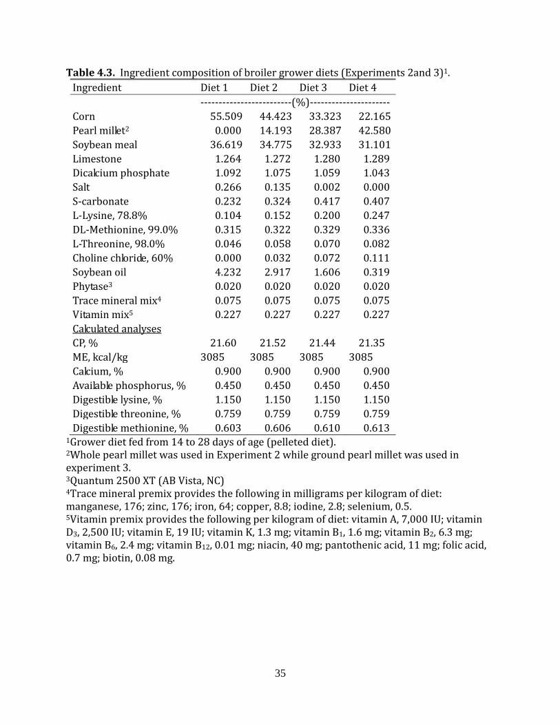
Table 4.3. Ingredient composition of broiler grower diets (Experiments 2and 3)1.
1Grower diet fed from 14 to 28 days of age (pelleted diet). 2Whole pearl millet was used in Experiment 2 while ground pearl millet was used in experiment 3. 3Quantum 2500 XT (AB Vista, NC) 4Trace mineral premix provides the following in milligrams per kilogram of diet: manganese, 176; zinc, 176; iron, 64; copper, 8.8; iodine, 2.8; selenium, 0.5. 5Vitamin premix provides the following per kilogram of diet: vitamin A, 7,000 IU; vitamin D3, 2,500 IU; vitamin E, 19 IU; vitamin K, 1.3 mg; vitamin B1, 1.6 mg; vitamin B2, 6.3 mg; vitamin B6, 2.4 mg; vitamin B12, 0.01 mg; niacin, 40 mg; pantothenic acid, 11 mg; folic acid, 0.7 mg; biotin, 0.08 mg.
Ingredient Diet 1 Diet 2 Diet 3 Diet 4 -------------------------(%)---------------------- Corn 55.509 44.423 33.323 22.165 Pearl millet2 0.000 14.193 28.387 42.580 Soybean meal 36.619 34.775 32.933 31.101 Limestone 1.264 1.272 1.280 1.289 Dicalcium phosphate 1.092 1.075 1.059 1.043 Salt 0.266 0.135 0.002 0.000 S-carbonate 0.232 0.324 0.417 0.407 L-Lysine, 78.8% 0.104 0.152 0.200 0.247 DL-Methionine, 99.0% 0.315 0.322 0.329 0.336 L-Threonine, 98.0% 0.046 0.058 0.070 0.082 Choline chloride, 60% 0.000 0.032 0.072 0.111 Soybean oil 4.232 2.917 1.606 0.319 Phytase3 0.020 0.020 0.020 0.020 Trace mineral mix4 0.075 0.075 0.075 0.075 Vitamin mix5 0.227 0.227 0.227 0.227
Calculated analyses CP, % 21.60 21.52 21.44 21.35 ME, kcal/kg 3085 3085 3085 3085 Calcium, % 0.900 0.900 0.900 0.900 Available phosphorus, % 0.450 0.450 0.450 0.450 Digestible lysine, % 1.150 1.150 1.150 1.150 Digestible threonine, % 0.759 0.759 0.759 0.759 Digestible methionine, % 0.603 0.606 0.610 0.613

36
Table 4.4. Ingredient composition of broiler finisher diets (Experiments 2and 3)1. Ingredient Diet 1 Diet 2 Diet 3 Diet 4 ----------------------(%)------------------------- Corn 64.694 53.705 42.595 31.410 Pearl millet2 0.000 14.193 28.387 42.580 Soybean meal 27.854 25.999 24.155 22.327 Limestone 1.228 1.236 1.244 1.252 Dicalcium phosphate 1.033 1.016 0.999 0.984 Salt 0.240 0.110 0.000 0.061 S-carbonate 0.231 0.322 0.397 0.339 L-Lysine, 78.8% 0.118 0.166 0.214 0.261 DL-Methionine, 99.0% 0.242 0.249 0.256 0.263 L-Threonine, 98.0% 0.045 0.057 0.069 0.081 Choline chloride, 60% 0.020 0.043 0.083 0.122 Soybean oil 3.972 2.584 1.278 0.000 Phytase3 0.020 0.020 0.020 0.020 Trace mineral mix4 0.075 0.075 0.075 0.075 Vitamin mix5 0.227 0.227 0.227 0.227
Calculated analysis CP, % 18.13 18.05 17.96 17.88 ME, kcal/kg 3152 3152 3152 3152 Calcium, % 0.850 0.850 0.850 0.850 Available phosphorus, % 0.425 0.425 0.425 0.425 Lysine, % 0.950 0.950 0.950 0.950 Threonine, % 0.637 0.636 0.636 0.636 Methionine, % 0.494 0.497 0.501 0.505
1Finisher diet fed from 28 to 42 days of age (pelleted diet). 2Whole pearl millet was used in Experiment 2 while ground pearl millet was used in experiment 3. 3Quantum 2500 XT (AB Vista, NC) 4Trace mineral premix provides the following in milligrams per kilogram of diet: manganese, 176; zinc, 176; iron, 64; copper, 8.8; iodine, 2.8; selenium, 0.5. 5Vitamin premix provides the following per kilogram of diet: vitamin A, 7,000 IU; vitamin D3, 2,500 IU; vitamin E, 19 IU; vitamin K, 1.3 mg; vitamin B1, 1.6 mg; vitamin B2, 6.3 mg; vitamin B6, 2.4 mg; vitamin B12, 0.01 mg; niacin, 40 mg; pantothenic acid, 11 mg; folic acid, 0.7 mg; biotin, 0.08 mg.
37
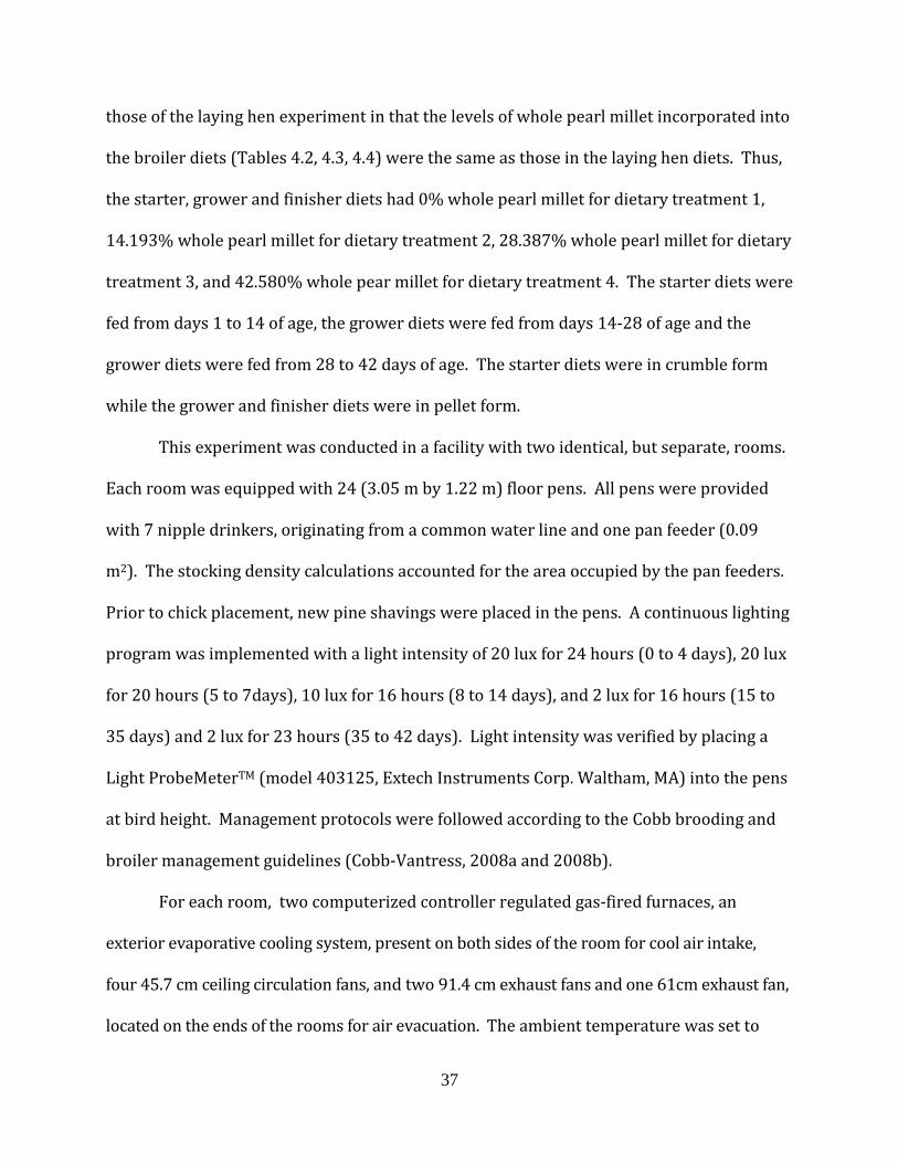
those of the laying hen experiment in that the levels of whole pearl millet incorporated into
the broiler diets (Tables 4.2, 4.3, 4.4) were the same as those in the laying hen diets. Thus,
the starter, grower and finisher diets had 0% whole pearl millet for dietary treatment 1,
14.193% whole pearl millet for dietary treatment 2, 28.387% whole pearl millet for dietary
treatment 3, and 42.580% whole pear millet for dietary treatment 4. The starter diets were
fed from days 1 to 14 of age, the grower diets were fed from days 14-28 of age and the
grower diets were fed from 28 to 42 days of age. The starter diets were in crumble form
while the grower and finisher diets were in pellet form.
This experiment was conducted in a facility with two identical, but separate, rooms.
Each room was equipped with 24 (3.05 m by 1.22 m) floor pens. All pens were provided
with 7 nipple drinkers, originating from a common water line and one pan feeder (0.09
m2). The stocking density calculations accounted for the area occupied by the pan feeders.
Prior to chick placement, new pine shavings were placed in the pens. A continuous lighting
program was implemented with a light intensity of 20 lux for 24 hours (0 to 4 days), 20 lux
for 20 hours (5 to 7days), 10 lux for 16 hours (8 to 14 days), and 2 lux for 16 hours (15 to
35 days) and 2 lux for 23 hours (35 to 42 days). Light intensity was verified by placing a
Light ProbeMeterTM (model 403125, Extech Instruments Corp. Waltham, MA) into the pens
at bird height. Management protocols were followed according to the Cobb brooding and
broiler management guidelines (Cobb-Vantress, 2008a and 2008b).
For each room, two computerized controller regulated gas-fired furnaces, an
exterior evaporative cooling system, present on both sides of the room for cool air intake,
four 45.7 cm ceiling circulation fans, and two 91.4 cm exhaust fans and one 61cm exhaust fan,
located on the ends of the rooms for air evacuation. The ambient temperature was set to
38
34°C on day 1 and decreased by 0.28°C until 24°C was reached and maintained. No
significant differences in temperature and humidity were noted throughout the study,
between the two rooms.
Prior to placing chicks, the 32 pens were assigned to one of the 4 dietary treatments
in a random block design [8 replicates per treatment (7 replicates in one room 1 replicate
in the other room)]. A total of 840 day of hatch Cobb 500 male broiler chicks, originating
from the same breeder flock, were obtained from a primary breeder hatchery. The chicks
were sorted and those with extreme weights were discarded before the remaining birds
were assigned to the 32 pens (21 birds per pen).
Feed and water were provided ad libitum throughout the duration of the
experiment. Diets were formulated on a digestible amino acid basis. All diets were
formulated to meet or exceed NRC (1994) requirements. All animal procedures were
approved by the University of Georgia Animal Care and Use Committee, Athens, GA.
For each room, humidity, temperature, water consumption, and pen mortality were
recorded twice daily. Birds and feed were weighed on days 0, 14, 28 and 42 to determine
body weight (BW), feed intake (FI), body weight gain (BWG), and feed conversion (FC). On
day 42, the mean bird weight for each pen was determined and 8 birds per pen, within 300
g above/below the mean weight of their pen, were selected for processing. Individual
weights for the selected birds were recorded and each bird was leg banded prior to
placement in a coop for an overnight feed withdrawal. On day 43, birds were weighed
again to determine their fasted live weight and processed at the University of Georgia's
Pilot Processing Plant as previously described (Hidalgo et al., 2004). During evisceration
the gizzard was removed. Once removed, fat tissue was removed from the outside of the
39
gizzard and then the gizzard was cut open to remove any contents and the koilin
membrane, before the gizzard was weighed. Subsequently, eviscerated hot carcass
weights were recorded for each bird prior to static chilling in an ice bath for 4 hours. After
a 4-hour chill, chilled carcasses were drained prior to cut up and deboning. Weights were
recorded for: drained chilled carcass, pectoralis major, pectoralis minor, wings, and leg
quarters of each bird. Percent yield calculations were based on the fasted, live weight of
the bird.
Experiment 3: Ground pearl millet in broilers
This experiment followed a very similar protocol as experiment 2 except that the
diets contained ground millet rather than whole millet. In addition, dietary treatment 3
(28.387% millet) was not included in this experiment, nor were gizzard weights recorded.
There were 10 replicates (5 replicate pens per room) per dietary treatment.
Ghrelin experiments
Experiment 4: Ghrelin expression in the hen ovary
To determine if ghrelin is expressed in the granulosa and theca tissue of chicken,
preovulatory follicles and initial characterization was completed using Hy-line W-36 laying
hens. Three 50 to 55 week old hens were killed by cervical dislocation 2 to 4 hours prior to
ovulation and the ovary was collected from each hen. The theca and granulosa layers from
each of the F1, F2, F3, and F4 follicles were manually separated from one another (Huang
and Nalbandov, 1979) while the theca and granulosa layers from the large small yellow
follicles (SYF1, 8 to 12 mm in diameter) small yellow follicles (SYF2, 5 to 8 mm in

40
diameter), large white follicles (LWF1, 2 to 5 mm in diameter) and small large white
follicles (LWF2, less than 2 mm in diameter) were separated enzymatically (Davis et al.,
2000a). The individual theca and granulosa tissue for each follicle size obtained from one
hen were combined with corresponding samples from the other two hens for RNA
extraction and subsequent RT-PCR detection of ghrelin mRNA. This procedure was
repeated two more times and a total of 3 sets (n = 3) of samples for each follicle size were
obtained. The theca layers collected from each individual F1 through F4 follicle and from
the individual pools of SYF and LWF were placed in 3 mL of guanidinium isothiocyanate
solution and homogenized for 30 seconds with a PowerGen 700 tissue disrupter (Fisher
Scientific, Pittsburg, PA). The single cell layer of granulosa tissue from each hierarchical
follicle and granulosa cells isolated from the individual pools of SYF and LWF was placed in
3 mL of guanidinium isothiocyanate solution and vortexed for 20 seconds. The tissue
solutions were stored at -80ºC for subsequent RNA extraction.
Experiment 5: Ghrelin in fed and fasted broiler breeder hens
This experiment was completed to determine if fasting influenced the mRNA
expression of ghrelin in the hierarchical or prehierarchical follicles of broiler breeder hens.
The Cobb 500 broiler breeder hens utilized for this experiment were between 45 and 55
weeks of age. The birds were reared as previously described (Spradley et al., 2008) using a
skip a day feed restriction program. At 21 weeks of age the pullets were placed in
individual cages and were photostimulated to initiate reproduction with a lighting program
that provided 14L:8D (lights on at 06:30 hours) per day. The hens were given free access
to water and were fed a standard broiler breeder layer diet each morning at 8am. The daily

41
amount of feed provided to the hens was determined using the guidelines set forth by the
primary breeder (Cobb-Vantress, 2005a and Cobb-Vantress 2005b) based on the weekly
body weight measurements and egg production rates of the hens. Eggs were collected
twice daily and individual hen egg production was recorded. All animal procedures were
approved by the University of Georgia Animal Care and Use Committee, Athens, GA.
Four hens in mid-laying sequence were divided into two treatment groups. The
hens in one treatment group continued to receive their daily allotment of feed while the
hens in the other treatment group did not receive food. After 72 hours of fasting, all 4 hens
were killed and the ovary was collected from each hen. The theca and granulosa layers
from each of the F1, F2, F3 and F4 follicles were collected as described in experiment 4.
The theca and granulosa layers from the SYF (5 to 10 mm in diameter) and LWF follicles (2
to 5 mm in diameter) were separated enzymatically (Davis et al., 2000a). The individual
theca and granulosa samples for each follicle size from one hen of each treatment were
combined with the corresponding samples from the other hen of that treatment for RNA
extraction. This collection procedure was repeated 2 more times to give 3 total replications
for each treatment (n = 3).
Approximately 300 mg of proventriculus was also collected from the 6 individual
fed and 6 individual fasted birds. Immediately after collection, each tissue was placed in 3
mL of guanidinium isothiocyanate solution and homogenized for 30 seconds with a
PowerGen 700 tissue disrupter (Fisher Scientific, Pittsburg, PA). Individual homogenized
tissue solutions were frozen and stored at -80ºC for future RNA extraction.

42
RNA Extraction
Total RNA was extracted from tissue samples using the guanidinium thiocyanate-
phenol-chloroform method (Chomczynski and Sacchi, 1987). RNA samples were stored at
-80ºC.
Real time RT-PCR
Extracted RNA samples were DNase treated using TURBO-DNA-free kit (Ambion,
Austin, TX) to remove any potential genomic DNA contamination. TaqMan minor groove-
binding probes and primers (Table 4.5) for detecting ghrelin (GenBank accession
#AY303688) and the endogenous control glyceraldehyde-3-phosphate dehydrogenase
(GAPDH, GenBank accession #M11213) were designed using Primer Express software
(Version 4.0, Applied Biosystems). Each probe was labeled at the 5’ end with FAM (6-
carboxyfluorescein) as the reporter dye and at the 3’ end with TAMRA (6-carboxy-
N,N,N’,N’-tetramethylrhodamine) as the quencher dye. Primer and probe sets were
validated for real-time PCR by determining the optimal amplification efficiency and
primer/probe concentration as described by the manufacturer (Applied Biosystems, Foster
City, CA).
Reverse-transcription cDNA synthesis reactions were performed using the TaqMan
Reverse Transcription Kit (Applied Biosystems) following the manufacturers protocol. For
two-step real-time PCR, 200 ng of cDNA was used for each sample for the ghrelin and
GAPDH amplification. The reactions were performed in a 25ul volume of reaction buffer
containing 1x TaqMan Universal PCR Master Mix (Applied Biosystems) and 900nM of
either, ghrelin or GAPDH primer pairs and 25nM of the appropriate probe. The reactions

43
were completed in an ABI 7500 thermocycler (Applied Biosystems). The thermocycler
conditions were 10 minutes at 95°C and 40 cycles each 15 seconds at 95°C and 1 minute at
60°C. The reactions for each sample were performed in duplicate for ghrelin and GAPDH
assays.

44
Table 4.5 Real-time RT-PCR primer and probe designs for ghrelin and GAPDH
Gene Primer Oligonucleotide Primer and Probe Sequence Product Size (Base Pairs)
Ghrelin Forward 5’-GGATACAAGAAAACCAACAGCAAGA-3’
71 Reverse 5’-CTGTTTCATCTGTATCCCAAAAGCT -3’ Probe 5’-TACATCGCCGAGGCAC-3’
GAPDH Forward 5’-TTGGCATTGTGGAGGGTCTT-3’
87 Reverse 5’-GGGCCATCCACCGTCTTC-3’ Probe 5’-TGACCACTGTCCATGCCAT-3’

45
The Ct (the cycle number at which the fluorescence exceeds the threshold level) was
determined for each reaction using the Sequence Detection Software (version 1.2.2,
Applied Biosystems) and quantification was completed using 2-ΔΔCt method (Livak and
Schmittgen, 2001). Briefly, the ghrelin samples Ct’s were determined and then normalized
to the GAPDH Ct from the same sample (GAPDH Ct subtracted from the ghrelin Ct to yield
the ΔCt). After all the ΔCt values were obtained for an experimental replicate, the ΔCt
values for each individual ghrelin sample were compared to the sample within the replicate
that had the highest mRNA expression for ghrelin using the 2-ΔΔCt method (Livak and
Schmittgen, 2001). Thus, all data for ghrelin is expressed as the fold-difference relative to
sample with the highest expression.
Statistics
Data from each experiment was subjected to ANOVA according to the General Linear
Model (GLM) with replicate and dietary treatment as factors in experiments 1, 2 and 3 and
replicate and tissue as factors in experiment 4 and replicate, feeding state and tissue as
factors in experiment 5. Tukey’s multiple-comparison procedure (Neter et al., 1990) was
used to detect significant differences among individual dietary treatments, tissues and
follicle sizes. Differences were considered significant when P < 0.05. All statistical
procedures were completed with the Minitab statistical software package (Release13, State
College, PA).
46
CHAPTER 5
RESULTS AND CONCLUSIONS
The protein content of the pearl millet varieties from Mali ranged from about 8.5 to
10.5%, while the nitrogen-corrected true metabolizable energy values ranged from about
3,400 to 3,750 kcal/kg on as-is basis (Table 5.1). The pearl millet variety Koutiala 1 had
among the best energy and protein content of the tested varieties, also had some of the best
essential amino acid concentrations (Table 5.2) and digestibility coefficients for these
essential amino acids (Table 5.3) relative to the other tested varieties. The mixed
industrial sample, which is a commercial blend of millet varieties grown in Mali, also had
very good available energy and amino acid contents relative to the individual varieties
tested.
Experiment 1
Laying hens fed diets in which the corn content of a standard laying hen diet had
been reduced by 25, 50 or 75% and replaced with the mixed industrial pearl millet in
whole form, that had been imported from Mali, performed equivalently to one another.
There were no differences in body weight gain (Table 5.4) or weekly hen day egg
production (Table 5.5) between the hens fed the 4 different dietary treatments from 36 to
52 weeks of age. The total hen housed egg production (mean ± SEM) was 100 ± 1, 100 ±
0.5, 98 ± 1 and 98 ± 0.5 eggs, during the 120 day experimental period for hens fed diets
containing 0, 14, 28 and 43% whole pearl millet, respectively. Total hen day egg
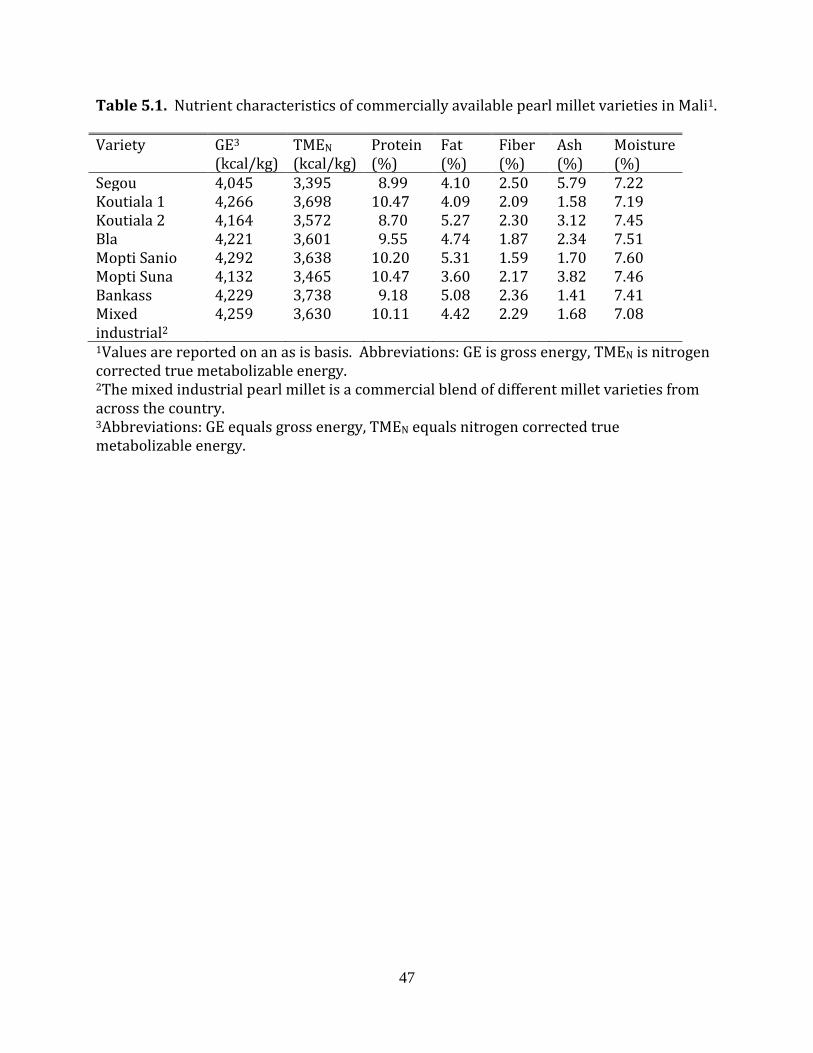
47
Table 5.1. Nutrient characteristics of commercially available pearl millet varieties in Mali1.
Variety GE3 (kcal/kg)
TMEN (kcal/kg)
Protein (%)
Fat (%)
Fiber (%)
Ash (%)
Moisture (%)
Segou 4,045 3,395 8.99 4.10 2.50 5.79 7.22 Koutiala 1 4,266 3,698 10.47 4.09 2.09 1.58 7.19 Koutiala 2 4,164 3,572 8.70 5.27 2.30 3.12 7.45 Bla 4,221 3,601 9.55 4.74 1.87 2.34 7.51 Mopti Sanio 4,292 3,638 10.20 5.31 1.59 1.70 7.60 Mopti Suna 4,132 3,465 10.47 3.60 2.17 3.82 7.46 Bankass 4,229 3,738 9.18 5.08 2.36 1.41 7.41 Mixed industrial2
4,259 3,630 10.11 4.42 2.29 1.68 7.08
1Values are reported on an as is basis. Abbreviations: GE is gross energy, TMEN is nitrogen corrected true metabolizable energy. 2The mixed industrial pearl millet is a commercial blend of different millet varieties from across the country. 3Abbreviations: GE equals gross energy, TMEN equals nitrogen corrected true metabolizable energy.
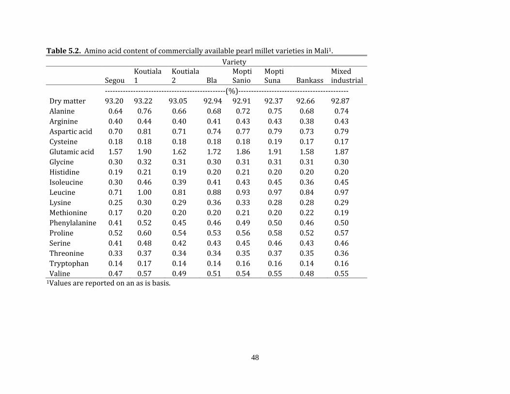
48
Table 5.2. Amino acid content of commercially available pearl millet varieties in Mali1. Variety
Segou Koutiala 1
Koutiala 2 Bla
Mopti Sanio
Mopti Suna Bankass
Mixed industrial
-----------------------------------------------(%)------------------------------------------- Dry matter 93.20 93.22 93.05 92.94 92.91 92.37 92.66 92.87 Alanine 0.64 0.76 0.66 0.68 0.72 0.75 0.68 0.74 Arginine 0.40 0.44 0.40 0.41 0.43 0.43 0.38 0.43 Aspartic acid 0.70 0.81 0.71 0.74 0.77 0.79 0.73 0.79 Cysteine 0.18 0.18 0.18 0.18 0.18 0.19 0.17 0.17 Glutamic acid 1.57 1.90 1.62 1.72 1.86 1.91 1.58 1.87 Glycine 0.30 0.32 0.31 0.30 0.31 0.31 0.31 0.30 Histidine 0.19 0.21 0.19 0.20 0.21 0.20 0.20 0.20 Isoleucine 0.30 0.46 0.39 0.41 0.43 0.45 0.36 0.45 Leucine 0.71 1.00 0.81 0.88 0.93 0.97 0.84 0.97 Lysine 0.25 0.30 0.29 0.36 0.33 0.28 0.28 0.29 Methionine 0.17 0.20 0.20 0.20 0.21 0.20 0.22 0.19 Phenylalanine 0.41 0.52 0.45 0.46 0.49 0.50 0.46 0.50 Proline 0.52 0.60 0.54 0.53 0.56 0.58 0.52 0.57 Serine 0.41 0.48 0.42 0.43 0.45 0.46 0.43 0.46 Threonine 0.33 0.37 0.34 0.34 0.35 0.37 0.35 0.36 Tryptophan 0.14 0.17 0.14 0.14 0.16 0.16 0.14 0.16 Valine 0.47 0.57 0.49 0.51 0.54 0.55 0.48 0.55
1Values are reported on an as is basis.
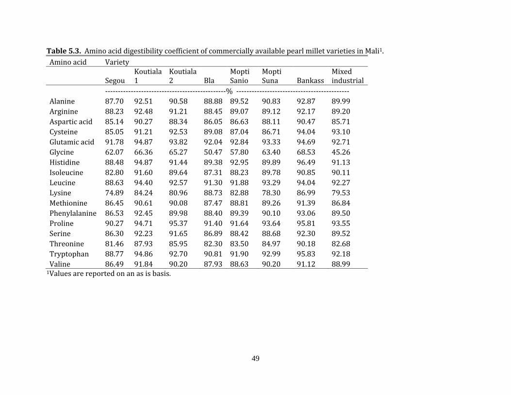
49
Table 5.3. Amino acid digestibility coefficient of commercially available pearl millet varieties in Mali1. Amino acid Variety
Segou Koutiala 1
Koutiala 2 Bla
Mopti Sanio
Mopti Suna Bankass
Mixed industrial
-----------------------------------------------% -------------------------------------------- Alanine 87.70 92.51 90.58 88.88 89.52 90.83 92.87 89.99 Arginine 88.23 92.48 91.21 88.45 89.07 89.12 92.17 89.20 Aspartic acid 85.14 90.27 88.34 86.05 86.63 88.11 90.47 85.71 Cysteine 85.05 91.21 92.53 89.08 87.04 86.71 94.04 93.10 Glutamic acid 91.78 94.87 93.82 92.04 92.84 93.33 94.69 92.71 Glycine 62.07 66.36 65.27 50.47 57.80 63.40 68.53 45.26 Histidine 88.48 94.87 91.44 89.38 92.95 89.89 96.49 91.13 Isoleucine 82.80 91.60 89.64 87.31 88.23 89.78 90.85 90.11 Leucine 88.63 94.40 92.57 91.30 91.88 93.29 94.04 92.27 Lysine 74.89 84.24 80.96 88.73 82.88 78.30 86.99 79.53 Methionine 86.45 90.61 90.08 87.47 88.81 89.26 91.39 86.84 Phenylalanine 86.53 92.45 89.98 88.40 89.39 90.10 93.06 89.50 Proline 90.27 94.71 95.37 91.40 91.64 93.64 95.81 93.55 Serine 86.30 92.23 91.65 86.89 88.42 88.68 92.30 89.52 Threonine 81.46 87.93 85.95 82.30 83.50 84.97 90.18 82.68 Tryptophan 88.77 94.86 92.70 90.81 91.90 92.99 95.83 92.18 Valine 86.49 91.84 90.20 87.93 88.63 90.20 91.12 88.99
1Values are reported on an as is basis.
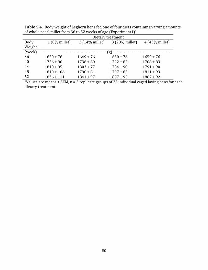
50
Table 5.4. Body weight of Leghorn hens fed one of four diets containing varying amounts of whole pearl millet from 36 to 52 weeks of age (Experiment1)1. Dietary treatment Body Weight
1 (0% millet) 2 (14% millet) 3 (28% millet) 4 (43% millet)
(week) ----------------------------------------------(g)------------------------------------------ 36 1650 ± 76 1649 ± 76 1650 ± 76 1650 ± 76 40 1756 ± 90 1736 ± 80 1722 ± 82 1708 ± 83 44 1810 ± 95 1803 ± 77 1784 ± 90 1791 ± 90 48 1810 ± 106 1790 ± 81 1797 ± 85 1811 ± 93 52 1836 ± 111 1841 ± 97 1857 ± 95 1867 ± 92 1Values are means ± SEM, n = 3 replicate groups of 25 individual caged laying hens for each dietary treatment.

51
Table 5.5. Weekly hen day egg production of leghorn hens fed one of four diets containing varying amounts of whole pearl millet from 36 to 52 weeks of age (Experiment 1)1. Dietary treatment
Age 1 (0% millet) 2 (14% millet) 3 (28% millet) 4 (43% millet)
(week) ------------------------------------------(%)---------------------------------------
37 95.2 ± 1.33 93.0 ± 1.49 91.6 ± 1.56 94.7 ± 1.33
38 92.2 ± 0.38 90.9 ± 0.66 89.1 ± 2.31 90.7 ± 0.50
39 93.9 ± 0.19 95.2 ± 1.66 92.0 ± 0.87 92.8 ± 1.16
40 89.9 ± 0.76 90.3 ± 2.01 92.0 ± 2.70 91.0 ± 0.19
41 93.1 ± 0.66 91.8 ± 0.69 89.5 ± 1.33 91.8 ± 1.16
42 91.6 ± 0.38 90.7 ± 0.38 90.1 ± 0.83 90.9 ± 1.44
43 91.6 ± 0.69 94.5 ± 1.06 91.4 ± 0.87 93.0 ± 0.19
44 89.1 ± 0.00 86.3 ± 0.87 88.3 ± 1.77 87.8 ± 1.63
45 87.4 ± 0.66 90.7 ± 1.90 87.8 ± 1.82 88.2 ± 0.76
46 90.3 ± 2.31 88.0 ± 0.87 88.1 ± 1.17 87.8 ± 0.69
47 90.1 ± 0.95 91.0 ± 0.19 88.2 ± 0.67 90.1 ± 0.50
48 89.3 ± 1.06 86.3 ± 0.87 85.5 ± 0.70 85.7 ± 1.51
49 84.8 ± 2.02 85.1 ± 1.87 80.5 ± 1.83 79.0 ± 1.44
50 79.6 ± 2.12 84.8 ± 2.29 80.9 ± 2.00 77.5 ± 4.14
51 83.8 ± 3.30 85.2 ± 2.36 85.5 ± 2.20 83.6 ± 2.43
52 88.6 ± 0.57 87.1 ± 0.53 86.5 ± 1.28 85.9 ± 1.57 1Values are means ± SEM, n = 3 replicate groups of 25 individual caged laying hens for each dietary treatment.
52
production for the experimental period was 100 ± 1, 100 ± 0.5, 100 ± 0.5 and 99 ± 1 eggs
for hens fed diets containing 0, 14, 28 and 43% whole pearl millet, respectively. Egg weight
(Table 5.6) and the specific gravity (Table 5.7) of the eggs produced by the hens did not
vary among the dietary treatments. The yellowness of the egg yolks from eggs produced
during the last week of the experiment was 79.3 ± 0.5, 78.6 ± 0.1, 77.1 ± 0.3 and 75.6 ± 0.5
for hens fed diets containing 0, 14, 28 and 43% whole pearl millet, respectively. The
degree of yellowness of egg yolks was significantly (P < 0.05) less in the eggs produced
from hens fed the two dietary treatments containing the most pearl millet compared to the
eggs produced by the hens fed the corn and soybean meal control diet. In addition, egg
yolk yellowness was also less in eggs produced from hens fed the highest level of dietary
pearl millet relative those fed the lowest level of pearl millet.
Daily feed consumption (mean ± SEM) was 103.00 ± 1.90, 104.11 ± 2.22, 105.56 ±
3.56 and 107.11 ± 1.64 grams per bird for hens fed diets containing 0, 14, 28 and 43%
whole pearl millet, respectively. There were no significant differences in feed intake
among the 4 dietary treatments.
Experiment 2 During the starter period (1 to14 days of age), broilers fed diets containing 28 and
43% whole millet gained less body weight and had a lower feed efficiency than those fed
diets containing 0 or 14% whole millet (Table 5.8). During the grower period, as in the
starter period, the inclusion of 14% whole millet in broiler diets did not affect bird
performance relative to the performance of birds fed the control diet, containing no whole
pearl millet (Table 5.9). Broilers fed a diet containing 43% whole pearl millet during the
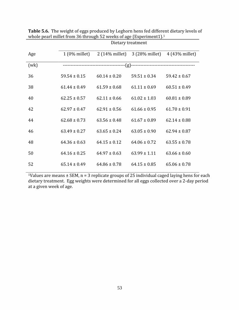
53
Table 5.6. The weight of eggs produced by Leghorn hens fed different dietary levels of whole pearl millet from 36 through 52 weeks of age (Experiment1).1 Dietary treatment
Age 1 (0% millet) 2 (14% millet) 3 (28% millet) 4 (43% millet)
(wk) ----------------------------------------(g)-----------------------------------------
36 59.54 ± 0.15 60.14 ± 0.20 59.51 ± 0.34 59.42 ± 0.67
38 61.44 ± 0.49 61.59 ± 0.68 61.11 ± 0.69 60.51 ± 0.49
40 62.25 ± 0.57 62.11 ± 0.66 61.02 ± 1.03 60.81 ± 0.89
42 62.97 ± 0.47 62.91 ± 0.56 61.66 ± 0.95 61.70 ± 0.91
44 62.68 ± 0.73 63.56 ± 0.48 61.67 ± 0.89 62.14 ± 0.88
46 63.49 ± 0.27 63.65 ± 0.24 63.05 ± 0.90 62.94 ± 0.87
48 64.36 ± 0.63 64.15 ± 0.12 64.06 ± 0.72 63.55 ± 0.78
50 64.16 ± 0.25 64.97 ± 0.63 63.99 ± 1.11 63.66 ± 0.60
52 65.14 ± 0.49 64.86 ± 0.78 64.15 ± 0.85 65.06 ± 0.78
1Values are means ± SEM, n = 3 replicate groups of 25 individual caged laying hens for each dietary treatment. Egg weights were determined for all eggs collected over a 2-day period at a given week of age.
54
Table 5.7. Specific gravities of eggs produced by Leghorn hens fed different dietary levels of whole pearl millet from 36 through 52 weeks of age (Experiment1).1 Dietary treatment
Age (wk) 1 (0% millet) 2 (14% millet) 3 (28% millet) 4 (43% millet)
46 1.081 ± 0.0003 1.080 ± 0.0013 1.083 ± 0.0008 1.083 ± 0.0008
48 1.079 ± 0.0003 1.078 ± 0.0006 1.080 ± 0.0010 1.081 ± 0.0003
50 1.082 ± 0.0001 1.088 ± 0.0069 1.082 ± 0.0008 1.082 ± 0.0008
52 1.080 ± 0.0003 1.079 ± 0.0007 1.080 ± 0.0010 1.079 ± 0.0007
54 1.080 ± 0.0003 1.079 ± 0.0009 1.081 ± 0.0008 1.081 ± 0.0006
56 1.077 ± 0.0003 1.078 ± 0.0006 1.076 ± 0.0011 1.077 ± 0.0004
1Values are means ± SEM, n = 3 replicate groups of 25 individual caged laying hens for each dietary treatment. Specific gravities were determined for all eggs collected over a 2 day period at a given week of age.

55
Table 5.8. Performance of male broilers from 0 to 14 days of age fed diets containing varying levels of whole pearl millet (Experiment 2)1. Dietary treatment Body weight Body weight gain Feed to gain
--------------- (g)--------------
1 (0% millet) 469 ± 3a 425 ± 3a 1.191 ± 0.007a
2 (14% millet) 471 ± 4a 427 ± 4a 1.203 ± 0.006a
3 (28% millet) 436 ± 5b 393 ± 5b 1.243 ± 0.012b
4 (43% millet) 442 ± 5b 399 ± 5b 1.256 ± 0.007b
a-bMeans within each column with different superscripts are statistically different, P <0.05. 1Values are means ± SEM, n = 8 replicate pens of 20 birds.

56
Table 5.9. Performance of male broilers from 14 to 28 days of age fed diets containing varying levels of whole pearl millet (Experiment 2)1. Dietary treatment Body weight Body weight gain Feed to gain
--------------------(g)------------------
1 (0% millet) 1,657 ± 11a 1,192 ± 8a 1.356 ± 0.008a
2 (14% millet) 1,655 ± 10a 1,185 ± 8a 1.367 ± 0.008a
3 (28% millet) 1.590 ± 17b 1,154 ± 14ab 1.399 ± 0.007b
4 (43% millet) 1,576 ± 13b 1,134 ± 10b 1.432 ± 0.006c
a-cMeans within each column with different superscripts are statistically different, P <0.05. 1Values are means ± SEM, n = 8 replicate pens of 20 birds.
57
finisher phase (28 to 42 days of age) gained less body weight and were less efficient in
converting ingested feed to body weight than those fed the control diet (Table 5.10). For
the overall experimental period, only the broilers fed the diet containing 43% millet had
lower body weight and body weight gains relative to the broilers fed the control diet (Table
5.11.).
Gizzard weight as a percent of fasted live body weight was greater in the broilers fed
diets containing 28 and 43% whole pearl millet than those fed a diet containing no whole
pearl millet (Table5.12). As a percent of fasted live body weight, the yields of wings, leg
quarters, and total white meat did not differ between the broilers fed the four different
dietary treatments. (Table 5.13).
Experiment 3
During the starter period, broilers fed diets containing 43% ground pearl millet
gained less body weight and were less efficient in converting ingested feed to body weight
gain than broilers fed a diet containing no ground pearl millet (Table5.14). In the grower
period, the broilers fed the diet containing 14% ground millet were more efficient in
converting ingested feed to body weight gain and weighed more at the end of this period
than the broilers fed a diet containing no ground millet (5.15). During the finisher period,
(Table 5.16) and in the overall period from 1 to 42 days of age (Table 5.17), there were no
differences in body weight or body weight gain between the broilers fed the different
dietary treatments, but the broilers fed the diet containing 43% millet had poorer feed
conversions during these time periods relative to the control broilers.

58
Table 5.10 Performance of male broilers from 28 to 42 days of age fed diets containing varying levels of whole pearl millet (Experiment 2)1. Dietary treatment Body weight Body weight gain Feed to gain
--------------(g)--------------
1 (0% millet) 3,131 ± 17a 1,490 ± 17 1.823 ± 0.012a
2 (14% millet) 3,150 ± 13a 1,496 ± 13 1.798 ± 0.010a
3 (28% millet) 3,038 ± 39a 1,474 ± 39 1.868 ± 0.012ab
4 (43% millet) 2,992 ± 37b 1,416 ± 27 1.903 ± 0.015b
a-bMeans within each column with different superscripts are statistically different, P <0.05. 1Values are means ± SEM, n = 8 replicate pens of 20 birds.

59
Table 5.11. Performance of male broilers from 0 to 42 days of age fed diets containing varying levels of whole pearl millet (Experiment 2)1.
Dietary treatment Body weight Body weight gain Feed to gain
-------------(g)------------
1 (0% millet) 3,131 ± 17a 3,088 ± 17a 1.553 ± 0.007a
2 (14% millet) 3,150 ± 13a 3,106 ± 13a 1.557 ± 0.015a
3 (28% millet) 3,038 ± 39ab 2,995 ± 39ab 1.611 ± 0.010b
4 (43% millet) 2,992 ± 37b 2,949 ± 37b 1.631 ± 0.006b
a-bMeans within each column with different superscripts are statistically different, P <0.05. 1Values are means ± SEM, n = 8 replicate pens of 20 birds.

60
Table 5.12. Processing yields from 42-day-old broilers that had been fed diets containing varying levels of whole pearl millet (Experiment 2)1. Dietary treatments Chilled carcass Hot carcass Gizzard
(g) (%)2 (g) (%)2 (g) (%)2
1 (0% millet) 2,211 ± 32a 74 ± 0.4 2,195 ± 31 74 ± 0.2 31 ± 1.0a 1.1 ± 0.04a
2 (14% millet) 2,237 ± 10a 75 ± 0.2 2,229 ± 10 74 ± 0.3 34 ± 0.8bc 1.1 ± 0.02ac
3 (28% millet) 2,164 ± 28ab 75 ± 0.4 2,152 ± 24 74 ± 0.6 35 ± 0.4bc 1.2 ± 0.01bc
4 (43% millet) 2,116 ± 19b 75 ± 0.7 2,105 ± 19 74 ± 0.8 33 ± 0.6ac 1.2 ± 0.02bc
a-cMeans within each column with different superscripts are statistically different, P <0.05. 1Values are means ± SEM, n = 8 replicate pens with 8 birds per pen processed. 2As a percent of live fasted weight.
61
Table 5.13. Processing yields from 42 day old broilers that had been diets containing varying levels of whole pearl millet (Experiment 2)1. Dietary Treatment
Pectoralis major Pectoralis minor Total white meat2 Wings Leg quarters
G %3 g %3 g %3 g %3 g %3
1 (0% millet) 562 ± 13 18.9 ± 0.3 106 ± 1ab 3.6 ± 0.03 667 ± 14b 22.5 ± 0.3 243 ± 5 8.0 ± 0.1 712 ± 10a 24.0 ± 0.1
2 (14% millet) 583 ± 5 19.5 ± 0.2 108 ± 1a 3.6 ± 0.06 695 ± 6a 23.2 ± 0.3 247 ± 3 8.3 ± 0.1 721 ± 5a 24.0 ± 0.1
3 (28% millet) 548 ± 12 18.9 ± 0.2 101 ± 2b 3.5 ± 0.07 650 ± 12b 22.4 ± 0.2 237 ± 5 8.2 ± 0.1 699 ± 8ab 24.1 ± 0.2
4 (43% millet) 549 ± 14 19.3 ± 0.3 103 ± 1ab 3.6± 0.03 650 ± 14b 22.9 ± 0.3 235 ± 3 8.3 ± 0.1 674 ± 7b 23.8 ± 0.1
a-cMeans within each column with different superscripts are statistically different, P <0.05. 1Values are means ± SEM, n = 8 replicate pens with 8 birds per pen processed. 2Pectoralis major plus Pectoralis minor. 3As a percent of live fasted weight.

62
Table 5.14. Performance of male broilers from 1 to 14 days of age fed diets containing varying levels of ground pearl millet (Experiment 3)1. Dietary treatments Body weight Body weight gain Feed to gain
---------------(g)---------------
1 (0% millet) 475± 5a 429 ± 5a 1.188 ± 0.010a
2 (14% millet) 463 ±6ab 418 ± 6ab 1.203 ± 0.006a
4 (43% millet) 455 ± 2b 410 ± 3b 1.247 ± 0.004b
a-bMeans within each column with different superscripts are statistically different, P <0.05. 1Values are means ± SEM, n = 10 replicate pens of 20 birds.
63
Table 5.15. Performance of male broilers from 14 to 28 days of age fed diets containing varying levels of ground pearl millet (Experiment 3)1. Dietary treatments Body weight Body weight gain Feed to gain
---------------(g)---------------
1 (0% millet) 1,713 ± 16 1,238 ±14a 1.418 ± 0.001a
2 (14% millet) 1,737 ± 16 1,275 ± 11b 1.384 ± 0.004b
4 (43% millet) 1,691 ± 9 1,237 ± 8a 1.445 ± 0.006c
a-cMeans within each column with different superscripts are statistically different, P <0.05. 1Values are means ± SEM, n = 10 replicate pens of 20 birds.

64
Table 5.16. Performance of male broilers from 28 to 42 days of age fed diets containing varying levels of ground pearl millet (Experiment 3)1. Dietary treatment Body weight Body weight gain Feed to gain
-----------------(g)----------------
1 (0% millet) 3,247 ± 22 1,533 ± 18 1.806 ± 0.014a
2 (14% millet) 3,239 ± 28 1,507 ± 20 1.867 ± 0.014b
4 (43% millet) 3,183 ± 16 1,495 ± 10 1.936 ± 0.015c
a-cMeans within each column with different superscripts are statistically different, P <0.05. 1Values are means ± SEM, n = 10 replicate pens of 20 birds.
65
Table 5.17. Performance of male broilers from 0 to 42 days of age fed diets containing varying levels of ground pearl millet (Experiment 3)1.
Dietary treatment Body weight Body weight gain Feed to gain
----------------(g)---------------
1 (0% millet) 3,247 ± 22 3,201 ± 22 1.573 ± 0.000a
2 (14% millet) 3,239 ± 28 3,193 ± 28 1.580 ± 0.010a
4 (43% millet) 3,183 ± 16 3,137 ± 16 1.653 ± 0.010b
a-cMeans within each column with different superscripts are statistically different, P <0.05. 1Values are means ± SEM, n = 10 replicate pens of 20 birds.

66
To the contrary, the percent carcass yield relative to live fasted weight was greater
in the broilers fed a diet containing 43% ground millet than those fed a diet containing no
millet (Table 5.18). In addition, as a percent of live fasted weight, total white meat yield
and pectoralis major were higher and wing yield was less, by weight and percent, for
broilers fed a diet containing 43% ground pearl millet relative to those consuming a diet
containing no pearl millet (Table 5.19).
Experiment 4
Ghrelin mRNA was detected in both the granulosa and theca cells of the
preovulatory follicles of the laying hen ovary (Figures 5.1 and 5.2). Overall, the mRNA
expression of ghrelin was greater in granulosa tissue than in theca tissue (Figure 5.1). The
mRNA expression of ghrelin was higher in granulosa cells obtained from 8 to 12mm
diameter small yellow follicles (SYG1) than from granulosa cells isolated from any of the
hierarchical follicles (Figure 5.2).
Experiment 5
There were no differences in the mRNA expression of ghrelin in the ovarian
preovulatory follicular tissue (Figure 5.3) or proventriculus (Figure 5.4) tissue between fed
and fasted broiler breeder hens. Since there were no difference in the expression of ghrelin
mRNA in the preovulatory follicle tissue with feeding state, the 3 fed and 3 fasted replicates
were combined for further analyses of ghrelin mRNA expression in the broiler breeder hen
ovary. Overall, ghrelin mRNA expression is greater in granulosa tissue than theca tissue
(Figure 5.5).

67
However, for any given follicle size there is not a difference in ghrelin expression
between the theca and granulosa cells and there were no differences in ghrelin mRNA
expression between different sized preovulatory follicles (Figure 5.6).
68
Table 5.18. Processing yields from 42 day old broilers that had been fed diets containing varying levels of ground pearl millet (Experiment 3)1. Dietary treatments Chilled carcass Hot carcass
(g) (%)2 (g) (%)2
1 (0% millet) 2290 ± 26 75 ± 0.2a 2273 ± 26 74 ± 0.2a
2 (14% millet) 2279 ± 18 75 ± 0.2ab 2261 ± 20 75 ± 0.2ab
4 (43% millet) 2285 ± 20 76 ± 0.2b 2261 ± 19 75 ± 0.2b
a-bMeans within each column with different superscripts are statistically different, P <0.05. 1Values are means ± SEM, n = 10 replicate pens with 8 birds per pen processed. 2As a percent of live fasted weight.

69
Table 5.19. Processing yields from 42 day old broilers that had been fed diets containing varying levels of ground pearl millet (Experiment 3)1. Dietary treatments
Pectoralis major Pectoralis minor Total white meat2 Wings Leg quarters
(g) (%)3 (g) (%)3 (g) (%)3 (g) (%)3 (g) (%)3
1 (0% millet) 596 ± 13 19.4 ± 0.2a 117 ± 2 3.8 ± 0.04 713 ± 14 23.3 ± 0.2a 246 ± 2a 8.0 ± 0.05a 708 ± 6 23.1 ± 0.2
2 (14% millet) 601 ± 9 19.8 ± 0.2ab 116 ± 2 3.8 ± 0.03 717 ± 10 23.7 ± 0.2ab 243 ± 3ab 8.0 ± 0.05ab 704 ± 5 23.2 ± 0.2
4 (43% millet) 616 ± 9 20.4 ± 0.3b 115 ± 1 3.8 ± 0.03 732 ± 10 24.2 ± 0.2b 237 ± 2b 7.9 ± 0.05b 698 ± 7 23.2 ± 0.1
a-bMeans within each column with different superscripts are statistically different, P <0.05. 1Values are means ± SEM, n = 10 replicate pens with 8 birds processed. 2Pectoralis major plus Pectoralis minor. 3As a percent of live fasted weight.
70
Theca Granulosa0.00
0.25
0.50
0.75
1.00
1.25
Rela
tive
fold
exp
ress
ion
of g
hrel
in m
RNA
b
a
Figure 5.1. The overall relative fold expression of ghrelin mRNA in theca and granulosa tissue collected from the four largest hierarchical (F1 through F4) follicles the large small yellow follicles (8 to 12 mm in diameter) small yellow follicles (5 to 8 mm in diameter), large white follicles (2 to 5 mm in diameter) and small large white follicles (less than 2 mm in diameter) from 50 to 55 week old Leghorn (Experiment 4). The mRNA expression data were normalized with glyceraldehyde-3-phosphate dehydrogenase and expressed as the mean fold difference (2-ΔΔCT) ± SEM, n = 48 [6 samples (3 theca and 3 granulosa) from each of the 8 follicle sizes].

71
F1T F1G F2T F2G F3T F3G F4T F4G SYT1 SYG1 SYT2 SYG2 LWT1 LWG1 LWT2 LWG20.00
0.25
0.50
0.75
1.00
1.25R
elat
ive
fold
exp
ress
ion
of g
hrel
in m
RN
A
d
d d
dd d d
cdcd
d
cd
bc
a
c
bc
ab
Figure 5.2. The relative fold expression of ghrelin in theca (T) and granulosa (G) tissue collected from the four largest hierarchical preovulatory follicles (F1 through F4), large small yellow follicles (SY1, 8 to 12 mm in diameter), small yellow follicles (SY2, 5 to 8 mm in diameter), large white follicles (LW1, 2 to 5 mm in diameter) and small large white follicles (LW2, less than 2 mm in diameter) from 50 to 55week old Leghorn hens (Experiment 4). The data is expressed as mean fold differences ± SEM, n = 3. a-dMeans with different letters differ, P < 0.05.
72
Fed Fasted0.00
0.25
0.50
0.75
1.00
1.25R
elat
ive
fold
exp
ress
ion
of g
hrel
in m
RN
A
Figure 5.3. The overall relative fold expression of ghrelin mRNA in theca and granulosa tissue collected from the four largest hierarchical (F1 through F4) follicles and the small yellow and large white follicles from 45 to 55 week old broiler breeder hens fed daily or fasted for 72 hours (Experiment 5). The mRNA expression data were normalized with glyceraldehyde-3-phosphate dehydrogenase and expressed as the mean fold difference (2-ΔΔCT) ± SEM, n = 36 [6 samples (3 theca and 3 granulosa) from each of the 6 follicle sizes].
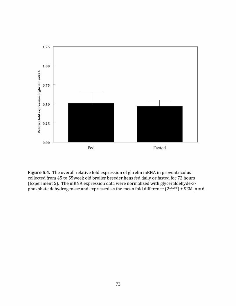
73
Fed Fasted0.00
0.25
0.50
0.75
1.00
1.25
Rela
tive
fold
exp
ress
ion
of g
hrel
in m
RNA
Figure 5.4. The overall relative fold expression of ghrelin mRNA in proventriculus collected from 45 to 55week old broiler breeder hens fed daily or fasted for 72 hours (Experiment 5). The mRNA expression data were normalized with glyceraldehyde-3-phosphate dehydrogenase and expressed as the mean fold difference (2-ΔΔCT) ± SEM, n = 6.
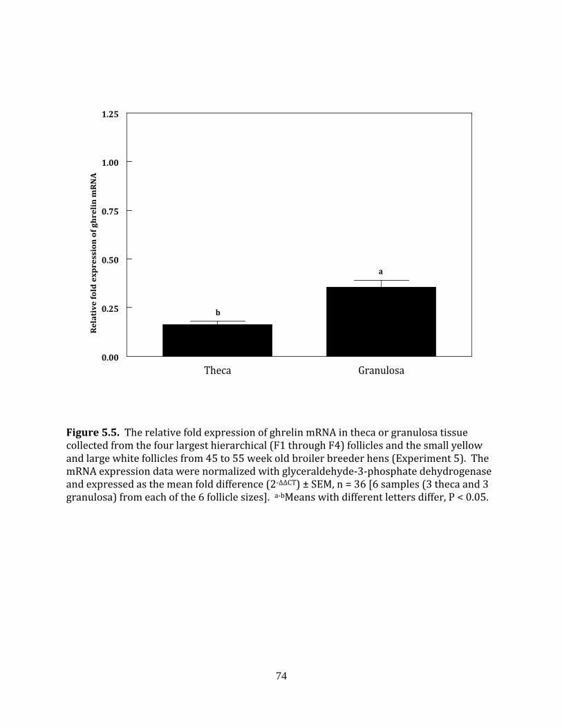
74
Theca Granulosa0.00
0.25
0.50
0.75
1.00
1.25
Rel
ativ
e fo
ld e
xpre
ssio
n of
ghr
elin
mR
NA
b
a
Figure 5.5. The relative fold expression of ghrelin mRNA in theca or granulosa tissue collected from the four largest hierarchical (F1 through F4) follicles and the small yellow and large white follicles from 45 to 55 week old broiler breeder hens (Experiment 5). The mRNA expression data were normalized with glyceraldehyde-3-phosphate dehydrogenase and expressed as the mean fold difference (2-ΔΔCT) ± SEM, n = 36 [6 samples (3 theca and 3 granulosa) from each of the 6 follicle sizes]. a-bMeans with different letters differ, P < 0.05.

75
F1T F1G F2T F2G F3T F3G F4T F4G SYT SYG LWT LWG0.00
0.25
0.50
0.75
1.00
1.25R
elat
ive
fold
exp
ress
ion
of g
hrel
in m
RN
A
Figure 5.6. The relative fold expression of ghrelin mRNA in theca (T) and granulosa (G) tissue collected from the four largest hierarchical (F1 through F4) follicles and the small yellow (SY) and large white (LW) follicles from 45 to 55 week old broiler breeder hens (Experiment 5). The mRNA expression data were normalized with glyceraldehyde-3-phosphate dehydrogenase and expressed as the mean fold difference (2-ΔΔCT) ± SEM, n = 6.

76
CHAPTER 6
DISCUSSION
Pearl millet
The overall goal of the pearl millet research was to test whether Malian grown pearl
millet would be an effective feed ingredient for poultry production. The current research
indicates that pearl millet grown in Mali could successfully replace corn in laying hen and
broiler diets. Furthermore, it indicates that performance of laying hens and broilers would
likely be unaffected even if the millet was incorporated into poultry diets in whole form.
Protein and energy content of Malian pearl millet
Genetic selection of pearl millet has been focused on improving its disease
resistance, yield and nutrient composition. Research from an animal feeding perspective
has been focused on testing these developed varieties, which have been produced and
grown almost exclusively in developed countries. Although variable, the crude protein
level of pearl millet typically ranges from 10-16% (Burton et al., 1972; Sullivan et al., 1990;
Adeola and Rogler, 1994; Amato and Forrester, 1995; Davis et al., 2003; Singh et al., 2005;
Vasan et al., 2008a, b). Protein levels vary from harvest to harvest and are different
depending on the variety of pearl millet and also agronomic differences, particularly the
nitrogen content in the soil from which it was harvested. Thus, given the arid conditions
and poorer soil fertility in Mali, it is not surprising that the protein concentration of the
most widely grown varieties of pearl millet were less or on the lower end of the
77
concentration range seen with the previously researched highly selected varieties of pearl
millet. However, all of the Malian pearl millet varieties tested had protein concentrations,
which were higher than corn and had similar apparent digestible amino acid coefficients as
corn.
The energy content of the tested Malian pearl millet varieties was also similar to or
slightly higher than corn and also similar to previously reported levels in the highly
genetically selected pearl millet varieties grown in developed countries. The nitrogen
corrected true metabolizable energy (TMEN of corn is typically about 3,350 kcal/kg
compared to 3,300 to 3,450 kcal/kg for pearl millet (Adeola et al., 1994; Collins et al., 1997;
Davis et al., 2003; Hidalgo et al., 2004) and the TMEN values for the tested Malian varieties
of pearl millet were all above 3,395 to as high as 3,738 kcal/kg on an as is basis.
Malian pearl millet in poultry production
In the present research, lowering the corn content of a control laying hen diet by 25,
50 or 75% by replacing it with whole millet imported from Mali, did not significantly affect
egg production in white leghorn hens during the 4-month experiment. Similarly, Collins et
al. (1995) reported that replacing 100% of the dietary corn (68% of the formulated diet)
with ground pearl millet caused no reduction in egg production for a 6-week trial period.
Similar results were also reported by Purushothaman and Thirumalai (1997) and Collins et
al. (1997). In contrast, Mehri et al. (2010) conducted a 12-week experiment and found that
100% replacement of corn (58% of the formulated diet) with whole millet decreased egg
production (P<0.05). However, replacing the corn content of the laying diet with 25, 50 or
75% millet did not affect egg production.
78
Egg weights and egg quality are typically unaffected by replacing dietary corn with
millet even when the dietary corn content is completely replaced by pearl millet (Collins et
al., 1997; Amini and Ruiz-Feria, 2007). Egg weight was not significantly affected by dietary
millet inclusion in the current research. However, yellow yolk pigmentation was reduced
in the eggs produced by the hens fed diets containing 28 and 43% whole millet. This result
is in line with previous research in which the yellowness of egg yolks was reduced when
dietary corn was replaced by pearl millet (Collins et al., 1997; Amini and Ruiz-Feria, 2007).
Thus, birds fed pearl millet based diets need additional dietary sources of xanthophylls to
obtain the degree of yellow pigmentation associated with feeding corn-based diets, for
consumer preference.
Although daily feed intake was not significantly different among the laying hens fed
0, 14, 28 or 43% whole millet, there was a clear trend that feed consumption increased as
the dietary levels of whole millet in the diet increased. The significance of this became
more apparent when broilers were fed the same levels of dietary whole pearl millet and
feed conversion ratios in these broilers increased as the level of dietary millet increased.
However, the degree to which body weight and body weight gain were affected by
increasing levels of dietary millet became less as the birds got older. Taken together, the
results suggested that the birds might be compensating for a nutrient deficiency in the
millet diets by eating more of the diet, rather than the idea that the whole millet seeds were
not completely digested or that the energy cost of digesting the whole millet was more than
anticipated.
Gizzard weight relative to body weight increased for the broilers fed the diets
containing 28 and 43% whole millet in the current research and this agreed with previous
79
research that our laboratory had conducted with a U.S. variety of pearl millet in which
broilers fed diets from 1 to 15 days of age containing 10, 15 or 20% whole millet had
increased relative gizzard weights to body size relative to birds fed diet with no whole
millet (Hidalgo et al., 2004). The anticipated increase in gizzard size in the current
research was actually expected to improve nutrient digestibility as previous research had
indicated that laying hens fed diets containing 15% whole millet versus 15% ground millet
had significantly higher starch digestibility of the diet than the hens fed the diet with
ground millet (Garcia and Dale, 2006). Previous studies had also indicated that broilers fed
diets containing whole wheat or barley had increased dietary starch digestibility (Hetland
et al., 2002; Plavnik et al., 2002; Svihus et al., 2004). The increased starch digestibility is
likely due to increased enzyme accessibility to starch granules, which may result from
enhanced gizzard function in birds fed whole grains.
Given the likely enhanced nutrient digestibility stimulated by feeding whole pearl
millet, our focus was not on the poor feed conversion ratios. Instead, we focused on a
possible amino acid deficiency. Although a representative sample of the pearl millet
utilized for the current research had been procured and analyzed for protein and digestible
amino acid content (mixed industrial variety, tables 5.1-5.3) prior to importation, another
sample of the imported millet remaining after completion of experiments 1 and 2 was
taken and analyzed. The protein content of the second analyzed sample was only 8.85%
compared to the original 10.11%. More importantly, while the digestible lysine,
methionine, and other essential amino acid contents between the two samples remained
essentially the same, the levels of digestible threonine and tryptophan decreased by 26 and
22%, respectively, which was enough to cause marginal deficiencies in the diets containing

80
28 and especially 43% pearl millet. This marginal deficiency would be especially apparent
for rapidly growing broilers that could not compensate by eating more of the less nutrient
diet.
Experiment 3 was conducted with the remaining pearl millet utilizing diets that
were equivalent in composition as to those used in Experiment 2 with whole pearl millet.
However, in this experiment, the millet was ground before being incorporated into the
diets. The results from Experiment 3 support the idea that the decrease in body weight
gain and efficiency seen in experiment 2 at the 43% dietary millet inclusion level were not
the result of feeding whole millet, but likely due to marginal amino acid deficiencies.,
because the diet containing 43% ground millet also resulted in broilers that were less
efficient in converting feed to body weight gain. However, the results from Experiment 3
also indicated that there could be some benefit to feeding ground pearl millet as it resulted
in improved carcass yields relative to the control diet.
Whole pearl millet
The utilization of whole pearl millet at a dietary level of 43% in laying hens and
broilers in the current research far exceeds the levels of whole millet utilized in previous
research. Hidalgo et al., (2004) reported the incorporation of up to 10% whole pearl millet
in diets given throughout an entire broiler production period of 42 days did not adversely
affect broiler performance, carcass yields or pellet quality. Similarly, Garcia and Dale
(2006) reported that laying hens fed a diet containing 10% whole millet for 4 weeks had no
adverse effects on feed consumption or egg production. The energy savings from not
having to grind pearl millet for inclusion in poultry diets would be significant and the

81
ability to feed millet in whole form eliminates the need for extra storage capacity for
ground millet.
In the Sahel Region of Africa where millet production is abundant and corn does not
thrive, millet has traditionally been used as a human food staple. Poultry production in this
region of Africa has not developed greatly in part because commercial producers use
imported corn and soybean meal to feed their birds which results in very elevated
consumer prices. However, the use of locally grown pearl millet in poultry diets as a corn
replacement could greatly reduce poultry production costs and stimulate commercial
development. The current research indicates that the widely grown millet varieties in Mali
are comparable or slightly better than corn in nutrient composition and would make an
ideal substitute for corn in poultry diets. In addition, the results from the current research
also indicates that pearl millet could be utilized in whole form when feeding broilers and
laying hens without compromising bird performance and the use of whole millet would
further reduce poultry production costs.
Ghrelin
Previously, our laboratory reported that the mRNA for ghrelin could not be detected
in theca and granulosa samples from broiler breeder hens (Freeman, 2008). In this
previous research using reverse transcriptase PCR followed by detection of product with
ethidium bromide staining, ghrelin production was confirmed in the proventriculus of the
broiler breeder hen. In the current research using a much more sensitive detection system
with real time reverse transcriptase PCR, ghrelin mRNA was detected in theca and
granulosa tissue from preovulatory follicles of all maturity stages in both the laying and
82
broiler breeder hen. In the laying hen, ghrelin mRNA expression was greater in granulosa
cells relative to theca cells and ghrelin expression was higher in granulosa cells from the
largest small yellow follicles than in granulosa cells isolated from any the hierarchical
follicles. While the overall expression of ghrelin mRNA was also greater in the granulosa
cells than theca cells of the broiler breeder hen ovary, there was not any differences in
ghrelin mRNA expression based on follicular size (maturity). This difference in the
expression of ghrelin relative to follicular development between laying and broiler breeder
hens needs to be further investigated in future research as does a direct determination of
ghrelin mRNA expression levels between the two types of hens.
In research from our laboratory, Dimova (2012) reported on the cDNA cloning of
GOAT in chickens and the tissue distribution of GOAT mRNA in broilers. GOAT is the
enzyme that acylates ghrelin, which allows it to bind to its receptor and it plays a critical
role in ghrelin biology. Expression of GOAT mRNA was detected in the proventriculus,
which is the primary site of endocrine ghrelin production, but most tested tissues had no
detectable GOAT mRNA expression. However, GOAT mRNA was detected in the granulosa
cells, but not the theca cells of hierarchical and prehierarchical preovulatory follicles
(Dimova, 2012). GOAT mRNA expression decreased with follicular maturity and was not
detected in granulosa cells from the F1 follicle (Dimova, 2012). Thus, it would appear that
is the granulosa cells of the small yellow and large white follicles that that are the primary
site for ovarian acylated ghrelin production.
Freeman (2008) reported that plasma acylated ghrelin levels increased when
broiler breeders were fasted as plasma acylated ghrelin levels, 72 hours after the last
feeding, were about double of the levels found 6 hours after the last feeding. Interestingly,

83
in the current research, ghrelin mRNA was not increased in broiler breeder hens fasted for
72 hours. However, in these same tissue samples GOAT mRNA expression was significantly
elevated after the broiler breeder hens had been fasted for 72 hours (Dimova, 2012). Thus,
in broiler breeder hens it would appear that GOAT may be the key regulator of the
production of acylated ghrelin.
The ghrelin receptor is also found in the theca and granulosa cells of prehierarchical
and hierarchical follicles of broiler breeder hens (Freeman, 2008). Furthermore, in broiler
breeder hens fasted for 72 hours the level of the mRNA for the ghrelin receptor is up-
regulated in theca tissue, while it remains unchanged in the granulosa tissue (Freeman,
2008). The increase in GOAT mRNA levels in the proventriculus at 72 hours of fasting and
the associated increase in plasma acylated ghrelin combined with the fasting induced up-
regulation of theca ghrelin receptor mRNA at 72 hours is important. The theca cells are
vascularized while the granulosa cells are avascular. Thus, ghrelin could be responsible for
communicating caloric insufficiency to the developing follicles through the theca cells,
which could trigger the initiation of atresia. These prehierarchical follicles are the most
sensitive to undergoing atresia with estimates that 90% of the follicles in these stages of
development undergo atresia (Gilbert et al., 1983) by apoptosis (Johnson et al., 1996).
Although the mRNA expression of GOAT and ghrelin in the granulosa cells of the
preovulatory follicles of broiler breeder hens was not affected by fasting, the very fact that
granulosa cells are one of the few tissues to even express GOAT and ghrelin mRNA is an
interesting indication that ghrelin is an important potential regulator of follicular
development.

84
In summary, the current research indicates that ghrelin mRNA is present in the
developing follicles of the broiler breeder hen ovary. This result combined with our
previous results indicates that the mRNA for all of the components of the ghrelin system
(ghrelin, GOAT, and ghrelin receptor) is expressed within the theca or granulosa cells of the
preovulatory follicles of the broiler breeder hen ovary. Therefore, ghrelin could be a key
regulator of follicular development during both the fed and fasted state and provide a link
between nutritional status and reproductive capability. However, further research is
needed to develop this hypothesis.

85
REFERENCES
Adeola, O., J. C. Rogler and T. W. Sullivan. 1994. Pearl millet in diets of white Pekin ducks.
Poult. Sci. 73:524-435. Adeola, O. and J. C. Rogler. 1994. Comparative extraction methods for spectrophometric
analysis of uric-acid in avian excreta. Archives of Animal Nutrition, 47:1-10. Ahmed, S. and S. Harvey. 2002. Ghrelin: a hypothalamic GH-releasing factor in domestic
fowl (Gallus domesticus). Journal of Endocrinology 172:117-125. Amato, S. V. and R. R. Forrester. 1995. Evaluation of pearl millet as a feed ingredient for
broiler rations. Pages 125-128 in : First Natl. Grain Pearl Millet Symp., Tifton, GA. I. D. Teare, ed. University of Georgia Coastal Plain Experiment Station, Tifton, GA.
Amini, K. and C. A. Ruiz-Feria. 2007. Evaluation of pearl millet and flaxseed effects on egg
production and n-3 fatty acid content. British Poultry Science, 48:661-668. Ariyasu, H., H. Takaya, H. Hosoda, et al. 2002. Delayed short term secretory regulation of
ghrelin in obese animals: evidenced by a specific RIA for the active form of ghrelin. Journal of Endocrinology 143:3341-3350.
Ariyasu, H., K. Takaya, T. Tagami, et al. 2001. Stomach is a major source of circulating
ghrelin, and feeding state determines plasma ghrelin-like immunoreactivity levels in humans. The Journal of Clinical Endocrinology and Metabolism 86:4753-4758.
Asakawa, A., A. Inui, R. Sakamaki, et al. 2005. Stomach regulates energy balance via
acylated ghrelin and desacyl ghrelin. Gut 54:18-24. Baldanzi, G, N. Filigheddu, S. Cutrupi, et al. 2002. Ghrelin and des-acyl ghrelin inhibit cell
death in cardiomyocytes and endothelial cells through ERK1/2 and IP#-kinase/AKT. Journal of Cell Biology 159:1029-1037.
Baltensperger, D. 2002. Progress with proso, pearl and other millets. Pages 100-103 in
Trends in New Crops and New Uses. J. Janick and A. Whipkey. Ed. ASHS Press, Alexandria, VA.
Barreiro, M., F. Gaytan, J. Castellano, et al. 2004. Ghrelin inhibits the proliferative activity
of immature leydig cells in vivo and regulates stem cell factor messenger ribonucleic acid expression in rat testes. Endocrinology 145:4825-4834.

86
Barreiro, M., J. Suominen, F. Gaytan, et al. 2003. Developmental, stage specific, and hormonally regulated expression of growth hormone secretagogue receptor messenger mRNA in rat testis. Biology of Reproduction 68:1631-1640.
Barreiro, M. and M. Tena-Sempere. 2004. Ghrelin and reproduction: a novel sign linking
energy status and fertility. Molecular and Cellular Endocrinology 226:1-9. Barazzoni, R., M. Zanetti, C. Ferreira, et al. 2007. Relationships between desacylated and
acylated ghrelin and insulin sensitivity in the metabolic syndrome. Journal of Clinical Endocrinology and Metabolism 92:3935-3940.
Baudet, M and S. Harvey. 2003. Ghrelin-induced GH secretion in the domestic fowl in vivo
and in vitro. Journal of Endocrinology 179:97-105. Baurhoo, N., B. Baurhoo and A. F. Mustata, et al. 2011. Comparison of corn-based and
Canadian pearl millet-based diets on performance, digestibility, villus morphology, and digestive microbial populations in broiler chickens. Poultry Science 90:579-586.
Brake, J. and G. R. McDaniels. 1981. Factors affecting broiler breeder performance:
Relationship of daily feed intake to performance of force molted broiler breeder hens. Poultry Science 60:313-316.
Bruggeman, V., O. Onagbesan, O. Ragot, et al. 2005. Feed allowance- genotype interactions
in broiler breeder hens. Poultry Science. 84: 298-306. Burton, G., A. T. Wallace, and K. O. Rachie. 1972. Chemical composition and nutritive value
of pearl millet (Pennisetum typhoides (Burm.) Stapf and E. C. Hubbard) grain. Crop Sci. 12:187-188.
Calvo, F. and J. Bahr. 1983. Adenylyl cyclase system of the small preovulatory follicles of
the domestic hen: responsiveness to follicle-stimulating hormone and luteinizing hormone. Biology of Reproduction 29:542-547.
Caminos, J., M. Tena-Sempere, F. Gaytan, et al. 2003. Expression of ghrelin in the cyclic and pregnant rat ovary . Endocrinology 144:1594-1602.
Cassoni, P., M. Papotti, C. Ghe, et al. 2004. Identification, characterization and biological
activity of specific receptors for natural (ghrelin) and synthetic growth hormone secretagogues and analogs in human breast carcinomas and cell lines. Journal of Clinical Endocrinology and Metabolism 86:1738-1745.
Chomczynski, P. and N. Sacchi. 1987. Single step method of RNA isolation by acid guanidinium thiocynate-phenol-choloform extraction. Analytical Biochemistry 162:156-159.
87
Cobb-Vantress Breeder Management Guide. 2008a and 2008b. Cobb-Vantress Inc. Siloam Springs, Arkansas.
Collins, V. P., A. H. Cantor, A. J. Pescatore, et al. 1997. Pearl millet in layer diets enhances
egg yolk n-3 fatty acids. Poult. Sci. 76:326-330. Collins, V. P., A. H. Cantor, A. J. Pescatore, et al. 1995. Effect of pearl millet in laying hen
diets on egg fatty acids. Poult. Sci. 74(supp. 1):4. Collins, V. P., A. H. Cantor, A. J. Pescatore, et al. 1994. Evaluation of pearl millet as a feed
ingredient in poultry. Fifteenth Annual Meeting of the Southern Poultry Science Society 73(supp. 1):132.
Dale, N. M. and H. L. Fuller. 1984. Correlation of protein content of feedstuffs with the
magnitude of nitrogen correction in true metabolizable energy determinations. Poult. Sci. 673:1008-1012.
Date, Y., M. Kojima, A. Sawaguchi, et al. 2000. Ghrelin, a novel growth hormone-releasing
acylated peptide, is synthesized in a distinct endocrine cell type in the gastrointestinal tracts of rats and humans. Endocrinology 141:4255-4261.
Davis, A., C. Brooks and P. Johnson. 2000a. Estradiol regulation of follistatin and inhibin a-
and bB- subunits in avian granulosa cells. General and Comparative Endocrinology 119:308-316.
Davis, A., C. Brooks and P. Johnson. 2001. Activin A and gonadotropins regulation of follicle-stimulating hormone and luteinizing hormone receptor messenger RNA in avian granulosa cells. Biology of Reproduction 65:1352-1358.
Davis, A. J., C. F. Brooks, and P. A. Johnson. 1999. Gonadotropin regulation of inhibin alpha-subunit mRNA and immunoreactive protein in cultured chicken granulosa cells. Gen. Comp. Endocrinol. 116:90-103.
Davis, A. J., C. F. Brooks and P. A. Johnson. 2000. Estradiol regulation of follistatin and inhibin alpha- and beta-subunit mRNA in avian granulose cells. General and Comparative Endocrinology 119:308-316.
Davis, A. J., N. M. Dale and F. J. Ferreira. 2003. Pearl millet as an alternative feed ingredient
in broiler diets. J. Appl. Poult. Res. 12:137-144. Dimova, M. 2012. Amino acid nutrition and ghrelin O-acetlytransferase mRNA
characterization in the broiler. Doctor of Philosophy thesis. University of Georgia. Duff, S. R. I. and P. M. Hocking. 1986. Chronic orthopedic disease in male broiler breeding
fowls. Research in Veterinary Science 41:340-348.

88
Etches, R. and K. Cheng. 1981. Changes in the plasma-concentrations of luteinizing-hormone, progesterone, estradiol and testosterone and in the binding of follicle-stimulating-hormone to the theca of follicles during the ovulation cycle of the hen (Gallus domestus). Journal of Endocrinology 91:11-22.
Fattori, T. R., H. R. Wilson, R. H. Harms, et al. 1991. Response of the broiler breeder females
to feed restriction below recommended levels: Growth and reproductive performance. Poultry Science 70:26-36.
Fernandez-Fernandez, R. , V. Navarro, M. Barreiro, et al. 2005a. Effect of chronic hyperghrelinemia on puberty onset and pregnancy outcome in the rat. Endocrinology 146:3018-3025.
Fernandez-Fernandez, R., M. Tena-Sempere, E. Aguilar, et al. 2004. Ghrelin effects on gonadotropin secretion in male and female rats. Neuroscience Letters 362:103-107.
Fernandez-Fernandez, R., M. Tena-Sempere, V. Navarro, et al. 2005b. Effects of ghrelin upon gonadotropin-releasing hormone and gonadotropin secretion in adult female rats: In vivo and in vitro studies. Neuroendocrinology 82:254-255.
Freeman, J. E. and B. J. Bocan. 1973. Pearl Millet: A potential crop for wet milling. Cereal Sci. Today 18:69.
Freeman, M. A. 2008. Ghrelins potential role in reproduction for the broiler breeder hen.
Masters thesis. University of Georgia.
Furata, M., T. Funabashi and F. Kimura. 2001. Intracerebroventricular administration of ghrelin rapidly suppresses pulsatile luteinizing hormone secretion in ovariectomized rats. Biochemical and Biophysical Communications 288:780-785.
Furuse, M., T. Tachibana, A. Ohgushi, et al. 2001. Intracerebroventricular injection of ghrelin and growth hormone releasing factor inhibits food intake in neonatal chicks. Neuroscience Letters 301:123-126.
Garcia, A. R. and N. M. Dale. 2006. Feeding of unground pearl millet to laying hens. J. Appl. Poult. Res. 15:574-578.
Gauna, C., P. Delhanty, L. Hofland, et al. 2005. Ghrelin stimulates, whereas des-octanoyl ghrelin inhibits, glucose output by primary hepatocytes. The Journal of Clinical Endocrinology and Metabolism 90:1055-1060.
Gaytan F., M. Barreiro, L. Chopin, et al. 2003. Immunolocalization of ghrelin and its functional receptor, the type 1a growth hormone secretagogue receptor, in the cyclic human ovary. The Journal of Clinical Endocrinology and Metabolism 88:879-887.

89
Geelissen, S., I. Beck, V. Darras, et al. 2003. Distribution and regulation of chicken growth hormone segretagogue receptor isoforms. General and Comparative Endocrinology 134:167-174.
Gibson, C. 2006. Influence of feeding methods during early lay in broiler breeder hens. Master’s thesis. University of Georgia, Athens GA.
Gilbert, A., M. Perry, D. Waddington, et al. 1983. Role of atresia in establishing the follicular hierarchy in the ovary of the domestic hen (Gallus domesticus). Journal of Reproduction and Fertility 69:221-227.
Gnanapavan, S., B. Kola, A. Bustin, et al. 2002. The tissue distribution of the mRNA of ghrelin and subtypes of its receptor, GHS-R, in humans. The Journal of Clinical Endocrinology and Metabolism 87:2988-2991.
Granata, R., F. Settanni, L. Trovato, et al. 2006. Unacylated as well as acylated ghrelin promotes cell survival and inhibit apaptosis in HIT-T15 beta cells. Journal of Endocrinological Investigation 29:RC19-RC22.
Granata, R., F. Settanni, L. Biancone, et al. 2007. Acylated and unactlyated ghrelin promotes proliferation and inhibit apoptosis of pancreatic beta-cells and human islets: Involvement of 3’, 5’-cyclic adenosine monophosphate/protein kinase A, extracellular signal-regulated kinase ½, and phoshatidyl inositol 3-kinase/Akt signaling. Endocrinology 148:512-529.
Gronberg, M., A. V. Tsolakis, L. Magnusson, et al. 2008. Distribution of obestatin and ghrelin in human tissues: immunoreactive cells in the gastrointestinal tract, pancreas, and mammary glands. Journal of Histochemisty and Cytochemistry 56:793-801.
Gualillo, O., J. Caminos, M. Blancos, et al. 2001a. Ghrelin, a novel derived placental hormone. Endocrinology 142:788-794.
Gualillo, O., F. Lago, J. Gomez-Reino, et al. 2003. Ghrelin, a widespread hormone: insights into molecular and cellular regulation of its expression and mechanisms of action. FEBS Letters 552:105-109.
Guan, X., H. Yu, Palyha O, et al. 1997. Distribution of mRNA encoding growth hormone
secretagogue receptor in the brain and in peripheral tissues. Brain Research: Molecular Brain Research 48:23-29.
Gutierrez, J. A., P. Solenberg, D. R. Perkins, et al. 2008. Ghrelin octanoylation mediated by
an orphan lipid transferase. Proceedings of the National Academy of Science of the United States of America 105:6320-6325.
90
Hataya, Y., T. Akamiza, K. Takaya, et al. 2001. A low dose of ghrelin stimulates growth hormone (GH) release synergistically with GH-releasing hormone in humans. The Journal of Clinical Endocrinology and Metabolism 86:4552-4555.
Hayashida, T., K. Nakahara, M. Mondal, et al. 2002. Ghrelin in neonatal rats: distribution in
stomach and its possible roles. Journal of Endocrinology 173:239-245. Heck, A., O. Onagbesan, K. Tona, et al. 2004. Effects of ad libitum feeding on performance of
different strains of broiler breeders. British Poultry Science. 45:695-703. Hetland, H., B. Svihus and V. Olaisen. 2002. Effect of feeding whole cereals on performance,
starch digestibility and duodenal particle size distribution in broiler chickens. Br. Poult. Sci. 43:416-423.
Hidalgo, M. A., A. J. Davis, N. M. Dale, et al. 2004. Use of whole pearl millet in broiler diets. J.
Appl. Poult. Res. 13:229-234. Hill, G. M. and W. W. Hanna. 1990. Nutritive characteristics of pearl millet grain in beef
cattle diets. J. Anim. Sci. 68:2061-2066. Hocking, P. M. 1987. Nutritional interactions with reproduction in birds. Proceedings of
the Nutrition Society 46:217-225. Hocking, P. M., D. Waddington, M. A. Walker, et al. 1989. Control of the development of the
ovarian follicular hierarchy in broiler breeder pullets by food restriction during rearing. Br. Poult. Sci. 30:161-173.
Hocking, P. M. and G. W. Robertson. 2005. Limited effect of intense genetic selection for
broiler traits on ovarian function and follicular sensitivity in broiler breeders as the onset of lay. Br. Poult. Sci. 46:354-360.
Hosoda, H., K. Doi, N. Nagaya, et al. 2004. Optimum collection and storage conditions for
ghrelin measurements: octanoyl modification of ghrelin is rapidly hydrolyzed to desacyl ghrelin in blood samples. Clinical Chemistry 50:1077-1080.
Hosoda, H., M. Kojima, H. Matsuo, et al. 2000. Ghrelin and des-acyl ghrelin: two major
forms of rat ghrelin peptide in gastrointestinal tissue. Biochemical and Biophysical Research Communications 279:909-913.
Hosoda, H., M. Kojima, T. Mizushima, et al. 2003. Structural divergence of human ghrelin.
The Journal of Biological Chemistry 278:64-70. Howard, A., S. Feighner, D. Cully, et al. 1996. A receptor in pituitary and hypothalamus that
functions in growth hormone release. Science 273:974-977.
91
Huang, E. and A. V. Nalbandov. 1979. Steroidogenesis of chicken granulosa and theca cells: in vitro incubation system. Biology of Reproduction 20:442-453.
Iqbal, J., Y. Kurose, B. Canny, et al. 2006. Effect of central infusion of ghrelin on food intake and plasma levels of growth hormone, luteinizing hormone, prolactin, and cortisol secretion in sheep. Endocrinology 147:510-519.
Inoue, Y., K. Nakahara, K. Kangawa, et al. 2010. Transitional change in rat fetal cell proliferation in response to ghrelin and des-acyl ghrelin during the last stage of pregnancy. Biomedical and Biophysical Research Communications 393:455-460.
Johnson, A., J. Bridgham, J. Witty, et al. 1996. Susceptibility of avian ovarian granulosa cells to apoptosis is dependent upon stage of follicle development and is related to endogenous levels of bcl-xlong gene expression. Endocrinology 137:2059-2066.
Johnson, A., J. Bridgham and D. Woods. 2004. Cellular mechanisms and modulation of activin A- and transforming growth factor b-mediated differentiation in cultured hen granulosa cells. Biology of Reproduction 71:1844-1851.
Johnson, A., and J. Bridgham. 2001. Regulation of steroidogenic acute regulatory protein and luteinizing hormone receptor messenger ribonucleic acid in hen granulosa cells. Endocrinology 142:3116-3124.
Jones, G. P. D., and R. D. Taylor. 2001. The incorporation of whole grain into pelleted broiler chicken diets: production and physiological responses. Br. Poult. Sci. 42:477-483.
Kaiya, H., E. Saito, T. Tachibana, et al. 2007. Changes in ghrelin levels of plasma and proventriculus and ghrelin mRNA in fasted and refed layer chicks. Domestic Animal Endocrinology 32:247-259.
Kaiya, H., S. Van der Geyten, M. Kojima, et al. 2002. Chicken ghrelin: purification, cDNA
cloning, and biological activity. Endocrinology 143:3454-3463. Kamegai, J., H. Tamura, T. Shimizu, et al. 2001. Chronic infusion of ghrelin increases
hypothalamic neuropeptide Y and agouti-related protein mRNA levels and body weight in rats. Diabetes 50:2438-2443.
Katanbaf, M. N., E. A. Dunnington and P. B. Siegel. 1989. Restricted feeding in early and
late-feathering chickens: growth and physiological responses. Poultry Science 68:334-351.
Kawamura, K., N. Sato, J. Fukuda, et al. 2003. Ghrelin inhibits the development of mouse
preimplantation embryos in vitro. Endocrinology 144:2623-2633. Kiiskinen, T. 1996. Feeding whole grain with pelleted diets to growing broiler chickens.
Agric. Food Sci. Finland 5:167-175.
92
Kinsella, J. E., B. Lokesh and R. A. Stone. 1990. Dietary n-3 polyunsaturated fatty acids and amelioration of cardiovascular disease: possible mechanisms. Am. J. Clin. Nutr. 52:1-28.
Kluge, M., P. Schusslet, M. Uhr, et al. 2007. Ghrelin suppresses secretion of luteinizing
hormone in humans. The Journal of Clinical Endocrinology and Metabolism 92:3202-3205.
Kojima, M., H. Hosoda, Y. Date, et al. 1999. Ghrelin is a growth hormone releasing acylated
peptide from stomach. Nature 402:656-660. Kojima, M., H. Hosoda, K. Kangawa. 2001. Purification and distribution of ghrelin: the
natural endogenous ligand for the growth hormone secretagogue receptor. Hormone Research 56 (supplement 1):93-97.
Kojima, M. and K. Kangawa. 2005. Ghrelin: structure and function. Physiological Reviews
25:426-457. Kojima, M. and K. Kangawa. 2010. Ghrelin: From gene to physiological function. Results
and Problems in Cell Differentiation 50:185-205. Korbonits, M., S. Bustin, M. Kojima, et al. 2001. The expression of the growth hormone
secretagogue receptor ligand in normal and abnormal human pituitary and other neuroendocrine tumors. The Journal of Clinical Endocrinology and Metabolism 86:881-887.
Kumar, A. M., V. R. Reddy, P. S. W. N. Reddy, et al. 1991. Utilization of pearl millet
(Pennisetum typhoided) for egg production. Br. Poult. Sci., 32:463-469.
Lebrethon, M., A. Aganina, M. Fournier, et al. 2006. Effect of in vivo and in vitro administration of ghrelin, leptin and neuropeptide mediators on the pulsatile gonadotrophin-releasing hormone secretion from the male rat hypothalamus before and after puberty. Journal of Neuroendocrinology 19:181-188.
Lee, D., W. Hanna, G. D. Buntin, et al. 2004. Pearl millet for grain. Cooperative Extension Service. University of Georgia
Lee, H. and J. Bahr. 1989. Inhibitory sites of androgens and estradiol in progesterone
biosynthesis in granulosa cells of the domestic hen. Endocrinology 125:760-765. Li, Z., and A. Johnson. 1993. Regulation of P450 cholesterol side-chain cleavage messenger
ribonucleic acid expression and progesterone production in hen granulosa cells. Biology of Reproduction 49:463-469.
Livak, K. and T.Schmittgen. 2001. Analysis of relative gene expression data using real-time quantitative PCR and the 2-DDCt method. Methods 25:402-408.

93
Manning, K., R. Pelling, T. Higham, et al. 2011. 4500-Year old domesticated pearl millet (Pennisetum glaucum) from the Tilemsi Valley, Mali: new insights into an alternative cereal domestications pathway. Journal of Archaeological Science 2:312-322.
Martini, A., R. Fernandez- Fernandez, S. Tovar, et al. 2006. Comparative analysis of the effects of ghrelin and unacylated ghrelin on luteinizing hormone secretion in male rats. Endocrinology 147:2374-2382.
Mehri, M., J. Pourreza and G. Sadeghi. 2010. Replacing maize with pearl millet in laying hens’ diets. Trop. Anim. Health Prod. 42:439-444.
Morris, T. and A. V. Nalbandov. 1961. Induction of ovulation in starving pullets using mammalian and avian gonadotropins. Endocrinology 68:687-697.
Moyle, W., and J. Ramachandran. 1973. Effect of LH on steriodogenesis and cyclic AMP accumulation in rat leydig cell preparations and mouse tumor Leydig cells. Endocrinology 93:137-134.
Muccioli, G., N. Pons, C. Ghe, et al. 2004. Ghrelin and des-acyl ghrelin both inhibit isoproterenol-induced lipolysis in rat adipocytes via a non-type 1a growth hormone secretatogue receptor. European Journal of Pharmacology 498:27-35.
Murakami ,N., T. Hayashida, T. Kuroiwa, et al. 2002. Role for central ghrelin on food intake
and secretion profile of stomach ghrelin in rats. Journal of Endocrinology 174:283-288.
Murata, M., Y. Okimura, K. Lida, et al. 2002. Ghrelin modulates the downstream molecules
of insulin signaling in hepatoma cells. Journal of Biological Chemistry 277:5667-5674.
Nakahara, K., M. Nakagawa, Y. Baba, et al. 2006. Maternal ghrelin plays an important role in
rat fetal development during pregnancy. Endocrinology 147:1333-1342. National Research Council. 1994. Nutrient requirements of poultry. 9th rev. ed. National
Academy Press, Washington, DC. Neter, J., W. Wassermann, and M. H. Kutner. 1990. Pages 519-561 in Applied Linear
Statistical Models. 3rd ed. Richard D. Irwin Inc., Boston, MA. Onagbesan, O., S. Metayer, K. Tona, et al. 2006. Effect of genotype and feed allowance on
plasma luteinizing hormones, follicle-stimulating hormones, progesterone, estradiol levels, follicle differentiation, and egg production rates of broiler breeder hens. Poultry Science 85:1245-1258.
Peino, R., R. Baldelli, J. Rodriguez-Garcia, et al. 2000. Ghrelin induced growth hormone secretion in humans. European Journal of Endocrinology 143:R11-R14.

94
Phillips, R. E. and C. S. Williams. 1944. Relationship of specific gravity and shell appearance to the hatchability of fertile turkey eggs. Poultry Science 23:110-113.
Plavnik, I., B. Macovsky and D. Sklan. 2002. Effect of feeding whole wheat on performance
of broiler chickens. Anim. Feed Sci. and Tech. 96:229-236. Purushothaman, M. R. and S. Thirumalai. 1997. Replacement of maize with rice waste in
layers. Indian Journal of Animal Science 67:829-830.
Qader, S., A. Salehi, R. Håkanson, et al. 2005. Long-term infusion of nutrients suppresses circulating ghrelin in food-deprived rats. Regulatory Peptides 131:82-88.
Rama Rao, S. V., M. R. Reddy, N. K. Prarharaj, et al. 2000. Laying performance of broiler breeder chickens fed various millets or broke rice as a source of energy at a constant nutrient intake. Tropical Animal Health and Production, 32:329-338.
Renema, R. A. and F. E. Robinson. 2004. Defining normal: comparison of feed restriction and full feeding of female broiler breeders. Worlds Poultry Science Journal 60:508-522.
Richards, M., S. Poch, and J. McMurtry. 2005. Characterization of turkey and chicken ghrelin genes, and regulation of ghrelin and ghrelin receptor mRNA levels in the broiler chicken. General and Comparative Endocrinology 145:298-310.
Robbins, K., G. McGhee, P. Osei, et al. 1986. Effect of feed restriction on growth, body composition, and egg production of broiler females through 68 weeks of age. Poultry Science 65:2226-2231.
Robinson, F., R. Etches, C. Anderson-Langmuir, et al. 1988. Steroidogenic relationships of
gonadotropins hormones in the ovary of the hen. General and Comparative Endocrinology 69:455-466.
Robinson, F. E., N. A. Robinson and T. A. Scott. 1991. Reproductive performance, growth
rate and body composition of full fed versus feed restricted broiler breeder hens. Canadian Journal of Anim. Sci. 71:549-556.
Rooney, L. W. 1978. Sorghum and pearl millet lipids. Cereal Chem. 55:584-590.
Saito, E., H. Kaiya, T. Tachibana, et al. 2005. Inhibitory effect of ghrelin on food intake is mediated by the corticotropin-releasing factor system in neonatal chicks. Regulatory Peptides 125:201-2008.
Sato, M., K. Nakahara, S. Goto, et al. 2006. Effects of ghrelin and des-acyl ghrelin on neurogenisis of the rat fetal spinal cord. Biomedical and Biophysical Research Communications 350:598-603.
95
Scanes, C. G., S. Harvey, and A. Chadwick. 1976. Plasma luteinizing hormone and follicle stimulating hormone concentration in fasted immature male chickens. IRCS Medical Science Library Compendium 4:371.
Scheideler, S. E., G. Froning, and S. Cuppert. 1994. Effect of dietary flaxseed and fish oil on egg components, sensory analysis, and oxidation products. Poult. Sci., 73(Suppl 1):118.
Senior, B. and B. Furr. 1975. Preliminary assessment of source of estrogen within the ovary of domestic fowl, Gallus domesticus. Journal of Reproduction and Fertility 43:241-247.
Sharma, B. D., V. R. Sadagopan and V. R. Reddy. 1979. Utilization of different cereals in broiler diets. Br. Poult. Sci. 20:371-378.
Shousha, S., K. Nakahara, M. Kojima, et al. 2005. Different effect of peripheral and central ghrelin on regulation of food intake in the Japanese quail. General and Comparative Endocrinology 141:178-183.
Sibbald, I. R. 1976. A bioassay for true metabolizable energy of feedingstuffs. Poult. Sci. 55:303-308.
Simopoulos, A. P., A. Leaf and N. Jr., Salem. 2000. Workshop on the essentiality of and recommended dietary intakes for Omega-6 and Omega-3 fatty acids 16:113-117.
Singh, D., A. K. Sharma, M. S. Saharan, et al. 2005. Status of resistance in popular wheat cultivars against major diseases in India. Symposium on Challenging Problems in Horticulture and Forrest Pathology 279-287.
Singh, D. and R. Perez-Maldonado. 2003. Pearl millet as an alternative feed grain for pigs and poultry. Asian-Pacific Journal of Clinical Nutrition 12(supp 1):S40.
Sirotkin, A., R. Grossman, M. Maria-Peon, et al. 2006. Novel expression and functional role of ghrelin in the chicken ovary. Molecular and Cellular Endocrinology 257-258:15-25.
Smith, R. L., L. S. Jenson, C. S. Hoveland, et al. 1989. Use of pearl millet, sorghum and triticale grain in broiler diets. J. Prod. Agric. 2:78-82.
Spradley, J. 2007. Influence of twice a day feeding regimen after photostimulation on the reproductive performance of broiler breeder hens. Master’s thesis. University of Georgia, Athens GA.
Spradley, J. M., M. E. Freeman, J. L. Wilson, et. al. 2008. The influence of twice-a-day feeding regimen after photostimulation on the reproductive performance of broiler breeder hens. Poultry Science 87:561-568.
96
Sullivan, T. W., J. H. Douglas, D. J. Andres, et al. 1990. Nutritional value of pearl millet for food and feed. Pages 83-94 in: Proc. Int. Conf. Sorghum Nutr. Qual. Perdue University, West Lafayette, IN.
Svihus, B., K. H. Klovstad, V. Perez, et al. 2004. Physical and nutritional effects of pelleting of broiler chicken diets made form ground wheat to different coarsenesses by the use of a roller mill and hammer mill. Anim. Feed Sci. and Tech. 117:281-293.
Svihus, B., O. Herstad, C. W. Newman, et al. 1997. Comparison of performance and
intestinal characteristics of broiler chickens fed on diets containing whole, rolled, or ground barley. Br. Poult. Sci. 38:524-529.
Tanabe, Y, T. Ogawa and T. Nakamura. 1981. The effect of short-term starvation on
pituitary and plasma LH, plasma estradiol and progesterone, and on pituitary response to LH-RH in the laying hen (Gallus domesticus). General and Comparative Endocrinology 43:392-398.
Tanaka, M., T. Miyazaki, I. Yamamota, et al. 2003. Molecular characterization of chicken
growth hormone secretagogue receptor gene. General and Comparative Endocrinology 134:198-202.
Tena-Sempere, M., M. Barreiro, L. Gonzaez, et al. 2002. Novel expression and functional
role of ghrelin in rat testis. Endocrinology 143:717-725. Tena-Sempere, M. 2005. Exploring the role of ghrelin as a novel regulator of gonadal
function. Growth Hormone and IGF Research 15:83-88.
Thompson, N., D. Gill, R. Davies, et al. 2004. Ghrelin and des-octanoyl ghrelin promote adipogenesis directly in vivo by a mechanism independent of the type 1a growth hormone secretatagogue receptor. Endocrinology 145: 234-242.
Toshinai, K., M. Mondal, M. Nakazato, et al. 2001. Upregulation of ghrelin expression in the stomach upon fasting, insulin induced hypoglycemia, and leptin administration. Biochemical and Biophysical Research Communications 281:1220-1225.
Toshinai, K., H. Yamaguchi, Y. X. Sun, et al. 2006. Des-acyl ghrelin induces food intake by a mechanism independent of the growth hormone secratagogue receptor. Endocrinology 5:2306-2314.
Tschop, M., D. Smiley and M. Meiman. 2000. Ghrelin induces adiposity in rodents. Nature 407:908-913.
Van der Lely, A., M. Tschop, M. L. Heiman, et al. 2004. Biological, physiological, pathophysiological, and pharmacological aspects of ghrelin. Endocrine Reviews 25:426-457.

97
Vasan, N. S., D. O. DeFouw and B. K. Holland. 2008a. Effect of grinding on apparent and true digestibilities of amino acids of pearl millet (Pennisetum typhoides) in adult roosters. Anim. Nutr. and Feed Tech. 8:111-114.
Vasan, N. S., N. Dutta, A. B. Mandal, et al. 2008b. Comparative digestibility of amino acids
of maize, sorghum, finger millet and pearl millet in cockerels and Japanese quails. British Poultry Science 49:176-180.
Volante, M., E. Allia, P. Gugliotta, et. al. 2002. Expression of ghrelin and of the GH
secretatogue receptor by pancreatic islet cells related endocrine tumors. Journal of Endocrinology and Metabolism 87:1300-1308.
Vulliemoz, N., E. Xiao, L. Xia-Zhang, et al. 2004. Decrease in luteinizing hormone pulse
frequency during a five-hour peripheral ghrelin infusion in the ovariectomized rhesus monkey. The Journal of Clinical Endocrinology and Metabolism 89:5718-5723.
Wada, R., I. Sakata, H. Kaiya, et al. 2003. Existence of ghrelin-immunopositive and –
expressing cells in the proventriculus of the hatching and adult chicken. Regulatory Peptides 111:123-128.
Williams, D., D. Cummings, H. Grill, et al. 2003. Meal-related ghrelin suppression requires
postgastric feedback. Endocrinology 144: 2765-2767. Wilson, J.P., W. W. Hanna, G. Gascho, et al. 1995. Pearl millet grain yield loss from rust
infection. Pages 54-56 in: First Natl. Grain Pearl Millet Symp. Proc., Tifton, GA. I.D. Teare, ed. University of Georgia Coastal Plain Experiment Station, Tifton, GA.
Wren, A., C. Small, H. Ward, et al. 2000. The novel hypothalamic peptide ghrelin stimulates food intake and growth hormone secretion. Endocrinology 141: 4325-4328.
Yang, J., T. J. Zhao, J. L. Goldstein, et al. 2008. Inhibition of ghrelin O-acetyltransferase (GOAT) by octanylated pentapeptides. Proceedings of the National Academy of Science of the United States of America 31:10750-10755.
Yoshimura, Y., K. Nagano, K. Subedi, et al. 2005. Identification of immunoreactive ghrelin and its mRNA in the oviduct of laying Japanese quail, Coturnix japonica. The Journal of Poultry Science 42:291-300.
Yu, M., F. Robinson, R. Charles, et al. 1992. Effect of feed allowance during rearing and breeding on female broiler breeders: Ovarian morphology and production. Poultry Science 71:1750-1761.
Yuan, J., J. Zhou, X. Hu, et al. 2007. Molecular cloning and comparison of avian preproghrelin genes. Biochemical Genetics 45:185-194.

98
Zhang, W., B. Chai, J. Li, et al. 2008. Effects of des-acyl ghrelin on adiposity and glucose metabolism. Endocrinology 149:4710-1716.
Zhu, X., Yun, C., K. Voodg, et al. 2006. On processing of proghrelin to ghrelin. Journal of
Biological Chemistry 281:38867-38870. Association of Official Analytical Chemists. 1984. Official Methods of Analysis. 14th ed.
AOAC, Arlington, VA. Analysis of variance was performed using the general linear model procedure of Minitab
(Release 8.2, State College, PA). Tukey’s multiple-comparison procedure was used to detect significant differences.