Embed Size (px)
Citation preview
Subscribe to ATOTW tutorials by visiting www.wfsahq.org/resources/anaesthesia-tutorial-of-the-week ATOTW 346 – PECS BLOCKS (31st Jan 2017) Page 1 of 6
R E G I O N A L A N A E S T H E S I A Tutorial 346
PECS BLOCKS Dr. Teresa Parras1 and Dr. Rafael Blanco2 1Consultant Anaesthetist, St George’s Hospital, London, UK 2Consultant Anaesthetist, Corniche Hospital, United Arab Emirates Edited by Dr, Kim Russon and Dr. Katharine Holmes Correspondence to [email protected]
INTRODUCTION The Pecs I and Pecs II blocks are superficial thoracic wall blocks which through blockade of the pectoral and intercostal nerves can be used to provide analgesia for breast surgery and other procedures / surgery involving the anterior chest wall. Using ultrasound guidance local anaesthetic is placed between the muscles of the thoracic wall. They are simple to perform, reduce postoperative analgesic requirements and avoid the use of more invasive techniques such as paravertebral blockade. Bilateral blocks and catheter insertions for continuous infusion can also be used for both Pecs blocks. It should be noted that blockade of the long thoracic and thoracodorsal nerves usually requires a serratus anterior plane block. [Serratus anterior plane block will be discussed in a separate tutorial]
QUESTIONS Before continuing, try to answer the following questions. The answers can be found at the end of the article, together with an explanation. Please answer True or False: 1. The following are indications for Pecs I or II block:
a. Breast expander surgery b. Portacath implantation c. Sentinel node biopsy d. Breast reconstruction e. Latissimus dorsi flap reconstruction
2. The following is the target in Pecs II block:
a. Between pectoralis major and minor muscles b. Between pectoralis minor and the serratus anterior muscles c. Between serratus anterior and latissimus dorsi muscles d. Below teres major muscle e. Above transverses abdominus
3. The following statement is true:
a. Pecs block can be performed as rescue analgesia. b. Catheter insertions are possible for continuous infusion analgesia. c. Bilateral blocks cannot be performed. d. Pecs II is performed at the level of the 5th-6th ribs e. Ultrasound is not needed to perform Pecs blocks.
31st Jan 2017
Key Points • Pecs blocks are thoracic regional fascial
plane blocks useful for breast surgery and procedures involving the chest wall
• Pecs I block - a single injection between pectoralis major and pectoralis minor muscles which blocks the lateral and medial pectoral nerves
• Pecs II block - Pecs I block and an additional injection between pectoralis minor and serratus anterior which blocks the intercostal and intercostobrachial nerves
• More extensive procedures may require blockade of the supraclavicular nerves or a serratus plane block.
• Ultrasound guidance is required for the block

Subscribe to ATOTW tutorials by visiting www.wfsahq.org/resources/anaesthesia-tutorial-of-the-week ATOTW 346 – PECS BLOCKS (31st Jan 2017) Page 2 of 6
ANATOMY The innervation of the breast is supplied mainly by the anterior branches of the 4th, 5th and 6th intercostal nerves which arise from the thoracic spinal nerves (T4-6). The apex of the axilla is supplied by the intercostobrachialis nerve; this is a cutaneous branch of the second intercostal nerve (T2). The pectoral major and minor muscles are innervated by the lateral pectoral nerve (C5-7) and medial pectoral nerve (C8-T1). The long thoracic nerve (C5-7) supplies the serratus anterior muscle. The thoracodorsal nerve (C6-8) supplies latissimus dorsi and this is relevant for more extensive procedures.
Figure 1. Anterior thoracic wall muscles. (Image supplied by Dr. M Fredrickson at ultrasound block.com)
Figure 2. Left: Innervation of the thoracic wall muscles at the axillary level; Right: Branches of the spinal innervating the chest wall. (Image supplied by Dr. M Fredrickson at ultrasound block.com)
Terminal branches of the supraclavicular nerves (C3-4) innervate the upper part of the breast and this should be taken into account when the surgical procedure involves this area (e.g. Portacaths and Hickman lines) because Pecs blocks will not block the supraclavicular nerve. Breast surgery however, is rarely performed at this level. PECS I BLOCK The Pecs I block is a single injection of local anaesthetic between pectoralis major and pectoralis minor muscles at the level of the 3rd rib to anaesthetise the lateral and medial pectoral nerves (LPN and MPN). Indications Surgery limited to pectoralis major e.g. unilateral surgery such as insertion of breast expanders and submuscular prostheses, portacaths and implantable cardiac defibrillators/pacemakers, anterior thoracotomies and shoulder surgery involving the deltopectoral groove.

Subscribe to ATOTW tutorials by visiting www.wfsahq.org/resources/anaesthesia-tutorial-of-the-week ATOTW 346 – PECS BLOCKS (31st Jan 2017) Page 3 of 6
Technique
Positioning Probe Depth Needle Needle approach Injection
Supine High frequency linear probe 38 mm, 6-13 MHz Usually 1-3 cm 22G regional block needle, 50-100mm In-plane 0.15 to 0.2 mls/kg of 0.25% of levobupivacaine. A minimum volume recommended is 10mls. (Figure 4)
Probe position and sonography for Pecs I
• Place the probe inferior to the clavicle (Figure 3) • Identify the pectoralis muscles with the axillary artery and axillary vein on sonography. The brachial plexus
should be visible underneath. (Pectoralis major is the most superficial, pectoralis minor lies underneath (Fig 4).
• Look for a vascular branch moving between the pectoral muscles from medial to lateral. This is the lateral pectoral branch of the thoraco-acromial artery.
• The author prefers to rotate the probe so that it is oblique to the spine and medial to the coracoid process. In this way, it will also be in a position suitable for Pecs II block (Fig 5). This is the original description for this block.
Figure 3: Pecs I block- probe and needle placement The needle can be introduced in-plane cephalic to inferior (Fig 3) or if you have rotated your probe it will be a medial to lateral approach. Use hydrolocation with saline or local anaesthetic to identify and open the space between the pectoralis muscles. It may be preferable to use saline for hydrolocation so as to not waste any local anaesthetic.
Subscribe to ATOTW tutorials by visiting www.wfsahq.org/resources/anaesthesia-tutorial-of-the-week ATOTW 346 – PECS BLOCKS (31st Jan 2017) Page 4 of 6
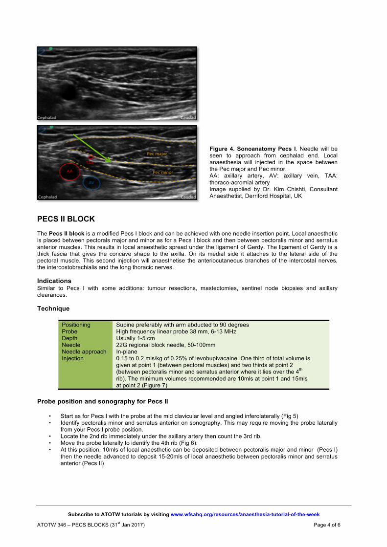
Figure 4. Sonoanatomy Pecs I. Needle will be seen to approach from cephalad end. Local anaesthesia will injected in the space between the Pec major and Pec minor. AA: axillary artery, AV: axillary vein, TAA: thoraco-acromial artery Image supplied by Dr. Kim Chishti, Consultant Anaesthetist, Derriford Hospital, UK
PECS II BLOCK The Pecs II block is a modified Pecs I block and can be achieved with one needle insertion point. Local anaesthetic is placed between pectorals major and minor as for a Pecs I block and then between pectoralis minor and serratus anterior muscles. This results in local anaesthetic spread under the ligament of Gerdy. The ligament of Gerdy is a thick fascia that gives the concave shape to the axilla. On its medial side it attaches to the lateral side of the pectoral muscle. This second injection will anaesthetise the anteriocutaneous branches of the intercostal nerves, the intercostobrachialis and the long thoracic nerves. Indications Similar to Pecs I with some additions: tumour resections, mastectomies, sentinel node biopsies and axillary clearances. Technique
Positioning Probe Depth Needle Needle approach Injection
Supine preferably with arm abducted to 90 degrees High frequency linear probe 38 mm, 6-13 MHz Usually 1-5 cm 22G regional block needle, 50-100mm In-plane 0.15 to 0.2 mls/kg of 0.25% of levobupivacaine. One third of total volume is given at point 1 (between pectoral muscles) and two thirds at point 2 (between pectoralis minor and serratus anterior where it lies over the 4th rib). The minimum volumes recommended are 10mls at point 1 and 15mls at point 2 (Figure 7)
Probe position and sonography for Pecs II
• Start as for Pecs I with the probe at the mid clavicular level and angled inferolaterally (Fig 5) • Identify pectoralis minor and serratus anterior on sonography. This may require moving the probe laterally
from your Pecs I probe position. • Locate the 2nd rib immediately under the axillary artery then count the 3rd rib. • Move the probe laterally to identify the 4th rib (Fig 6). • At this position, 10mls of local anaesthetic can be deposited between pectoralis major and minor (Pecs I)
then the needle advanced to deposit 15-20mls of local anaesthetic between pectoralis minor and serratus anterior (Pecs II)
Subscribe to ATOTW tutorials by visiting www.wfsahq.org/resources/anaesthesia-tutorial-of-the-week ATOTW 346 – PECS BLOCKS (31st Jan 2017) Page 5 of 6
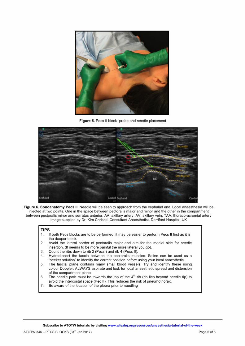
Figure 5. Pecs II block- probe and needle placement
Figure 6. Sonoanatomy Pecs II. Needle will be seen to approach from the cephalad end. Local anaesthesia will be injected at two points. One in the space between pectoralis major and minor and the other in the compartment
between pectoralis minor and serratus anterior. AA: axillary artery, AV: axillary vein, TAA: thoraco-acromial artery Image supplied by Dr. Kim Chrishti, Consultant Anaesthetist, Derriford Hospital, UK
TIPS 1. If both Pecs blocks are to be performed, it may be easier to perform Pecs II first as it is
the deeper block. 2. Avoid the lateral border of pectoralis major and aim for the medial side for needle
insertion. (It seems to be more painful the more lateral you go). 3. Count the ribs down to rib 2 (PecsI) and rib 4 (Pecs II). 4. Hydrodissect the fascia between the pectoralis muscles. Saline can be used as a
“seeker solution” to identify the correct position before using your local anaesthetic.. 5. The fascial plane contains many small blood vessels. Try and identify these using
colour Doppler. ALWAYS aspirate and look for local anaesthetic spread and distension of the compartment plane.
6. The needle path must be towards the top of the 4th rib (rib lies beyond needle tip) to avoid the intercostal space (Pec II). This reduces the risk of pneumothorax.
7. Be aware of the location of the pleura prior to needling

Subscribe to ATOTW tutorials by visiting www.wfsahq.org/resources/anaesthesia-tutorial-of-the-week ATOTW 346 – PECS BLOCKS (31st Jan 2017) Page 6 of 6
SUMMARY
Pecs blocks are a useful addition to analgesia for chest wall surgery, potentially avoiding more invasive procedures such as paravertebral blockade. Serratus plane or supraclavicular nerve blocks may also be needed for complete analgesia. An ultrasound machine is necessary for the performance of this regional nerve block. ANSWERS TO QUESTIONS 1. The following are indications for Pecs I or II block:
a. True b. True c. True d. True e. False: This would require a serratus plane block
2. The following is the target in Pecs II block: a. True: In Pecs II the first injection is between pectoralis major and minor b. True: In Pecs II the second injection is between pectoralis minor and the serratus anterior muscle towards
the 4th rib. c. False: This is the serratus plane block d. False: In the scanning to perform the serratus plane block we could see the teres major muscle, but it is not
our target in Pecs II. e. False: This is the transversus abdominis plane (TAP) block for the abdomen.
3. The following statement is true:
a. True b. True: The placement of catheters are possible and useful, providing continuous analgesia; the insertion is
between pectoralis major and minor muscles. c. False: Bilateral blocks can be performed. The maximum safe dose of local anaesthetic needs to be
calculated. d. False: Pecs II is performed at the level of the 4th rib. e. False: Ultrasound is needed to perform Pecs blocks.
ACKNOWLEGEMENT We would like to thank Dr. Kim Chishti for reviewing the article and for contributing the photographs and ultrasound images and Dr. M Fredrickson for the permission to use the images from ultrasound block.com.
REFERENCES and FURTHER READING 1. Blanco R. The ‘pecs block’: a novel technique for providing analgesia after breast surgery. Anaesthesia 2011; 66 (9): 847-848. 2. Blanco R, Fajardo M, Parras T. Ultrasound description of Pecs II (modified Pecs I). A novel approach to breast surgery. Rev Esp Anestesiol Reanim 2012; 59 (9): 470-475. http://www.csen.com/pecs.pdf 3. U-tube video; Dr Amit Pawa, LSORA PECS II Block https://www.youtube.com/watch?v=YFWneF4pwOA (accessed 11.10.16)
This work is licensed under the Creative Commons Attribution-NonCommercial 3.0 Unported License. To view a copy of this license, visit http://creativecommons.org/licenses/by-nc/3.0/


![Imagens pecs[1]](https://img.pdfslide.net/doc/110x75/55849a20d8b42a33688b4a64/imagens-pecs1.jpg)