Embed Size (px)
Citation preview
1
Pediatric Fractures
Nicholas White, MD
Assistant Professor of Pediatrics
Eastern Virginia Medical School
Attending, Pediatric Emergency Department
Children’s Hospital of The King’s Daughters
Objectives
• Pediatric bone anatomy and physiology
• ED assessment and management
• Fracture description
• Fractures unique to pediatrics
• Upper extremity pediatric fractures
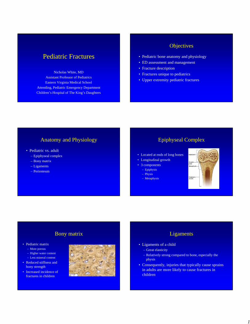
Anatomy and Physiology
• Pediatric vs. adult– Epiphyseal complex
– Bony matrix
– Ligaments
– Periosteum
Epiphyseal Complex
• Located at ends of long bones
• Longitudinal growth
• 3 components– Epiphysis
– Physis
– Metaphysis
Bony matrix
• Pediatric matrix – More porous
– Higher water content
– Less mineral content
• Reduced stiffness and bony strength
• Increased incidence of fractures in children
Ligaments
• Ligaments of a child– Great elasticity
– Relatively strong compared to bone, especially the physis
• Consequently, injuries that typically cause sprains in adults are more likely to cause fractures in children
2
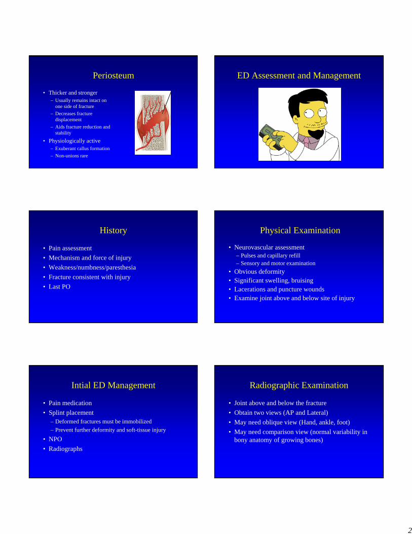
Periosteum
• Thicker and stronger– Usually remains intact on
one side of fracture
– Decreases fracture displacement
– Aids fracture reduction and stability
• Physiologically active– Exuberant callus formation
– Non-unions rare
ED Assessment and Management
History
• Pain assessment
• Mechanism and force of injury
• Weakness/numbness/paresthesia
• Fracture consistent with injury
• Last PO
Physical Examination
• Neurovascular assessment – Pulses and capillary refill– Sensory and motor examination
• Obvious deformity• Significant swelling, bruising• Lacerations and puncture wounds• Examine joint above and below site of injury
Intial ED Management
• Pain medication
• Splint placement– Deformed fractures must be immobilized
– Prevent further deformity and soft-tissue injury
• NPO
• Radiographs
Radiographic Examination
• Joint above and below the fracture
• Obtain two views (AP and Lateral)
• May need oblique view (Hand, ankle, foot)
• May need comparison view (normal variability in bony anatomy of growing bones)
3
Speaking With Orthopedics
• Accurate description using appropriate terminology
Clinical Fracture Description
• Age and sex
• Mechanism of injury
• Anatomic location
• Neurovascular status
• Extent of associated soft tissue injury– Open
– Closed
Radiographic Fracture Description
• Anatomic location of fracture
• Pattern of fracture
• Relationship of fracture fragments
• Physeal involvement (Salter-Harris Type)
• Joint involvement--dislocation
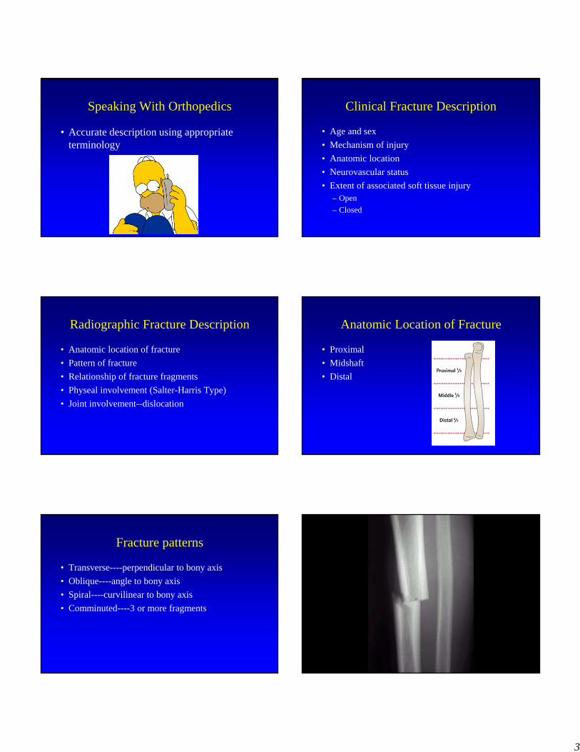
Anatomic Location of Fracture
• Proximal
• Midshaft
• Distal
Fracture patterns
• Transverse----perpendicular to bony axis
• Oblique----angle to bony axis
• Spiral----curvilinear to bony axis
• Comminuted----3 or more fragments

4
Relationship of fracture fragments
• Angulation
• Shortening
• Distraction
• Displacement
Fractures Unique To Children
• Physeal fractures
• Torus (Buckle) fractures
• Greenstick fractures
• Bowing fractures
• Avulsion fractures
• Toddler’s fracture
5
Physeal Fractures
• Weakest point of pediatric skeleton
• Up to 30% of pediatric fractures involve the growth plate
• Most common in early adolescence
• Ligaments stronger than growth plate
• Physeal fractures occur in children where sprains commonly occur in adults
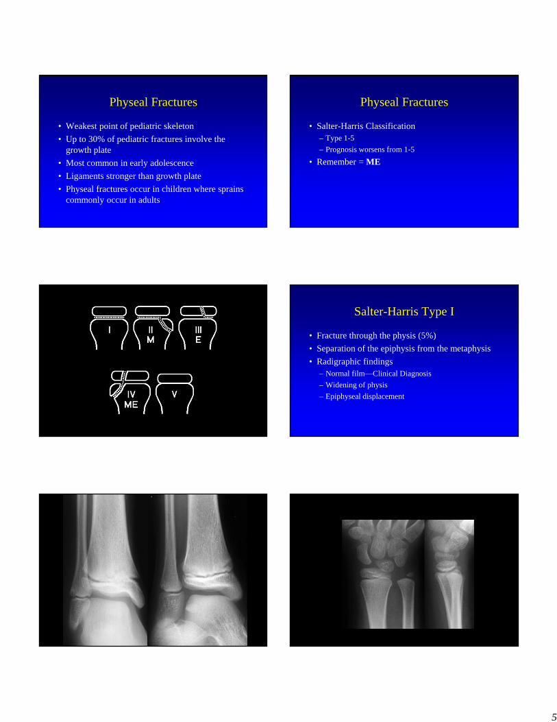
Physeal Fractures
• Salter-Harris Classification– Type 1-5
– Prognosis worsens from 1-5
• Remember = ME
Salter-Harris Type I
• Fracture through the physis (5%)
• Separation of the epiphysis from the metaphysis
• Radigraphic findings – Normal film—Clinical Diagnosis
– Widening of physis
– Epiphyseal displacement
6
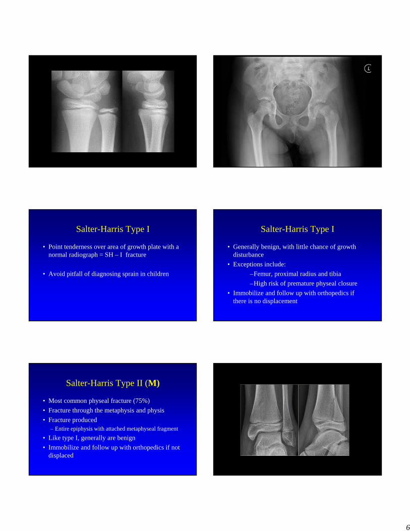
Salter-Harris Type I
• Point tenderness over area of growth plate with a normal radiograph = SH – I fracture
• Avoid pitfall of diagnosing sprain in children
Salter-Harris Type I
• Generally benign, with little chance of growth disturbance
• Exceptions include:
–Femur, proximal radius and tibia
–High risk of premature physeal closure
• Immobilize and follow up with orthopedics if there is no displacement
Salter-Harris Type II (M)
• Most common physeal fracture (75%)
• Fracture through the metaphysis and physis
• Fracture produced– Entire epiphysis with attached metaphyseal fragment
• Like type I, generally are benign
• Immobilize and follow up with orthopedics if not displaced
7
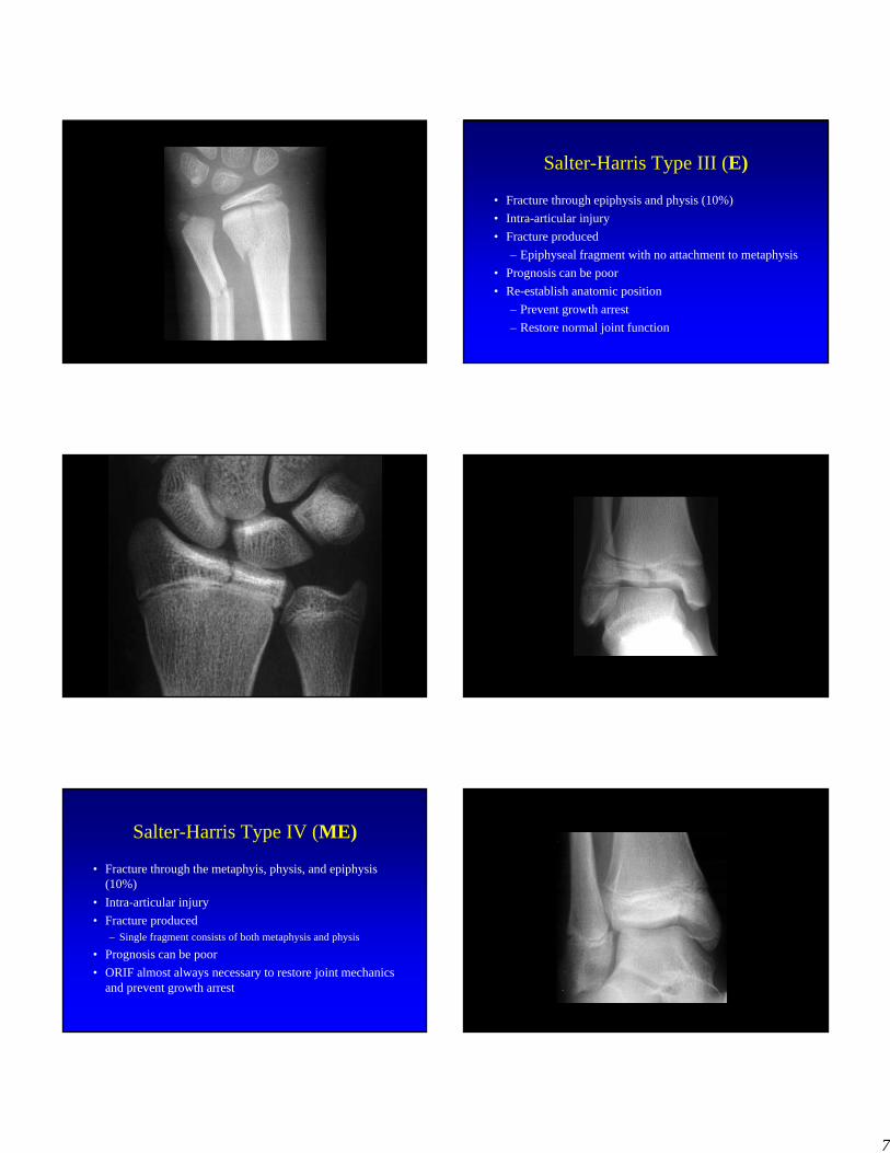
Salter-Harris Type III (E)
• Fracture through epiphysis and physis (10%)
• Intra-articular injury
• Fracture produced
– Epiphyseal fragment with no attachment to metaphysis
• Prognosis can be poor
• Re-establish anatomic position
– Prevent growth arrest
– Restore normal joint function
Salter-Harris Type IV (ME)
• Fracture through the metaphyis, physis, and epiphysis (10%)
• Intra-articular injury
• Fracture produced– Single fragment consists of both metaphysis and physis
• Prognosis can be poor
• ORIF almost always necessary to restore joint mechanics and prevent growth arrest
8
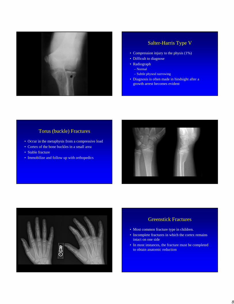
Salter-Harris Type V
• Compression injury to the physis (1%)
• Difficult to diagnose
• Radiograph – Normal
– Subtle physeal narrowing
• Diagnosis is often made in hindsight after a growth arrest becomes evident
Torus (buckle) Fractures
• Occur in the metaphysis from a compressive load
• Cortex of the bone buckles in a small area
• Stable fracture
• Immobilize and follow up with orthopedics
Greenstick Fractures
• Most common fracture type in children.
• Incomplete fractures in which the cortex remains intact on one side
• In most instances, the fracture must be completed to obtain anatomic reduction

9
Bowing Fractures
• Force exceeds elastic limits of bone
• Bowed deformation
• Fracture line not apparent radiographically
• Histologically see multiple oblique microfractures
• Reduction depends on age and degree of angulation
Avulsion Fractures
• Apophysis-bone growth center that has a strong muscular attachment
• During intense muscular contraction (sports), fractures occur through the apophyseal plate.
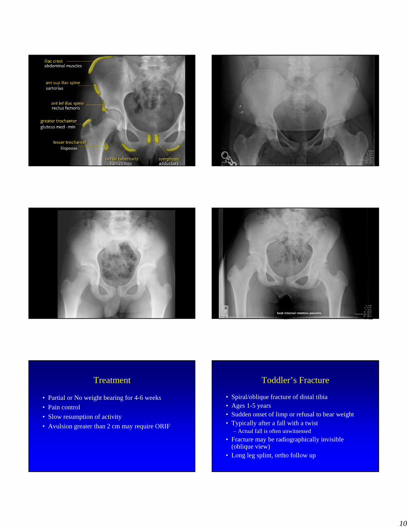
• Pelvis most common site
Pelvic Avulsion Fractures
• Athletic teenagers– Running, kicking
– Hip pain, point tenderness, decreased range of motion
10
Page: 1 of 2Page: 1 of 2 IM: 1001IM: 1001Compressed 32:1Compressed 32:1
W: 1024W: 1024C: 512C: 512
Z: 0.48 Z: 0.48 S: 605S: 605
cm cm
Page: 1 of 2Page: 1 of 2 IM: 1001IM: 1001Compressed 32:1Compressed 32:1
W: 1024W: 1024C: 512C: 512
Z: 0.48 Z: 0.48 S: 183S: 183
cm cm
Treatment
• Partial or No weight bearing for 4-6 weeks
• Pain control
• Slow resumption of activity
• Avulsion greater than 2 cm may require ORIF
Toddler’s Fracture
• Spiral/oblique fracture of distal tibia• Ages 1-5 years• Sudden onset of limp or refusal to bear weight• Typically after a fall with a twist
– Actual fall is often unwitnessed
• Fracture may be radiographically invisible (oblique view)
• Long leg splint, ortho follow up
11
Common upper extremity fractures
• Clavicular fracture
• Proximal & Mid-shaft humerus fracture
• Supracondylar fracture
• Forearm fractures
• Hand fractures
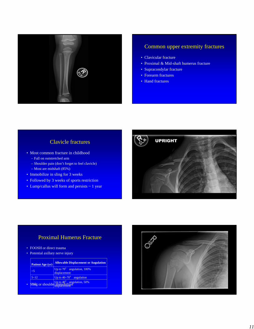
Clavicle fractures
• Most common fracture in childhood– Fall on outstretched arm
– Shoulder pain (don’t forget to feel clavicle)
– Most are midshaft (85%)
• Immobilize in sling for 3 weeks
• Followed by 3 weeks of sports restriction
• Lump/callus will form and persists ~ 1 year
Proximal Humerus Fracture
• FOOSH or direct trauma
• Potential axillary nerve injury
• Sling or shoulder immobilizer
Patient Age (yr)Allowable Displacement or Angulation
<5Up to 70° angulation, 100% displacement
5–12 Up to 40–70° angulation
>12Up to 40° angulation, 50% displacement
12
CompComp
cm cm
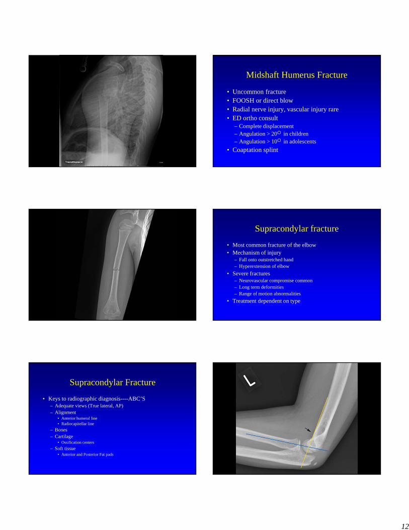
Midshaft Humerus Fracture
• Uncommon fracture• FOOSH or direct blow• Radial nerve injury, vascular injury rare• ED ortho consult
– Complete displacement– Angulation > 20○ in children– Angulation > 10○ in adolescents
• Coaptation splint
Supracondylar fracture
• Most common fracture of the elbow • Mechanism of injury
– Fall onto outstretched hand– Hyperextension of elbow
• Severe fractures– Neurovascular compromise common – Long term deformities– Range of motion abnormalities
• Treatment dependent on type
Supracondylar Fracture
• Keys to radiographic diagnosis----ABC’S– Adequate views (True lateral, AP) – Alignment
• Anterior humeral line• Radiocapitellar line
– Bones– Cartilage
• Ossification centers
– Soft tissue• Anterior and Posterior Fat pads
13
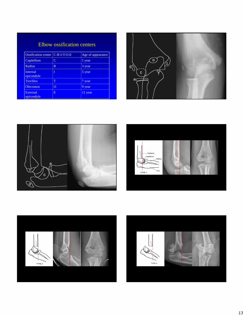
Elbow ossification centers
Ossification center C-R-I-T-O-E Age of appearance
Capitellum C 1 year
Radius R 3 year
Internal epicondyle
I 5 year
Trochlea T 7 year
Olecranon O 9 year
External epicondyle
E 11 year
14
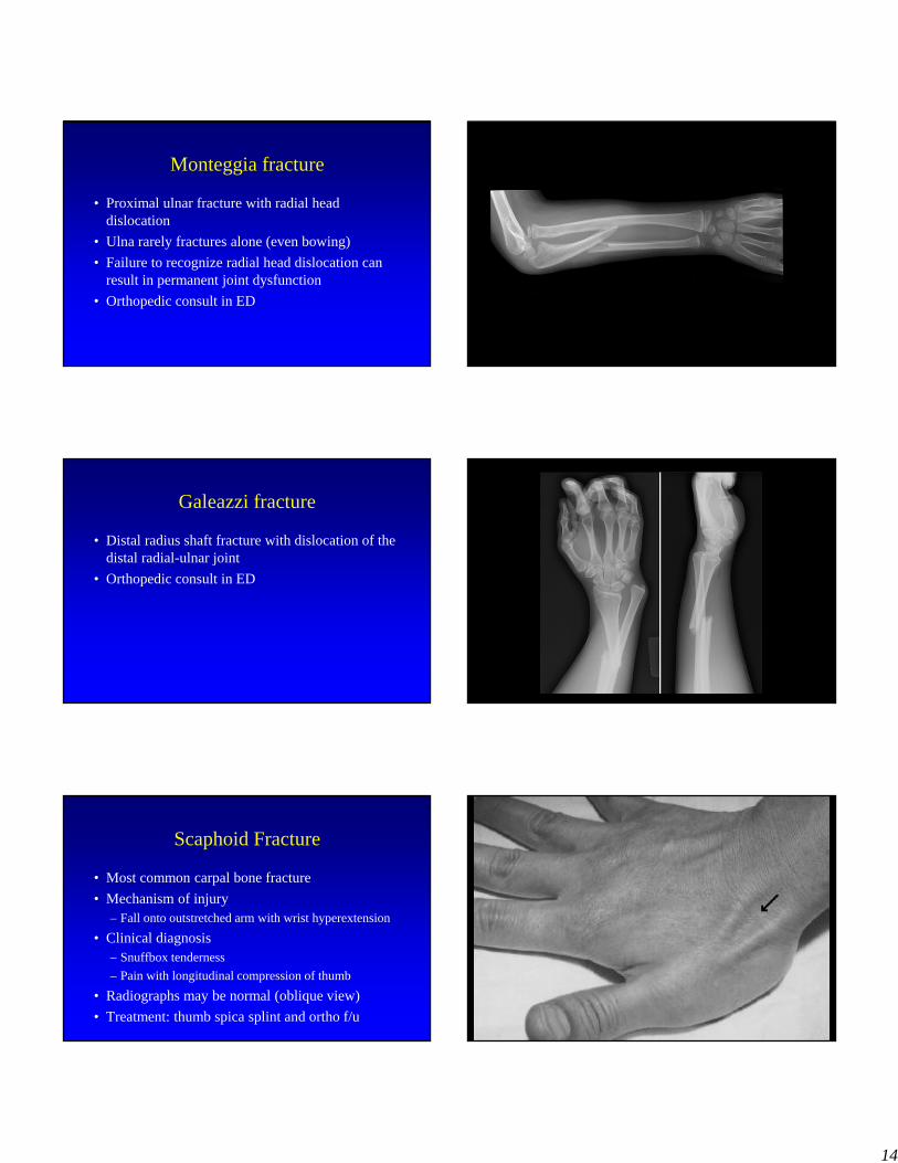
Monteggia fracture
• Proximal ulnar fracture with radial head dislocation
• Ulna rarely fractures alone (even bowing)
• Failure to recognize radial head dislocation can result in permanent joint dysfunction
• Orthopedic consult in ED
Galeazzi fracture
• Distal radius shaft fracture with dislocation of the distal radial-ulnar joint
• Orthopedic consult in ED
Scaphoid Fracture
• Most common carpal bone fracture
• Mechanism of injury– Fall onto outstretched arm with wrist hyperextension
• Clinical diagnosis– Snuffbox tenderness
– Pain with longitudinal compression of thumb
• Radiographs may be normal (oblique view)
• Treatment: thumb spica splint and ortho f/u
15
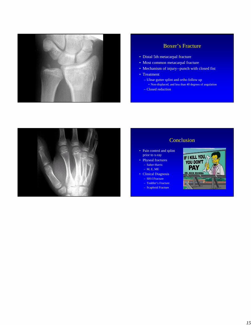
Boxer’s Fracture
• Distal 5th metacarpal fracture
• Most common metacarpal fracture
• Mechanism of injury--punch with closed fist
• Treatment– Ulnar gutter splint and ortho follow up
• Non-displaced, and less than 40 degrees of angulation
– Closed reduction
Conclusion
• Pain control and splint prior to x-ray
• Physeal fractures– Salter-Harris
– M, E, ME
• Clinical Diagnosis– SH-I Fracture
– Toddler’s Fracture
– Scaphoid Fracture