Embed Size (px)
Citation preview
1
PEDIATRIC GI EMERGENCIES
Ghazala Q. Sharieff, MD
PEDIATRIC ABDOMINAL PAINHow Common Is It?
• 5% of unscheduled visits• 2% of patients are admitted• 1% need operative intervention
EXAMINATION TIPS
• Palpate between cries• Examine on parent• Palpate with pt’s hand
• Head of stethoscope• Parent examination• Have pt jump/climb
AGE-RELATED DIAGNOSISEarly Infancy
• Malrotation with or without volvulus• Pyloric Stenosis• Hirschsprung Disease• Necrotizing Enterocolitis• Non-accidental Trauma• Colic• Incarcerated hernia• Gastroenteritis

2
AGE-RELATED DIAGNOSISInfancy ( >3 months)
• Intussusception• Hirschsprung Disease• Hernia• Non-accidental trauma• Gastroenteritis
AGE-RELATED DIAGNOSISEarly Childhood
• Appendicitis• Constipation• Peptic Ulcer Disease• Hemolytic-uremic syndrome
AGE-RELATED DIAGNOSISSchool Age
• Appendicitis• Constipation• Peptic Ulcer Disease• Inflammatory Bowel Disease• Pancreatitis / Biliary Tract Disease• Henoch-Schoenlein Purpura• Functional Pain
AGE-RELATED DIAGNOSISAdolescence-other
Considerations
• Pregnancy• Complications of pregnancy• Mittelschmerz• PID• HSP• Ovarian torsion/cyst

3
DISEASE PROCESSES REQUIRING SURGERY
• Malrotation with midgut volvulus• Pyloric stenosis• Intussusception• Appendicitis• Hirschsprung Disease• Incarcerated Hernia
CASE PRESENTATION
• A 10 day old female presents with bilious vomiting and abdominal distension. She is afebrile and has no ill contacts.
• HR 200, RR 70, O2 sat 99%• What are your management priorities?• What is the most likely diagnosis?
MALROTATION WITH MIDGUT VOLVULUS- What is it?
• Congenital malrotation of the midgut• During 5th-8th embryonic week, intestine
projects out of abdominal cavity, rotates 270 degrees and then returns
• If rotation is incomplete, the intestine does not anchor at the mesentery
• Volvulus is twisting of a loop of bowel about the mesenteric attachment
MALROTATION Signs & Symptoms
• Usually presents in the first year of life• Child looks sick => shock• Bilious vomiting• Abdominal distension may be present• Hematochezia may develop

4
MALROTATIONDiagnosis
• Abdominal films- Classic double bubble sign
• Upper GI- Gold standard. “cork-screwing”, spiraling of the SI around the SMA
• Ultrasound- distended, fluid filled duodenum, with dilated loops of bowel to the right of the spinal column
MALROTATION Treatment
• IV Hydration• Correction of electrolytes• NG tube• Surgical correction

5
CASE PRESENTATION
• A 1-year old female presents with vomiting, intermittent abdominal cramping, and an episode of blood-streaked stool. She has been very lethargic today.
• You palpate a mass in the RUQ, and the patient has heme-positive stools
• What is the most likely diagnosis?
INTUSSUSCEPTIONWhat is it?
• Prolapse of one part of the intestine into the lumen of an immediately adjacent distal part
• Most common location: ileo-colic• Mesentery and venous supply obstruct=>
mucosal edema and increased pressure+> arterial flow obstruction
INTUSSUSCEPTIONWhat causes it?
• Idiopathic in children less than 2 years• Meckel’s Diverticulum• HSP• Polyps• Tumors Ex-lymphoma• Post-rotaviral vaccination
HENOCH-SCHOENLEIN PURPURA“ARENA”
• A- abdominal pain, +/- bloody stools• R- purpuric rash• E- edema• N-nephritis• A- arthralgias/ arthritis

6
INTUSSUSCEPTIONSigns & Symptoms
• Intermittent, colicky abdominal pain• Currant jelly stools are a late finding• May have RUQ mass• Emesis bilious• Heme-positive stools• ALOC
INTUSSUSCEPTIONDiagnosis
• Abdominal films- may be normal initially, but then may see signs of obstruction, paucity of air and dilated loops of bowel
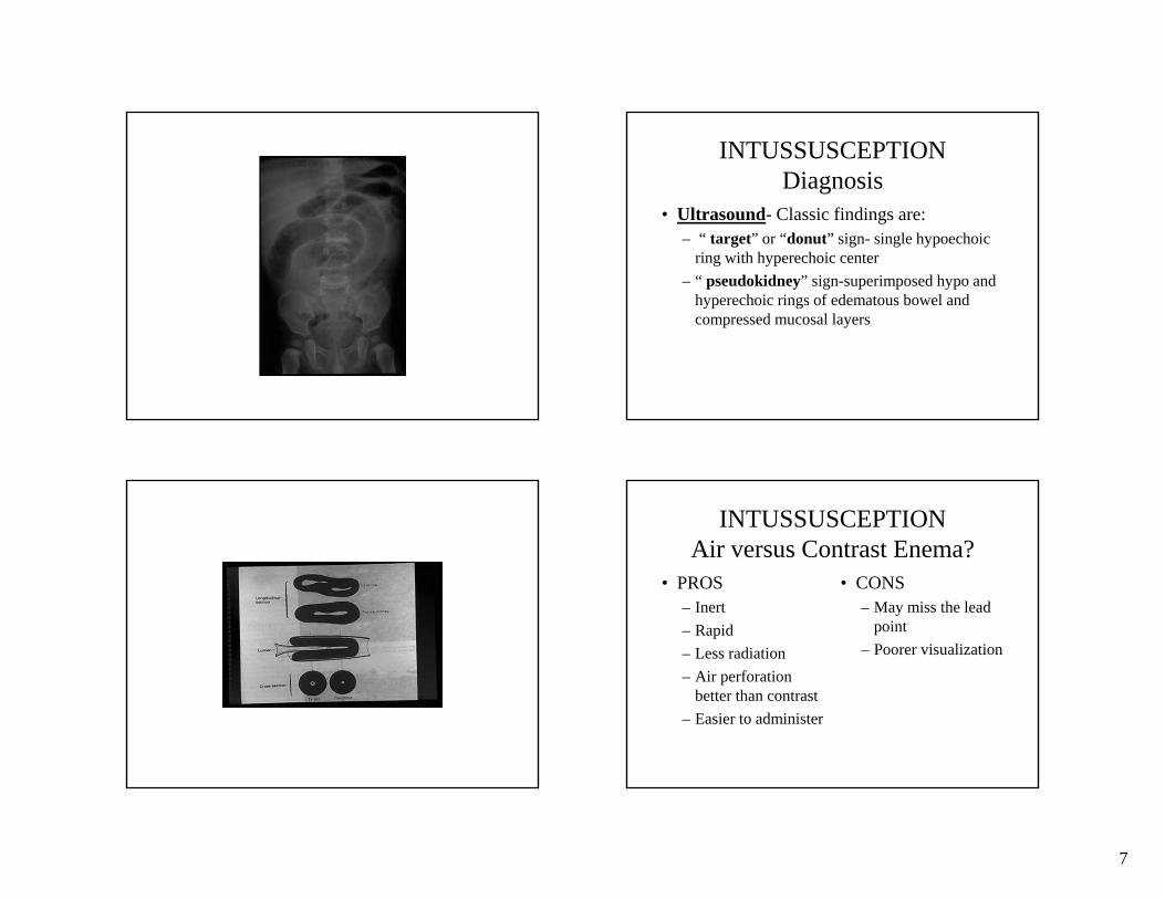
7
INTUSSUSCEPTIONDiagnosis
• Ultrasound- Classic findings are:– “ target” or “donut” sign- single hypoechoic
ring with hyperechoic center– “ pseudokidney” sign-superimposed hypo and
hyperechoic rings of edematous bowel and compressed mucosal layers
INTUSSUSCEPTIONAir versus Contrast Enema?
• PROS– Inert– Rapid– Less radiation– Air perforation
better than contrast– Easier to administer
• CONS– May miss the lead
point– Poorer visualization

8
CASE PRESENTATION
• A 10-year old male presents with vague, epigastric pain and maroon colored stools, since yesterday. He has no fever, no diarrhea, and no other ill contacts. His HCT is 17, coags are normal
• An endoscopy and colonoscopy is performed, and these are normal. What other study should you consider? What could the diagnosis be?
MECKEL’S DIVERTICULUMWhat Is It?
• Most common congenital abnormality of the small intestine, remnant of the omphalomesenteric duct.
• This duct should disappear by the 7th
gestational week• 2% of the population• Most symptomatic patients are males and
less than 2 years of age
MECKEL’S DIVERTICULUM
• Located 40-100cm proximal to the ileocecal valve
• Consists of al layers of bowel wall• Ectopic gastric mucosa is the most common
finding

9
MECKEL’S DIVERTICULUM,Pathophysiology
• Intestinal obstruction may occur from:• Intussusception• Herniation• Volvulus
• GI Bleeding may occur from peptic ulcer formation within the diverticulum or in the adjacent ileum
MECKEL’S DIVERTICULUMSigns & Symptoms
• Isolated rectal bleeding frequent in children less than 5 years of age
• Bleeding is usually painless• Bleeding is acute and severe and lead to
anemia and cardiovascular instability• Diverticulitis occurs in older children and
can be confused with appendicitis
MECKEL’S DIVERTICULUMDiagnosis
• Meckel’s scan-diverticuli that take up the Technetium Pertechnitate have gastric mucosa ( High sensitivity and specificity)
• Technetium tagged RBC’s- can detect the site of active bleeding ( not specific for Meckel’s)

10
MECKEL’S DIVERTICULUMTreatment
• ABC’s• Large bore IV’s• May need transfusion• NG tube• Initiate antibiotics if signs of peritonitis• Diverticulotomy versus more extensive
resection if ischemic bowel is present
CASE PRESENTATION
• A 6-week old male presents with non-bilious emesis after meals, there is no blood in the stools and he has no fever. He appears to be hungry at all times. The emesis has become projectile over the past few days.
• What is the most likely diagnosis?
PYLORIC STENOSISOverview
• Most common disorder requiring surgery in infancy
• Occurs in 1 /250 births• M:F ratio=4:1• Whites> Blacks• ? Increased incidence in first born males
PYLORIC STENOSISPathophysiology
• Hypertrophy and hyperplasia of the pyloric musculature leading to obstruction
• Occurs after birth• Unknown etiology

11
PYLORIC STENOSISSigns & Symptoms
• Usually presents 2 weeks to 2 months after birth
• Most commonly presents as non-bilious vomiting, which may become projectile
• Patients may have a voracious appetite• Constipation is common• Jaundice may also be present
PYLORIC STENOSISPhysical Examination
• Dehydration “ old man” appearance• Peristaltic waves from left to right• Palpable mass ( olive) in RUQ, lateral to
the right rectus muscle (70-90%)
PYLORIC STENOSISVERSUS GER?
Smith GA et al. Am J Emerg Med, l999
• 75 consecutive infants with OR diagnosis of pyloric stenosis and 75 consecutive infants with diagnosis of GE reflux. All patients were less than 12 weeks of age
• Serum HCO3 mean was 27 meq for pts with PS and 22 for GER
• HCO3 >29 and CL <98 : PPV 96% and 97%, specificity 99%, sensitivity 36% and 50%

12
PYLORIC STENOSISDiagnosis
• UGI-string sign, mushroom sign, shoulder sign
• Ultrasound– Pyloric wall thickness > 4mm ( normal <2mm)– Canal length is >14mm ( normal <10mm)
PYLORIC STENOSISDiagnosis
Mandell et al. Pediatrics, l999
• 89 vomiting pts age 11-120 days• NG tube placed after at least 1 NPO hour• If NG aspirate greater than 5cc, US was
performed. If negative then onto UGI• If NG aspirate < 5cc, UGI performed first
PYLORIC STENOSISIMAGING APPROACH
• 23 / 89 had PS ( 25%)• 66 / 89 had UGI- 79% had reflux• Sensitivity of aspirate criteria = 91%;
specificity of 88%.• 6 false positives due to recent meal and 2
due to antral dysmotility• If NPO for 3-4 hours, specificity increases
to 94% and accuracy improves to 96%

13
PYLORIC STENOSISTreatment
• Electrolyte correction• IV hydration• Definitive surgery- widening of the pylorus
via pylorotomy
CASE PRESENTATION
• A 6-year old male presents with diffuse abdominal pain, decreased appetite, fever, vomiting, and increased pain with motion. He has had 2 loose stools today.
• What diagnosis must you consider?
APPENDICITISOverview
• Most common etiology for surgical abdomen in children
• False positive rate of 15-20%• Perforation rate 15-40% in younger
children due to delays in diagnosis
APPENDICITISDiagnosis
• Helical CT: inflamed appendix, fecalith, abscess, stranding of the peri-appendiceal fat
• Labs: elevated WBC not 100% sensitive, elevated CRP

14
Acute Appendicitis-Other Tests?Gavela T. Peds EM Care 2012
• 111 consecutive pts admitted with a diagnosis of acute appendicitis between July 2009 and February 2010
• Age, sex, time since diagnosis, laboratory data, complications (abscess, intestinal obstruction), presence of hemodynamic instability, mortality, length of stay, and need for admission to the pediatric intensive care unit.
Acute Appendicitis-Other Tests?Gavela T. Peds EM Care 2012
• Patients divided into 2 groups (group 1, appendicitis; group 2, localized or generalized peritonitis).
Acute Appendicitis- Other tests?
• CRP and PCT predict the outcome of pediatric patients with appendicitis.
• Children with CRP greater than 3 mg/dL and/or PCT greater than 0.18 ng/mL have a greater risk of complications
Risk Factors For PerforationHung. Peds EM Care, 2012
• 228 patients < 17 years of age• 140 had a postoperative pathological
diagnosis of a nonperforated appendix, • 88 had perforated appendix, resulting in a
perforation rate of 38.6%.

15
Risk Factors For Perforation Hung. Peds EM Care, 2012
• Younger age, longer duration of pain, fever, muscle guarding, and elevated CRP significantly associated with perforation
Scoring for AppendicitisBhatt AM. Acad Emerg Med 2009
• Convenience sample of children, 4-18 years old with abdominal pain of less than 3 days' duration
• Score components: – right lower quadrant and hop tenderness,– anorexia,– pyrexia, – emesis, – pain migration, – leukocytosis,– neutrophilia
Scoring for AppendicitisBhatt AM. Acad Emerg Med 2009
• When a PAS of >or=8 determined the need for appendectomy, the score's specificity was 95.1%
• Negative appendectomy rate would have been 8.8%, missed appendicitis rate would have been 2.4%, and 41% of imaging investigations would have been avoided.
Scoring for AppendicitisBhatt AM. Acad Emerg Med 2009
• Scores of <or=4 help rule out appendicitis, scores of >or=8 help predict appendicitis. Patients with a PAS of 5-7 may need further radiologic evaluation.

16
Imaging and AppendicitisAnn Emerg Med 2012
• 1810 children, 3-18 years, mean 10.9 yrs• 49% males• 1216 had CT, 832 had US, 238 had both• Sensitivity of US increased with duration of
pain – 0.81 for <12 hours;0.87 for 24-35 hours,0.92
for 36-47 hours and 0.92 for 48-71 hours
Conclusions• Sensitivity of US for appendicitis increases
with longer duration of abdominal pain• CT demonstrates high sensitivity regardless
of pain duration• CT scans are less likely to be equivocal
with longer duration of pain
A Practical Approach To Appendicitis
• RLQ pain and good history, WBC and CRP– + => OR
• +/- history and equivocal labs=> Ultrasound• US positive=> OR• US negative, but worrisome exam/history or
labs=> CT or MRI• CT or MRI neg but pt still with pain=>
admission for observation!
CASE PRESENTATION
• A 2-day old male presents with a distended abdomen, delayed capillary refill, tachypnea, and a heart rate greater than 210. His mother reports that he has not passed a meconium stool yet.
• What is the most likely diagnosis?• Should you perform a rectal exam in this
patient?

17
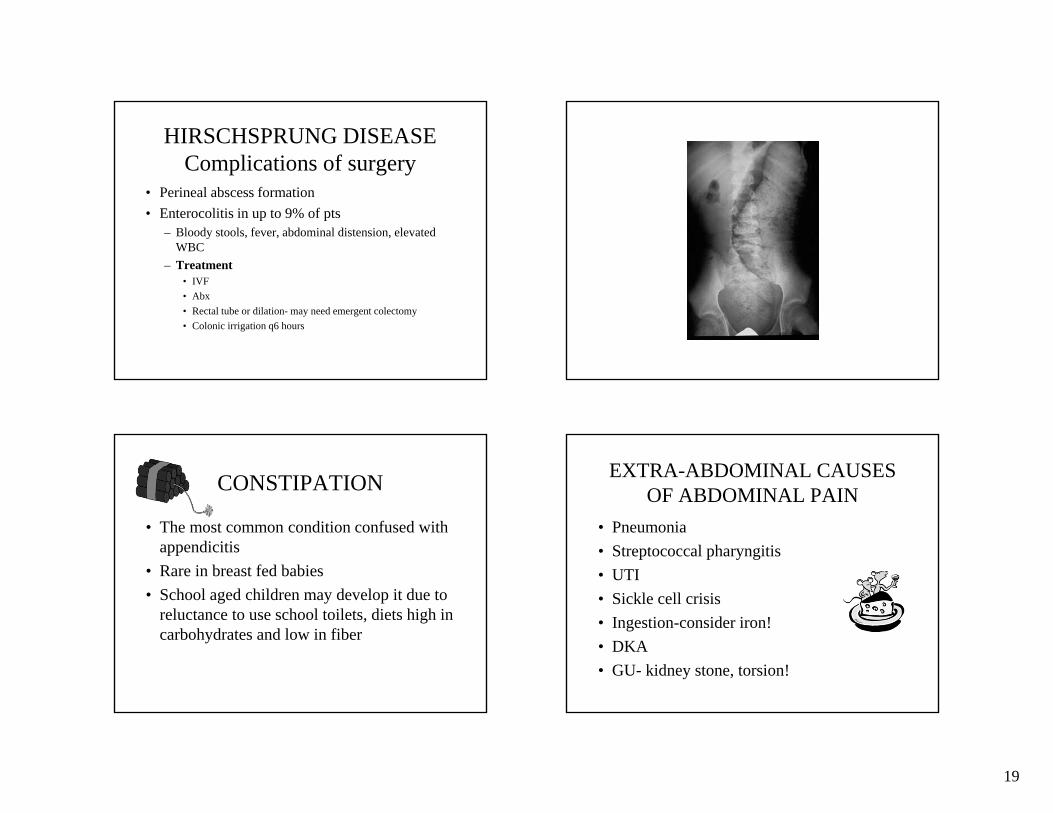
HIRSCHSPRUNG DISEASEWhat is it?
• Absence of the intramural ganglion cells in the rectum, extends to the sigmoid in 77%, & involves the entire colon in 15% of pts
• 1/5,000 births. No ethnic predilection• Male to female ratio 4:1
HIRSCHSPRUNG DISEASEClinical Presentation
• Neonates present with failure to pass a meconium stool
• Infants and children present with constipation and obstipation, no history of encopresis
HIRSCHSPRUNG DISEASEPhysical Examination
• Abdominal mass is frequently palpated• Vomiting may be present• May see signs of obstruction• Rectal shows no stool in the vault, but if
you suspect this diagnosis..DO NOT DO RECTAL!!

18
HIRSCHSPRUNG DISEASEDiagnosis
• Barium enema: Cone-shaped transition zone with dilated segment of proximal colon
• Rectal manometry: Paradoxical contraction of the internal anal sphincter
• Rectal Biopsy: Gold standard. Reveals the lack of ganglion cells in the rectal submucosa
HIRSCHSPRUNG DISEASETreatment
• IV hydration• Surgical repair options:
– Immediate repair within the first few weeks of life
19
HIRSCHSPRUNG DISEASEComplications of surgery
• Perineal abscess formation• Enterocolitis in up to 9% of pts
– Bloody stools, fever, abdominal distension, elevated WBC
– Treatment• IVF• Abx• Rectal tube or dilation- may need emergent colectomy• Colonic irrigation q6 hours
CONSTIPATION
• The most common condition confused with appendicitis
• Rare in breast fed babies• School aged children may develop it due to
reluctance to use school toilets, diets high in carbohydrates and low in fiber
EXTRA-ABDOMINAL CAUSES OF ABDOMINAL PAIN
• Pneumonia• Streptococcal pharyngitis• UTI• Sickle cell crisis• Ingestion-consider iron!• DKA• GU- kidney stone, torsion!

20
21
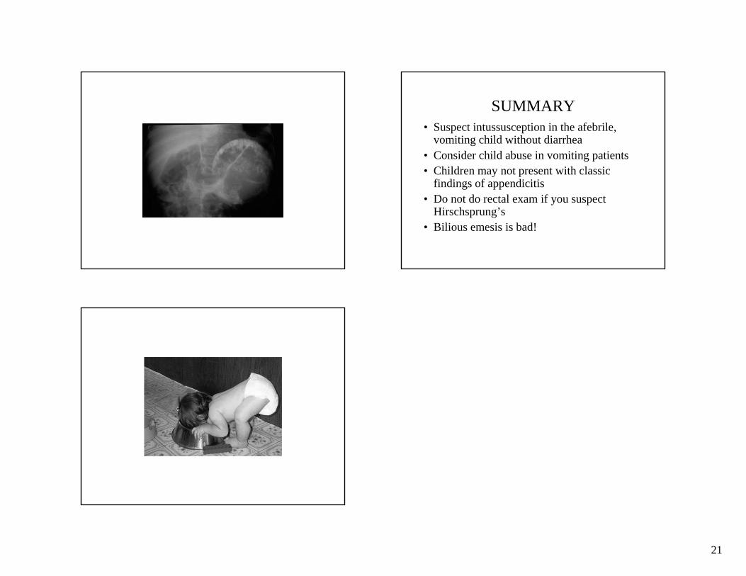
SUMMARY• Suspect intussusception in the afebrile,
vomiting child without diarrhea• Consider child abuse in vomiting patients• Children may not present with classic
findings of appendicitis• Do not do rectal exam if you suspect
Hirschsprung’s• Bilious emesis is bad!