Embed Size (px)
Citation preview

PEDIATRIC HEMATOLOGY-ONCOLOGY
CASE-BASED LECTURE SERIES
Carl Koschmann, M.D.
Pediatric Hematology-Oncology
University of Michigan
With additional help from:
Julie Park, M.D.1, Douglas S. Hawkins, M.D. 1, Michael Bender, M.D. 1, Laura Winter, Pharm.D. 1, Chris Pendergrass1, Andy Luks, M.D. 2
1Seattle Children’s Hospital, 2University of Washington

How to use this lecture
This content is based on the inpatient management
of commonly seen scenarios in pediatric
hematology-oncology patients
The content will be ideal for medical students,
pediatric residents, and first-year pediatric
hematology-oncology fellows
Some definitions (when to stop antibiotics) may
differ at different treating institutions, but attempt
has been made to make information generalizable

How to use this lecture
Main menu listed on left side of lecture to navigate
between topics
Use arrows below to change slides
Hyperlinks on each page to navigate between
pages, and for additional information on certain
concepts
Questions for participant in blue/italics
Total of 4 cases
Should take about 15 minutes per case

Cases
1. Fever and Neutropenia
2. Tumor Lysis Syndrome
3. Nausea and Vomiting
4. Acute Chest Syndrome

CASE 1 –
FEVER AND NEUTROPENIA

Location: Emergency Department
• HPI: 7 year old female previously diagnosed with standard-risk acute lymphoblastic leukemia (ALL) currently receiving chemotherapy now on day 15 of her protocol (last chemo received – vincristine, doxorubicin day 8)
• Presents to ED with a history of fever to 102.1, feels tired, decreased oral intake, no other symptoms
• Hematocrit (Hct) 25.4%, platelets (plt) 179, white blood count (WBC) 0.6 K, absolute neutrophil count (ANC) 83
• ER draws blood cultures, starts antibiotics
• What else do you want to know?

Get a thorough history
History of present illness
Patient/parent focal complaints/concerns
Is the patient hydrated, any concerns about perfusion, respiratory status
Chills (symptom - feeling of cold/mild shivering) vs. rigors (sign – intense,
uncontrollable shivering; often seen with worsening bacterial infection/sepsis;
consider broadening antibiotics)
Review of systems
No focal concerns or other symptoms, appears hydrated and perfused per
family and ED physician
Past medical history
Patient has a history of previous line infection with Coagulase-Negative Staph
(CONS) which required a 14 day IV vancomycin course, and a history of
Pseudomonas urinary tract infection (UTI) earlier during therapy
No recent surgery, but underwent lumbar puncture two weeks prior for
intrathecal methotrexate

Physical exam
Very important to attempt to localize infection in
neutropenic patient
Localizing signs may be subtle (may not be able to
develop erythema with low neutrophil count), so close
attention should be given to mild signs
Full exam including central line site area, perianal
area, ear exam are normal, patient looks tired but is
cooperative, vitals signs normal
Any other changes or studies at this point?

Management – additional studies
With history of UTI – and questionable age for reporting
dysuria, obtain a non-catheterized urinalysis (UA) (+culture)
Lack of WBCs on UA does not rule out UTI while neutropenic
Should consider UA in all febrile neutropenic patients, but
would not delay antibiotics for sample
DO NOT instrument neutropenic patient outside of special
circumstances (high risk of infection with mucosal break).
NO RECTAL TEMPERATURES.
Other studies as symptoms direct (chest X-ray,
respiratory viral panel, stool studies – clostridium
difficile toxin, rotavirus assay, etc.)

F + N – definition
F(ever) = 38.3˚C (101˚F) one time (or 38.0˚C twice
within defined time period, e.g., 8 or 24 hours, at some
centers)
N(eutropenia) = ANC < 500 (or 200 at some centers)
OR predicted to fall below 500 in next 24-48 hours
For febrile, non-neutropenic cancer patient with ill-
appearance, treat as if neutropenic (immunosuppressed
patient with indwelling central line is at high-risk for
infection)

Location of documented infections
Most (79%) have fever of unknown origin
Of the 21% with documented infection:
Other 10%
Perineal region 6%
Mouth 6%
Blood 65%Lungs
8%
Bowel 5 %
Castagnola et al. CID. 2007.

Isolated pathogens
Coagulase
negative staph
species 28%
Other
11%
Fungi 7%
Pseudomonas
6%E. coli 13%
Other
streptococci
(including
viridans) 28%
Staph
aureus 7%
Anti-pseudomonal agent such as
ceftazidime or cefepime is necessary due
to frequency of pseudomonas in
neutropenic pediatric patients Castagnola et al. CID. 2007.

Choice of antibiotic
Start with cephalosporin with anti-pseudomonal
activity (e.g., ceftazidime or cefepime depending
on center), and add additional antibiotic(s) if
situation arises:
Concern for abdominal or perineal infection: provide
additional coverage for abdominal anaerobes
Severe mucositis: provide additional coverage for oral
anaerobes
Hypotension, rigors, or other signs of hemodynamic
instability: additional coverage for gram negative and
positive infection in the setting of possible beta-lactamase
resistance

Case 1 (cont)
The patient is stable appearing after receiving one
dose of antibiotic
The medical student on service wonders if the
(neutropenic) patient should empirically receive
coverage for resistant gram positives, such as
methicillin-resistant staph aureus (MRSA)
Are there additional antibiotics you would add?

Adding vancomycin empirically
Prospective trial of febrile neutropenic patients randomized for initial empiric therapy ceftazidime alone vs. ceftazidime plus vancomycin (Ramphal R et al. Antimicrob Agents Chemother. 1992.)
similar outcomes and survival rates; more renal and cutaneous toxicities in patients receiving vancomycin
Increased rates of VRE (vancomycin-resistant enterococcus)
Most institutions DO NOT support adding vancomycin empirically for hemodynamically stable patients
History of previous line infection with resistant gram positive also DOES NOT mean the patient needs empiric vancomycin
Consider vancomycin if:
focal skin findings or severe mucositis
soft tissue pain beyond proportion of exam (sign of necrotizing fasciitis)
hemodynamic instability
Recent administration of cytarabine
Add vancomycin if blood culture shows gram positives (keep until susceptibilities further direct therapy)

Case 1 (cont)
Day 3 of admission
Blood culture and urine culture have no growth to date
(NGTD.) Patient has been afebrile for 36 hours.
Patient has ANC of 0.
Can you stop antibiotics? If not, when?
Can you discharge the patient? If not, when?

Stopping antibiotics / discharge
Consider (there is considerable institutional variability)
switching low-risk patients to oral antibiotics for discharge
while neutropenic if the following are met:
Afebrile for 24 hours AND
Negative cultures for 48 hours AND
Well appearing, no concerning symptoms, can remain hydrated
Some centers also require minimum ANC or signs of marrow recovery
– neutrophils or monocytes on differential
Regimens often contain an oral fluoroquinolone (sometimes with
additional antibiotics) until recovery of counts
Consider stopping antibiotics once patient has met above
criteria and is no longer neutropenic by institutional definition
(ANC > 200 or 500)

Case 1 - part 2 (6 months later)
Patient has since developed relapse of his leukemia – is now getting treated with high dose cytarabine
In clinic for evaluation, Hct 24, Plt 60, WBC 2.3, ANC 15. Febrile to 39.0˚C.
Patient well appearing, blood pressure (BP) 110/50, HR 110
What antibiotic to start?

Viridans Group Streptococcus
Important part of the normal microbial flora Upper respiratory tract, gastro-intestinal tract especially oral
cavity
Common in cancer patients Shock syndrome 10 - 25%
Mortality about 6 -12%
Risk factors:
Female gender
High-dose cytarabine (included in AML and certain relapsed ALL re-inductions) OR clofarabine
Mucositis
Severe neutropenia
Prophylactic co-trimoxazole/quinolones
Poor dentition

Cefepime vs. Ceftazidime
Regimen Activity against Viridans
Streptococci
Cefepime Excellent
Vancomycin Excellent
Ceftazidime Poor
Ceftazidime does not provide good coverage for Strep Viridans and
therefore may not be the empiric agent of choice for patients at high risk
for this infection. Cefepime will provide better coverage for patients at
risk for this virulent organism.

Case 1 – part 2 (cont)
Patient receives antibiotics – now appears drowsy,
but just got benadryl for nausea. Vital signs: BPs
70s/20s. HR 150s. Cap refill (CR) 3 sec.
What do you want to do?

Case 1 – part 2 management
Blood culture, urine culture (don’t hold antibiotics),
add additional coverage for resistant organisms (if
not already started), normal saline (NS) bolus 20
ml/kg – all ordered stat, and verbalize plan with
nurse (RN).
RN asks how to long to hang bolus over? When to re-
assess fluid status?

Fluid resuscitation in pediatric septic shock
• For pediatric patients with confirmed septic shock (positive blood culture, BP < 2 SD from mean, and tachycardia/poor perfusion), correlation between receiving >40 ml/kg normal saline (NS) (more than 2 boluses) in first hour and survival
0
2
4
6
8
Correct bolus schedule:NS bolus 20 ml/kg over 20 minutes
re-assess (5 minutes) next bolus (20 minutes) re-assess (5 min) repeat as needed
• Non-Survivors
• Survivors> 20 ml/kg NS
(> 1 bolus/hour) 20-40 ml/kg NS
(1-2 boluses/hour) > 40 ml/kg NS
(>2 boluses/hour)Carcillo JA. JAMA. 1991.

Case 1 – part 2 (cont)
Day 7 - Patient stabilizes hemodynamically – all
blood cultures were negative.
Patient still intermittently febrile, but eating more
and interacting with family.
ANC still 0.
Any changes to regimen?

Fungal coverage
Persistent fever on 5-7 days therapy
Consider adding antifungal therapy
Voriconazole or Ambisome
◼ Randomized trial of patients with fever and neutropenia for
four days (Walsh TJ. N Engl J Med. 2002.)
◼ voriconazole vs. ambisome (800 patients, 33 children)
◼ voriconazole – less breakthrough infections, less infusion reactions,
less renal toxicity (more hepatic toxicity), more transient visual
disturbances and hallucinations than the ambisome
◼ voriconazole/posaconazole are typically held (or switched
to ambisome) a few days before and after administration of
vincristine and/or anthracyclines due to interactions

Case 1 – part 2 (cont)
Family has read that giving their son a
Granulocyte-Colony Stimulating Factor (G-CSF)
may hasten recovery of neutrophils – and wonders
if this is an option.
Is there a role for G-CSF in this patient?

G-CSF?
G-CSF (Granulocyte-Colony Stimulating Factor) –medication administered subcutaneously or IV to encourage myeloid line proliferation
Up front with chemo (primary prophylaxis)
Meta-analysis of 16 studies (Sung L et al. J Clin Oncol. 2004.)
◼ CSFs were associated with 20% reduction in febrile neutropenia +
two-day decrease in hospitalization duration
◼ NO difference in parenteral IV antibiotic therapy + NO difference
in infection-related mortality rate
ASCO guidelines – given if rate of fever and neutropenia with chemotherapy predicted to be greater than 20%

G-CSF once febrile?
When patient becomes febrile
67 patients randomized to antibiotics + G-CSF or antibiotics alone (Ozkaynak MF et al. Pediatr Blood Cancer. 2005.)
◼ Group with G-CSF had faster resolution of neutropenia (4 days vs. 13 days), slightly shorter hospital stays (4 days vs. 5 days), but no difference in duration of fever, antibiotic therapy or incidence of
shock
Controversial role, but considered if patient is very ill (intensive care, etc.) with prolonged neutropenia
◼ For this well appearing patient, most oncologists would not add G-CSF, and would wait for natural recovery of neutrophils

REFERENCE SLIDES
Fever and Neutropenia Case

ALL chemotherapy
ALL is treated in multiple stages of multi-agent
chemotherapy (induction, consolidation/interim
maintenance, delayed intensification, maintenance)
usually over two to three years
Patients may or may not be neutropenic throughout
therapy depending on timing from last
chemotherapy, response to chemotherapy,
concurrent illness, and other factors
BACK TO CASE

Lab values
Absolute Neutrophil Count (ANC) = total white
blood cell count (cells/microL) x [percent
polymorphonuclear leukocytes (PMNs) + percent
bands)]
Absolute Phagocyte Count (APC) = total white
blood cell count (cells/microL) x (percent PMNs +
bands + monocytes)
BACK TO CASE

Bacteria
Coagulase-Negative Staph (CONS)
Gram positive bacteria, most commonly staph epidermidis, found as normal flora on skin; frequently isolated in blood stream infections in patients with indwelling catheters
Distinct (and less virulent) from (coagulase-positive) staph aureus
Frequently resistant to cephalosporins, often requires treatment with vancomycin
Pseudomonas species
Frequently virulent gram negative bacteria, usually resistant to lower generation cephalosporins
BACK TO CASE

Normal vitals in children
age Weight (kg) HR RR Systolic
blood
pressure
Diastolic
blood
pressure
Newborn 3 100-160 30-60 50-70 29-45
6 mo 7 110-170 25-40 80-100 50-70
1-2 years 12 90-150 20-30 80-100 50-90
3-4 years 16 70-140 20-30 80-100 39-89
5-6 years 20 65-130 20-30 85-115 45-85
7-8 years 26 60-130 18-25 85-115 50-70
Adolescent 50 60-120 15-20 90-120 50-70
BACK TO CASE

REFERENCE MATERIAL
Fever and Neutropenia Case

1. Aquino VM et al. Early discharge of low-risk febrile neutropenic children and adolescents with cancer. Clin Infect Dis. 1997 Jul;25(1):74-8.
2. Carcillo JA. Role of early fluid resuscitation in pediatric septic shock. JAMA. 1991;266(9):1242-5.
3. Castagnola et al. A Prospective Study on the Epidemiology of Febrile Episodes during Chemotherapy-Induced Neutropenia in Children with Cancer or after Hemopoietic Stem Cell Transplantation. CID. 2007:45 (15 November).
4. Freifeld AG et al. Infectious Diseases Society of America. Clinical practice guideline for the use of antimicrobial agents inneutropenic patients with cancer: 2010 update by the infectious diseases society of america. Clin Infect Dis. 2011 Feb 15;52(4):e56-93.
5. Henning KJ et al. Vancomycin-resistant Enterococcus faecium on a pediatric oncology ward. Pediatr Infect Dis J. 1996;15(10):848-54.
6. Klastersky J. Science and pragmatism in the treatment and prevention of neutropenic infection. Journal of Antimicrobial Chemotherapy (1998) 41, Suppl. D, 13–24.
7. Ozkaynak MF et al. Randomized comparison of antibiotics with and without granulocyte colony-stimulating factor in children with chemotherapy-induced febrile neutropenia: a report from the Children's Oncology Group. Pediatr Blood Cancer. 2005;45(3):274-80.
8. Ramphal R et al. Vancomycin is not an essential component of the initial empiric treatment regimen for febrile neutropenic patients receiving ceftazidime: a randomized prospective study. Antimicrob Agents Chemother. 1992;36(5):1062-7.
9. Seattle Children’s Hospital Standard Practice meeting. “Infection in the Pediatric Oncology Patient.” August 2010.
10. Smith TJ et al. 2006 update of recommendations for the use of white blood cell growth factors: an evidence-based clinical practice guideline. J Clin Oncol. 2006;24(19):3187-205.
11. Sung L et al. Prophylactic granulocyte colony-stimulating factor and granulocyte-macrophage colony-stimulating factor decrease febrile neutropenia after chemotherapy in children with cancer: a meta-analysis of randomized controlled trials. J Clin Oncol. 2004;22(16):3350-6.
12. Walsh TJ. Voriconazole compared with liposomal amphotericin B for empirical antifungal therapy in patients with neutropenia and persistent fever. N Engl J Med. 2002;346(4):225-34.

CASE 2 -
TUMOR LYSIS SYNDROME

Location: Emergency Department
15 year old male presents to ED with fatigue and bone pain. CBC shows Hematocrit (Hct) 22%, Platelets 64K, white blood cell count (WBC) 60.8 K with 39% blasts.
Uric acid 5.0 (normal 2-6), LDH 1555 (<700), K 3.6, Phos 3.8 (3-5.5) Ca 9.5 (8.5-10.5) Cr 0.7
Patient is 77 kg
ER resident asks what you want for fluids/labs/any medications to start. . . .

Tumor lysis prevention – the basics
IV Fluids at 1.5 to 2 times maintenance rate – initial IVFs differ
at different institutions (we will discuss choices later)
D5 ¼ NS (hypotonic)
D5 ½ NS (isotonic)
D5 ½ NS + 40mEq/L sodium bicarbonate (NaHCO3)
Do NOT add potassium (or use lactated ringer’s, which includes
potassium) with any concern for tumor lysis
Consider medication for uric acid reduction (more on this later)
Check “tumor lysis labs”
Electrolytes (lytes), BUN, creatinine (Cr), calcium (Ca), magnesium (Mg),
phosphorous (Phos), uric acid, twice a day (or more)

Tumor lysis syndrome
The electrolyte changes and clinical effects from rapid turnover and lysis of neoplastic cells
Laboratory tumor lysis syndrome (LTLS) defined as two or more of the following:
uric acid ≥8 mg/dL (or 25 % increase)
potassium (K) ≥6.0 mmol/L (or 25 % increase)
Phos ≥6.5 mg/dL (or 25% increase)
Ca ≤7 mg/dL (or 25% decrease)
Clinical TLS (CTLS)
LTLS + Creatinine increase to 1.5x normal, arrhythmia or seizure
Bottom line – tumor lysis can result in:
Uric acid or CaPhos crystals in kidneys -> kidney failure
Elevated potassium /arrhythmia

Epidemiology
Incidence of tumor lysis syndrome
Retrospective review of 788 oncology patients /332 children. (Annemans
et al. Leuk Lymphoma. 2003).
◼ Acute Myeloid Leukemia (AML) - 14.7% LTLS and 3.4% CTLS
◼ Acute Lymphoblastic Leukemia (ALL) - 21.4% LTLS and 5.2% CTLS
◼ Non-Hodgkin’s Lymphoma (NHL) - 19.6% LTLS and 6.1% CTLS
Retrospective review of 614 AML patients during induction chemo
(Mosteninos et al. Haematologica. 2008.)
◼ 17% LTLS and 5% CTLS
◼ LTLS not associated with death (p= 0.51)
◼ CTLS associated with death (p=<0.001), cause of death in 14
patients

Prevention
Fluid works: Increasing urine flow rate most important strategy With high risk patient or elevated uric acid: Aim for 4 to 6 ml/kg/hr
urine and Urine specific gravity <1.010
Be careful to not fluid overload patient (especially in context of mediastinal mass or severe anemia), by assessing that urine output is matched with fluid input
Use diuretics (furosemide increases the excretion of potassium and uric acid) as hemodynamics allows
Increased urine ph with NaHCO3 – theoretically increases uric acid solubility in urine
◼ 15 mg/dl at urine ph of 5
◼ 200 mg/dl at urine ph of 7
This effect is only theoretic: In mice models, increasing urine pH DOES NOT prevent uric acid crystals
◼ NaHCO3 carries with it risks (discussed below)

Allopurinol
Efficacious
When used prophylactically in
children at risk for tumor lysis,
prevents increase in uric acid
in 92% of children
Well tolerated
Side effects – rash/allergy in
< 10% patients
Why not?
Increases uric acid precursors
that may also form crystals
Use is center-dependant
DNA (purine) breakdown:
increased up to 7-fold in
blasts (immature WBCs)
Xanthine
Uric acid – can
crystallize in kidneys
Allantoin – highly soluble /
safely excreted in urine
allopurinol

Case 2 (cont)
Patients’ bone marrow shows blasts on smear.
Parents have read about tumor lysis online and are
asking if their son is at high risk for tumor lysis
syndrome when treatment starts.

Risk factors for tumor lysis syndrome
Tumor
Lymphoma
ALL/AML
Solid tumors with fast proliferation and response to treatment
Baseline clinical status Bulky disease
Elevated uric acid/LDH, WBC (>25,000)
Oliguria, elevated creatinine
This patient has a leukemia with an elevated WBC and LDH, but normal kidney function and uric acid, so is at a moderate risk when treatment starts

Case 2 (continued)
Patient on IVFs at 200 ml/hr
Treatment hasn’t begun
TLS labs come back:
Uric acid 5.0 (nml 2-6), LDH 3800 (<700), K 3.8,
Phos 6.0 (3-5.5) Ca 10.0 (8.5-10.5) Cr 0.8
Any changes you want to make?

Watch out for the phos!
Ca and phos rising – elevated Calcium and
phosphorous can lead to CaPhos crystals in urine
Risk can be estimated sum of serum Ca x serum Phos
◼ Ca x Phos > 50, at higher risk for CaPhos crystals
In order to decrease risk:
◼ Discontinue NaHCO3 from fluids if being used (see next
slide)
◼ Add sevalemer or alternative phosphate binder

Urine pH – a tricky game
High urine pH (> 8.5)
GOOD for uric acid solubility (excreted without crystal formation)
BAD for Ca/Phos solubility (CaPhoscrystals more likely)
urine pH
Low urine pH (< 6.5)
GOOD for Ca/Phos solubility
BAD for uric acid solubility
◼ If used at all, NaHCO3 is generally dropped once chemo starts
(leukemic cells, particularly ALL cells, are high in phosphorous)
◼ Should be dropped at any point with concern for Ca/Phos crystals
◼ Consider following urine pH with each void to assess for risk

Hyper phos – how to treat
Fluids – keep urine output high
Phosphate binders – enterally
Sevelamer (renvela)
Aluminum hydroxide (amphogel) - can lead to aluminum
toxicity
DON’T use Calcium Carbonate (fuel for crystals)
Dialysis

Case (cont)
36 hours into treatment. TLS labs comes back at
2am:
WBC 12 K
Uric acid 8.5 (nml 2-6), K 3.2, Phos 4.0 (3-5.5) Ca 8.5
(8.5-10.5) Cr 1.2
Any changes to medications?

Uric acid management
Urine - 4 to 6 ml/kg/hr
Rasburicase (recombinant frog urate oxidase)
IV – given as a single dose
Draw uric acid on ice (otherwise enzyme keeps working on sample)
Stop alkalinized fluids
Causes hemolysis if G6PD (unknown status not a reason to hold up administration)
What’s the catch?
Expensive
Anaphylaxis
Increased liver function tests (LFTS)
DNA (purine) breakdown:
increased up to 7-fold in blasts
(immature WBCs)
Xanthine
Uric acid – can
crystallize in kidneys
Allantoin – highly soluble /
safely excreted in urine
Urate oxidase /
rasburicase

REFERENCE SLIDES
Tumor Lysis Syndrome Case

IV fluids
In general, we hydrate pediatric patients with
D5 (5% dextrose)
+ ¼ or ½ 0.9% NaCl (Normal Saline or NS)
◼ ¼ NS (38 mEq/L NaCl in 1 L) - hypotonic
◼ ½ NS (77 mEq/L NaCl in 1 L) - isotonic
+ 20 mEq/L KCl (potassium chloride) – avoided in patients with any concern for tumor lysis syndrome due to accumulation of potassium as cells lyse (potassium moves to extracellular environment)
Rate
◼ Maintenance = 4/2/1 rule (4 ml/kg/hour for first 10 kg, 2 ml/kg/hr for next 10 kg, and 1 ml/kg/hr beyond 20 kg)
◼ For example, the maintenance rate for a 24 kg patient would be:◼ 40 (for first 10 kg) + 20 (for next 10 kg) + 4 (for next 4 kg) = 64 ml/hr
◼ 2x maintenance rate would be = 128 ml/hr
BACK TO CASE

REFERENCE MATERIAL
Tumor Lysis Syndrome Case

1. Annemans et al. Incidence and pathogenesis of tumor lysis syndrome. Leuk Lymphoma. 44:77-83, 2003.
2. Coiffier et al. Guidelines for the Management of Pediatric and Adult Tumor Lysis Syndrome: An Evidence-Based Review. J Clin Oncol. 2008 Jun 1;26(16):2767-78.
3. Pizzo and Poplack. “Tumor Lysis syndrome.” Principles and Practice of Pediatric Oncology Fifth Edition. Pp 1222 –1224. 2006. Lippincott.
4. Mosteninos et al. Tumor lysis syndrome in patients with acute myeloid leukemia: identification of risk factors and development of a predictive model. Haematologica. 2008 Jan;93(1):9-13.

CASE 3 –
NAUSEA AND VOMITING

Location: Floor
Nurse (RN) calling to ask for assistance with a patient experiencing increased nausea/vomiting
Patient is a 5 year old male on day 2 of chemotherapy according to the center’s high-risk brain tumor chemotherapy protocol including cylcophosphamide, cisplatin, vincristine, etoposide
As part of his therapy, patient receives big doses of ondansetron (0.45 mg/kg) days 1-3 and IV dexamethasone (0.3 mg/kg) days 1-3 both every 24 hours to reduce nausea. No as needed (PRN) medications for nausea are prescribed.
Anything else you want to know? What would you like to do?

History!
Before assuming nausea is chemo related –
Make sure nausea is not related to other treatable
medical condition
◼ infection – urinary tract infection (UTI), gastroenteritis
◼ obstruction/typhlitis/pancreatitis
◼ intracranial pressure/shunt problem
◼Gastro-esophageal reflux (GER)
Make sure the patient is hydrated

History – patient had tumor resection and placement of venticulo-peritoneal (VP) shunt at diagnosis (3 months ago) Underwent radiation and first cycle without issue, other than e. coli
UTI treated with ceftriaxone
Repeat brain MRI one month ago showed no residual tumor, no hydrocephalus
According to RN and mom, patient has been a bit more sleepy today, but this is normal when he gets chemo. Seems to have more vomiting with this chemo.
Ins/Outs (last 18 hours) - 1000/1200, urine output 3 cc/kg/hr.
Last vital signs - HR 70, BP 135/85, RR 20
Has not drank or eaten today, emesis x 4, multiple episodes of retching in last hour
Anything you are concerned about, in particular? What do you want to do at this point?
Patient history

Physical Exam
Examine the patient!
Exam reassuring, patient able to cooperate with exam. Looks well hydrated. No dysuria.
Repeat vitals HR 100, BP 110/60, RR 20.
Based on the exam, you are (appropriately) re-assured that the patient is well hydrated, and that the nausea is chemo-related.
Parents want to know – when is chemo-related nausea going to peak?

Chemo – Emetogenic potential
Potential for chemo-induced vomiting graded by chance of emesis – effects are additive High (>90% chance of emesis) – cisplatin, high dose Ara-
C (cytarabine) Mod High – cyclophosphamide, ifosfamide, carboplatin,
high dose methotrexate, idarubicin, dactinomycin Mod (30-90%)– doxorubicin, daunorubicin, intrathecal
(IT) cytarabine Mod-Low – etoposide Low – (10-30%)- vincristine, oral thiaguanine (TG), oral
mercaptopurine (6-MP), corticosteroids
This patient’s regimen has very high emetogenic potential – hence the prophylactic high-dose ondansetron and dexamethasone - but break-through nausea is very possible.

Chemotherapy Induced Nausea/ Vomiting (CINV)
Time course Acute – within first 24 hours (usually 1-2 hours after
chemo administration)
Delayed – greater than 24 hours (cisplatin has a second peak at 24-48 hours, can last 5 days)
Anticipatory – conditioned response to chemo
Principles Use scheduled anti-emetics when possible
Use what has worked for the patient before

Ondansetron (Zofran)
5-HT3 antagonist – extremely effective at preventing and treating acute CINV
Dose (IV/by mouth (PO)/sub-lingual) For highly emetogenic chemo – 0.45 mg/kg IV (max 24
mg) q 24 hours, ok to give one breakthrough dose (0.15 mg/kg) at 18 hours
Otherwise – dose is 0.15 mg/kg q 8 hours (0.1 mg/kg q 6 hours acceptable if patient prefers)
Side effects – Headache, malaise/fatigue, constipation, increased liver function tests (LFTs)
Considered a very safe and effective anti-emetic

Dexamethasone (Decadron)
Dexamethasone - corticosteroid with great evidence to support
its synergistic benefit to ondansetron
Meta-analysis showing it increased the chance of no acute vomiting by
25% (compared to ondansetron alone)
Dosing:
Per chemotherapy protocol, consider 0.3 mg/kg IV daily
In determining use, benefit is weighed against additional
immunosuppression
Some centers avoid in conditions where dexamethasone is
part of chemotherapy regimen (ALL, etc.)
Back to patient, he is not due for the breakthrough ondansetron
dose – what can we do for his nausea?

Case 3- continued
Diphendydramine/metoclopramide
A frequently used combination for chemotherapy-
associated nausea and vomiting with good evidence
of efficacy, generally well tolerated
Often used with oncology patients as second tier
approach after ondansetron

Metoclopramide (reglan)
Dopamine receptor antagonist
higher doses probably inhibit 5-HT3, has good evidence of anti-
emetic effect, but with neurologic side effects
Dosing
0.5 mg/kg IV/PO q 4-6 hours; must always be given with
diphenhydramine at this dose
Side effects
Drowsiness, hypotension, diarrhea
dystonic reactions / extra-pyramidal symptoms (restlessness,
agitation, spasms of neck/tongue/jaw – increased in children but
not a contra-indication, but decreased with anti-histamine)

Diphenhydramine (benadryl)
Anti-histamine
Not a true anti-emetic (effect is sedation) though often used as one with varying effect
Dosing 1 mg/kg (max 50 mg) IV/PO, no age minimum while
inpatient, every 4-6 hours
Side effects
sedation (not hypotension), may cause excitation – trial during daytime

Case 3 – Part 2
After initial response to chemo, had local relapse
requiring local radiation. Initially outpatient, but
now being admitted due to increased
nausea/vomiting
On ondansetron, metoclopramide/diphenhydramine
combination (staggered) as needed at home.
What else can you use for refractory nausea?

Lorazepam (ativan)
Benzodiazepine
Amnestic and anxiolytic properties, good for reduction of anticipatory nausea
Lower range of dosing decreases risk of hallucinations
Dosing
Appropriate low dose is 0.03 mg/kg/dose IV/PO q 4-6 hours
Side effects
sedation, respiratory suppression, hypotension. Some evidence of increase in N/V in children <6 yo.

Promethazine (phenergan)
Antihistamine /anti-dopaminergic
Can cause dystonic reactions due to dopamine
blockade – lessened with additional anti-histamine,
sedation
Dosing
12.5 to 25 mg PO/IV q 4 to 6 hours
Cannot be used in children <2 years (reports
of death)

Scopalamine patch
Anticholinergic – effective for prophylaxis for
motion sickness
Dosing – transdermal patch every 72 hours for
patients age 10 and up
Side effects – dry mouth, drowsiness, vision
disturbance, urinary retention, dilated pupil if
patient rubs patch then their eye
Avoid use during radiation therapy

Dronabinal (marinol)
Cannabinoid
Unclear benefit not better than placebo to improve appetite, less efficacious
than metoclopramide for chemo-related nausea
Dosing PO - 5 to 15 mg/m2 every 4-6 hrs up to 6 doses/day
(start at low dose less frequently to ensure tolerability)
Recommend using only in adolescent & young adults
Side effects
vertigo, xerostomia, hypotension, dys- or euphoria, sedation

Case 3 – Part 2 (cont)
Patient has less nausea, but has little appetite due to nausea. Patient is on:
ondansetron Q 8 hrs
diphenhydramine/metoclopramide Q 6 hrs
lorazepam Q 4 hrs
scopalamine patch Q 72 hrs
Any room for increasing? (What does a maxed out regimen look like?)

What does a maxed out regimen look like?
• Ondansetron 0.15 mg/kg IV q 8 hours
• Staggering every 2 hours:
– Diphenhydramine 1 mg/kg + Metoclopramide 0.5 mg/kg q 6 hours
– Promethazine 1 mg/kg q 6 hours
– Lorazepam 0.03 mg/kg q 6 hours
• Scopalamine patch
• Can also
– Substitute prochlorperazine [compazine] (dopamine receptor antagonist) for metoclopramide 2.5 mg PO q 8-12 hours (not in less than 2 year olds.)
– Drop one of the above agents if not tolerated and up another agents to q4

What’s new?
Aprepitant (Emend) – PO/IV
NK1 receptor antagonist
VERY efficacious for cisplatin containing regimen prevention and treatment of acute CINV
Side effects: Fatigue, Weakness, Hiccups, complicated metabolism –P450 effects
$$
Polanosetron (Aloxi) – PO/IV
2nd generation 5-HT3 receptor (40 fold increased affinity and longer half life than ondansetron – better for delayed emesis?)
Possibly more effective than ondansetron
Less of an improvement as combo with decadron
? use in children
$$$

REFERENCE SLIDES
Nausea and Vomiting Case

Pediatric brain tumor chemotherapy
Most pediatric brain tumors are treated with combinations of surgery (if amenable), radiation and chemotherapy in multiple stages of multi-agent regimen (induction and maintenance) usually over about a year For infants (< 3 years), radiation therapy is avoided if
possible to protect neurocognitive development
Pediatric brain tumors are generally considered higher risk if there are malignant cells found in the CSF (stage M1), metastastasis (stage M2-M4) or residual disease after surgery
BACK TO CASE

Shunt placement for brain tumors
Most pediatric brain tumors are found in the posterior fossaand are frequently associated with obstructive hydrocephalus and elevated intracranial pressure (ICP) at the time of diagnosis Signs of elevated ICP - vomiting, somnolence, Cushing’s triad
(hypertension, bradycardia, abnormal respirations)
Shunt placement is delayed until surgical removal of brain tumor (if possible) as this often relieves the pressure
If pressure concerns persist, ventriculo-peritoneal (VP) shunts are placed during or after the initial surgery to allow drainage of CSF during brain tumor therapy
BACK TO CASE

Normal vitals in children
age Weight (kg) HR RR Systolic
blood
pressure
Diastolic
blood
pressure
Newborn 3 100-160 30-60 50-70 29-45
6 mo 7 110-170 25-40 80-100 50-70
1-2 years 12 90-150 20-30 80-100 50-90
3-4 years 16 70-140 20-30 80-100 39-89
5-6 years 20 65-130 20-30 85-115 45-85
7-8 years 26 60-130 18-25 85-115 50-70
Adolescent 50 60-120 15-20 90-120 50-70
BACK TO CASE

REFERENCE MATERIAL
Nausea and Vomiting Case

Resources
1. de Jong, FA et al. Medicinal cannabis in oncology practice: still a bridge too far? J Clin Oncol. 2005; 23:2886.
2. Hesketh, PJ. Chemotherapy-induced nausea and vomiting. N Engl J Med. 2008;358(23):2482-94.
3. Ioannidis JP et al. Contribution of dexamethasone to control of chemotherapy-induced nausea and vomiting: a meta-analysis of randomized evidence. J Clin Oncol. 2000;18(19):3409-22.
4. Pizzo and Poplack. “Pharmacolgic approaches to Control of Acute Symptoms.” Principles and Practice of Pediatric Oncology Fifth Edition. Pp 1368-1373. 2006. Lippincott.
5. UptoDateOnline: “Characteristics of antiemetic drugs.” Accessed 2/8/11.
6. Winter L, Pharm D. “Nausea and vomiting.” SCH Heme-onc resident guide. April 2005.

CASE 4 –
ACUTE CHEST SYNDROME

Location: Emergency Department
9-year-old female visiting from Morocco (arriving 2 weeks ago) presents to the ED with diffuse pain
Per family she has been treated at a hospital in Morocco for multiple events per year that entailed pain and transfusion requirement, but the family was not aware of a diagnosis

Exam
Exam - Temperature to 37.3, heart rate 97, respiratory rate 20, blood pressure 119/69, 99% on room air
General: Sclerae are mildly icteric. Lungs: Clear bilaterally. Heart: Normal S1/S2, no murmurs. Spleen is palpable, non tender. Musculoskeletal exam: Global discomfort with range of motion of joints in all extremities. No local erythema or swelling of any joints.
What further tests would you like?

Labs
Complete blood count (CBC)
Hematocrit (hct) 21.3%, platelets (plt) 423, white blood
count (wbc) 21.4 (81% polys, 1% bands)
Blood smear showing sickled cells
Liver function tests (LFTs)
Bilirubin unconjugated 3.5, LDH 2300
Sickle screen positive
Any other studies you want at this point?

Additional sickle cell labs
Reticulocyte count – sickle cell patients maintain their hematocrit with brisk red cell production; if this falls to “normal” or low levels during illness or stress, the baseline hemolysis will result in relative anemia Raw reticulocyte count 11.7%
Hematocrit corrected reticulocyte count 6.6%
Reticulocyte production index 3.3%
Ok to hold on blood culture (if afebrile and well-appearing) and chest x-ray for now (no respiratory symptoms)
Formal electrophoresis to confirm diagnosis of sickle cell disease pending . . .

Case 4 – part 2
RN calls to report that patient has new-onset
tachypnea, low grade fever, oxygen saturation
between 85% and 90%
Any interventions or studies at this point?

Chest X-ray – anterior/posterior view

Chest X-ray – lateral view

Acute Chest Syndrome (ACS)
A new (non-atelectatic) pulmonary infiltrate detected by chest
radiograph in a sickle cell patient
Definition varies in different texts and at different institutions
◼ some require lobar infiltrate and/or additional clinical findings (see next page)

Associated clinical findings
These findings are occasionally (not always) present in
some combination:
Chest pain
Temperature >38.5ºC
Tachypnea, wheezing, cough, sputum production
Increased work of breathing (i.e., retractions)
Hypoxemia relative to baseline measurements

ACS – Epidemiology / Management
Epidemiology:
Most common in:
◼ Younger children (more susceptible to infection?)
◼ Hemoglobin SS genotype /Sickle beta zero (S-B0) thalassemia
High fetal hemoglobin (HbF) protective
Higher leukocyte count correlates with higher mortality with acute chest episode
Management:
Goal is to improve factors leading to deoxygenation of HbS
and injury to lung tissue
What are these factors?

Improving oxygenation in ACS
1. Hydration
2. Infection
3. Pulmonary
4. Pain
5. Anemia
6. Inflammation

1. Hydration
What is the GOAL?
Euvolemia! OK to be active to maintain this, but use gentle steps and re-assess
◼Dehydrated - gentle boluses of IV fluids (10 ml/kg at a time)
◼Fluid overloaded – diuretic (furosemide)
◼If roughly euvolemic – DO NOTHING, start IV fluids at a rate of:
◼ PO (oral intake) + IVF = maintenance

2. Infection
What labs/what antibiotics?
Labs: blood culture, other labs with symptoms
Antibiotics: Ceftriaxone and azithromycin (for atypical
bacteria such as mycoplasma pneumonia)
◼ And vancomycin if very ill-appearing for coverage of
resistant gram positives
Medical student asks whether acute chest is caused by
infection or vaso-occlusion. . .

Could be one or more of any of the following (will
not be able to differentiate based on clinical
appearance/radiograph):
Infection (viral, bacterial, atypical)
Vaso-occlusive
Ischemic/hemorrhagic
Embolic
Edema
Hypoventilation
Pathophysiology

3. Pulmonary
What tools do you have?
Oxygen – Maintain oxygen saturation > 92% (> 94%
at some centers)
Albuterol – very low threshold to start, continue with
any evidence of airway reactivity or improvement after
treatments
Incentive spirometry – q 2 hours, avoid
hypoventilation/atelectasis
Avoid inactivity/oversedation leading to
hypoventilation

4. Pain
Under-treating pain is a bigger problem than over-treating pain – stay ahead of the pain to allow patient to breath more deeply and comfortably
Ketorolac – [better analgesia] up to 3 to 5 days
OR ibuprofen [better anti-inflammatory]
Morphine –
low threshold to start patient-controlled analgesia (PCA) – can use lower narcotic doses overall by keeping on top of pain
Consider nalbuphine

Case 4 – part 3
Patient is clinically stable – on room air and
antibiotics. Follow-up CBC comes back at 6 am.
Hct 17%, retic 12%
Any intervention you want?

6. Anemia – transfusion
Theoretic benefit (increased oxygen carrying capacity, less proportional HbS)
Simple transfusion; Partial exchange transfusion
Cochrane’s review (Alhashimi D et al. 2010): no clear evidence to support transfusion
Concerns
Alloimmunization/delayed hemolytic transfusion reaction/hyperhemolysis (DHTR/H), seen in 5% to 35% of transfused sickle cell patients (Talano J. et al.
Pediatrics. 2003.)
Increased with ethnic/antigen mismatch between donor/recipient
Indications for simple transfusion vary at different institutions
Contraindications to consider include high hematocrit/hemoglobin (e.g., Hct >25%/Hgb > 9), or concerns for fluid overload
Consider exchange transfusion if continued decline despite simple transfusion (with at least a day to assess for response)
Can lead to fluid shifts and hemodynamic instability

6. Inflammation – steroids
Potential benefit in mild/moderate ACS
Largest retrospective study
Steroid use resulted in longer hosp stay and higher re-
admission rate
May be beneficial with patients with documented
asthma – should wean after steroid course to
prevent “rebound ACS”

REFERENCE SLIDES
Acute Chest Syndrome Case

Sickle cell screen
Hemoglobin S (HbS) Solubility test – tests for
presence of HbS by looking for sickling with
reduced oxygen availability
Screening test - not sensitive or specific
Doesn’t distinguish trait from disease
Diagnosis needs to be confirmed with electrophoresis
BACK TO CASE

Fetal hemoglobin (HbF)
Oxygen is carried in red blood cells by hemoglobin – a tetramer made of two alpha and two beta chains
Fetal hemoglobin (HbF) is the main form in late fetal circulation, and has two gamma chains instead of beta chains (where the sickle mutation is found)
Persistence of HbF in sickle cell patients provides non-sickling RBCs and helps with oxygen delivery
HbF production is increased with hydroxyurea
BACK TO CASE

IV fluids
In general, we hydrate pediatric patients with
D5 (5% dextrose)
+ ¼ or ½ 0.9% NaCl (Normal Saline or NS)◼ ¼ NS (38 mEq NaCl in 1 L) - hypotonic
◼ ½ NS (77 mEq NaCl in 1 L) - isotonic
+ 20 mEQ KCl (potassium chloride)
Rate
◼ Maintenance = 4/2/1 rule (4 ml/kg/hour for first 10 kg, 2 ml/kg/hr for next 10 kg, and 1 ml/kg/hr beyond 20 kg)
◼ For example, the maintenance rate for a 24 kg patient would be:
◼ 40 (for first 10 kg) + 20 (for next 10 kg) + 4 (for next 4 kg) = 64 ml/hr
◼ 2x maintenance rate would be = 128 ml/hr
BACK TO CASE
Reticulocyte count
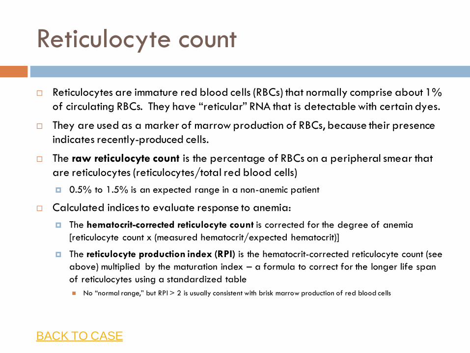
Reticulocytes are immature red blood cells (RBCs) that normally comprise about 1%
of circulating RBCs. They have “reticular” RNA that is detectable with certain dyes.
They are used as a marker of marrow production of RBCs, because their presence
indicates recently-produced cells.
The raw reticulocyte count is the percentage of RBCs on a peripheral smear that
are reticulocytes (reticulocytes/total red blood cells)
0.5% to 1.5% is an expected range in a non-anemic patient
Calculated indices to evaluate response to anemia:
The hematocrit-corrected reticulocyte count is corrected for the degree of anemia
[reticulocyte count x (measured hematocrit/expected hematocrit)]
The reticulocyte production index (RPI) is the hematocrit-corrected reticulocyte count (see
above) multiplied by the maturation index – a formula to correct for the longer life span
of reticulocytes using a standardized table
◼ No “normal range,” but RPI > 2 is usually consistent with brisk marrow production of red blood cells
BACK TO CASE

REFERENCE MATERIAL
Acute Chest Syndrome Case

Resources
1. Alhashimi D et al. Blood transfusions for treating acute chest syndrome in people with sickle cell disease. Cochrane Database Syst Rev. 2010 Jan 20;(1):CD007843.
2. Buchanan et al. Opioid selection in ACS. Pediatr Blood Cancer. 2005 Oct 15;45(5):716-24.
3. Castro et al. The acute chest syndrome in sickle cell disease: incidence and risk factors. The Cooperative Study of Sickle Cell Disease. Blood. 1994 Jul 15;84(2):643-9.
4. Sobota A. Corticosteroids in ACS. Am J Hematol. 2010 Jan;85(1):24-8
5. Talano J. et al. Delayed hemolytic transfusion reaction/hyperhemolysissyndrome in children with sickle cell disease. Pediatrics. 2003 Jun;111(6 Pt 1):e661-5.