Embed Size (px)
Citation preview
J Pediatr Rev. In Press(In Press):e7785.
Published online 2016 November 21.
doi: 10.17795/jpr-7785.
Review Article
Pediatric Nephrolithiasis: Trend, Evaluation and Management: A
Systematic Review
Emadoddin Moudi,1 Rahman Ghaffari,2 and Asaad Moradi3,*
1Department of Urology, Babol University of Medical Sciences, Babol, Iran2Department of Cardiac Surgery, Mazandaran Heart Center, Hazrat Fatima Hospital, Sari, Iran3Department of Urology, Firozgar Hospital, Iran University of Medical Sciences, Tehran, Iran
*Corresponding author: Asaad Moradi, Department of Urology, Firozgar Hospital, Iran University of Medical Sciences, Postal Code: 4716681451, Tehran, Iran. Tel: +98-2132256285,Fax: +98-2132254392, E-mail: [email protected]
Received 2016 July 05; Revised 2016 October 30; Accepted 2016 November 06.
Abstract
Context: Pediatric nephrolithiasis is a noticeable cause of morbidity among children. Although, nephrolithiasis is a common disease in adults,its incidence has had a rising trend in children. Here in, we reviewed the current state regarding evaluation and management of children withnephrolithiasis in the last decade.Evidence Acquisition: The current literature regarding incidence and trend, along with diagnostic evaluation and management of children agedless than 18 years old with nephrolithiasis was searched on MEDLINE and Google scholar from January 2005 to March 2016. The search terms included,“kidney stone OR nephrolithiasis OR urolithiasis and Pediatric OR children”. English language and human studies were included. Expert opinions,editorials and case reports were excluded. Consequently, the authors independently reviewed the abstracts and the papers, which matched theinclusion criteria.Results: From a total of 1050 studies identified through the database search, 71 articles were selected for the review. According to the results of thisreview, pediatric nephrolithiasis has an increasing rate worldwide. Recent studies in this regard indicated that the number of girls with nephrolithi-asis has increased. The change of life style, diet, obesity and metabolic syndrome and popular imaging study along with referral of the patients topediatric centers could be contributed to this trend.Conclusions: Evaluation of patients, particularly young children, includes metabolic assessment. Stone analysis and 24-urine collection analysisare very helpful for determining underlying diseases. Medical as well as surgical approaches are used to prevent kidney injuries.
Keywords: Nephrolithiasis, Children, Incidence, Management
1. Context
Over the past decades, the prevalence of pediatricnephrolithiasis has increased by 70% in adults and hasbecome the third top urinary tract disease, and the gen-der gap between males and females is narrowing in manycountries, as about 10% and 5% of males and females, re-spectively, complain of this condition during their life (1-3). Sas et al. (4) reported in their study that during the lastcouple of decades, the incidence of pediatric nephrolithi-asis has increased by approximately 6 to 10% annually andhas actually reached 50 per 100000 in adolescents.
This situation may cause some complications and hassubsequent health care and medical costs. Pearle et al.(5) estimated that more than two billion dollars is in-curred annually for nephrolithiasis in the United States.This condition causes further chronic conditions requir-ing health care services in pediatric medicine. Moreover,recent studies revealed that this disorder has a gender-related distribution in this context by more inclinationtoward nephrolithiasis. In addition, race and geographiclocation may play a role in nephrolithiasis development.
African countries have low prevalence and the Middle East,Pakistan and India have higher incidence of urolithiasis (6,7). This review article tried to provide appropriate infor-mation about pediatric nephrolithiasis in different aspectssuch as epidemiology, risk factors and etiology, clinical pre-sentation and management.
2. Evidence Acquisition
We reviewed the current literature regarding inci-dence and trend, diagnostic evaluation and managementof children with nephrolithiasis. For this purpose, wesearched MEDLINE and Google scholar from January 2005to March 2016. The search terms or key worlds included“kidney stone OR nephrolithiasis OR urolithiasis and Pedi-atric OR children”. The studies were included if their sub-jects were human aged less than 18 years and were writtenin English language. Also, expert opinions, editorials, andcase reports were excluded. Moreover, the bibliography ofrelevant review articles recognized in our search was readand appropriate articles were selected. Consequently, the
Copyright © 2016, Mazandaran University of Medical Sciences. This is an open-access article distributed under the terms of the Creative CommonsAttribution-NonCommercial 4.0 International License (http://creativecommons.org/licenses/by-nc/4.0/) which permits copy and redistribute the material just innoncommercial usages, provided the original work is properly cited.
Uncorr
ected
Proo
f
Moudi E et al.
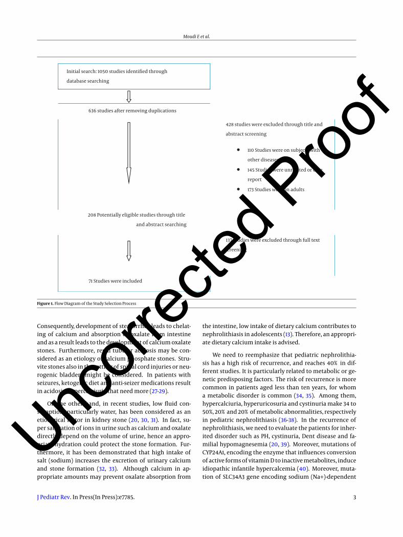
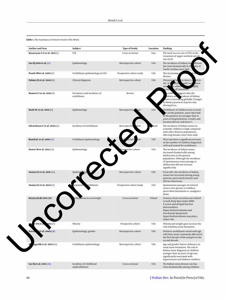
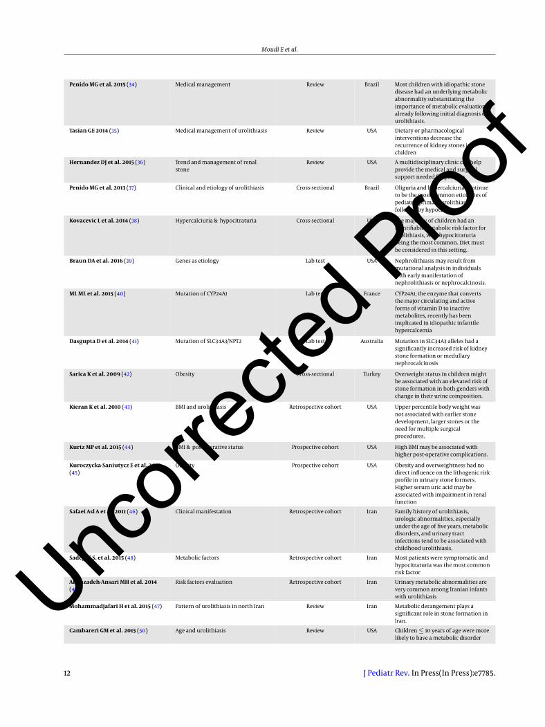
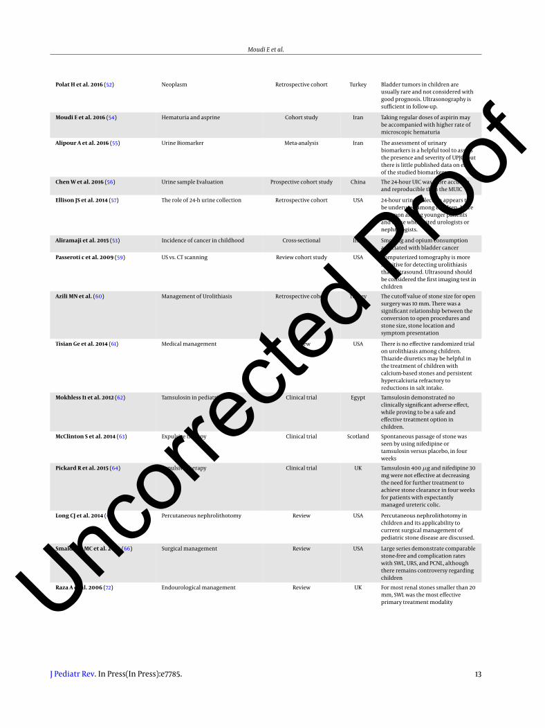
authors independently reviewed the abstracts and the pa-pers, which matched the inclusion criteria and selectedmore recent publications. The flow diagram of the studyselection process is shown in Figure 1. Reviewed articles aresummarized in Table 1.
3. Results
From the total of 1050 studies identified throughdatabase searching, 71 articles were selected for the review.Among 71 reviewed articles, 11 were cross-sectional, 23 ret-rospective cohort, 11 prospective studies, 20 review articles,three lab studies, and three were clinical trials. The quali-tative results derived from the reviewed articles related toincidence and trend, risk factors and causes of nephrolithi-asis, clinical presentation, evaluation of the patients in-cluding history taking and physical examination, urinaly-sis and other laboratory evaluations, imaging studies, ther-apeutic approaches such as acute management, surgicalmanagement, prevention of recurrence are summarizedin Table 1, and discussed here.
4. Discussion
4.1. Incidence and Trend
The real incidence of nephrolithiasis in children re-mains non-conclusive. This fact is relatively secondary tomultiple etiological factors and nonspecific clinical pre-sentations. Multiple studies over the past decade havedocumented an increased incidence of nephrolithiasis indifferent countries (8). In the United States, the resultof the national health and nutrition examination survey(NHANES), as a cross-sectional survey presented an increas-ing estimation of nephrolithiasis in adults of this country.Furthermore, recent studies revealed that the incidence ofhospital admissions increased from 1 in 7600 to 1 in 685admissions from 1999 to 2007 among children. The an-nual increase was 10.6% on average (9). These epidemi-ologic changes could be due to life style modifications,particularly in eastern countries, and increased diagnosisof nephrolithiasis by using ultrasonography evaluation inchildren with specific or nonspecific symptoms. Besides,adult urologists are now referring more patients to pedi-atric centers. Yasui et al. (10) from Japan displayed an in-cidence of 12.4/100000 to 17.7/100000 in female and malechildren aged 10 - 19 years old, respectively. Additionally,Edvardsson et al. (11) in Iceland reported an incidence of5.6/100000 in patients younger than 18 years old. Further-more, Routh et al. (12) in their study, by using nationalhealth information system database, declared an elevated
incidence of nephrolithiasis in children. In an epidemio-logic project in Minnesota from 1998 to 2008, the overallincidence of nephrolithiasis in adolescents was increasedfrom 7 to 14 per 100000 patients aged 12 to 17 years old(13). Tasian et al. (14)in a large population study indicatedthat between 1997 and 2012, the rate of nephrolithiasis in-creases annually by one percent. On the other hand, therehas been a change regarding the gender of patients. Pre-vious reports demonstrated that nephrolithiasis was usu-ally more common in boys than in girls, but recent studieshave shown a noticeable rising incidence of nephrolithi-asis in girls and some studies even substantiated thatnephrolithiasis are more common among girls (4, 9, 10,12, 15, 16). Usually girls in the second decade are moreprone to hospital administration for kidney stone (12).The possible relationship between nephrolithiasis, obesity,and metabolic syndrome was demonstrated in previous re-ports but it is not conclusive (16, 17). As mentioned before,geographic location may affect nephrolithiasis develop-ment. Former studies revealed that nephrolithiasis is morecommon among non-Hispanic white individuals as com-pared with non-Hispanic black individuals in adults andpediatric populations (18-20). In addition, age of childrenmay have a role in this setting as in some studies, it wasdemonstrated that the incidence of nephrolithiasis has in-creased from 0.6 per 100000 in children aged less thanthree years old to 34.9 per 100000 among adolescents agedless than 18. They indicated that the risk of nephrolithiasisin age group of 14 to 18 years is 10 times greater. The hospi-talization rate also follows the same pattern (9, 12, 21, 22). Ina Tasian study, the greatest change belonged to 15 to 19 yearolds, and the incidence of nephrolithiasis increased by 26%per five years. The incidence increased by 15% per five yearsamong females in general (14).
4.2. Risk Factors and Causes of Nephrolithiasis
In general, nephrolithiasis in many cases is associatedwith a metabolic abnormality including hypercalciuria,hyperoxaluria, hypocitraturia, cystinuria, and hyperurico-suria, in which hypercalciuria or hypocitraturia are morefrequent. However, we need to pay attention to some dis-orders such as cystic fibrosis (CF), spina bifida or inflamma-tory bowel disease, which may prone the patient to risk ofstone recurrence or raise the possibility of stone formationsuch as oxalosis or Primary Hyperoxaluria (PH). This disor-der has two types PH-1 and PH-2 (7, 23-25). Tubular disorderssuch as cystinuria increased the risk of stone formation,which accounted for 3% of renal stones (26). Some systemicdiseases such as CF and inflammatory bowel diseases (IBD)may be associated with nephrolithiasis. These diseasesusually affect the intestine and induce fat mal-absorption.
2 J Pediatr Rev. In Press(In Press):e7785.
Uncorr
ected
Proo
f
Moudi E et al.
Initial search: 1050 studies identified through
database searching
636 studies after removing duplications
428 studies were excluded through title and
abstract screening
110 Studies were on subjects with
other diseases
145 Studies were unrelated or case
report
173 Studies were on adults
208 Potentially eligible studies through title
and abstract searching
137 Studies were excluded through full text
screening
71 Studies were included
Figure 1. Flow Diagram of the Study Selection Process
Consequently, development of steatorrhea leads to chelat-ing of calcium and absorption of oxalate from intestineand as a result leads to the development of calcium oxalatestones. Furthermore, renal tubular acidosis may be con-sidered as an etiology of calcium phosphate stones. Stru-vite stones also in the setting of spinal cord injuries or neu-rogenic bladders might be considered. In patients withseizures, ketogenic diet and anti-seizer medications resultin acidosis, hypercalciuria that need more (27-29).
On the other hand, in recent studies, low fluid con-sumption, particularly water, has been considered as anetiological factor in kidney stone (20, 30, 31). In fact, su-per saturation of ions in urine such as calcium and oxalatedirectly depend on the volume of urine, hence an appro-priate hydration could protect the stone formation. Fur-thermore, it has been demonstrated that high intake ofsalt (sodium) increases the excretion of urinary calciumand stone formation (32, 33). Although calcium in ap-propriate amounts may prevent oxalate absorption from
the intestine, low intake of dietary calcium contributes tonephrolithiasis in adolescents (13). Therefore, an appropri-ate dietary calcium intake is advised.
We need to reemphasize that pediatric nephrolithia-sis has a high risk of recurrence, and reaches 40% in dif-ferent studies. It is particularly related to metabolic or ge-netic predisposing factors. The risk of recurrence is morecommon in patients aged less than ten years, for whoma metabolic disorder is common (34, 35). Among them,hypercalciuria, hyperuricosuria and cystinuria make 34 to50%, 20% and 20% of metabolic abnormalities, respectivelyin pediatric nephrolithiasis (36-38). In the recurrence ofnephrolithiasis, we need to evaluate the patients for inher-ited disorder such as PH, cystinuria, Dent disease and fa-milial hypomagnesemia (20, 39). Moreover, mutations ofCYP24A1, encoding the enzyme that influences conversionof active forms of vitamin D to inactive metabolites, induceidiopathic infantile hypercalcemia (40). Moreover, muta-tion of SLC34A3 gene encoding sodium (Na+)-dependent
J Pediatr Rev. In Press(In Press):e7785. 3
Uncorr
ected
Proo
f
Moudi E et al.
phosphate cotransporter 2c (NPT2c) causes hereditary hy-pophosphatemic rickets with hypercalciuria that conse-quently may increase the risk of nephrolithiasis (41). Thereare not enough studies regarding obesity and pediatricnephrolithiasis. Obesity probably leads to a decreasedurinary pH and increases the risk of uric acid stone. Ina study in Turkey on 96 patients, the author concludedthat overweight children with alteration of urine composi-tions, such as higher oxalate and uric acid and lower urinevolume, are at the risk of forming of stone (42). Kieranet al. (43) also revealed that 41% of their patients wereoverweight or obese. Kurtz et al. (44), in a population-based study on 2871 patients aged 2 months to 18 years re-vealed that 420 (14.6%) were overweight and 440 (15.3%)were obese. They concluded that BMI in children was as-sociated with increased overall complications. Generally,there is no consensus about obesity as a risk factor fornephrolithiasis and this association remains under ques-tion (15, 45).
In Iran there are limited reports regarding the etiol-ogy of nephrolithiasis, but to the best of our knowledge,the pattern of nephrolithiasis and its related factors followother reports. However, we need to focus on the fact thatthe Middle East region, including Iran, is a warm regionwith a rapidly changing life style to western nations. In thissetting, Safaei Asl et al. (46) in their report on 78 patientsindicated that 52.6% had a metabolic risk factor includ-ing normocalcemic hypercalciuria (21.7%), hyperuricosuria(11.5%), cystinuria (3.8%), and hyperoxaluria (5.1%). In addi-tion, Mohammadjafari in a 10-year prospective study on 217children, two months to 16 years old revealed that 35.1%,10% and 4.1% of patients had metabolic, infective and ob-structive problems. In this study, 110 children had no recog-nized etiology; furthermore hypercalciuria hyperoxaluriaand hypocitraturia were seen in 25.5%, 18.4% and 18.1% ofpatients (47). In addition, Sadeghi et al. (48) in a study inthe south east of Iran on 100 patients reported that hyper-calciuria and hypocitraturia were seen in 56% and 64% ofpatients, respectively, followed by anatomic malformation(32%) and Urinary Tract Infections (UTI) (9%). Alemzadeh-Ansari et al. (49) also in a study on 152 infants from south-west of Iran revealed that the most common metabolicrisk factors were hypercalciuria (79.6%) and hypocitraturia(40.9%). In this study hyperoxaluria and hypomagnesuriawere found in about 28% of patients.
4.3. Clinical Presentation
The presentations of nephrolithiasis in children arevariable. The classic adult presentation with flank painradiated to the groin is not common and almost age-dependent (33, 44, 50). Although, adolescents usuallypresent colicky pain similarity to adults, younger children
have nonspecific symptoms including abdominal pain,nausea and vomiting or irritability. Hematuria, micro-scopic or macroscopic can occur in up to 90% of cases.In addition, urinary infection (UTI) in children less thanfive years old could be the main presentation (36). Non-specific and localized pain is typical of infants and youngchildren with nephrolithiasis. Hematuria and uncharac-teristic abdominal pain are common in only 10% to 14%of all pediatric cases (33, 37, 51). However, nephrolithiasisis discovered incidentally, during imaging evaluation forother reasons, and remains asymptomatic for a long time(24, 33). In our region, the symptoms generally follow thesame pattern. Sadeghi et al. reported that the most com-mon clinical presentations were irritability (62%), flankpain (33%) and gross hematuria (4%), while Sepahi et al.(48, 51) reported that of one hundred patients, 54% pre-sented urinary tract infection (UTI), while in this study,fever, pain, irritability, dysuria and hematuria were themain clinical presentations. It seems that along with non-specific abdominal pain, UTI may be an important sign ofnephrolithiasis. Furthermore, in adolescents, hematuriamay need greater evaluation to eliminate other seriousproblems (52). Moudi et al. (52-54) also demonstrated thatlong-term usage of aspirin might induce occult hematuria.
Several reports indicated that most nephrolithiasis inchildren are calcium based, in which calcium oxalate is themost common type of calculi with about 50% followed bycalcium phosphate (10% - 20%). Mixed calcium oxalate andcalcium phosphate are present in more than 10% and mag-nesium ammonium phosphate (infection related) stone isreported in more than 17% of patients (33, 36, 37).
4.4. Evaluation
4.5. History and Physical Examination
Obtaining an appropriate medical history and physicalexamination is a milestone for an accurate diagnosis andidentification of those who are at risk and need treatment.In this regard, family history of urinary tract diseases maygive a clue for metabolic and inherited conditions. Thepresence of a family history was reported in 23% - 75% of pa-tients. In addition, focusing on diet by emphasizing on saltand fluid intake and having a special diet are important.Ketogenic diet increases the risk of uric acid stones. Fur-thermore, some medications, particularly corticosteroids,diuretics protease inhibitors, vitamins C and D or supple-ments are associated with nephrolithiasis; hence, there isa need to clarify their performance. Patients with historyof prematurity, IBD, CF, anatomical abnormalities or UTIare prone to nephrocalcinosis. Physical exam may be help-ful to identify the dysmorphic shape of the body, rickets orother abnormalities of systemic diseases and genetics as-sociated with nephrolithiasis (Table 2) (23).
4 J Pediatr Rev. In Press(In Press):e7785.
Uncorr
ected
Proo
f

Moudi E et al.
4.6. Urinalysis
Urinalysis is the main part of initial assessment ofnephrolithiasis. It provides good information of urinegravity as a marker of urine concentration and fluid in-take, urine pH, hematuria and pyuria. Microscopic analy-sis may give good information regarding crystalluria thathelp clarify the origin of the problem. Tubular dysfunctionis presented by glucosuria and proteinuria.
4.7. Other Laboratory Evaluations
To evaluate renal function, assessment of electrolytelevels including potassium, sodium, magnesium, calciumand phosphorus are highly recommended. In case of hy-perkalemia, hyperparathyroidism may be considered. Hy-pervitaminose D can lead to hypercalciuria.
The second step is to determine the underlying dis-eases such as metabolic disorder. It has been demonstratedthat in children, metabolic risk factors affect the majorityof cases therefore a complete workup in this setting is re-quired. On the other hand, the risk of recurrence is highand in line with metabolic abnormalities. Hence, stoneanalysis takes an important place and could canalize thephysician to identify the risk factors of nephrolithiasis. Infact, in children due to rare spontaneous passage or sur-gical stone retrieval, stone analysis is not common. Fur-thermore, Aliramaji et al. (53) in a meta-analysis revealedthat urine biomarkers may helpful in diagnosis of uretero-pelvic junction obstruction, as a predicting factor. As analternative, a 24-hour urine collection is a recommendedworkup. This test permits the screening of elements con-tributing to the stone-formation process. Due to variabil-ity of daily diet and fluid intake regimen, it is required tocollect a 24-hour urine sample for evaluating creatinine,sodium, calcium, phosphorus, magnesium, oxalate, cit-rate, cysteine, uric acid and urine volume. Some authorsrecommend two 24-hour urine collections. Nevertheless,recent studies indicated the low volume of using this test.In case that the collection is not possible, the urine spoturine samples may be helpful but cannot replace 24-hoururine collection (33, 53-56). Normal urinary values in spoturine samples and 24-hour samples are shown in Tables 3and 4.
4.8. Imaging
Imaging studies during or after the initial labora-tory evaluation were required in pediatric nephrolithia-sis. In adults, because of high sensitivity and specificityof computerized tomography (CT) scanning and regard-ing a good image of anatomy, taking a CT without contrastis usually recommended. In children, radiation of CT has
been concerned in many recommendations, therefore ul-trasonography is recommended by many pediatric asso-ciations as an initial imaging evaluation. Ultrasonogra-phy with low radiation and low cost simply identifies anechogenic mass and hydronephrosis as a consequence ofstone in the kidney. Studies on adults demonstrated thatthere is no significant difference between CT and US as ini-tial imaging evaluation. However, a pediatric study indi-cated that sensitivity and specificity of US compared withCT was 76% and 100%, respectively (33, 36, 57).
4.9. Management
4.9.1. Acute Management
The initial steps in therapeutic approach innephrolithiasis are control of acute renal colic pain,recognition of the necessity of emergency intervention,and trying to pass the stone if possible. The second step isto prevent new stone formation.
The pain is usually controlled by analgesics such asnon-steroidal anti-inflammatory drug (NSAID) or opium.Children may need hospitalization in case of initiation ofparenteral analgesic or hydration. Usually, renal stone inchildren is not obstructing and emergency decompressionis not indicated. However, the size and location of stonemust be considered seriously as a stone less than 4 mmhas a chance to pass spontaneously in all patients. In chil-dren, renal stones pass spontaneously in 50% of cases (36,58). In this situation, conservative management may beenough. Besides, medical expulsive therapy such as alpha-blocker (tamsulosin, terazosin) or calcium channel block-ers (nifedipine) may facilitate the passage of small stonesby relaxing ureteral smooth muscle. Actually, tamsulosinis wildly prescribed for pediatric nephrolithiasis (59-62).The stone passage may take more than one month and inthis period, the patients need appropriate care and follow-up.
4.9.2. Surgical Management
In case of obstructing or large stones, acute interven-tion and decompression may be required. In this context,urethral stent, nephrostomy, Shock Wave Lithotripsy (SWL)or even removal of stone via ureteroscopy is advised. Previ-ous studies demonstrated that in children, the stone witha size more than 5 mm rarely passes spontaneously. Dif-ferent studies revealed that about 22% of patients wouldneed surgical therapeutic approach during the next sixmonths after initiation of disease (36, 61, 63). The choice ofeach procedure depends on patient’s characteristic. WhileSWL and ureteroscopy are considered for small stones, per-cutaneous nephrolithotomy and pyelolithotomy are re-served for large calculi (> 1.5 cm) (36, 63). Generally, by
J Pediatr Rev. In Press(In Press):e7785. 5
Uncorr
ected
Proo
f
Moudi E et al.
the improvement of endourological techniques, interven-tions have shifted to minimally invasive procedures. Inthis setting, ureteroscopy is used commonly and is be-coming the therapeutic approach of choice in lower andalso upper tract stones (64-66). Tejwani et al. (67) ina large study to compare SWL and ureteroscopy demon-strated that patients, who benefit from ureteroscopy, hadless stone-related procedures in the next 12 months afterthe procedure, but a higher rate of readmissions withinsubsequent month after the initial intervention. Compar-ison of treatment modalities for surgical management ofpediatric nephrolithiasis is summarized in Table 5 (36).
4.10. Prevention of Recurrence
Prevention of nephrolithiasis recurrence in children isan effort to determine the potential risk factors of stoneformation and their modification. Previous reports indi-cated that children with known underling metabolic dis-order have a 50% risk of recurrence of nephrolithiasis,compared to 10% in patients without these risk factors (58,66, 68). Appropriate fluid intake, about 1.5 l/ m2/d, and lim-ited dietary intake should be advised to help decrease su-persaturation of urinary elements such as calcium, oxalateand uric acid. The fluid intake could be enough to pro-vide urine volume more than 750 mL/d in infants and morethan 1000 mL/d for children less than five years old, 1500mL/d for children 5 to 10 years, and 2000 mL/d in childrenmore than 10 years. Children need appropriate dietary cal-cium and calcium restriction is not recommended. Exces-sive protein intake can lead to hypercalciuria and hypoci-traturia; hence a sufficient protein intake is recommended(20, 30, 31, 36). Furthermore, avoidance of medication thatmay cause nephrolithiasis should be considered. Medica-tion may be a part of management of children with re-current nephrolithiasis, solitary kidney, or metabolic dis-orders. In this context, the result of stone analysis or a24-hour urine collection analysis plays an important role(33, 39, 69, 70). Thiazides and potassium-sparing diuret-ics are on top of the list. In a case with hypocitraturia,potassium and citrate can increase the urinary Ph and pre-vent uric acid crystallization. Patients with hypercalciuriabenefit from thiazide diuretics, which decreases urinarycalcium excretion. In this condition, a low sodium dietand adequate fluid intake must be encouraged. However,in presence of hypercalcemia, the prescription of thiazideis forbidden and combination of thiazide and potassium-sparing diuretics can increase calcium absorption (36, 66).The treatment of hyperoxaluria includes prevention of ox-alate absorption by reducing the intake of oxalate –richfoods and preventing crystallization of calcium oxalatein the urine. Usually, pyridoxine can be empirically pre-scribed. In fact, hyperoxaluria, secondary to malabsorp-
tion syndromes, requires treatment of underlying disease(15, 31, 33). Moreover, magnesium and pyrophosphate mayprevent calcium oxalate crystallization. In case of uric acidstones, the increase of excretion of uric acid by urinary al-kalization is a helpful modality. Allopurinol can be usedin patients with purine metabolism disorders. Further-more, alkalization can be used for the treatment of cys-teine stone. The aim is to reach a urinary pH of 7 - 7.5. Potas-sium citrate is recommended. Similarly, D-penicillamineand Tiopronin (Alpha-mercaptopropionylglycine) can beused in cystinuria. However, generally speaking, conserva-tive management remains the first line of management ofpatients with nephrolithiasis.
4.11. Conclusions
Pediatric nephrolithiasis, because of changing trendand underlying risk factors is different in adults. The aimof the treatment is to preserve the function of kidney andprevent the recurrence of disease. Metabolic disorders areimportant in this setting and work up is recommended.Despite advances in technology and medication, the riskfactor modifications remain the basis for the treatment ofnephrolithiasis.
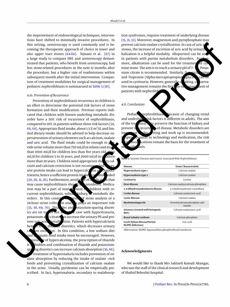
Table 2. Systemic Diseases and Genetic Associated With Nephrolithiasis
Disease Stone Characteristic
Hyperoxaluria type 1 Calcium oxalate
Hyperoxaluria type 2 Calcium oxalate
Cystinuria Cystine
Dent disease Calcium oxalate/calcium phosphate
2, 8-dihydroxyadeninuria disease 2, 8-hydroxyadenine crystalluria
Crohn disease Calcium oxalate/uric acid
Cystic fibrosis Calcium oxalate
Myelomeningocele Struvite/calcium phosphate andoxalate
Seizures (treated with ketogenicdiet)
Calcium oxalate/uric acid
Renal tubular acidosis Calcium phosphate
Lesch-Nyhan disease/PartialHGPRT deficiency
Uric acid
Abbreviation: HGPRT, hypoxanthine phosphoribosyl transferase.
Acknowledgments
We would like to thank Mrs Sakineh Kamali Ahangar,who was the staff of the clinical research and developmentof Shahid Beheshti hospital.
6 J Pediatr Rev. In Press(In Press):e7785.
Uncorr
ected
Proo
f
Moudi E et al.
Table 3. Normal Urinary Values in Spot Urine Samplesa
Urine Constituent Age Value, mg/mg Creatinine
Calcium
< 7 month < 0.86
7 - 18 month < 0.6
19 month - 6 year < 0.42
> 6 year < 0.2
Oxalate
< 6 month < 0.29
6 month - 2 year < 0.20
>2 - 5 year < 0.11
6 - 12 year < 0.06
Citrate< 5 year > 0.42
> 5 year > 0.25
Cystine All ages < 0.07
Uric acid All ages < 0.56 per GFRa
Abbreviation: GFR, glomerular filtration rate.aCalculated by multiplying the ratio of uric acid to creatinine (mg/mg) with serum creatinine (mg/dL) (71).
Table 4. Normal Urinary Values in 24-Hour Urine Collection
Urine Constituent Age Timed Collection Urine Constituent Age Timed Collection Urine Constituent Age Timed Collection
Calcium All ages < 4 mg/kg
Oxalate > 2 y < 0.45 mg/1.73 m2
CitrateAll ages, male > 365 mg/1.73 m2
All ages, female > 310 mg/1.73 m2
Cystine All ages < 60 mg/1.73 m2
Uric acid All ages < 815 mg/1.73 m2
Creatininea
3 - 5 y 12 - 20 mg
6 - 13 15 - 25
14 - 18 male 18 - 27
14 - 18 female 17 - 24aAdequacy of urine collection can be assessed by checking urinary creatinine. Excretion based on age and gender (55, 56).
Table 5. Comparison of Treatment Modalities for Surgical Management of Pediatric Nephrolithiasis
Treatment Indication Stone Stone Free Rate (%) Limitation Complications
Extracorporeal shock wavelithotripsy
Smaller stones, ureter andkidney
80 - 83 Lacks direct visualization ofstone, high bretreatment rate
Steinstrasse, perirenalhematoma
Ureteroscopy Smaller stones, ureter andkidney
85 - 88 Adult-sized instruments,surgical expertise needed
Infection, ureteral obstruction,ureteral stricture
Percutaneousnephrolithotomy
Larger stones, abnormalanatomy
70 - 97 Adult-sized instruments,surgical expertise needed,higher complication rate,inpatient procedure
Bleeding (8%-16% require bloodtransfusion), urine leak, urinaryobstruction, sepsis
Open pyelolithotomy Larger stones 79 - 98 Invasive, long postoperativeconvalescence
As for any open surgery
Minimally invasivepyelolithotomy
Larger stones No data Surgical expertise needed,learning curve
As for any minimally invasivesurgery
References
1. Kasaeeyan AA, Moudi E, Aliramaji A, Yousefnia P. The results of Trans-Ureteral Lithotripsy (TUL) for Upper Ureteral Stones: a single centerexperience. Caspian J Applied Sci Res. 2015;4(4):1–5.
2. Schissel BL, Johnson BK. Renal stones: evolving epidemiologyand management. Pediatr Emerg Care. 2011;27(7):676–81. doi:10.1097/PEC.0b013e3182228f10. [PubMed: 21730811].
3. Moudi E. A radiographic correlation between the presence of pulpstones and kidney stones Cas. J Applied Sci Res. 2015;4(3):1–7.
4. Sas DJ, Hulsey TC, Shatat IF, Orak JK. Increasing incidence of kid-ney stones in children evaluated in the emergency department. J
J Pediatr Rev. In Press(In Press):e7785. 7
Uncorr
ected
Proo
f
Moudi E et al.
Pediatr. 2010;157(1):132–7. doi: 10.1016/j.jpeds.2010.02.004. [PubMed:20362300].
5. Pearle MS, Calhoun EA, Curhan GC, Urologic Diseases of Amer-ica P. Urologic diseases in America project: urolithiasis. J Urol.2005;173(3):848–57. doi: 10.1097/01.ju.0000152082.14384.d7. [PubMed:15711292].
6. Palmer JS, Donaher ER, O’Riordan MA, Dell KM. Diagnosis of pedi-atric urolithiasis: role of ultrasound and computerized tomography.J Urol. 2005;174(4 Pt 1):1413–6. doi: 10.1097/01.ju.0000173133.79174.c8.[PubMed: 16145452].
7. Copelovitch L. Urolithiasis in children: medical approach. Pediatr ClinNorth Am. 2012;59(4):881–96. doi: 10.1016/j.pcl.2012.05.009. [PubMed:22857835].
8. Romero V, Akpinar H, Assimos DG. Kidney stones: a global picture ofprevalence, incidence, and associated risk factors. Rev Urol. 2010;12(2-3):86–96. [PubMed: 20811557].
9. Bush NC, Xu L, Brown BJ, Holzer MS, Gingrich A, Schuler B, et al. Hos-pitalizations for pediatric stone disease in United States, 2002-2007.J Urol. 2010;183(3):1151–6. doi: 10.1016/j.juro.2009.11.057. [PubMed:20096871].
10. Yasui T, Iguchi M, Suzuki S, Kohri K. Prevalence and epidemi-ological characteristics of urolithiasis in Japan: nationaltrends between 1965 and 2005. Urology. 2008;71(2):209–13. doi:10.1016/j.urology.2007.09.034. [PubMed: 18308085].
11. Edvardsson V, Elidottir H, Indridason OS, Palsson R. High incidence ofkidney stones in Icelandic children. Pediatr Nephrol. 2005;20(7):940–4. doi: 10.1007/s00467-005-1861-5. [PubMed: 15912382].
12. Routh JC, Graham DA, Nelson CP. Epidemiological trends in pedi-atric urolithiasis at United States freestanding pediatric hospitals.J Urol. 2010;184(3):1100–4. doi: 10.1016/j.juro.2010.05.018. [PubMed:20650479].
13. Dwyer ME, Krambeck AE, Bergstralh EJ, Milliner DS, Lieske JC, RuleAD. Temporal trends in incidence of kidney stones among children:a 25-year population based study. J Urol. 2012;188(1):247–52. doi:10.1016/j.juro.2012.03.021. [PubMed: 22595060].
14. Tasian GE, Ross ME, Song L, Sas DJ, Keren R, Denburg MR, et al. AnnualIncidence of Nephrolithiasis among Children and Adults in SouthCarolina from 1997 to 2012. Clin J Am Soc Nephrol. 2016;11(3):488–96.doi: 10.2215/CJN.07610715. [PubMed: 26769765].
15. Penido MG, Tavares MS. Pediatric primary urolithiasis: Symptoms,medical management and prevention strategies. World J Nephrol.2015;4(6):444–54. doi: 10.5527/wjn.v4.i4.444.
16. Kirejczyk JK, Korzeniecka-Kozerska A, Baran M, Porowska H, PorowskiT, Wasilewska A. Dyslipidaemia in overweight children and adoles-cents is associated with an increased risk of kidney stones. Acta Paedi-atr. 2015;104(9):407–13. doi: 10.1111/apa.13079. [PubMed: 26096629].
17. Taylor EN, Stampfer MJ, Curhan GC. Obesity, weight gain,and the risk of kidney stones. JAMA. 2005;293(4):455–62. doi:10.1001/jama.293.4.455. [PubMed: 15671430].
18. Novak TE, Lakshmanan Y, Trock BJ, Gearhart JP, Matlaga BR. Sexprevalence of pediatric kidney stone disease in the United States:an epidemiologic investigation. Urology. 2009;74(1):104–7. doi:10.1016/j.urology.2008.12.079. [PubMed: 19428065].
19. Matlaga BR, Schaeffer AJ, Novak TE, Trock BJ. Epidemiologic insightsinto pediatric kidney stone disease. Urol Res. 2010;38(6):453–7. doi:10.1007/s00240-010-0327-9. [PubMed: 20967433].
20. Sas DJ. An update on the changing epidemiology and metabolicrisk factors in pediatric kidney stone disease. Clin J Am Soc Nephrol.2011;6(8):2062–8. doi: 10.2215/CJN.11191210. [PubMed: 21737846].
21. Seitz C, Fajkovic H. Epidemiological gender-specific aspects inurolithiasis. World J Urol. 2013;31(5):1087–92. doi: 10.1007/s00345-013-1140-1. [PubMed: 23942884].
22. Prezioso D, Illiano E, Piccinocchi G, Cricelli C, Piccinocchi R, Saita A, etal. Urolithiasis in Italy: an epidemiological study. Arch Ital Urol Androl.2014;86(2):99–102. doi: 10.4081/aiua.2014.2.99. [PubMed: 25017588].
23. Valentini RP, Lakshmanan Y. Nephrolithiasis in children. Adv Chronic
Kidney Dis. 2011;18(5):370–5. doi: 10.1053/j.ackd.2011.07.002. [PubMed:21896379].
24. VanDervoort K, Wiesen J, Frank R, Vento S, Crosby V, Chandra M, et al.Urolithiasis in pediatric patients: a single center study of incidence,clinical presentation and outcome. J Urol. 2007;177(6):2300–5. doi:10.1016/j.juro.2007.02.002. [PubMed: 17509344].
25. Seyedzadeh A, Momtaz HE, Moradi MR, Moradi A. Pediatric cystinecalculi in west of Iran: a study of 22 cases. Urol J. 2006;3(3):134–7.[PubMed: 17559028].
26. Hoppe B, Kemper MJ. Diagnostic examination of the child withurolithiasis or nephrocalcinosis. Pediatr Nephrol. 2010;25(3):403–13.doi: 10.1007/s00467-008-1073-x. [PubMed: 19104842].
27. Matlaga BR, Kim SC, Watkins SL, Kuo RL, Munch LC, Lingeman JE.Changing composition of renal calculi in patients with neurogenicbladder. J Urol. 2006;175(5):1716–9. doi: 10.1016/S0022-5347(05)01015-3.[PubMed: 16600738].
28. Sampath A, Kossoff EH, Furth SL, Pyzik PL, Vining EP. Kidney stonesand the ketogenic diet: risk factors and prevention. J Child Neurol.2007;22(4):375–8. doi: 10.1177/0883073807301926. [PubMed: 17621514].
29. McNally MA, Pyzik PL, Rubenstein JE, Hamdy RF, Kossoff EH. Empiricuse of potassium citrate reduces kidney-stone incidence with the ke-togenic diet. Pediatrics. 2009;124(2):300–4. doi: 10.1542/peds.2009-0217. [PubMed: 19596731].
30. Saez-Torres C, Grases F, Rodrigo D, Garcia-Raja AM, Gomez C, Fron-tera G. Risk factors for urinary stones in healthy schoolchildrenwith and without a family history of nephrolithiasis. Pediatr Nephrol.2013;28(4):639–45. doi: 10.1007/s00467-012-2368-5.
31. Prezioso D, Strazzullo P, Lotti T, Bianchi G, Borghi L, Caione P, et al.Dietary treatment of urinary risk factors for renal stone formation. Areview of CLU Working Group. Arch Ital Urol Androl. 2015;87(2):105–20.doi: 10.4081/aiua.2015.2.105.
32. Cogswell ME, Yuan K, Gunn JP, Gillespie C, Sliwa S, Galuska DA, et al. Vi-tal signs: sodium intake among U.S. school-aged children - 2009-2010.MMWRMorbMortalWkly Rep. 2014;63(36):789–97. [PubMed: 25211544].
33. Sas DJ, Becton LJ, Tutman J, Lindsay LA, Wahlquist AH. Clinical, demo-graphic, and laboratory characteristics of children with nephrolithi-asis. Urolithiasis. 2016;44(3):241–6. doi: 10.1007/s00240-015-0827-8.[PubMed: 26467033].
34. Penido MG, Tavares Mde S. Pediatric primary urolithiasis: Symptoms,medical management and prevention strategies. World J Nephrol.2015;4(4):444–54. doi: 10.5527/wjn.v4.i4.444. [PubMed: 26380196].
35. Tasian GE, Copelovitch L. Evaluation and medical managementof kidney stones in children. J Urol. 2014;192(5):1329–36. doi:10.1016/j.juro.2014.04.108. [PubMed: 24960469].
36. Hernandez JD, Ellison JS, Lendvay TS. Current Trends, Evalua-tion, and Management of Pediatric Nephrolithiasis. JAMA Pediatr.2015;169(10):964–70. doi: 10.1001/jamapediatrics.2015.1419. [PubMed:26302045].
37. Penido MG, Srivastava T, Alon US. Pediatric primary urolithiasis:12-year experience at a Midwestern Children’s Hospital. J Urol.2013;189(4):1493–7. doi: 10.1016/j.juro.2012.11.107. [PubMed: 23201378].
38. Kovacevic L, Wolfe-Christensen C, Edwards L, Sadaps M, Laksh-manan Y. From hypercalciuria to hypocitraturia–a shifting trendin pediatric urolithiasis?. J Urol. 2012;188(4 Suppl):1623–7. doi:10.1016/j.juro.2012.02.2562. [PubMed: 22910255].
39. Braun DA, Lawson JA, Gee HY, Halbritter J, Shril S, Tan W, et al. Preva-lence of Monogenic Causes in Pediatric Patients with Nephrolithia-sis or Nephrocalcinosis. Clin J Am Soc Nephrol. 2016;11(4):664–72. doi:10.2215/CJN.07540715. [PubMed: 26787776].
40. Figueres ML, Linglart A, Bienaime F, Allain-Launay E, Roussey-KesslerG, Ryckewaert A, et al. Kidney function and influence of sunlightexposure in patients with impaired 24-hydroxylation of vitamin Ddue to CYP24A1 mutations. Am J Kidney Dis. 2015;65(1):122–6. doi:10.1053/j.ajkd.2014.06.037. [PubMed: 25446019].
41. Dasgupta D, Wee MJ, Reyes M, Li Y, Simm PJ, Sharma A, et al.
8 J Pediatr Rev. In Press(In Press):e7785.
Uncorr
ected
Proo
f
Moudi E et al.
Mutations in SLC34A3/NPT2c are associated with kidney stonesand nephrocalcinosis. J Am Soc Nephrol. 2014;25(10):2366–75. doi:10.1681/ASN.2013101085. [PubMed: 24700880].
42. Sarica K, Eryildirim B, Yencilek F, Kuyumcuoglu U. Role of over-weight status on stone-forming risk factors in children: a prospectivestudy. Urology. 2009;73(5):1003–7. doi: 10.1016/j.urology.2008.11.038.[PubMed: 19193407].
43. Kieran K, Giel DW, Morris BJ, Wan JY, Tidwell CD, Giem A, et al.Pediatric urolithiasis–does body mass index influence stone pre-sentation and treatment?. J Urol. 2010;184(4 Suppl):1810–5. doi:10.1016/j.juro.2010.03.111. [PubMed: 20728147].
44. Kurtz MP, McNamara ER, Schaeffer AJ, Logvinenko T, Nelson CP. As-sociation of BMI and pediatric urologic postoperative events: Re-sults from pediatric NSQIP. J Pediatr Urol. 2015;11(4):224 e1–6. doi:10.1016/j.jpurol.2015.04.014. [PubMed: 26139160].
45. Kuroczycka-Saniutycz E, Porowski T, Protas PT, Pszczołkowska M. ,Porowska H. , Kirejczyk J. K. , et al. Does obesity or hyperuricemia in-fluence lithogenic risk profile in children with urolithiasis?. PediatrNephrol. 2015;30(5):797–803. doi: 10.1007/s00467-014-2999-9.
46. Safaei Asl A, Maleknejad S. Pediatric urolithiasis: an experience of asingle center. Iran J Kidney Dis. 2011;5(5):309–13. [PubMed: 21876306].
47. Mohammadjafari H, Barzin M, Salehifar E, Khademi Kord M, AalaeeA, Mohammadjafari R. Etiologic and epidemiologic pattern ofurolithiasis in north iran;review of 10-year findings. Iran J Pediatr.2014;24(1):69–74. [PubMed: 25793048].
48. Sadeghi S, Fazeli F, Zarifi E. Clinical Characteristics and MetabolicAbnormalities in Pediatric Urolithiasis in South East Iran. J PediatrNephrol. 2015;3(4):149–54.
49. Alemzadeh-Ansari MH, Valavi E, Ahmadzadeh A. Predisposing factorsfor infantile urinary calculus in south-west of Iran. Iran J Kidney Dis.2014;8(1):53–7. [PubMed: 24413722].
50. Cambareri GM, Kovacevic L, Bayne AP, Giel D, Corbett S, Schurtz E, et al.National multi-institutional cooperative on urolithiasis in children:Age is a significant predictor of urine abnormalities. J Pediatr Urol.2015;11(4):218–23. doi: 10.1016/j.jpurol.2015.04.021.
51. Sepahi MA, Heidari A, Shajari A. Clinical manifestations and etiologyof renal stones in children less than 14 years age. Saudi J Kidney DisTranspl. 2010;21(1):181–4. [PubMed: 20061721].
52. Polat H, Utangac MM, Gulpinar MT, Cift A, Erdogdu IH, Turkcu G.Urothelial neoplasm of the bladder in childhood and adolescence:a rare disease. Int Braz J Urol. 2016;42(2):242–6. doi: 10.1590/S1677-5538.IBJU.2015.0200. [PubMed: 27256177].
53. Aliramaji A, Kaseean A, Yousefnia Pasha YR, Shafi H, Kamali S, SafariM, et al. Age distribution types of bladder cancers and their relation-ship with opium consumption and smoking. Caspian J Intern Med.2015;6(2):82–6. [PubMed: 26221505].
54. Moudi E, Hosseini S, Bijani A. Higher rate of microscopic hematuria inelderly patients who take regular doses of aspirin: Result from AHAPStudy. Caspian J Intern Med. 2016;7(4):278–82.
55. Alipour A, Mohammadjafar M, Rafiei A, Amjadi O. The Role of UrinaryBiomarker Levels in Assessing the Presence and Severity of Uretero-pelvic Junction Obstruction in Children: A Systematic Review andMeta-Analysis. J Pediatr Rev. 2016;4(2):7567. doi: 10.17795/jpr-7567.
56. Chen W, Wu Y, Lin L, Tan L, Shen J, Pearce EN, et al. 24-Hour Urine Sam-ples Are More Reproducible Than Spot Urine Samples for Evaluationof Iodine Status in School-Age Children. J Nutr. 2016;146(1):142–6. doi:10.3945/jn.115.215806. [PubMed: 26609173].
57. Ellison JS, Kaufman SR, Kraft KH, Wolf JS, Hollenbeck BK,Hollingsworth JM. Underuse of 24-hour urine collection among chil-dren with incident urinary stones: a quality-of-care concern?. Urol-ogy. 2014;84(2):457–61. doi: 10.1016/j.urology.2014.04.035. [PubMed:24958480].
58. Milose JC, Kaufman SR, Hollenbeck BK, Wolf JS, Hollingsworth JM.Prevalence of 24-hour urine collection in high risk stone formers.
J Urol. 2014;191(2):376–80. doi: 10.1016/j.juro.2013.08.080. [PubMed:24018242].
59. Passerotti C, Chow JS, Silva A, Schoettler CL, Rosoklija I, Perez-Rossello J, et al. Ultrasound versus computerized tomographyfor evaluating urolithiasis. J Urol. 2009;182(4 Suppl):1829–34. doi:10.1016/j.juro.2009.03.072. [PubMed: 19692054].
60. Azili MN, Ozturk F, Inozu M, Cayci FS, Acar B, Ozmert S, et al. Man-agement of stone disease in infants.Urolithiasis. 2015;43(6):513–9. doi:10.1007/s00240-015-0788-y. [PubMed: 26036325].
61. Tasian GE, Cost NG, Granberg CF, Pulido JE, Rivera M, Schwen Z, etal. Tamsulosin and spontaneous passage of ureteral stones in chil-dren: a multi-institutional cohort study. J Urol. 2014;192(2):506–11. doi:10.1016/j.juro.2014.01.091. [PubMed: 24518765].
62. Mokhless I, Zahran AR, Youssif M, Fahmy A. Tamsulosin for themanagement of distal ureteral stones in children: a prospec-tive randomized study. J Pediatr Urol. 2012;8(5):544–8. doi:10.1016/j.jpurol.2011.09.008. [PubMed: 22099477].
63. McClinton S, Starr K, Thomas R, McLennan G, McPherson G, McDonaldA. Use of drug therapy in the management of symptomatic uretericstones in hospitalized adults (SUSPEND), a multicentre, placebo-controlled, randomized trial of a calcium-channel blocker (nifedip-ine) and anα-blocker (tamsulosin): study protocol for a randomizedcontrolled trial. Trials. 2014;15:238. doi: 10.1186/1745-6215-15-238.
64. Pickard R, Starr K, MacLennan G, Lam T, Thomas R, Burr J, et al. Medi-cal expulsive therapy in adults with ureteric colic: a multicentre, ran-domised, placebo-controlled trial. Lancet. 2015;386(9991):341–9. doi:10.1016/S0140-6736(15)60933-3. [PubMed: 25998582].
65. Long CJ, Srinivasan AK. Percutaneous nephrolithotomy andureteroscopy in children: evolutions. Urol Clin North Am. 2015;42(1):1–17. doi: 10.1016/j.ucl.2014.09.002. [PubMed: 25455168].
66. Smaldone MC, Docimo SG, Ost MC. Contemporary surgical manage-ment of pediatric urolithiasis. Urol Clin North Am. 2010;37(2):253–67.doi: 10.1016/j.ucl.2010.03.006. [PubMed: 20569803].
67. Tejwani R, Wang HH, Wolf S, Wiener JS, Routh JC. Outcomesof Shock Wave Lithotripsy and Ureteroscopy for Treatmentof Pediatric Urolithiasis. J Urol. 2016;196(1):196–201. doi:10.1016/j.juro.2016.02.2975. [PubMed: 26997313].
68. Wang HH, Huang L, Routh JC, Nelson CP. Shock wave lithotripsy vsureteroscopy: variation in surgical management of kidney stonesat freestanding children’s hospitals. J Urol. 2012;187(4):1402–7. doi:10.1016/j.juro.2011.12.010. [PubMed: 22341283].
69. Schwarz RD, Dwyer NT. Pediatric kidney stones: long-term out-comes. Urology. 2006;67(4):812–6. doi: 10.1016/j.urology.2005.10.020.[PubMed: 16566973].
70. Elmaci AM, Ece A, Akin F. Pediatric urolithiasis: metabolic risk fac-tors and follow-up results in a Turkish region with endemic stonedisease. Urolithiasis. 2014;42(5):421–6. doi: 10.1007/s00240-014-0682-z. [PubMed: 25022263].
71. Murphy DP, Hsu CY. Estimating glomerular filtration rate: is it goodenough? And is it time to move on?. Curr Opin Nephrol Hyper-tens. 2013;22(3):310–5. doi: 10.1097/MNH.0b013e32836041e4. [PubMed:23571811].
72. Raza A, Turna B, Smith G, Moussa S, Tolley DA. Pediatric urolithia-sis: 15 years of local experience with minimally invasive endourolog-ical management of pediatric calculi. J Urol. 2005;174(2):682–5. doi:10.1097/01.ju.0000164749.32276.40. [PubMed: 16006948].
73. Akhavan-Sepahi M, Sharifian M, Mohkam M, Vafadar M, Hejazi S. Bio-chemical risk factors for stone formation in healthy school children.Acta Med Iran. 2012;50(12):814–8. [PubMed: 23456523].
74. Dauw CA, Alruwaily AF, Bierlein MJ, Asplin JR, Ghani KR, Wolf JS, etal. Provider variation in the quality of metabolic stone management.J Urol. 2015;193(3):885–90. doi: 10.1016/j.juro.2014.09.111. [PubMed:25286012].
J Pediatr Rev. In Press(In Press):e7785. 9
Uncorr
ected
Proo
f
Moudi E et al.
Table 1. The Summary of Articles Used in This Work
Author and Year Subject Type of Study Location Finding
Kasaeeyan A A et al. 2015 (1) TUL Cross-sectional Iran The total success rate of TUL in thetreatment of upper urethral calculiwas 93.6%
Sas DJ 2010 et al. (4) Epidemiology Retrospective cohort USA The incidence of kidney stone diseasehas risen dramatically in the state ofSouth Carolina since 1996
Pearle MSet al. 2005 (5) Urolithiasis epidemiology in USA Prospective cohort study USA The increasing trend of renal stonedisease
Palmer JS et al. 2005 (6) Clinical diagnosis Retrospective cohort USA Ultrasound failed to detect calculi in41% of the children with urolithiasissymptoms, whereas CT was highlyaccurate in all situations
Romero V et al. 2010 (8) Prevalence and incidence ofurolithiasis
Review USA The evidence suggests that theincidence and prevalence of kidneystones is increasing globally. Changesin dietary practices may be a keydriving force.
Bush NC et al. 2010 (9) Epidemiology Retrospective cohort USA Urolithiasis of children now accountfor 1 in 685 pediatric, more than halfof the patients are younger than 13years at hospitalization. Gender andlocation did not risk factor S.
Edvardsson V et al. 2005 (11) Incidence of urolithiasis Retrospective cohort Iceland The incidence of kidney stones inIcelandic children is high comparedwith other Western populations,affecting females more than male
Routh JC et al. 2010 (12) Urolithiasis Epidemiology Retrospective cohort USA There has been a significant increasein the number of children diagnosedwith and treated for urolithiasis
Dawer M et al. 2012 (13) Epidemiology Prospective cohort USA The incidence of kidney stonesincreased dramatically amongadolescents in the generalpopulation. Although the incidenceof spontaneous stone passage inadolescents did not increasesignificantly.
Tasian GE et al. 2016 (14) Epidemiology Retrospective cohort USA From 1997, the incidence of kidneystones has increased among youngpatients, particularly females andAfrican Americans.
Tasian GE et al. 2014 (15) Treatment of urolithiasis Prospective cohort study USA Spontaneous passages of ureteralstones were greater in childrenprescribed tamsulosin vs. analgesicsalone.
Kirejczyk JK 2015 (16) Dyslipidemia in overweight Cross-sectional Poland Urinary citrate excretion was relatedto both Body Mass Index (BMI)Z-scores and all lipid fractionabnormalities.Hypercholesterolaemia andlow-density lipoproteinhypercholesterolemia may play amajor role.
Taylor EN et al. 2005 (17) Obesity Prospective cohort USA Obesity and weight gain increase therisk of kidney stone formation
Novak TE et al. 2009 (18) Epidemiology; gender Retrospective cohort USA Pediatric urolithiasis varied with age,with boys more commonly affected inthe first decade of life and girls in thesecond decade.
Matlaga BR et al. 2010 (19) Urolithiasis epidemiology Retrospective cohort USA Age and gender had an influence onrenal stone formation. The risk ofkidney stone diagnosis in childrenyounger than six years of age wassignificantly associated withhypertension and diabetes mellitus.
Sas Dj et al. 2011 (20) Incidence of childhoodnephrolithiasis
Cross-sectional USA The kidney stone disease rate hasrisen dramatically among children
10 J Pediatr Rev. In Press(In Press):e7785.
Uncorr
ected
Proo
f

Moudi E et al.
Seitz C et al. 2013 (21) Role of gender Cohort Austria Female gender was associated withsignificantly different risk ratio ofstone development in differentvariables. Lifestyle must beconsidered.
Prezioso D et al. 2015 (22) Diet and urolithiasis Systematic review Italy Moderate dietary salt restriction andimplementation of potassium intakeare useful in limiting urinary calciumexcretion whereas dietary calciumrestriction is not recommended forchildren with nephrolithiasis
Schissel BL et al. 2011 (2) Urolithiasis epidemiology Review USA Changing epidemiology and etiologyof renal stone in children as well asproviding a framework forappropriate clinical evaluation
Penido 2015 (15) Pediatric primary urolithiasis Review Brazil Most children with idiopathic stonedisease have an underlying metabolicabnormality
Copelovitch L et al. 2012 (7) Medical treatment of urolithiasis Review USA Metabolic diseases are common andmetabolic evaluation is essential forall children with renal calculi
Yasui T et al. 2008 (10) Incidence of urolithiasis Retrospective cohort study Japan Increasing incidence of urolithiasis
Valentini et al. RP 2011 (23) Epidemiology of nephrolithiasis Review USA Aggressive fluid intake is themainstay of prevention for all formsof stone disease, but specific therapytargeted to the most likely underlyingmetabolic abnormality is often used.
VanDervoort K et al. 2007 (24) Clinical presentation Cross-sectional USA The primary diagnostic test could bea 24-hour urine collection. The mostcommon metabolic abnormality washypocitraturia, followed byhypercalciuria. Recurrence of stonesis common
Seyedzadeh A et al. 2006 (25) Cystine urolithiasis Cross-sectional Iran Metabolic workup of childhoodurolithiasis and appropriate medicalmanagement of its underlyingdisease.
Bernd Hoppe et al. 2013 (26) Diagnostic examination Review UK The stone is not the disease itself; it isonly one serious sign, so it needs acomplete evaluation includingphysical exam, history taking and labevaluations.
Matlaga BR et al. 2006 (27) Composition of calculi Retrospective cohort USA Patients with neurogenic bladderexperience urolithiasis secondary toinfection and metabolic diseases.
Sampath A et al. 2007 (28) Ketogenic diet and renal stone Retrospective cohort study USA There is no association between thesetwo items.
McNally MA et al. 2009 (29) Ketogenic diet Cross-sectional USA Oral potassium citrate is an effectivepreventive supplement againstkidney stones in children who receiveKD
Saez-Torres C et al. 2013 (30) Risk factors for urinary stones inchildren
Prospective cohort study Spain Family history and no adequate fluidintake are important for lithiasisformation
Prezioso D et al. 2015 (31) Diet and urolithiasis Review Italy Adequate fluid intake in children aswell as moderate dietary saltrestriction and implementation ofpotassium intake are useful inlimiting urinary calcium excretionwhereas dietary calcium restriction isnot recommended for children
Cogswell ME et al. (32) Sodium intake in urolithiasis Prospective cohort study USA Sodium intake among school-agedchildren is much higher thanrecommended, which requiresreduction based on New nationalnutrition standards
Sas DJ et al. 2016 (33) Clinic and laboratory characteristics Review USA Low urine volume and high level ofurinary calcium was associated withlithiasis although BMI was not. Moregirls presented their first stoneduring adolescence.
J Pediatr Rev. In Press(In Press):e7785. 11
Uncorr
ected
Proo
f
Moudi E et al.
Penido MG et al. 2015 (34) Medical management Review Brazil Most children with idiopathic stonedisease had an underlying metabolicabnormality substantiating theimportance of metabolic evaluationalready following initial diagnosis ofurolithiasis.
Tasian GE 2014 (35) Medical management of urolithiasis Review USA Dietary or pharmacologicalinterventions decrease therecurrence of kidney stones inchildren
Hernandez DJ et al. 2015 (36) Trend and management of renalstone
Review USA A multidisciplinary clinic can helpprovide the medical and surgicalsupport needed for patients
Penido MG et al. 2013 (37) Clinical and etiology of urolithiasis Cross-sectional Brazil Oliguria and hypercalciuria continueto be the most common etiologies ofpediatric primary urolithiasis,followed by hypocitraturia
Kovacevic L et al. 2014 (38) Hypercalciuria & hypocitraturia Cross-sectional USA The majority of children had anidentifiable metabolic risk factor forurolithiasis, with hypocitraturiabeing the most common. Diet mustbe considered in this setting.
Braun DA et al. 2016 (39) Genes as etiology Lab test USA Nephrolithiasis may result frommutational analysis in individualswith early manifestation ofnephrolithiasis or nephrocalcinosis.
ML ML et al. 2015 (40) Mutation of CYP24A1 Lab test France CYP24A1, the enzyme that convertsthe major circulating and activeforms of vitamin D to inactivemetabolites, recently has beenimplicated in idiopathic infantilehypercalcemia
Dasgupta D et al. 2014 (41) Mutation of SLC34A3/NPT2 Lab test Australia Mutation in SLC34A3 alleles had asignificantly increased risk of kidneystone formation or medullarynephrocalcinosis
Sarica K et al. 2009 (42) Obesity Cross-sectional Turkey Overweight status in children mightbe associated with an elevated risk ofstone formation in both genders withchange in their urine composition.
Kieran K et al. 2010 (43) BMI and urolithiasis Retrospective cohort USA Upper percentile body weight wasnot associated with earlier stonedevelopment, larger stones or theneed for multiple surgicalprocedures.
Kurtz MP et al. 2015 (44) BMI & postoperative status Prospective cohort USA High BMI may be associated withhigher post-operative complications.
Kuroczycka-Saniutycz E et al. 2015(45)
Obesity Prospective cohort USA Obesity and overweightness had nodirect influence on the lithogenic riskprofile in urinary stone formers.Higher serum uric acid may beassociated with impairment in renalfunction
Safaei Asl A et al. 2011 (46) Clinical manifestation Retrospective cohort Iran Family history of urolithiasis,urologic abnormalities, especiallyunder the age of five years, metabolicdisorders, and urinary tractinfections tend to be associated withchildhood urolithiasis.
Sadeghi S. et al. 2015 (48) Metabolic factors Retrospective cohort Iran Most patients were symptomatic andhypocitraturia was the most commonrisk factor
Alemzadeh-Ansari MH et al. 2014(49)
Risk factors evaluation Retrospective cohort Iran Urinary metabolic abnormalities arevery common among Iranian infantswith urolithiasis
Mohammadjafari H et al. 2015 (47) Pattern of urolithiasis in north Iran Review Iran Metabolic derangement plays asignificant role in stone formation inIran.
Cambareri GM et al. 2015 (50) Age and urolithiasis Review USA Children ≤ 10 years of age were morelikely to have a metabolic disorder
12 J Pediatr Rev. In Press(In Press):e7785.
Uncorr
ected
Proo
f
Moudi E et al.
Polat H et al. 2016 (52) Neoplasm Retrospective cohort Turkey Bladder tumors in children areusually rare and not considered withgood prognosis. Ultrasonography issufficient in follow-up.
Moudi E et al. 2016 (54) Hematuria and asprine Cohort study Iran Taking regular doses of aspirin maybe accompanied with higher rate ofmicroscopic hematuria
Alipour A et al. 2016 (55) Urine Biomarker Meta-analysis Iran The assessment of urinarybiomarkers is a helpful tool to assessthe presence and severity of UPJO, butthere is little published data on eachof the studied biomarkers.
Chen W et al. 2016 (56) Urine sample Evaluation Prospective cohort study China The 24-hour UIC was more accurateand reproducible than the MUIC
Ellison JS et al. 2014 (57) The role of 24-h urine collection Retrospective cohort USA 24-hour urine collection appears tobe underused among children. Morecommon among younger patientsand those who visited urologists ornephrologists.
Aliramaji et al. 2015 (53) Incidence of cancer in childhood Cross-sectional Iran Smoking and opium consumptionassociated with bladder cancer
Passeroti c et al. 2009 (59) US vs. CT scanning Review cohort study USA Computerized tomography is moresensitive for detecting urolithiasisthan ultrasound. Ultrasound shouldbe considered the first imaging test inchildren
Azili MN et al. (60) Management of Urolithiasis Retrospective cohort Turkey The cutoff value of stone size for opensurgery was 10 mm. There was asignificant relationship between theconversion to open procedures andstone size, stone location andsymptom presentation
Tisian Ge et al. 2014 (61) Medical management Review USA There is no effective randomized trialon urolithiasis among children.Thiazide diuretics may be helpful inthe treatment of children withcalcium-based stones and persistenthypercalciuria refractory toreductions in salt intake.
Mokhless I1 et al. 2012 (62) Tamsulosin in pediatrics Clinical trial Egypt Tamsulosin demonstrated noclinically significant adverse effect,while proving to be a safe andeffective treatment option inchildren.
McClinton S et al. 2014 (63) Expulsive therapy Clinical trial Scotland Spontaneous passage of stone wasseen by using nifedipine ortamsulosin versus placebo, in fourweeks
Pickard R et al. 2015 (64) Expulsive therapy Clinical trial UK Tamsulosin 400 µg and nifedipine 30mg were not effective at decreasingthe need for further treatment toachieve stone clearance in four weeksfor patients with expectantlymanaged ureteric colic.
Long CJ et al. 2014 (65) Percutaneous nephrolithotomy Review USA Percutaneous nephrolithotomy inchildren and its applicability tocurrent surgical management ofpediatric stone disease are discussed.
Smaldone MC et al. 2010 (66) Surgical management Review USA Large series demonstrate comparablestone-free and complication rateswith SWL, URS, and PCNL, althoughthere remains controversy regardingchildren
Raza A et al. 2006 (72) Endourological management Review UK For most renal stones smaller than 20mm, SWL was the most effectiveprimary treatment modality
J Pediatr Rev. In Press(In Press):e7785. 13
Uncorr
ected
Proo
f
Moudi E et al.
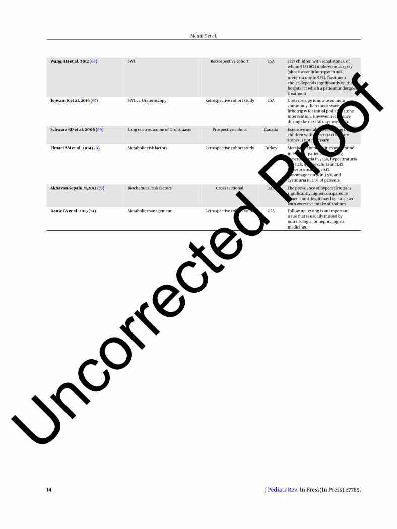
Wang HH et al. 2012 (68) SWL Retrospective cohort USA 3377 children with renal stones, ofwhom 538 (16%) underwent surgery(shock wave lithotripsy in 48%,ureteroscopy in 52%). Treatmentchoice depends significantly on thehospital at which a patient undergoestreatment
Tejwani R et al. 2016 (67) SWL vs. Ureteroscopy Retrospective cohort study USA Ureteroscopy is now used morecommonly than shock wavelithotripsy for initial pediatric stoneintervention. However, recurrenceduring the next 30 days was high.
Schwarz RD et al. 2006 (69) Long term outcome of Urolithiasis Prospective cohort Canada Extensive metabolic screening ofchildren with upper tract urinarystones is not necessary
Elmaci AM et al. 2014 (70) Metabolic risk factors Retrospective cohort study Turkey Metabolic abnormalities were foundin 79.2% of patients, includinghypercalciuria in 31.5%, hypocitraturiain 24.2%, hyperoxaluria in 11.4%,hyperuricosuria in 9.1%,hypomagnesuria in 3.9%, andcystinuria in 3.1% of patients.
Akhavan-Sepahi M,2012 (73) Biochemical risk factors Cross sectional Iran The prevalence of hypercalciuria issignificantly higher compared toother countries, it may be associatedwith excessive intake of sodium
Dauw CA et al. 2015 (74) Metabolic management Retrospective cohort study USA Follow up testing is an importantissue that is usually missed bynon-urologist or nephrologistsmedicines.
14 J Pediatr Rev. In Press(In Press):e7785.
Uncorr
ected
Proo
f