Embed Size (px)
Citation preview
22Pediatric Nerve Conduction Studies and EMG
Peter B. Kang
SummaryA variety of neuromuscular conditions affect children, ranging from severe, usually fatal disorders,
such as spinal muscular atrophy type I (Werdnig–Hoffman syndrome) to relatively mild problems, suchas benign congenital hypotonia. The evaluation of children in the EMG laboratory requires special carebecause of the discomfort of the tests. Moreover, other considerations, such as slower baseline nerveconduction velocities and conditions that generally do not present in adulthood, such as congenitalmyasthenic syndromes, can make the pediatric neurophysiological examination especially challenging.This chapter reviews both the common pediatric neuromuscular conditions and their assessment in theEMG laboratory.
Key Words: Electromyography; Guillain–Barré syndrome; hereditary neuropathies; pediatrics; rootavulsion; spinal muscular atrophy.
1. INTRODUCTION
EMG is a useful diagnostic tool in children suspected of having acquired or inheritedneuromuscular disease and complements the advances in molecular genetic testing duringthe past decade. The technical limitations of performing EMG in children require that theelectromyographer be selective in deciding how to approach the study. In evaluating forgeneralized processes, examination of one or two extremities is often adequate to narrowthe differential diagnosis or, in some cases, suggest a specific disorder. EMG may be usedto evaluate hypotonia in infants, assess the severity and localization of perinatal brachialplexus injuries, and distinguish between different possible causes of gait difficulties inolder children. In children, EMG may contribute to the diagnosis of many disorders,including spinal muscular atrophy (SMA), brachial plexus injury, hereditary polyneuropathy,acquired polyneuropathy, disorders of neuromuscular transmission, myopathy, musculardystrophy, and myotonic disorders.
2. TECHNICAL ISSUES
2.1. General Approach
Electrodiagnostic studies are highly dependent on technique in adults, but are even moredependent on technique in children. Infants and toddlers do not understand the purpose of thestudy, have a limited tolerance for discomfort, and often squirm and withdraw during testing.In selected cases, sedation or general anesthesia is required to obtain adequate data. Topicalanesthetic creams are used in some pediatric EMG laboratories, which can help reduce pain,
From: The Clinical Neurophysiology PrimerEdited by: A. S. Blum and S. B. Rutkove © Humana Press Inc., Totowa, NJ
369
but they require that the electromyographer either guess which sites are most likely candi-dates for needle EMG before the nerve conduction studies, or impose a stressful half-hourwait between the nerve conduction studies and needle EMG on the patient. Despite theselimitations, it is usually possible to perform a successful and informative study.
As in adults, a brief history and focused physical examination is critical in directing the testsperformed, especially because they may be terminated prematurely. If the child is old enoughto understand the procedure, it is important to explain it to the child as well as the parent. It issometimes possible to convince younger children that the EMG machine is a “tickling” machinefor nerve conduction studies—the power of suggestion being quite effective. Infants may bemore comfortable sitting in a parent’s lap on the examination table or in a chair. Pacifiers andtoys are often effective in calming infants and toddlers. Even school-age children and adoles-cents may feel more comfortable if a parent sits or stands near them during the study.
2.2. Nerve Conduction Studies
The temperature of the extremity during nerve conduction studies is as important in chil-dren as in adults. An excessively cold extremity will yield falsely slow conduction velocitiesand increased amplitudes. Upper extremity skin temperatures, measured at first dorsalinterosseous, should be at least 32°C. Lower extremity skin temperatures, measured at lowergastrocnemius, should be at least 30°C. Warming may be achieved with the use of towelsdampened with hot water and wrapped around the distal extremity for approx 5 min. Caremust be taken not to overheat the towels; a child’s skin is more delicate than that of an adult,and is more susceptible to scalding injury. It is also important to wring out the towel beforeapplying to the skin and to dry the skin after warming; a wet extremity will rapidly cool.Some electromyographers use disposable hot packs that produce heat via a chemical reaction.If used in children, these packs should be wrapped in towels to prevent burns, for they maybecome very hot. The temperature should be measured again after warming to confirm that itis in the acceptable range.
Surface active and reference recording electrodes may need to be trimmed or even cut inhalf for infants. Full-sized ground electrodes should be used whenever possible, however,because their placement is more flexible. Pediatric stimulators with small cathodes and anodesare useful for this age group. Standard adult distal distances cannot be used in infants andyoung children because of the small size of the extremities involved; thus, evaluation ofmotor distal latencies must take the patient’s age into account (see Table 1). The initial stim-ulation should always be less than 10 mA. It can be very reassuring to a child when the firststimulation is barely perceptible.
Questions of generalized processes, such as polyneuropathies and myopathies, are commonin pediatric EMG laboratories. Limited patient tolerance often makes it practical to performonly a motor and sensory study in an upper and lower extremity. In such cases, the electromyo-grapher may perform median motor and sensory studies in an upper extremity, and peronealmotor and a sural or medial plantar (depending on the age) sensory study in a lower extremity.In a child who is uncooperative or anxious, it may be best to perform the sensory studies first,because those require less stimulation intensity and are, thus, better tolerated.
The median motor study is performed as in an adult, with the E1 recording electrodeplaced over the abductor pollicis brevis and stimulation occurring at the wrist and cubital
370 Kang
Pediatric EMG 371
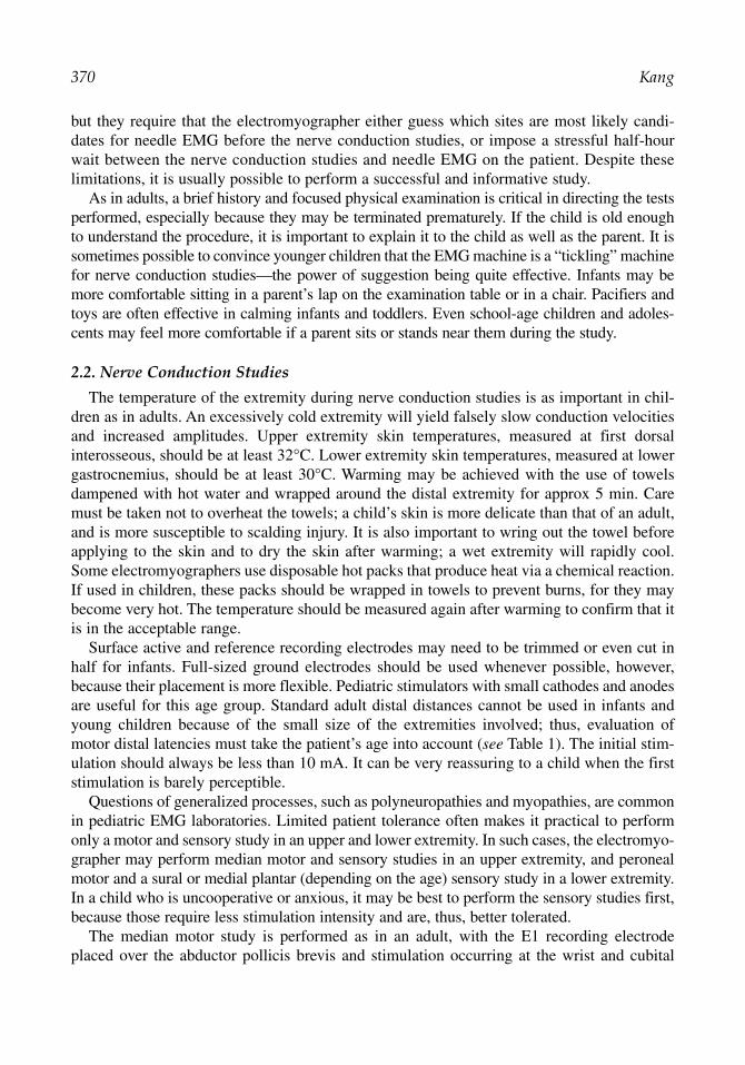
Table 1Motor Nerve Conduction Studies, Suggested Values
Age A (mV) CV (m/s) DL (ms)
Median nervePreterm (33–39 wk) — ≥18 —0–1 mo ≥2.5 ≥20 ≤3.51–6 mo ≥3.5 ≥25 ≤3.07–12 mo ≥2.5 ≥30 ≤3.01–2 yr ≥3.5 ≥35 ≤2.52–3 yr — ≥40 ≤2.53–4 yr — ≥45 ≤2.54+ yr — ≥50 ≤3.0Adult ≥4.0 ≥50 ≤4.0
Ulnar nervePreterm (33–39 wk) — ≥18 ≤3.30–1 mo ≥1.5 ≥20 ≤3.01–6 mo ≥2.5 ≥25 ≤3.37–12 mo ≥3.0 ≥35 ≤2.51–2 yr ≥2.5 ≥40 ≤2.52–3 yr — ≥40 —3–4 yr — ≥45 —4+ yr — ≥50 —Adult ≥6.0 ≥50 ≤3.3
Peroneal nerve0–1 mo ≥1.5 ≥20 ≤3.01–6 mo ≥1.5 ≥25 ≤2.57–12 mo ≥2.0 ≥30 ≤3.51–2 yr ≥1.5 ≥35 ≤3.52–3 yr — ≥40 —3–4 yr — ≥40 —4+ yr — ≥40 —Adult ≥2.0 ≥40 ≤6.5
Tibial nervePreterm (33–36 wk) — ≥14 —Preterm (37–39 wk) — ≥18 —0–1 mo — ≥20 ≤4.51–6 mo — ≥20 ≤4.07–12 mo — ≥25 ≤3.51–2 yr — ≥30 ≤3.02–3 yr — ≥35 ≤4.03–4 yr — ≥40 ≤4.04–6 yr — ≥40 ≤4.56+ yr — ≥40 ≤5.0Adult ≥4.0 ≥40 ≤5.8
A, suggested amplitude; CV, suggested conduction velocity; DL, suggested distallatency; —, data not available.
aAdapted from refs. 1, 2, 4–6, 23–25.
fossa. The peroneal motor study is performed as in an adult, with the E1 recording electrodeplaced over the extensor digitorum brevis and stimulation occurring at the ankle, fibular head,and popliteal fossa. Recording of tibialis anterior may be used when no response or a verysmall response is obtained from extensor digitorum brevis. One of the proximal stimulationsites may be omitted in children, unless, of course, there is a question of a peroneal neuropa-thy at the fibular head, such neuropathy being quite rare in children.
In infants, the most difficult aspect of the median antidromic sensory study involves theplacement of the active and reference electrodes. The active electrode may be placed on thesecond digit and the reference on the third, or the electrodes can be cut in half and both placedon the second or third digit. If this is unsuccessful, ring electrodes may be used instead. Thestimulation site is, as usual, at the wrist. In neonates and some infants, surface recordings ofsural nerve sensory action potentials are often obscured by artifact caused by high skinimpedance and short interelectrode distances. In this age group, the most technically reliablesensory study to obtain in the lower extremity is the orthodromic medial plantar study, recordingthe tibial nerve at the ankle and stimulating the medial plantar region of the sole.
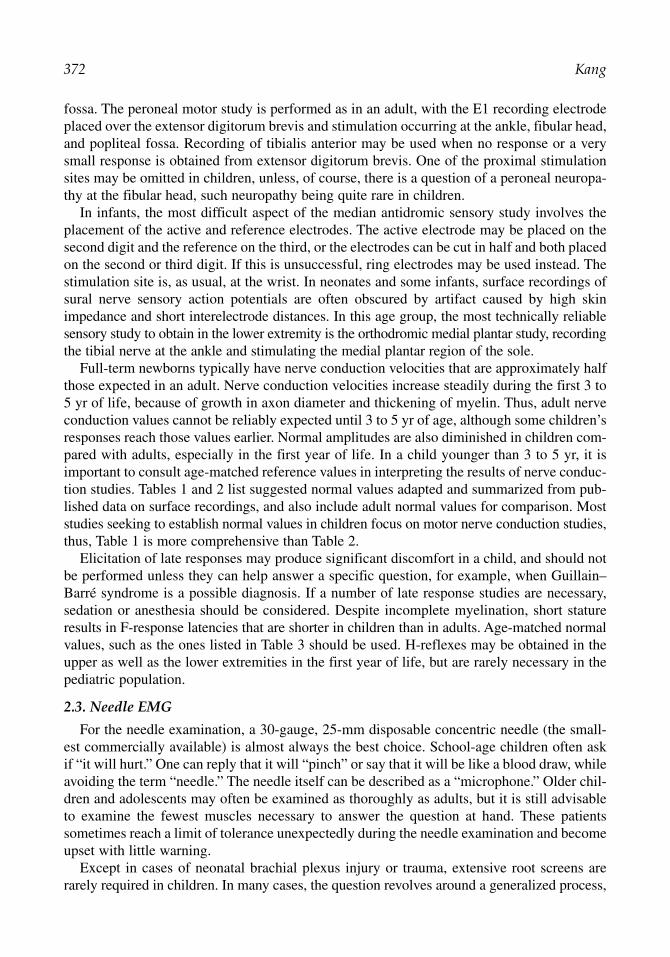
Full-term newborns typically have nerve conduction velocities that are approximately halfthose expected in an adult. Nerve conduction velocities increase steadily during the first 3 to5 yr of life, because of growth in axon diameter and thickening of myelin. Thus, adult nerveconduction values cannot be reliably expected until 3 to 5 yr of age, although some children’sresponses reach those values earlier. Normal amplitudes are also diminished in children com-pared with adults, especially in the first year of life. In a child younger than 3 to 5 yr, it isimportant to consult age-matched reference values in interpreting the results of nerve conduc-tion studies. Tables 1 and 2 list suggested normal values adapted and summarized from pub-lished data on surface recordings, and also include adult normal values for comparison. Moststudies seeking to establish normal values in children focus on motor nerve conduction studies,thus, Table 1 is more comprehensive than Table 2.
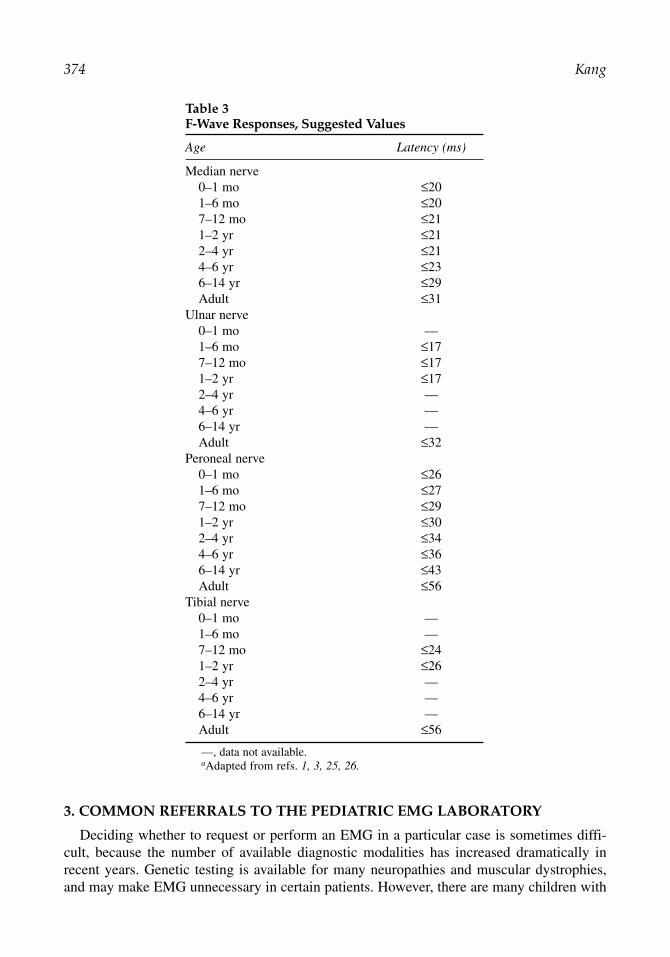
Elicitation of late responses may produce significant discomfort in a child, and should notbe performed unless they can help answer a specific question, for example, when Guillain–Barré syndrome is a possible diagnosis. If a number of late response studies are necessary,sedation or anesthesia should be considered. Despite incomplete myelination, short statureresults in F-response latencies that are shorter in children than in adults. Age-matched normalvalues, such as the ones listed in Table 3 should be used. H-reflexes may be obtained in theupper as well as the lower extremities in the first year of life, but are rarely necessary in thepediatric population.
2.3. Needle EMG
For the needle examination, a 30-gauge, 25-mm disposable concentric needle (the small-est commercially available) is almost always the best choice. School-age children often askif “it will hurt.” One can reply that it will “pinch” or say that it will be like a blood draw, whileavoiding the term “needle.” The needle itself can be described as a “microphone.” Older chil-dren and adolescents may often be examined as thoroughly as adults, but it is still advisableto examine the fewest muscles necessary to answer the question at hand. These patientssometimes reach a limit of tolerance unexpectedly during the needle examination and becomeupset with little warning.
Except in cases of neonatal brachial plexus injury or trauma, extensive root screens arerarely required in children. In many cases, the question revolves around a generalized process,
372 Kang
therefore, a limited needle examination often yields the necessary data. In infants and tod-dlers, poor cooperation often makes it impossible to evaluate both insertional activity and vol-untary activity in the same muscle. In the upper extremities, insertional activity is most easilyobserved in triceps and first dorsal interosseous, whereas motor unit activity is better assessedin biceps and flexor carpi radialis. In the lower extremities, medial gastrocnemius and vastuslateralis are better for insertional activity, whereas tibialis anterior and iliopsoas are preferredfor motor unit potential (MUP) analysis.
As in adults, if there is a question of a polyneuropathy, it is important to examine distalmuscles, although it is rarely necessary to study intrinsic foot muscles in young children.Children with possible myopathy should have several proximal muscles evaluated, but oneextremity (preferably lower) should be spared so that histological examination of a musclebiopsy performed at a later time will be free of potential artifacts from needle injury.
Pediatric EMG 373
Table 2Sensory Nerve Action Potentials, Suggested Values
Age A (mV) CV (m/s)
Median nerve (antidromic)0–1 mo ≥5 ≥251–6 mo ≥10 ≥357–12 mo ≥15 ≥301–2 yr ≥15 ≥402–3 yr — —3–4 yr — —4+ yr — —Adult ≥20 ≥50
Sural nerve (antidromic)0–1 mo ≥6 ≥201–6 mo ≥6 ≥207–12 mo ≥6 ≥251–2 yr ≥6 ≥302–3 yr ≥6 ≥353–4 yr ≥6 ≥404+ yr ≥6 ≥40Adult ≥6 ≥40
Medial plantar nerve orthodromic in children0–1 mo ≥10 —1–6 mo ≥15 ≥357–12 mo ≥15 ≥351–2 yr ≥15 ≥352–4 yr — —4+ yr — —Adult (antidromic) ≥2 ≥35
A, suggested amplitude; CV, suggested conduction velocity; —, data notavailable. Sural sensory responses may be obscured by subcutaneous tissuein neonates and infants; medial plantar studies may be more accurate in thisage group.
aAdapted from refs. 1–3, 6, 25.
3. COMMON REFERRALS TO THE PEDIATRIC EMG LABORATORY
Deciding whether to request or perform an EMG in a particular case is sometimes diffi-cult, because the number of available diagnostic modalities has increased dramatically inrecent years. Genetic testing is available for many neuropathies and muscular dystrophies,and may make EMG unnecessary in certain patients. However, there are many children with
374 Kang
Table 3F-Wave Responses, Suggested Values
Age Latency (ms)
Median nerve0–1 mo ≤201–6 mo ≤207–12 mo ≤211–2 yr ≤212–4 yr ≤214–6 yr ≤236–14 yr ≤29Adult ≤31
Ulnar nerve0–1 mo —1–6 mo ≤177–12 mo ≤171–2 yr ≤172–4 yr —4–6 yr —6–14 yr —Adult ≤32
Peroneal nerve0–1 mo ≤261–6 mo ≤277–12 mo ≤291–2 yr ≤302–4 yr ≤344–6 yr ≤366–14 yr ≤43Adult ≤56
Tibial nerve0–1 mo —1–6 mo —7–12 mo ≤241–2 yr ≤262–4 yr —4–6 yr —6–14 yr —Adult ≤56
—, data not available. aAdapted from refs. 1, 3, 25, 26.
acquired conditions and unusual presentations of inherited disorders in whom EMG is anessential test, and the volume of EMG referrals, in our experience, has remained steady.
The differential diagnosis of hypotonia in infancy and early childhood is vast, andincludes a number of peripheral nervous system processes. However, it is important toremember that the majority of cases of infant hypotonia arise from central causes. An exten-sive peripheral nervous system evaluation should be pursued only if supported by the his-tory and examination. Peripheral nervous system lesions may localize to the anterior horncell, peripheral nerve, neuromuscular junction, or muscle. If a congenital myopathy or mus-cular dystrophy lies in the differential diagnosis, it is often useful to obtain a set of muscleenzymes (creatine kinase, aldolase, alanine aminotransferase, aspartate aminotransferase,and lactate dehydrogenase) before requesting or performing an EMG or muscle biopsy. It isimportant to remember that muscle enzymes may be artifactually elevated immediately afterneedle EMG is performed. A significant elevation of one or more enzymes (the elevatedenzymes should include either creatine kinase or aldolase, because these are the ones thatare most specific to muscle) will justify a muscle biopsy as the next step. Mild abnormali-ties or normal levels of these enzymes do not always exclude a congenital myopathy; inthose cases, an EMG may be helpful in directing further studies. The precise sensitivity ofEMG in the evaluation of the hypotonic infant is unclear. One study reported an 80% over-all sensitivity for the correct diagnosis, whereas another recorded a 65% sensitivity for SMAand 10% for myopathy.
A neonate with a birth injury to the brachial plexus may present with a weak or flaccidupper extremity. It is rare for alternative diagnoses to be plausible, therefore, the question forthe electromyographer usually revolves around issues of severity and prognosis. Except in themost severe cases, an EMG is most helpful after the first few months of life, when the pres-ence or absence of chronic reinnervation and axonal continuity may best be assessed.
Toddlers and older children may present with delayed motor milestones or difficulty walk-ing. As in the evaluation of hypotonia, the differential diagnosis is broad, and may includealmost any segment of the peripheral nervous system. Proximal weakness, often detected bythe presence of Gowers sign, Trendelenburg gait, or both, typically suggests the possibility ofDuchenne, Becker, or limb–girdle muscular dystrophy. If such a patient has significantly ele-vated muscle enzyme levels, either molecular genetic testing or muscle biopsy will yield adiagnosis in most cases. When such an evaluation is unrevealing, an EMG may be useful inassessing the child for a neuropathic process, such as SMA type III (also known asKugelberg–Welander disease). SMA type III may resemble a muscular dystrophy with aproximal distribution of weakness and a mild elevation of the creatine kinase level.
Abrupt onset gait difficulty may be caused by acute inflammatory demyelinating polyneu-ropathy (AIDP; Guillain–Barré syndrome), an inflammatory myopathy, tick paralysis, spinalcord conditions (mass lesion or transverse myelitis), or central causes. Although poliomyelitis,fortunately, has almost been eradicated in much of the world, West Nile virus has also beenfound to produce a similar syndrome of acute motor paralysis. A detailed skin and scalp exam-ination is important in such acutely weak children, because tick paralysis is readily cured byremoving the tick. Because ataxia is often observed in AIDP, it may be difficult in some casesto distinguish between that condition and other causes of ataxia, such as acute cerebellitis. Ifa particular diagnosis cannot be supported by other findings, such as areflexia, cytoalbumino-logical dissociation, rash, elevated creatine kinase level, or nerve root enhancement on MRIimaging of the spine, EMG may be helpful in distinguishing between the possibilities.
Pediatric EMG 375
Chronic gait difficulty associated with areflexia, sensory loss, pes cavus, distal weakness,or a combination of these suggests the presence of an isolated inherited polyneuropathy, suchas Charcot–Marie–Tooth (CMT) disease, also called hereditary motor sensory neuropathy. Insome instances, a demyelinating polyneuropathy may be associated with a more generalizedneurodegenerative disorder, such as metachromatic leukodystrophy, especially if there is anassociated cognitive decline.
4. IMPORTANT FINDINGS IN THE PEDIATRIC EMG LABORATORY
4.1. Motor Neuron Disease
Because of the development of the polio vaccine, SMA has become the dominant anteriorhorn cell disease affecting children in developed countries. The onset of the most commonvariant, type I (Werdnig–Hoffman disease), is typically in the first 6 mo of life. The infantspresent with hypotonia, weakness, and delayed motor milestones. Mothers may reportdecreased fetal movements. Examination is notable for areflexia or hyporeflexia, hypotonia,weakness, and preserved extraocular movements. Tongue fasciculations may sometimes beobserved, but are not a reliable finding, especially in young infants, and the lack of tonguefasciculations should never be used to exclude SMA. These children never sit independentlyand never walk. The majority do not survive the second year of life.
The onset of SMA type II is typically from 6 to 18 mo, with some cases beginning as earlyas the neonatal period. These children often present with motor delays. They eventually sitindependently but never walk, and survive only to adolescence or early adulthood. The gaitdifficulties that are often the first signs of SMA type III usually begin after 18 mo. Thesepatients walk independently for most of their lives. Life expectancy is normal in most casesof SMA type III, although some patients require wheelchair assistance as adults.
SMA is caused by a deletion in the survival motor neuron (SMN) gene on chromosome 5q13.Until this discovery, EMG and muscle biopsy were the principal means of confirming the diag-nosis during life. Genetic testing for deletions in SMN is now readily available, but EMG is stillhelpful in many cases, especially if the presentation is mild or otherwise unusual, or if an infantdevelops respiratory failure before the results of genetic testing become available.
As in adult motor neuron disease, it is critical to document normal sensory responses onnerve conduction studies; if these are abnormal, the diagnosis should remain in serious doubt.Compound motor action potential (CMAP) amplitudes are often diminished in SMA type I,but may be minimally reduced or normal in SMA types II and III. During the needle exami-nation, at least three limbs should be studied. Cranial muscles may be substituted for one limb,especially if tongue fasciculations are observed. Examination of paraspinal muscles is not gen-erally helpful and should be avoided. Both ongoing denervation and chronic reinnervation maybe observed. In adults, chronic reinnervation should be demonstrated in most muscles of atleast three limbs to meet criteria for motor neuron disease, but in children it is not always pos-sible to do this extensive a needle examination, and any findings should be confirmed bygenetic testing. Fasciculation potentials are observed more rarely in SMA as compared withamyotrophic lateral sclerosis, and the MUP abnormalities are generally symmetrical in SMA.
4.2. Sensory Neuropathy and Neuronopathy
It is rare to find an isolated sensory neuropathy or sensory neuronopathy in childhood. Ininfants, the sural and superficial peroneal sensory responses are often difficult to record.Thus, in children of that age, the medial plantar sensory responses and upper extremity
376 Kang
sensory responses must be checked before concluding that the patient has a true loss of sen-sory responses. Age-specific normal values must be used in children younger than 3 to 5 yr.
If a true sensory neuropathy or sensory neuronopathy is present, the most likely causes areFriedreich’s ataxia and the hereditary sensory and autonomic neuropathies. Because genetictesting for the triplet-repeat expansion in Friedreich’s ataxia is available, patients with thetypical clinical presentation are often diagnosed without the assistance of an EMG.Associated findings include ataxia, absent lower extremity reflexes, extensor plantarresponses, thinning of the spinal cord on spine MRI, normal brain MRI early in the course,onset before 20 yr, pes cavus, dysarthria, distal sensory loss, optic atrophy, diabetes, andcardiomyopathy.
4.3. Brachial Plexus
Brachial plexus injuries in children most commonly occur at birth, as a result of shoulderdystocia (difficulty in extracting the shoulder from the birth canal). Risk factors includedifficult delivery and large birthweight. As might be expected from the mechanism of injury,upper plexus injuries (Erb’s palsy) predominate. Lower brachial plexus lesions (Klumpke’spalsy) and total plexus injuries (Erb-Klumpke paralysis) are less common. The neonate willhave flaccid weakness of one upper extremity.
The history and examination are, in most cases, diagnostic of perinatal brachial plexusinjury. A careful examination will suggest the root levels involved. In upper plexus lesions, theposture known as the “waiter’s tip” is usually found, consisting of arm adduction, elbow exten-sion, forearm pronation, and wrist flexion. In lower plexus lesions, wrist and finger flexors andintrinsic hand muscles are weak. A Horner’s syndrome is typically present. A combination ofthese findings or complete upper extremity paralysis suggests total plexus involvement.
Initial management includes gentle immobilization of the affected extremity against theabdomen and range of motion exercises, preferably under the direction of a trained physicaltherapist. In almost all cases, observation and conservative management are recommendeduntil 3 to 6 mo of age. If there is no improvement at that point, evaluation for possible micro-surgical repair may be indicated, including EMG and MRI.
In this setting, the purpose of performing an EMG study is to determine the localizationand severity of the injury. The severity is largely dependent on the localization, becausediscontinuity of the axon, most commonly caused by avulsion of the nerve root, is associatedwith a much poorer prognosis than “stretching” of the brachial plexus structures.
If brachial plexus injury and root avulsion were mutually exclusive conditions, the studywould be relatively simple, because, in the former condition, sensory responses would beabsent, whereas, in the latter, they would be preserved. However, in many cases, the twolesions coexist, making abnormal sensory nerve action potentials (SNAPs) less reassuringthan they would be under other circumstances.
The needle study is the key to determining prognosis. Because examination of the cervi-cal paraspinal muscle is impractical in most infants, the presence of MUPs on needle EMGat various root levels confirms continuity of the axon and, thus, the likelihood of at least par-tial recovery of function. This assessment can only be made several months after birth, afterreinnervation has begun to occur, because a premature examination may yield an inaccuratelygrim prognosis.
Precise localization among the root, trunk, division, cord, and nerve structures requiresthe evaluation of a large number of muscles, but unless the infant is anesthetized, this is
Pediatric EMG 377
not usually possible. Thus, because of the high likelihood that the study will be terminatedprematurely, the choice of muscles is critical. If an infant has a typical upper plexus pres-entation, the deltoid, biceps, and triceps muscles should be studied first. Comparing del-toid and biceps can help distinguish between root and plexus involvement; both muscleswill be abnormal when a C5–C6 root lesion is present, but only one may be abnormal inlesions of the brachial plexus. Unfortunately, examination of the rhomboids and serratusanterior is rarely practical in infants, limiting the precision of localization within thebrachial plexus.
When a Horner’s syndrome or other signs of lower plexus involvement are present, adistal muscle, such as the first dorsal interosseous, should be among the first musclesexamined. Depending on the clinical picture and the tolerance of the patient, other mus-cles that may be studied include the supraspinatus, extensor digitorum communis, flexordigitorum superficialis, and abductor digiti quinti. If the triceps has been successfullyexamined, needle EMG of extensor digitorum communis may not be necessary. As formedian-innervated nerve muscles, examination of the flexor digitorum superficialis isusually more informative than that of abductor pollicis brevis, because the infant is morelikely to activate the former.
Muscle activation in infants is often fleeting. For flexor muscles, tickling or gentle pinch-ing of the fingers and hand may stimulate withdrawal when the needle itself fails to inducethis response. The infant is less likely to activate extensor muscles under these circum-stances. It is sometimes difficult to decide how long to examine a muscle, especially if thechild is crying and it seems that the time remaining in the study is rapidly diminishing.When no MUPs are present, tickling or pinching to activate muscle contractions should beperformed at least once for each major level (C5–C6, C7, and C8–T1), and preferably inmultiple muscles. Because a decision whether or not to perform surgery may hinge partlyon the study results, any examination that is too truncated to be reliable should be repeatedunder anesthesia.
4.4. Polyneuropathies
AIDP (Guillain–Barré syndrome) may occur at almost any age in childhood, from 1 moto adulthood. The classic clinical presentation is the same as in adults, with an acute onsetof ascending paralysis, areflexia, and cytoalbuminological dissociation in the cerebrospinalfluid, with protein values often ranging from 80 to 200. An EMG may be very helpful inconfirming or casting doubt on the diagnosis. The use of spine MRI with gadoliniumenhancement to detect nerve root enhancement is also becoming more widespread. The ini-tial symptoms may be atypical in some patients, consisting primarily of numbness, pares-thesias, or pain.
Nerve conduction studies are most useful in the evaluation of possible AIDP. In the clas-sic demyelinating form of AIDP, the diagnostic findings are temporal dispersion and con-duction block in the setting of slow conduction velocities and prolonged distal latencies. Thecriteria for these findings are generally the same in children as in adults: at least 15% pro-longation of CMAP duration on proximal vs distal stimulation for temporal dispersion, atleast 50% diminution of CMAP amplitude on proximal vs distal stimulation for partial con-duction block, and an absent CMAP on proximal stimulation for complete conduction block.The presence of temporal dispersion excludes partial or complete conduction block. Anothercriterion for nonuniform slowing suggesting an acquired demyelinating process is a significant
378 Kang
disparity in nerve conduction velocities between different nerves, usually at least 10 m/s dif-ference within the upper or lower extremities, and at least 15 m/s difference between anupper and a lower extremity. The usual adult standards stipulating the number of nervesinvolved may not always be practical to apply in children, because the study may be termi-nated early. Evidence for axonal loss may be present, but unless severe, this is usually a sec-ondary phenomenon.
In some early or mild cases, temporal dispersion, conduction block, and conduction velo-city slowing may not be present. Prolonged or absent F-responses and H-reflexes may beearly signs of AIDP, as may be reduced recruitment on an otherwise normal needle EMGstudy. If tolerated, F-waves should be performed on several nerves in multiple extremities. H-reflexes are extremely uncomfortable, especially for a child, and, thus, should be avoided.These early electrophysiological signs of AIDP may be subtle and nonspecific; if the diagnosiscannot clearly be supported by other data, a repeat study later in the course may provide moredefinite evidence.
The less common, primarily axonal form of Guillain–Barré, known as acute motor axonalneuropathy, was first recognized in China, and most commonly occurs there, but has alsobeen described in the United States, Europe, and other parts of Asia. In adults, the prognosisis clearly worse in axonal variants. Children with acute motor axonal neuropathy have aworse course acutely (many require assisted ventilation) and recover more slowly than thosewith AIDP, but the long-term prognosis is generally good if they survive the acute phase.Conduction velocities are normal or mildly slow. There is evidence for axonal loss both onnerve conduction studies and needle EMG.
A generalized sensorimotor polyneuropathy in the setting of long-standing symptoms maybe caused by chronic inflammatory demyelinating polyradiculoneuropathy (CIDP) or an inher-ited polyneuropathy, such as CMT (also known as hereditary sensory and motor neuropathy),metachromatic leukodystrophy, Cockayne syndrome, Pelizaeus-Merzbacher disease, oradrenomyeloneuropathy. An EMG can determine whether a polyneuropathy is present, andmay, in some cases, help distinguish between acquired and genetic causes. Traditionally,signs of nonuniform slowing of nerve conduction, such as temporal dispersion, conductionblock, and significant disparities in nerve conduction velocities between different nerves haveindicated the presence of an acquired polyneuropathy, whereas uniform slowing has beenassociated with an inherited polyneuropathy. However, numerous reports have demonstratednonuniform slowing in some instances of inherited polyneuropathies, including metachro-matic leukodystrophy, hereditary neuropathy with liability to pressure palsies, the X-linkedvariant of CMT (CMTX), and adrenomyeloneuropathy.
Children with CIDP generally have manifestations of motor delay or impairment. Theweakness may show a distal predominance or may be diffusely distributed. Other findingsinclude sensory loss in some patients, normal cranial nerve function, and hyporeflexia or are-flexia. CIDP may develop at any age, and has even been reported to be present at birth.
The electrophysiological findings in CIDP are similar to those in fully developed AIDP.Nerve conduction velocities may be as slow as 2.3 m/s. Temporal dispersion and conductionblock are classic findings. Needle EMG reveals evidence for secondary axonal loss. Thechronicity or relapsing nature of the course distinguishes CIDP from AIDP. It is sometimes dif-ficult to confirm CIDP based on electrophysiology alone, especially in light of the exceptionsto the uniform vs nonuniform slowing rule that have recently been described. Palpable enlargednerves and pes cavus may be present in CIDP as well as in CMT. Elevated protein in cerebrospinal fluid
Pediatric EMG 379
is found in CIDP, but also in inherited disorders such as metachromatic leukodystrophy andCMT. The absence of cognitive involvement, MRI abnormalities, and long-tract signs is sug-gestive of CIDP rather than an inherited leukodystrophy, but nerve biopsy is still required to dis-pel remaining doubts, especially if there is a question of CMT.
CMT type I is the most common inherited polyneuropathy. It is characterized by a predom-inantly demyelinating pattern on EMG, with nerve conduction velocities typically in theteens. The pattern of inheritance is autosomal dominant. Initial symptoms may include distallower extremity weakness and wasting, with slow progression over several decades.Hyporeflexia, palpable enlarged nerves, pes cavus, distal weakness and wasting, and distalsensory loss are classic physical findings.
Primary axonal loss is found in CMT type II. Nerve conduction velocities are normal ormildly slow. Onset is typically in early adulthood, and the course is mild. Inheritance is usu-ally autosomal dominant, although autosomal recessive kindreds with moderately slow nerveconduction velocities have been described.
CMTX is characterized by primary axonal loss combined with secondary demyelination.The most prominent EMG findings are reduced CMAP and SNAP amplitudes, but nerve con-duction velocities are moderately slow, which helps distinguish CMTX from CMT type IIelectrophysiologically, and there may be heterogeneity of conduction parameters, includingconduction block, giving it an “acquired” appearance. Heterozygote females have mild symp-toms, whereas affected males have the full spectrum of manifestations, including pes cavus,distal muscle weakness and atrophy, and distal sensory loss.
Type III (Dejerine–Sottas disease) is associated with severe demyelination. EMGdemonstrates profound slowing of nerve conduction velocities, typically in the single digits,with temporal dispersion of CMAPs. Onset may occur anytime between birth and 2 yr.Delayed motor milestones, weakness, areflexia, and hypotonia typically occur. Elevatedcerebrospinal fluid protein may be present. Palpably enlarged nerves are a classic butinconsistent finding.
4.5. Mononeuropathies
Mononeuropathies are rare in children compared with adults. Median neuropathies at thewrist may be associated with idiopathic carpal tunnel syndrome, but may also occur in thesetting of acute trauma, chronic trauma via sports, mucopolysaccharidosis (Hurler/Scheie,Hunter, and Maroteux–Lamy), mucolipidosis (types II or III), or scleroderma. In somecases, the median neuropathy may be the first specific finding that indicates a systemic con-dition. Thus, the possibility of a mucopolysaccharidosis or mucolipidosis should always beconsidered if a child is diagnosed with a distal median neuropathy. Proximal median neu-ropathies seem to occur at least as often as median neuropathies at the wrist, and are typi-cally caused by trauma, such as a fracture of the humerus or a laceration. The classic clinicalpicture of a median neuropathy occurs in some of these patients but is not universal. A care-ful needle EMG is important in cases of median neuropathy to investigate the possibility ofa proximal lesion.
An isolated ulnar neuropathy may occur at the level of the elbow, forearm, wrist, or hand.It is most commonly caused by trauma, including fracture in the supracondylar area or fore-arm, laceration anywhere along the course of the nerve, acute compression during surgery,and chronic compression during activity (e.g., pressure from bicycle handlebars and wheel-chair armrests). Cubital tunnel syndrome occurs occasionally. Because direct trauma is more
380 Kang
common in children than compression, axonal pathophysiology is correspondingly observedmore often in these patients.
In contrast to median and ulnar mononeuropathies, radial neuropathy may be observed atany age, from newborns to 17-yr-old children, and generally has a good prognosis. Becauseof the location of the radial nerve, it is susceptible to prenatal or perinatal intrauterine com-pression. Other causes of compression and entrapment, as well as direct trauma from frac-tures and lacerations have also been identified. Weakness of extensor muscles and wristdropare the most common signs. Radial sensory nerve conduction studies should be performedin such cases. If normal values are not available for children younger than 3 to 5 yr and thesymptoms are unilateral, a study in the contralateral arm may be used for comparison.Radial motor studies are difficult because of the technical challenges in young children andtheir limited tolerance for the strong stimuli that may be required. The needle EMG studyis critical to localize the lesion. With adequate sedation or anesthesia, detailed needleexamination is possible even in neonates. Unless symptoms progress or there is evidencefor entrapment, surgical exploration is generally unnecessary, and most patients may bemanaged conservatively.
Peroneal neuropathy is the most common lower extremity mononeuropathy. It may becaused by direct trauma during sports activities, compression from casts and other devicesattached to the leg, or entrapment from fibrous bands. Patients whose lesions were predomi-nantly demyelinating had a better outcome than those with significant axonal loss, as indi-cated by a low-amplitude or absent peroneal CMAP.
4.6. Neuromuscular Junction
Disorders of neuromuscular transmission are caused by impaired transmission of acetyl-choline across the neuromuscular junction. In children, the most common causes of these dis-orders are myasthenia gravis (of which there are three types: neonatal, congenital, andjuvenile) and botulism. Lambert–Easton myasthenic syndrome is rare in this age group.
In neonatal myasthenia gravis, maternal antibodies to the acetylcholine receptor cross theplacenta and enter the fetal circulation. This occurs in 12% of infants born to mothers withsymptomatic or quiescent autoimmune myasthenia gravis. Soon after birth, an affectedneonate develops respiratory distress, feeding difficulties, and weakness. In one-third ofcases, ventilatory support and nasogastric tube feedings are required until the baby clears theantibodies from the circulation, which usually takes several weeks. Oral pyridostigmine maybe helpful in severe cases.
Congenital myasthenic syndrome is caused by genetic mutations affecting the neuromus-cular junction. The most common mutations are in genes coding for the acetylcholine recep-tor, but presynaptic lesions may also occur. The inheritance is usually autosomal recessive.These patients do not have antibodies to the acetylcholine receptor. Onset is typically in thefirst year. Compared with juvenile myasthenia gravis, there are fewer fluctuations in weak-ness and more prominent ocular features. Episodes of apnea may be present in some subtypessuch as choline acetyltransferase deficiency and rapsyn deficiency. Cholinesterase inhibitorsmay alleviate symptoms, but immune modulating therapies are ineffective, as would beexpected from the pathophysiology.
Juvenile myasthenia gravis has the same autoimmune origins as adult myasthenia gravis,but the onset is during childhood or adolescence. Symptoms such as ophthalmoplegia, ptosis(bilateral or unilateral), orbicularis oculi or facial weakness, dysphagia, dysarthria, and dyspnea
Pediatric EMG 381
in a fluctuating pattern may suggest the diagnosis. Isolated generalized weakness may pres-ent a challenge, because the differential diagnosis is broad. The evaluation is essentially thesame as in adult myasthenia gravis. Diagnosis may be made through acetylcholine receptorantibody testing, EMG, the edrophonium test, or a combination of these. Once the diagnosisis confirmed, a chest CT study should be performed, although the incidence of thymomais lower in children than in adults. Symptomatic therapy with acetylcholinesterase inhibitorsand immune modulating medications may be used, as in adults. Medications known to exac-erbate myasthenia gravis should be avoided.
The most common form of botulism in childhood is infant botulism, caused by ingestionof Clostridium botulinum spores from the soil. Endemic states include Pennsylvania, Utah,and California. Botulinum toxin inhibits the release of acetylcholine from the presynaptic ter-minal. Infants typically present with the acute onset of constipation, extremity weakness, bul-bar weakness, sluggish pupillary responses, and oculomotor palsies. Reflexes may be eitherpreserved or diminished. Because the infants ingest spores rather than preformed toxin as inadult botulism, the illness is caused by low levels of subacute toxin production rather than anoverwhelming single dose. Thus, stool samples rather than blood samples should be sent forbotulinum toxin testing in patients suspected of having infant botulism.
Routine nerve conduction studies are an important component of an evaluation for disor-ders of neuromuscular transmission. Not only must a generalized polyneuropathy beexcluded, but certain findings may be suggestive of particular neuromuscular transmissiondefects, such as low-amplitude CMAPs in presynaptic disorders and repetitive CMAPs onsingle supramaximal stimulation in slow channel syndrome (a form of congenital myasthenicsyndrome). During repetitive stimulation, the accessory nerve should be the first one studied,unless there is focal or maximal weakness in the distribution of another nerve, such as theulnar or median nerve. Accessory nerve studies are more sensitive than ulnar studies, and inchildren, the study may need to be terminated at any time because of patient discomfort. Ininfants, repetitive nerve stimulation should only be performed with the patient under sedationor anesthesia.
In postsynaptic disorders, such as neonatal, juvenile, or postsynaptic congenital myas-thenia, the characteristic finding on low-frequency (2–3 Hz) repetitive stimulation is agreater than 10% decrement in the CMAP amplitude. The nadir typically occurs at thefourth or fifth stimulus. If the nadir occurs earlier or later, the decrement is most likely arti-factual. In presynaptic disorders, such as botulism and the presynaptic forms of congenitalmyasthenia, low-frequency repetitive stimulation may also produce a decremental response,but it is not observed as consistently as in postsynaptic disorders. The more sensitive testin suspected disorders of presynaptic transmission is high-frequency repetitive stimulationat 20 to 50 Hz, which yields an incremental response in nearly all such patients. This pat-tern may also be observed occasionally in patients with severe postsynaptic defects. Thethreshold for determining abnormal facilitation of the CMAP amplitude on high fre-quency repetitive stimulation is typically considered a 100% increment. On the rare occa-sion when an older child is suspected of having a presynaptic disorder, such as adultbotulism or Lambert–Eaton myasthenic syndrome, a single supramaximal stimulus pre-and then again post- 10 seconds of maximal contraction of the muscle being tested maysubstitute for high-frequency repetitive stimulation. Because high-frequency stimulation iseven more uncomfortable than low-frequency stimulation, this procedure is generally per-formed under anesthesia in any age group.
382 Kang
Abnormalities in at least two nerves should be identified before diagnosing a child with adisorder of neuromuscular transmission. This may be difficult in a child who is uncomfort-able or uncooperative. Single-fiber EMG, a technique used often in adults, is impractical inmost children. If the study is technically inadequate, other diagnostic modalities (acetyl-choline receptor antibody levels and edrophonium testing) may be considered, or the studymay be repeated under anesthesia.
Low-amplitude, short-duration MUPs suggestive of a myopathy are often observed on nee-dle EMG in patients with disorders of neuromuscular transmission. Fibrillation potentials maybe observed in cases of botulism and severe cases of autoimmune myasthenia gravis. Someinvestigators have found neurogenic findings in patients with botulism, which may be explainedby the functional denervation that can occur in this disorder. In a child with myopathic EMGfindings of unclear etiology, it is important to consider the possibility of a disorder of neuro-muscular transmission and, if indicated, perform repetitive stimulation studies.
4.7. Muscle
Before the discovery of dystrophin and the subsequent development of genetic testing forDuchenne and Becker muscular dystrophy, EMGs were commonly used in the diagnosticevaluation of boys who had gait difficulties. The question of possible myopathy or musculardystrophy does still arise in the pediatric EMG laboratory, but the patients who are referredoften have atypical presentations or rare conditions. Congenital myopathies include centronu-clear myopathy, nemaline myopathy, and central core disease. Muscular dystrophies includecongenital muscular dystrophy, Emery–Dreifuss dystrophy, facioscapulohumeral musculardystrophy, limb–girdle muscular dystrophy, Duchenne muscular dystrophy, and Becker mus-cular dystrophy. Children may also present with symptoms suggestive of a metabolic myopa-thy, categorized as glycogenosis or fatty acid oxidation disorder. Mitochondrial myopathiesare also considered a form of metabolic myopathy. Glycogenoses include acid maltase defi-ciency (glycogenosis type II, Pompe’s disease), myophosphorylase deficiency (glycogenosistype V, McArdle’s disease), and phosphofructokinase deficiency (glycogenosis type VII).Inflammatory myopathies include dermatomyositis and polymyositis.
The evaluation of a possible primary muscle disorder in the EMG laboratory can be diffi-cult. Because normal MUPs are smaller in infants and children than in adults, the threshold fordefining a unit as myopathic is different. In infants, MUPs are typically biphasic, with dura-tions of 1 to 4 ms and amplitudes less than 100 µV. In addition, inconsistent muscle activationin some children may make it difficult to detect short-duration MUPs and early recruitmentwith confidence. When in doubt, it is always more prudent to conclude that the results of astudy are normal rather than overemphasize potentially false positive findings. The EMGreport should indicate that normal study results do not exclude a myopathic process.
In cases of possible myopathy or muscular dystrophy, it is important to examine proximalmuscles and any other muscles that are weak. Iliopsoas should be studied whenever possible—in infants and younger children, it is crucial to locate the femoral pulse and ensure that theneedle is not placed in the femoral artery. If the patient is so agitated that this cannot be guar-anteed, it is advisable to defer study of that muscle. Other proximal muscles in the upper andlower extremities should also be studied.
In many cases of noninflammatory myopathy and muscular dystrophy, needle EMG willdemonstrate low-amplitude, short-duration MUPs, sometimes accompanied by fibrillationpotentials and positive sharp waves. These findings may also be observed in disorders of
Pediatric EMG 383
neuromuscular transmission. Abnormal spontaneous activity will typically be more promi-nent in cases of inflammatory myopathy. Early recruitment, in which multiple MUPs are acti-vated despite minimal force generation, is another sign of myopathy, but is very difficult toassess in infants and children. Myopathic findings do not generally help to distinguishbetween the various myopathic disorders, unless there is very prominent spontaneous activ-ity, myotonia, or both.
A myotonic discharge is a distinct finding of abnormal spontaneous activity that may bestbe described as a series of rapidly firing fibrillation potentials or positive sharp waves, withfluctuating amplitude and frequency and a duration of 2 to 20 s. The fluctuations produce asound that has been described as resembling that of a “dive bomber,” but the sound of amotorcycle engine may be a better analogy. Myotonic discharges are often elicited with smallinsertions of the EMG needle. These discharges may be missed, however, especially in chil-dren, if the needle EMG study is technically limited or only performed on a few muscles. Theabsence of myotonic discharges on a needle EMG study does not entirely exclude that entity.
Classically, myotonic discharges are associated with myotonic dystrophy, myotonia con-genita, paramyotonia congenita, and hyperkalemic periodic paralysis. However, because it is, ina sense, a severe manifestation of positive sharp waves and fibrillation potentials, it may also beobserved in some myopathic disorders not caused by channelopathies, such as acid maltasedeficiency, centronuclear myopathy, polymyositis, and dermatomyositis.
4.8. Myotonic Disorders
There are four classic primary myotonic disorders: myotonic dystrophy, myotonia con-genita, paramyotonia congenita, and hyperkalemic periodic paralysis. There are two types ofmyotonic dystrophy (type 1 and type 2). Type 1 is the most common form in childhood; type 2is mainly an adult disorder. Onset of symptoms of type 1 myotonic dystrophy is in late child-hood or adolescence; there is, however, a variant that is symptomatic at birth. A fully devel-oped case of myotonic dystrophy has many distinctive features, including frontal balding,temporal wasting, distal weakness, and myotonia on examination. The neonatal variant maypresent with arthrogryposis multiplex congenita. These patients are often diagnosed directlyby DNA testing. However, the findings may be more subtle early in the course, therefore, anEMG may be helpful in those cases. Routine motor and sensory nerve conduction studies areusually normal, although some reports demonstrate mild slowing of conduction velocities.Repetitive stimulation produces CMAP decrements. Needle EMG reveals, in addition tomyotonic discharges, other abnormal spontaneous activity, and short-duration, low-amplitudeMUPs, demonstrative of an underlying myopathic process.
There are autosomal dominant (Thomsen’s disease) and recessive (Becker’s disease) vari-ants of myotonia congenita, which is caused by mutations in the chloride channel. Musclestiffness may be present at rest, but is relieved on activity. Muscle hypertrophy occurs in bothdisorders. Motor strength is normal in Thomsen’s disease, but weakness typically occurs inBecker’s disease. Routine motor and sensory nerve conduction studies are normal, but repe-titive stimulation and short exercise will lead to CMAP decrements. Needle EMG demons-trates myotonic discharges, but there are usually no other abnormal findings.
Paramyotonia congenita and hyperkalemic periodic paralysis are both caused by mutationsin the sodium channel. Signs of paramyotonia congenita are often present in infancy. Theremay be delayed eye opening after sneezing. In later years, the child may experience stiffen-ing of the extremities, weakness, and falling with activity. This pattern of “paradoxical”
384 Kang
myotonia with activity is the reverse of what is typically found in other myotonic disorders,leading to the name paramyotonia. Symptoms may also be triggered by cold weather. Eatinghard-to-chew or cold foods, such as bagels or ice cream, may trigger dysphagia. The resultsof standard motor and sensory nerve conduction studies are normal. The response to repeti-tive stimulation or exercise is normal in most cases, but some patients may develop CMAPdecrements, especially with cooling of the extremity. Myotonic discharges and other abnor-mal spontaneous activity may be accentuated by briefly cooling the extremity before needleEMG. There may be electrical silence on needle EMG during an episode.
ACKNOWLEDGMENT
The author thanks Basil T. Darras, M.D., for helpful comments on the manuscript.
REVIEW QUESTIONS
1. You are attempting to perform a sural sensory nerve conduction study in a 6-mo-old infant,but despite several attempts cannot record a reproducible SNAP. The best course at this pointis to:A. Move on to the next patient.B. Record a superficial peroneal sensory response.C. Record a saphenous sensory response.D. Record a medial plantar sensory response.E. Proceed to the motor studies.
2. You are performing nerve conduction studies and EMG on a full term neonate with hypotoniaand hyporeflexia. The upper extremity nerve conduction study results are as follows:
Distal latency Amplitude Velocity
Median motor 2.3 ms 3.1 mV 27 m/sMedian sensory 14 µV 30 m/s
You conclude that:A. The study is normal so far.B. The patient has a demyelinating neuropathy.C. The patient has an axonal neuropathy.D. The patient could have either a motor neuron disorder or a myopathy.E. Electrode placement is deficient.
3. Nerve conduction velocities are different in infants and children compared with adults mainlybecause of:A. The shorter distances between electrodes.B. Persistence of fetal isoforms of ion channels in the axons.C. Incomplete myelination of axons.D. Smaller surface area of electrodes used in infants and children.E. Differences in axon diameters.
4. While performing needle EMG in infants:A. It is almost always possible to record information on both spontaneous activity and voluntary
activity from each muscle.B. Insertional activity is best recorded in extensor muscles, whereas voluntary units are best
observed in flexor muscles.C. Fibrillation potentials and positive sharp waves may often be regarded as normal findings.D. It is important to examine all relevant muscles despite the patient’s level of discomfort.E. Needle placement is often incorrect because the infant cannot follow directions.
Pediatric EMG 385
5. A 4-mo-old full term infant whose birthweight was 9 lb, 11 oz has a flaccid, pronated left upperextremity with flexed fingers. He becomes agitated during nerve conduction studies and youanticipate that he will only tolerate needle EMG on one muscle. Which muscle would youchoose?A. Biceps.B. Triceps.C. Flexor digitorum superficialis.D. First dorsal interosseous.E. Abductor pollicis brevis.
6. A 10-yr-old girl has had difficulty walking with frequent falls for 4 yr, and is slowly worsening.On examination, she has proximal weakness with a Gowers sign. Her serum creatine kinase levelis normal. Nerve conduction studies reveal low-amplitude CMAPs and normal SNAPs, and nee-dle EMG demonstrates large motor units with significantly decreased recruitment. The next stepin the evaluation should be:A. Muscle biopsy.B. Genetic testing for PMP22 duplication.C. Genetic testing for Duchenne muscular dystrophy deletion.D. Genetic testing for SMN deletion.E. Lumbar puncture for protein level.
7. A 15-yr-old boy is teased in school for lifting his legs up high “like a robot” when he walks. Hismother reports that he has always walked this way, but that his symptoms have been worsening.He has calf atrophy, high arches, and foot drop on examination, with loss of vibration sensationand proprioception in his toes. His mother also has high arches and uses a cane, and says that“stumbling” runs on her side of the family. Based on the subtype of his suspected disease that ismost common in the general population, the most likely findings on nerve conduction studieswill be:A. Mildly slow conduction velocities, in the 35 to 40 m/s range.B. Low amplitude CMAPs.C. Low amplitude SNAPs.D. Conduction block.E. Markedly slow conduction velocities, in the 20 to 30 m/s range.
8. An 18-yr-old woman with a long history of diabetes has developed progressive difficulty walk-ing and numbness in her toes during the past 5 yr. She was initially thought to have diabetic neu-ropathy and was referred with that presumptive diagnosis, but when you examine her beforenerve conduction studies and EMG, you find that she has marked dysarthria, absent lowerextremity reflexes, and extensor plantar responses. What electrophysiological findings wouldyou expect?A. Low-amplitude motor and sensory responses.B. Low-amplitude or absent sensory responses.C. Markedly slow conduction velocities.D. Myopathic motor units on needle EMG.E. Temporal dispersion.
9. A 12-yr-old girl is referred for numbness in her fingers. She has markedly prolonged medianmotor distal latencies, abnormal median SNAPs, and evidence for active denervation andchronic reinnervation at the abductor pollicis brevis. The remainder of her electrodiagnosticexamination is normal, but you notice during the study that she has a large-appearing head withcoarse facial features, short hands and fingers, and a protruding abdomen. She wears hearingaids and sometimes has difficulty understanding your directions during the study. Her parentstell you that previous doctors have just said that she is “different.” If you were to include a clin-ical correlation in your report, you would say:A. That median neuropathies are quite common in childhood.B. She might have CMT disease.
386 Kang
C. It may be helpful to send urine for mucopolysaccharides.D. A chromosome analysis may be indicated.E. She should have serum amino acids and urine organic acids checked.
10. You are in practice in Pennsylvania when you are asked to perform nerve conduction studies andEMG on an inpatient at your local hospital, a 4-mo-old infant boy with an acute onset of weak-ness and hypotonia. On examination, he is intubated and quite flaccid, with ophthalmoparesis,sluggish pupillary responses, and hyporeflexia. His mother tells you that the first sign of diffi-culty was constipation 4 d ago. His father is not present because he is a foreman on a crew break-ing ground for the new hospital outpatient building across the street. The highest yield studieson nerve conduction studies and EMG will be:A. Median and peroneal motor studies.B. Sensory studies.C. 2- to 3-Hz repetitive stimulation.D. 20- to 50-Hz repetitive stimulation.E. Needle EMG.
REVIEW ANSWERS
1. The correct answer is D. The medial plantar sensory response is often easier to obtain than thesural sensory response in infants, in part, because of the substantial amount of subcutaneous tissue over the sural nerve at that age.
2. The correct answer is A. Nerve conduction studies and EMG normal values in infants and children differ markedly from those in adults, but follow a consistent age-dependent pattern. Ageneral rule of thumb is that nerve conduction velocities in newborns are half those of adults.
3. The correct answer is C. Myelination of axons continues after birth, often not reaching maturityuntil the third to fifth year of life. Smaller axon diameters in infants and children also slow con-duction velocities to some extent, but have a more modest effect than incomplete myelination.
4. The correct answer is B. An infant’s typical reflexive response to a painful stimulus is withdrawalof the extremity, thus, needle insertion in an extensor muscle will usually result in relaxation of thatmuscle, whereas insertion in a flexor muscle will lead to activation. Fibrillation potentials and pos-itive sharp waves are abnormal findings in infants, just as in adults. The needle study in an infant isoften abbreviated compared with an equivalent study in an adult. It is unnecessary to subject aninfant to excessive discomfort, and the electromyographer must carefully triage the muscles studied.In the hands of a skilled electromyographer, needle placement is almost always correct. Anatomicallandmarks are critical in infant studies, and larger muscles are studied whenever possible.
5. The correct answer is A. The infant most likely has an Erb’s palsy affecting the upper trunk ofthe brachial plexus caused by traction on the neck and shoulders during a difficult delivery,which can be deduced from the large birthweight and clinical presentation. Thus, a muscle inner-vated by the upper trunk of the brachial plexus would be ideal if only one muscle can be stud-ied. The biceps, innervated by the musculocutaneous nerve, lateral cord, upper trunk, andC5–C6, is ideal. The other options all indicate muscles innervated by lower portions of thebrachial plexus. The deltoid is another upper trunk muscle frequently studied in this setting, butis less likely than the biceps to be activated by an uncooperative infant. At the age of 4 mo, themost likely abnormal finding is chronic reinnervation rather than active denervation, making thebiceps a better choice. If the infant were 1-mo old, the deltoid might be preferable. In an infantwho tolerates the study well, as full a root/plexus screen as possible should be performed,because the extent and severity of injury can vary significantly.
6. The correct answer is D. This patient most likely has SMA type III (Kugelberg–Welander disease), with the proximal weakness, normal creatine kinase levels, and evidence for motor neuron disease on electrophysiological studies. In almost all cases, the patient will have ahomozygous deletion in exon 7, exon 8, or both, of the SMN gene. A muscle biopsy shoulddemonstrate fiber type grouping suggesting a neurogenic lesion, but is more invasive than
Pediatric EMG 387
genetic testing on blood lymphocytes and does not reveal a molecular diagnosis. The patientdoes not fit the picture of hereditary motor and sensory neuropathy (CMT), which is associatedwith distal weakness, sensory loss, and a duplication in the PMP22 gene. This presentation isalso not consistent with Duchenne or Becker muscular dystrophy, nor with an inflammatorydemyelinating polyneuropathy, which is associated with elevations in cerebrospinal fluid pro-tein. If this patient were a severely hypotonic infant with the same creatine kinase levels andelectrodiagnostic results, she would most likely have SMA type I (Werdnig–Hoffman disease).
7. The correct answer is E. This patient most likely has CMT disease. Type 1, a demyelinatingform, is most common and statistically would be most likely in this patient without furtherdemographic information, especially with the history of autosomal dominant inheritance.Conduction velocities are markedly slow in most cases. Amplitudes are typically normal in thisform, although they can be markedly abnormal in type 2, the axonal form, of CMT. Evidence formultifocal slowing, such as conduction block, is classically found with acquired polyneu-ropathies, although multifocal slowing in inherited polyneuropathies, including some forms ofCMT, does occur.
8. The correct answer is B. This patient seems to have Friedreich’s ataxia, which is often associ-ated with diabetes and cardiomyopathy. Onset occurs before age 20 yr. A sensory neuronopathy,with severe reduction in sensory response amplitudes is common, and preserved CMAPs andconduction velocities help distinguish it from CMT.
9. The correct answer is C. This girl most likely has Hurler disease, a mucopolysaccharidosis. Aurine screen for mucopolysaccharides is indicated, followed by or concurrent with blood lyso-somal enzyme testing. Median neuropathies, including carpal tunnel syndrome, are exceedinglyrare in children in the absence of a systemic underlying condition. Assuming that she has nolower extremity symptoms, this presentation is inconsistent with CMT. Chromosome analysis,an amino acid panel, and an organic acid panel would not diagnose Hurler disease.
10. The correct answer is D. Pennsylvania, along with Utah and California, are states with high inci-dences of infant botulism, because of the presence of botulinum spores in the soil. A parent ofan affected infant typically works in construction or is otherwise in close and frequent contactwith newly disrupted soil. Constipation is a frequent early sign. The most important studies toperform are high-frequency repetitive stimulation studies at 20 to 50 Hz, which should yield anincremental response. It is important to perform such studies with the patient under anesthesia,because the studies are quite painful. Because this infant is already intubated, this should not bedifficult. There may also be a decrement on low-frequency repetitive stimulation, but this find-ing may not always be present. Other electrodiagnostic studies, including routine nerve conduc-tion studies and needle EMG, abnormalities such as diminished CMAP amplitudes will yieldnormal results or nonspecific abnormalities.
SUGGESTED READING
Baer RD, Johnson EW. Motor nerve conduction velocities in normal children. Arch Phys MedRehabil. 1965;46:698-704.
Cornblath DR, Sladky JT, Sumner AJ. Clinical electrophysiology of infantile botulism. MuscleNerve. 1983;6:448-452.
Cruz Martinez A, Perez Conde MC, Ferrer MT. Motor conduction velocity and H-reflex in infancyand childhood: 1.-study in newborns, twins and small-for-dates. Electromyogr ClinNeurophysiol. 1977;17:493-505.
Darras BT, Jones HR. Diagnosis of pediatric neuromuscular disorders in the era of DNA analysis.Pediatr Neurol. 2000;23:289-300.
Deymeer F, Jones HR, Jr. Pediatric median mononeuropathies: a clinical and electromyographicstudy. Muscle Nerve. 1994;17:755-762.
Escolar DM, Jones HR, Jr. Pediatric radial mononeuropathies: a clinical and electromyographic studyof sixteen children with review of the literature. Muscle Nerve. 1996;19:876-883.
388 Kang
Felice KJ, Royden Jones H, Jr. Pediatric ulnar mononeuropathy: report of 21 electromyography-documented cases and review of the literature. J Child Neurol. 1996;11:116–120.
Gamstorp I. Normal Conduction Velocity of Ulnar, Median and Peroneal Nerves in Infancy,Childhood and Adolescence. Acta Paediatr. 1963;14:SUPPL146:168–176.
Jones HR, Jr., Bolton CF, Harper CM, Jr. Pediatric Clinical Electromyography. Philadelphia-NewYork: Lippincott-Raven, 1996:1–36.
Jones HR, Jr., Felice KJ, Gross PT. Pediatric peroneal mononeuropathy: a clinical and electromyo-graphic study. Muscle Nerve. 1993;16:1167–1173.
Jones HR, Jr. Guillain-Barre syndrome in children. Curr Opin Pediatr. 1995;7:663–668. Kang PB, Finkel RS. Myasthenia gravis. In: Burg FD, Ingelfinger JR, Polin RA, Gershon AA, eds.
Current Pediatric Therapy. Philadelphia: Saunders Elsevier, 2006;1028–1033. Martinez AC, Ferrer MT, Conde MC, Bernacer M. Motor conduction velocity and H-reflex in infancy
and childhood. II. -Intra and extrauterine maturation of the nerve fibres. Development of theperipheral nerve from 1 month to 11 years of age. Electromyogr Clin Neurophysiol. 1978; 18:11–27.
Miller RG, Gutmann L, Lewis RA, Sumner AJ. Acquired versus familial demyelinative neuropathiesin children. Muscle Nerve. 1985;8:205–210.
Miller RG, Kuntz NL. Nerve conduction studies in infants and children. J Child Neurol. 1986;1:19–26.
Namba T, Brown SB, Grob D. Neonatal myasthenia gravis: report of two cases and review of the literature. Pediatrics. 1970;45:488–504.
Nicolas G, Maisonobe T, Le Forestier N et al. Proposed revised electrophysiological criteria forchronic inflammatory demyelinating polyradiculoneuropathy. Muscle Nerve. 2002;25:26–30.
Packer RJ, Brown MJ, Berman PH. The diagnostic value of electromyography in infantile hypotonia.Am J Dis Child. 1982;136:1057–1059.
Parano E, Uncini A, De Vivo DC, Lovelace RE. Electrophysiologic correlates of peripheral nervoussystem maturation in infancy and childhood. J Child Neurol. 1993;8:336–338.
Preston DC, Shapiro BE. Electromyography and Neuromuscular Disorders. 2nd ed. Boston:Butterworth-Heinemann, 2005;704.
Russell JW, Afifi AK, Ross MA. Predictive value of electromyography in diagnosis and prognosis ofthe hypotonic infant. J Child Neurol. 1992;7:387–391.
Sladky JT. Neuropathy in childhood. Semin Neurol. 1987;7:67–75.Sladky JT, Brown MJ, Berman PH. Chronic inflammatory demyelinating polyneuropathy of infancy:
a corticosteroid-responsive disorder. Ann Neurol. 1986;20:76–81.Streib EW. AAEE minimonograph #27: differential diagnosis of myotonic syndromes. Muscle Nerve.
1987;10:603–615.Swoboda KJ, Edelbol-Eeg-Olofsson K, Harmon RL, et al. Pediatric electromyography. In: Jones HR,
Jr., De Vivo DC, Darras BT, eds. Neuromuscular Disorders of Infancy, Childhood, andAdolescence: a clinician's approach. Boston: Butterworth-Heinemann, 2003;35–74.
Zerres K, Rudnik-Schoneborn S. Natural history in proximal spinal muscular atrophy. Clinical analy-sis of 445 patients and suggestions for a modification of existing classifications. Arch Neurol.1995;52:518–523.
Pediatric EMG 389