Embed Size (px)
Citation preview
Traumatic Orthopedics
Peds RC Exam ReviewFebruary 28, 2019
Dr. Naminder Sandhu, FRCPCPediatric Emergency Medicine
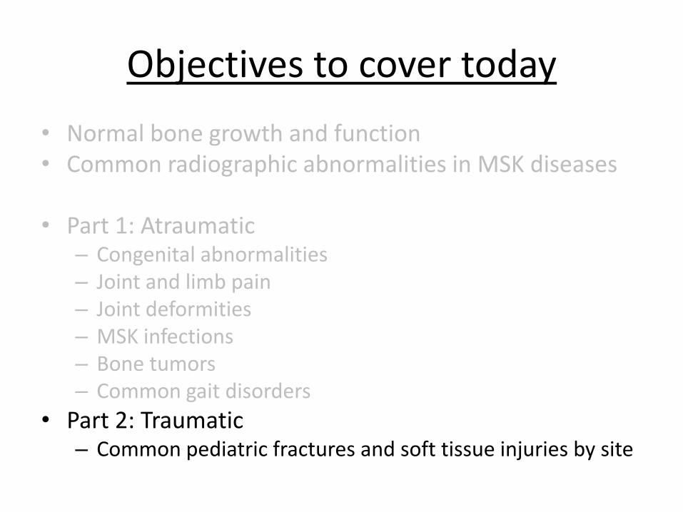
Objectives to cover today
• Normal bone growth and function• Common radiographic abnormalities in MSK diseases
• Part 1: Atraumatic– Congenital abnormalities – Joint and limb pain– Joint deformities– MSK infections– Bone tumors– Common gait disorders
• Part 2: Traumatic– Common pediatric fractures and soft tissue injuries by site

Overview of traumatic MSK pain
Acute injuries• Fractures• Joint dislocations
– Most common in ED: patella, digits, shoulder, elbow
• Muscle strains – Eg. groin/adductors
• Ligament sprains– Eg. Ankle, ACL/MCL, acromioclavicular joint separation
Chronic/ overuse injuries• Stress fractures• Tendonitis• Bursitis• Fasciitis• Apophysitis
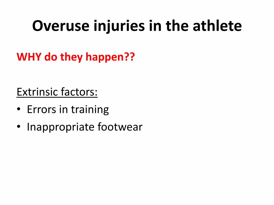
Overuse injuries in the athlete
WHY do they happen??
Extrinsic factors:
• Errors in training
• Inappropriate footwear
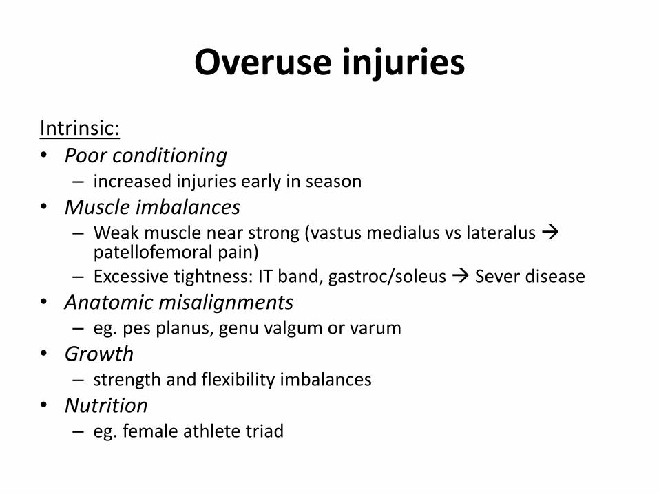
Overuse injuries
Intrinsic:• Poor conditioning
– increased injuries early in season
• Muscle imbalances– Weak muscle near strong (vastus medialus vs lateralus
patellofemoral pain)– Excessive tightness: IT band, gastroc/soleus Sever disease
• Anatomic misalignments – eg. pes planus, genu valgum or varum
• Growth– strength and flexibility imbalances
• Nutrition– eg. female athlete triad
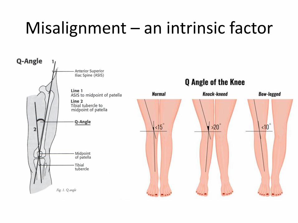
Misalignment – an intrinsic factor
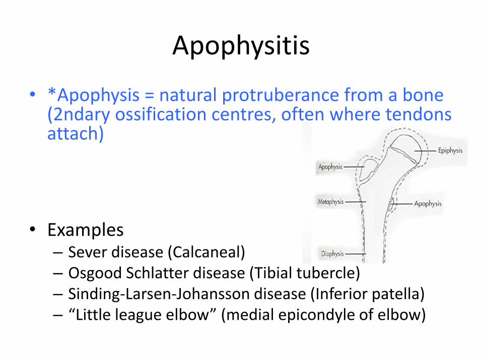
Apophysitis
• *Apophysis = natural protruberance from a bone (2ndary ossification centres, often where tendons attach)
• Examples– Sever disease (Calcaneal)– Osgood Schlatter disease (Tibial tubercle)– Sinding-Larsen-Johansson disease (Inferior patella)– “Little league elbow” (medial epicondyle of elbow)
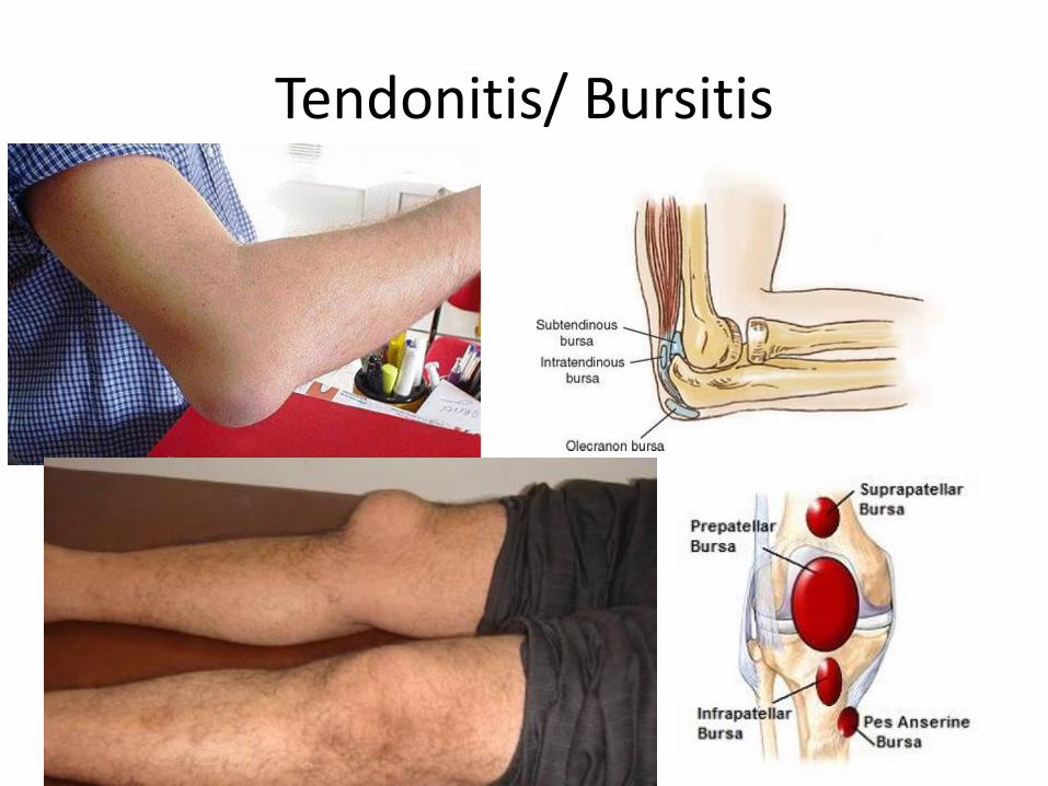
Tendonitis/ Bursitis
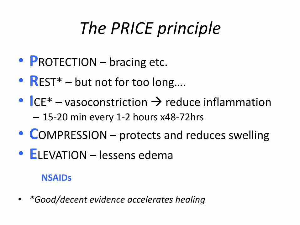
The PRICE principle
• PROTECTION – bracing etc.
• REST* – but not for too long….
• ICE* – vasoconstriction reduce inflammation– 15-20 min every 1-2 hours x48-72hrs
• COMPRESSION – protects and reduces swelling
• ELEVATION – lessens edema
• *Good/decent evidence accelerates healing
NSAIDs
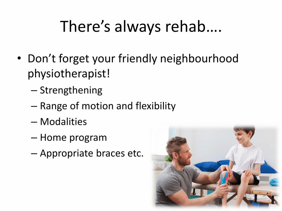
There’s always rehab….
• Don’t forget your friendly neighbourhoodphysiotherapist!
– Strengthening
– Range of motion and flexibility
– Modalities
– Home program
– Appropriate braces etc.
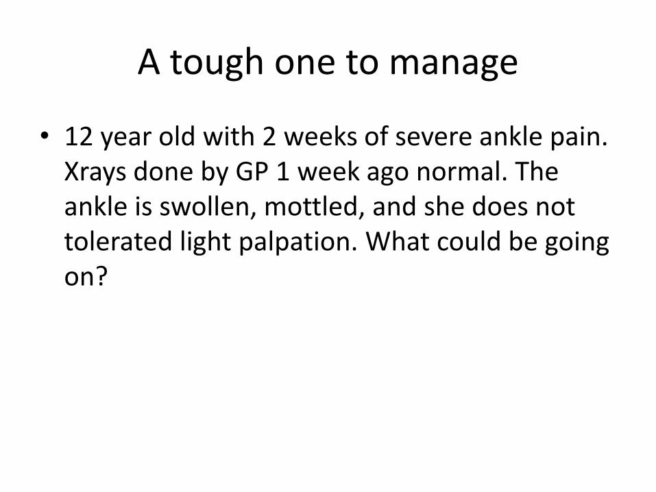
A tough one to manage
• 12 year old with 2 weeks of severe ankle pain. Xrays done by GP 1 week ago normal. The ankle is swollen, mottled, and she does not tolerated light palpation. What could be going on?
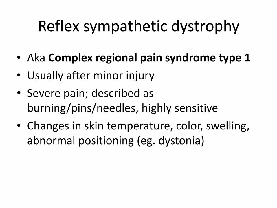
Reflex sympathetic dystrophy
• Aka Complex regional pain syndrome type 1
• Usually after minor injury
• Severe pain; described as burning/pins/needles, highly sensitive
• Changes in skin temperature, color, swelling, abnormal positioning (eg. dystonia)

Management
• Prognosis highly variable
• Management principles:• Rehab/physio – keeping the painful limb moving
(reduce circulatory symptoms, improve ROM, strength, function, lessen chance of more chronic symptoms)
• Psychotherapy – can develop mood disturbances that heighten perception of symptoms
• Medications?
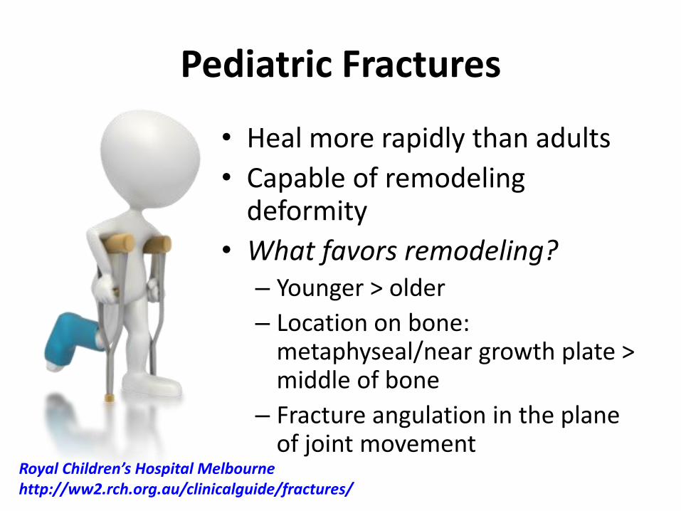
Pediatric Fractures
• Heal more rapidly than adults
• Capable of remodeling deformity
• What favors remodeling?– Younger > older
– Location on bone: metaphyseal/near growth plate > middle of bone
– Fracture angulation in the plane of joint movement
Royal Children’s Hospital Melbournehttp://ww2.rch.org.au/clinicalguide/fractures/
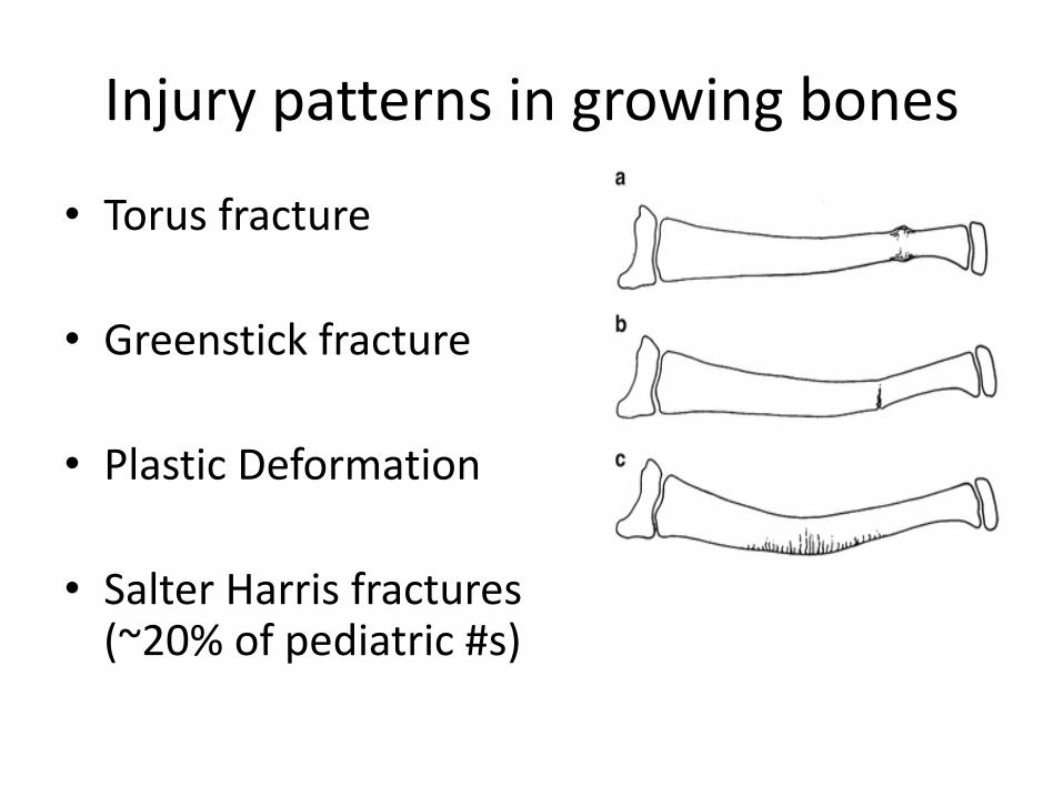
Injury patterns in growing bones
• Torus fracture
• Greenstick fracture
• Plastic Deformation
• Salter Harris fractures (~20% of pediatric #s)
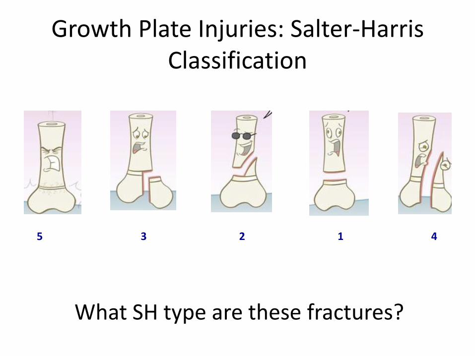
Growth Plate Injuries: Salter-Harris Classification
What SH type are these fractures?
5 3 2 1 4
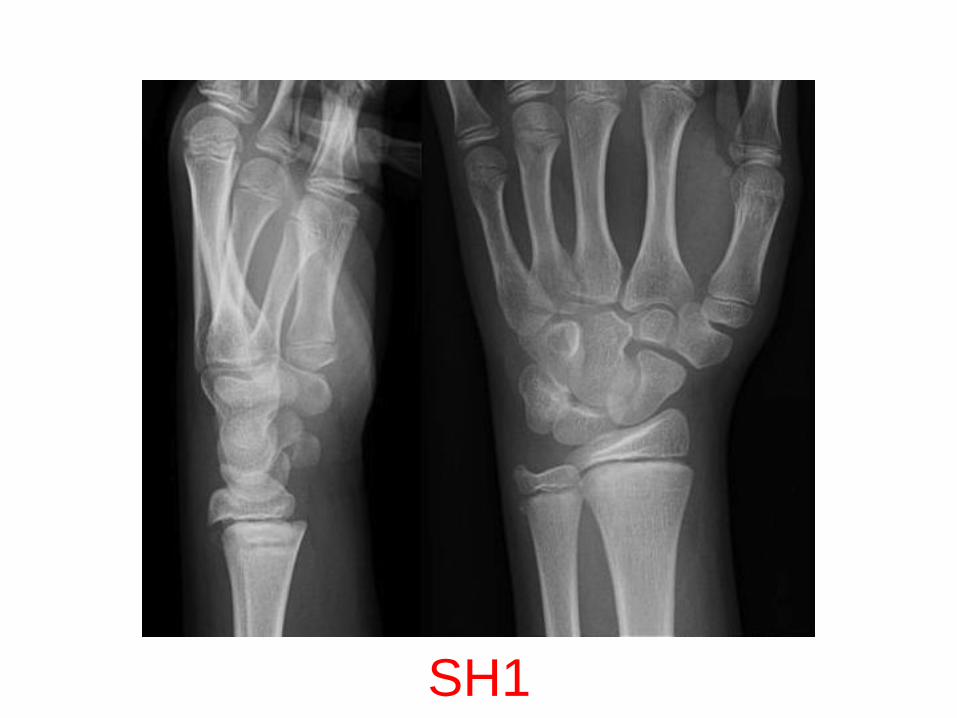
SH1
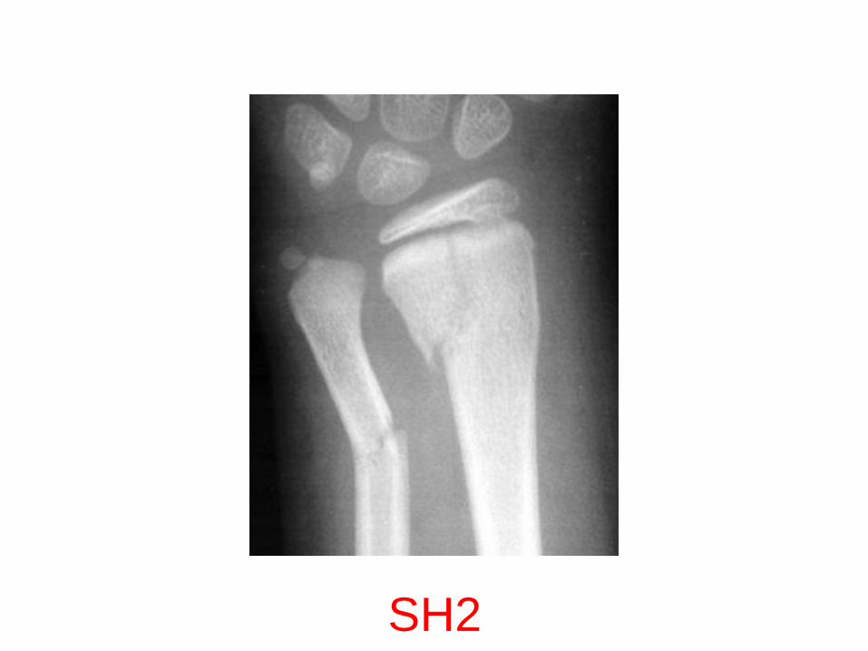
SH2
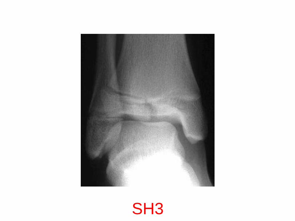
SH3
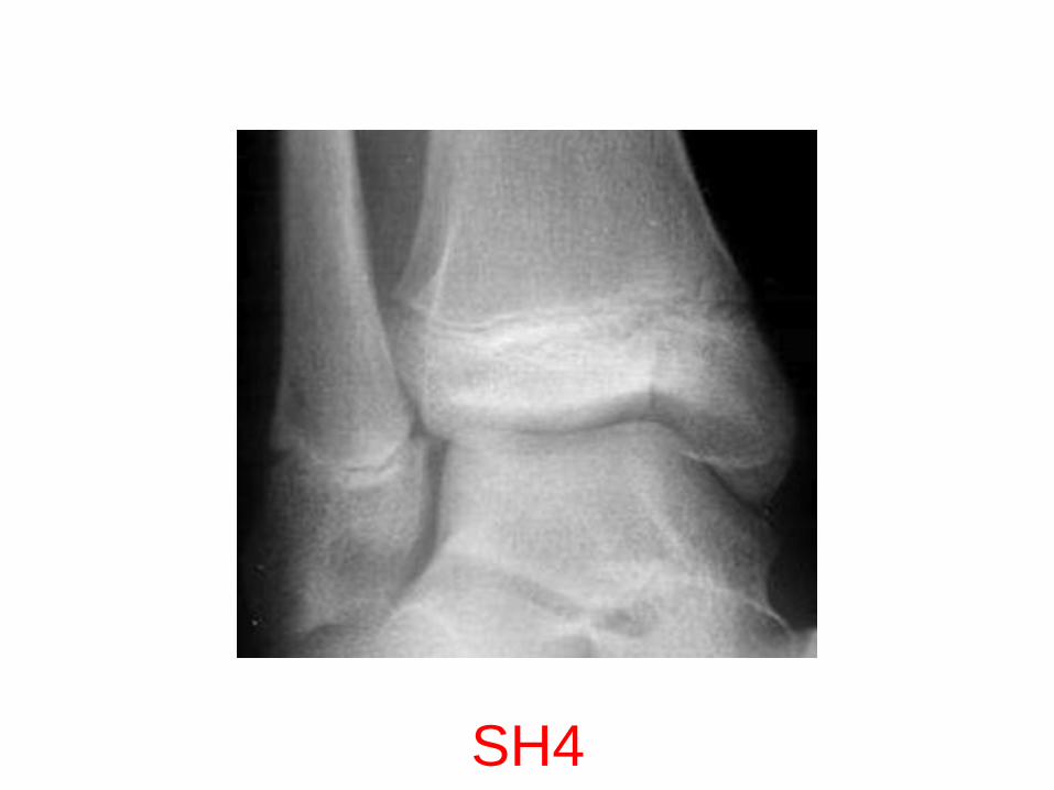
SH4
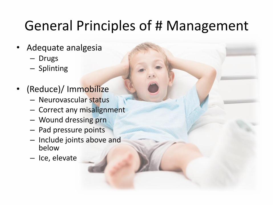
General Principles of # Management
• Adequate analgesia– Drugs– Splinting
• (Reduce)/ Immobilize– Neurovascular status– Correct any misalignment– Wound dressing prn– Pad pressure points– Include joints above and
below– Ice, elevate
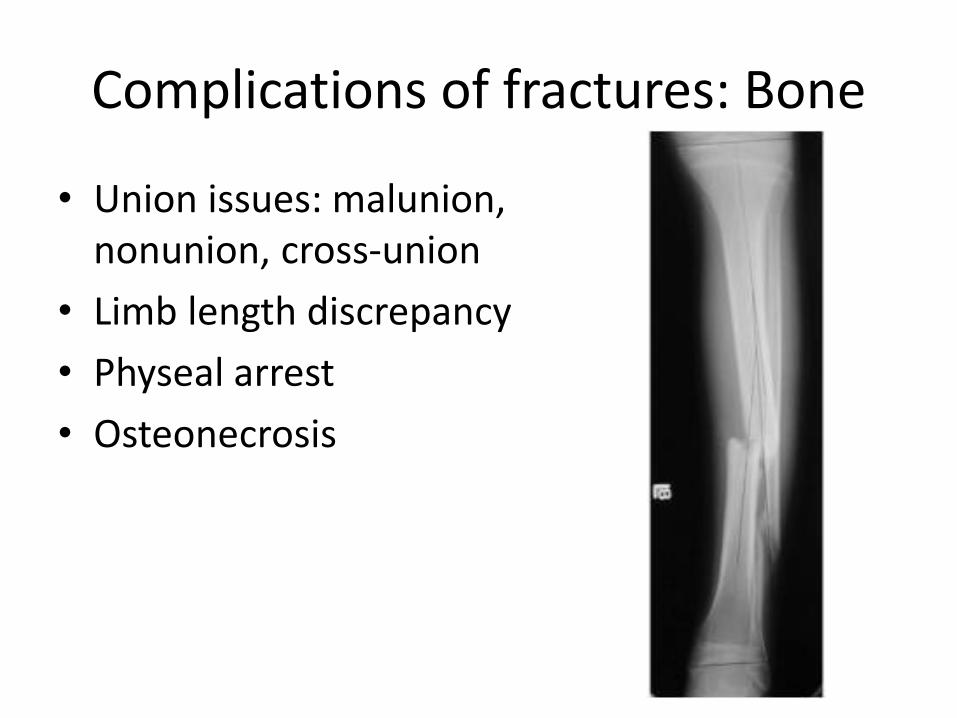
Complications of fractures: Bone
• Union issues: malunion, nonunion, cross-union
• Limb length discrepancy
• Physeal arrest
• Osteonecrosis
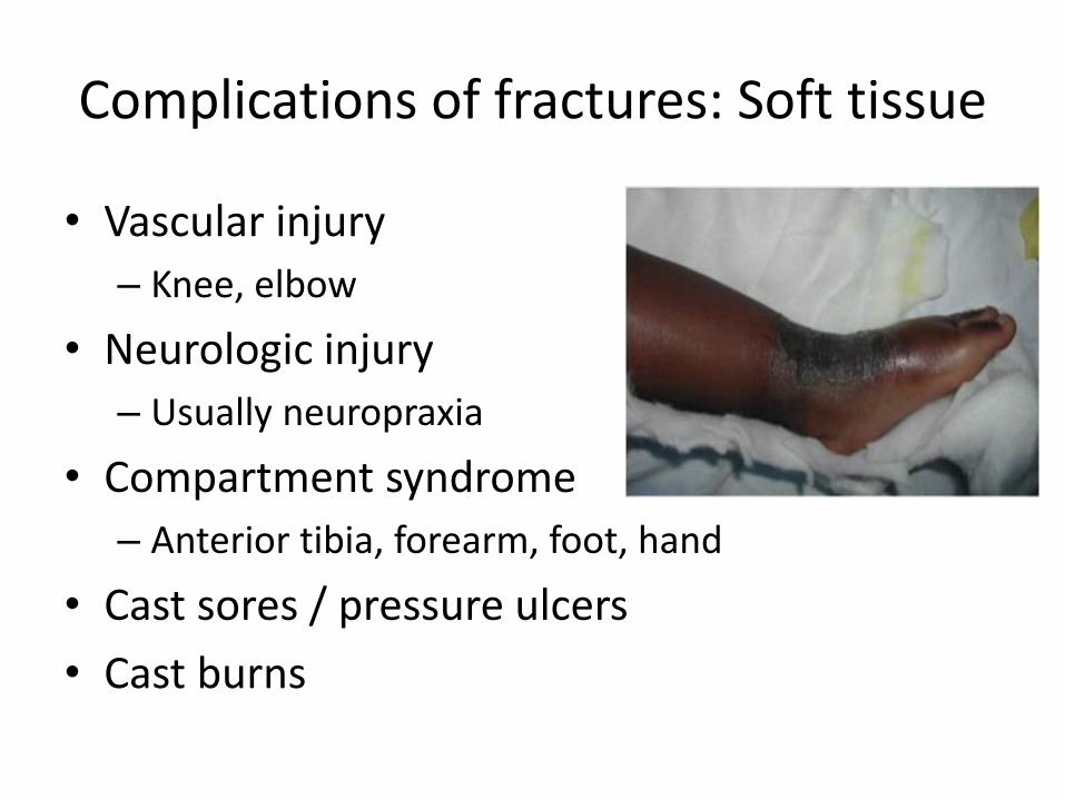
Complications of fractures: Soft tissue
• Vascular injury
– Knee, elbow
• Neurologic injury
– Usually neuropraxia
• Compartment syndrome
– Anterior tibia, forearm, foot, hand
• Cast sores / pressure ulcers
• Cast burns
Fractures…
. let’s
practice!
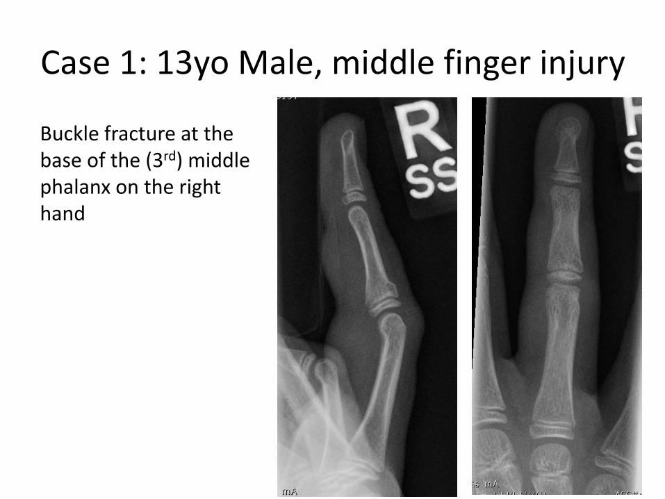
Case 1: 13yo Male, middle finger injury
Buckle fracture at the base of the (3rd) middle phalanx on the right hand
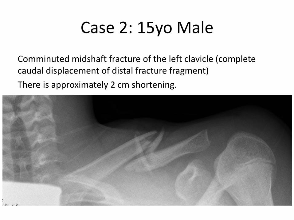
Case 2: 15yo Male
Comminuted midshaft fracture of the left clavicle (complete caudal displacement of distal fracture fragment)
There is approximately 2 cm shortening.
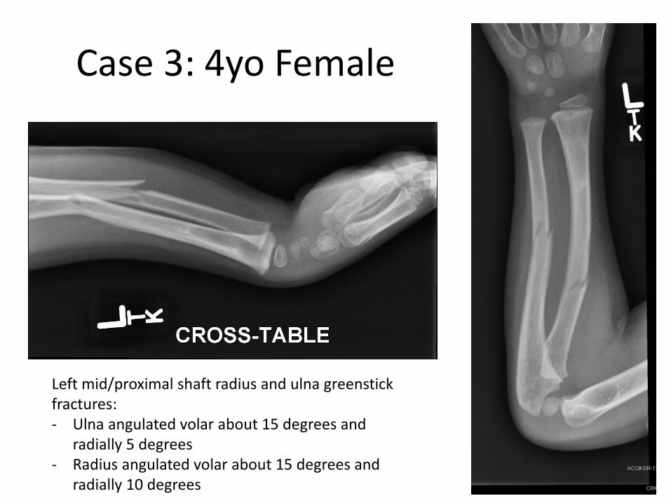
Case 3: 4yo Female
Left mid/proximal shaft radius and ulna greenstick fractures:- Ulna angulated volar about 15 degrees and
radially 5 degrees- Radius angulated volar about 15 degrees and
radially 10 degrees
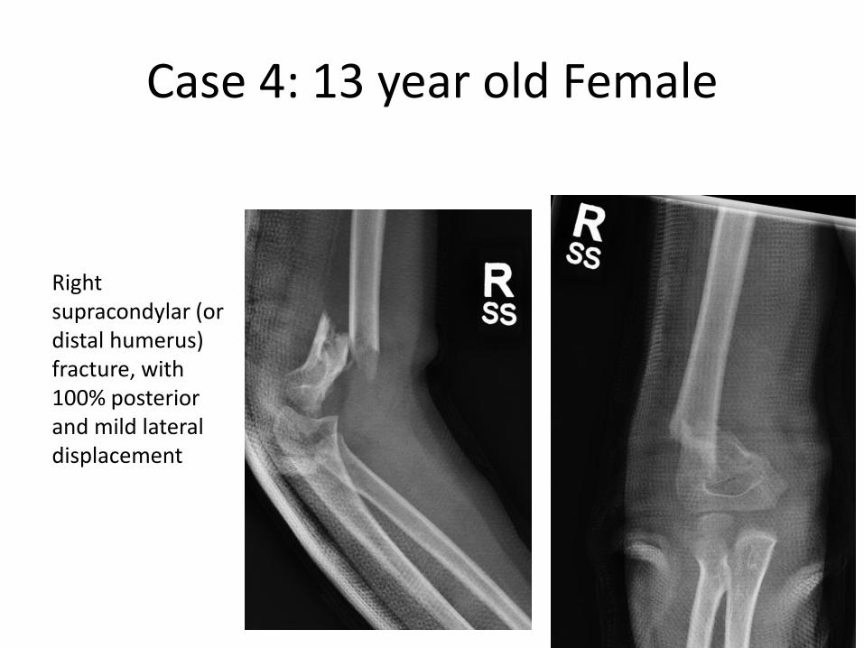
Case 4: 13 year old Female
Right supracondylar (or distal humerus) fracture, with 100% posterior and mild lateral displacement

ELBOWFOREARM
CLAVICLE/ SHOULDER
LOWER LEG/ ANKLE
MSK injuries:
Let’s get
specific…
BACK
KNEE
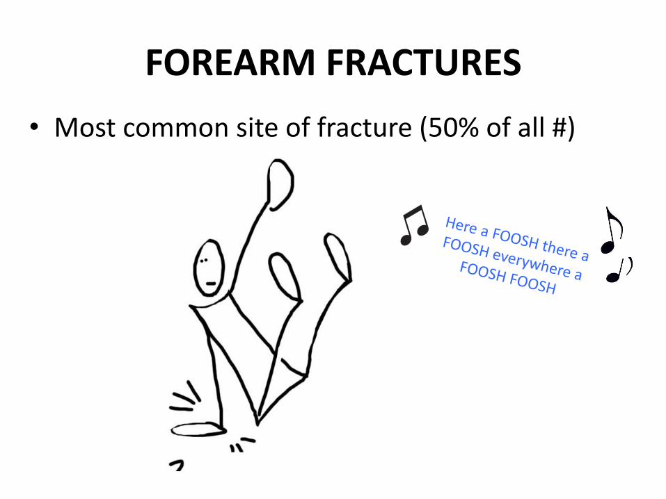
FOREARM FRACTURES
• Most common site of fracture (50% of all #)
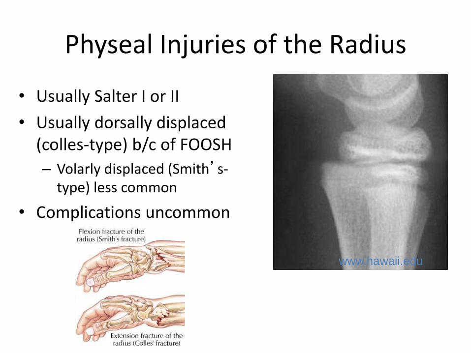
Physeal Injuries of the Radius
• Usually Salter I or II
• Usually dorsally displaced (colles-type) b/c of FOOSH
– Volarly displaced (Smith’s-type) less common
• Complications uncommon
www.hawaii.edu
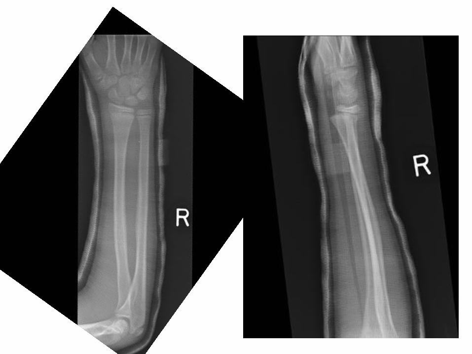
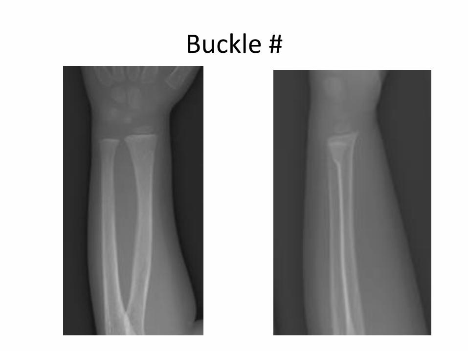
Buckle #
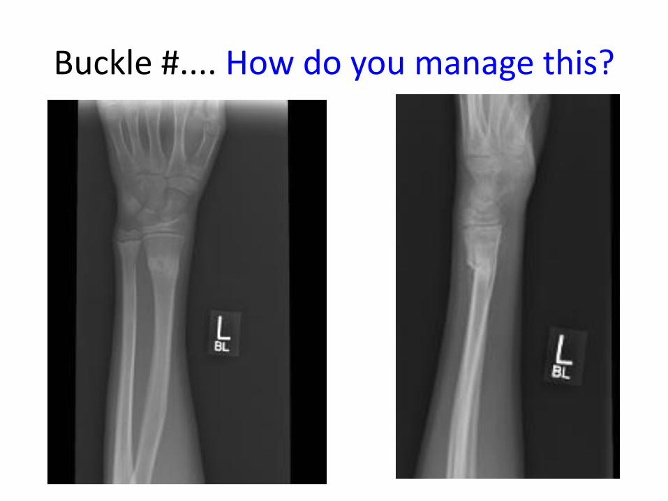
Buckle #.... How do you manage this?
Distal Forearm Buckle #
• Stable fractures, management has evolved…
• A randomized controlled trial of removable splinting vs casting for wrist buckle fractures in children. Plint et al. Pediatrics 2006;117(3):691-7– Splint = better physical functioning, easier to bath
– Cast = increased return visits for cast problems
– Pain scores same, no fracture complications in either group
• Who might benefit from cast? More severe buckle, very young, very active
• How long do we immobilize? 3 wks
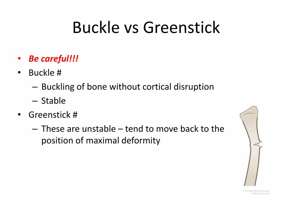
Buckle vs Greenstick
• Be careful!!!
• Buckle #
– Buckling of bone without cortical disruption
– Stable
• Greenstick #
– These are unstable – tend to move back to the position of maximal deformity
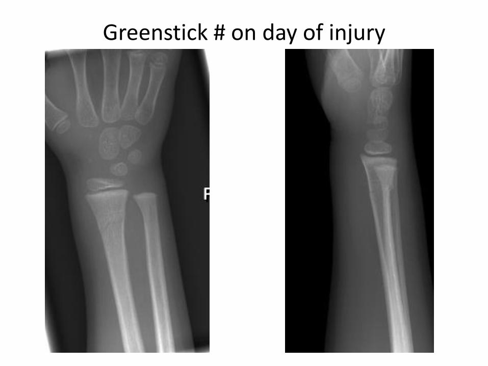
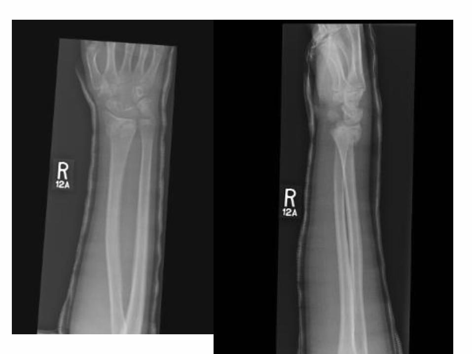
Greenstick # on day of injury
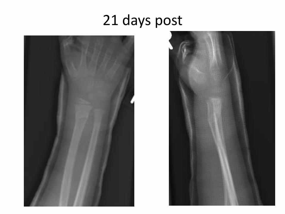
21 days post
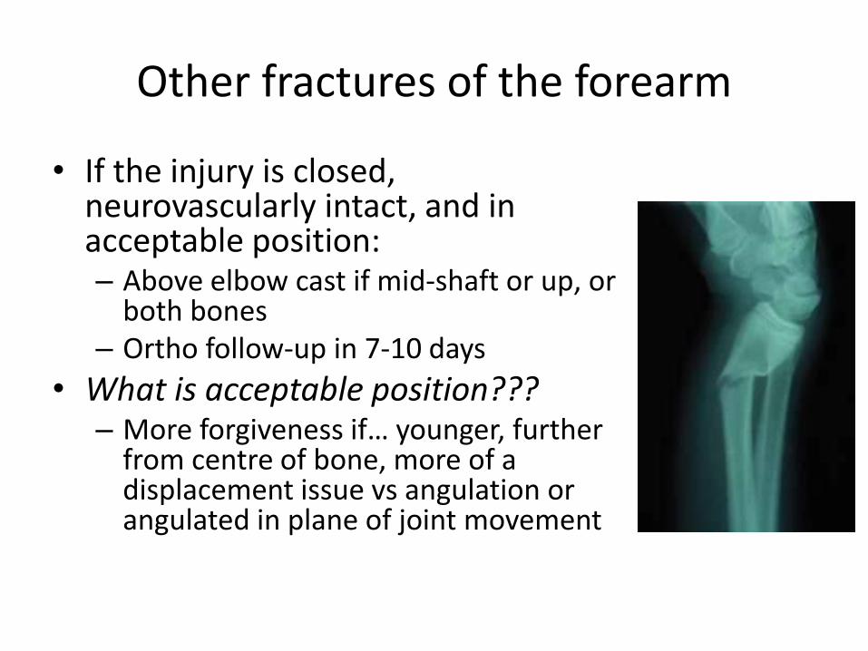
Other fractures of the forearm
• If the injury is closed, neurovascularly intact, and in acceptable position:– Above elbow cast if mid-shaft or up, or
both bones– Ortho follow-up in 7-10 days
• What is acceptable position???– More forgiveness if… younger, further
from centre of bone, more of a displacement issue vs angulation or angulated in plane of joint movement
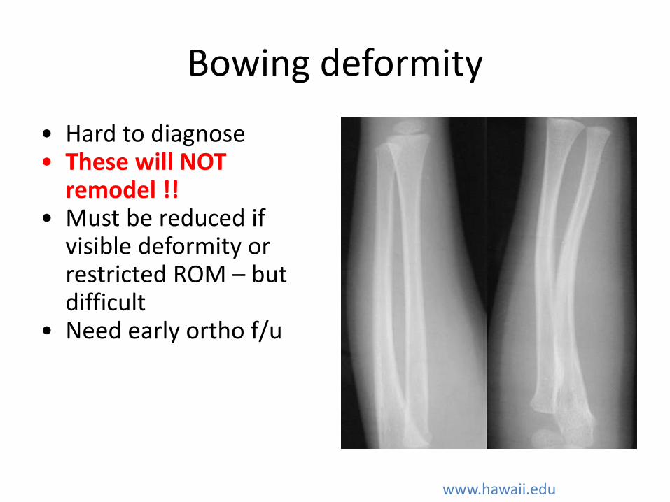
Bowing deformity
• Hard to diagnose• These will NOT
remodel !! • Must be reduced if
visible deformity or restricted ROM – but difficult
• Need early ortho f/u
www.hawaii.edu

ELBOWFOREARM
CLAVICLE/ SHOULDER
LOWER LEG/ ANKLE
MSK injuries:
Let’s get
specific…
BACK
KNEE
THE (DREADED) ELBOW
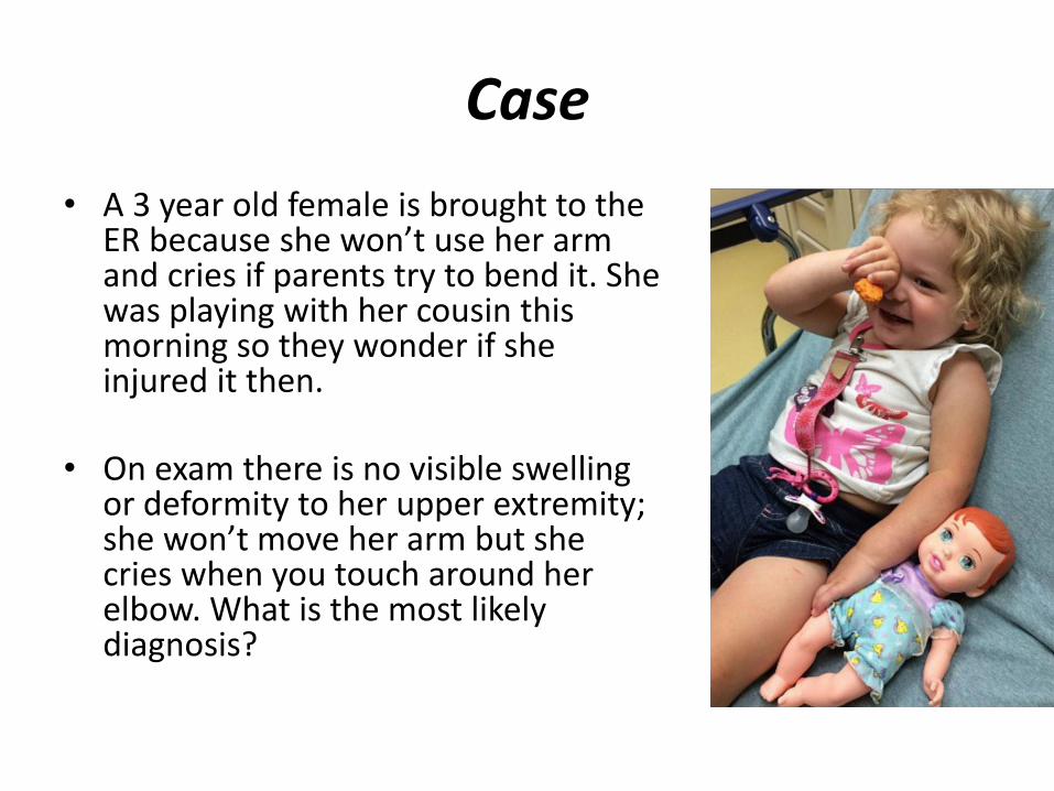
Case
• A 3 year old female is brought to the ER because she won’t use her arm and cries if parents try to bend it. She was playing with her cousin this morning so they wonder if she injured it then.
• On exam there is no visible swelling or deformity to her upper extremity; she won’t move her arm but she cries when you touch around her elbow. What is the most likely diagnosis?
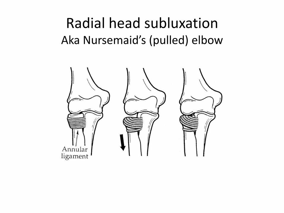
Radial head subluxationAka Nursemaid’s (pulled) elbow
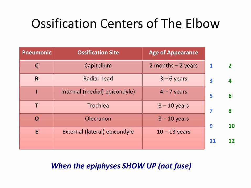
Ossification Centers of The Elbow
Pneumonic Ossification Site Age of Appearance
C Capitellum 2 months – 2 years
R Radial head 3 – 6 years
I Internal (medial) epicondyle) 4 – 7 years
T Trochlea 8 – 10 years
O Olecranon 8 – 10 years
E External (lateral) epicondyle 10 – 13 years
1
3
5
7
9
11
2
4
6
8
10
12
When the epiphyses SHOW UP (not fuse)
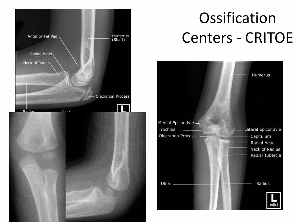
Ossification Centers - CRITOE
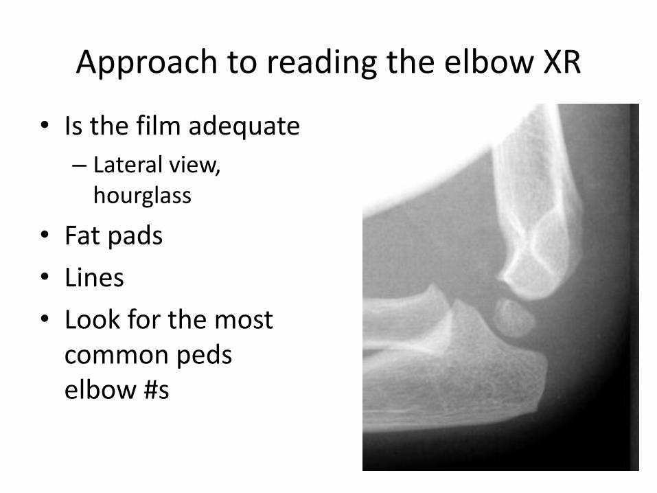
Approach to reading the elbow XR
• Is the film adequate
– Lateral view, hourglass
• Fat pads
• Lines
• Look for the most common pedselbow #s
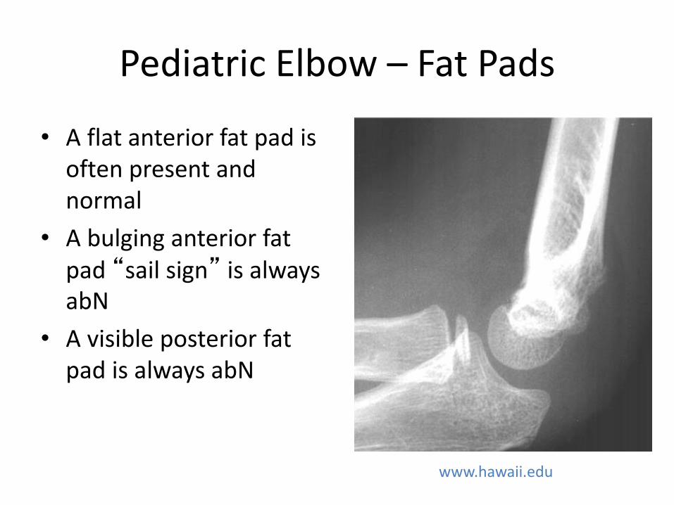
Pediatric Elbow – Fat Pads
• A flat anterior fat pad is often present and normal
• A bulging anterior fat pad “sail sign” is always abN
• A visible posterior fat pad is always abN
www.hawaii.edu
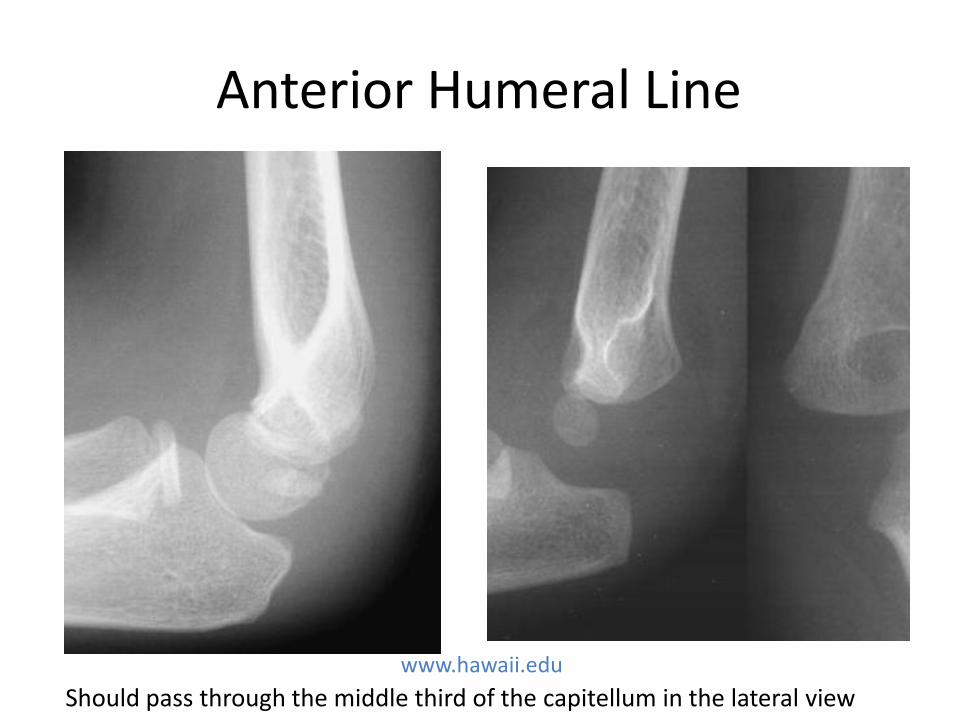
Anterior Humeral Line
Should pass through the middle third of the capitellum in the lateral view
www.hawaii.edu
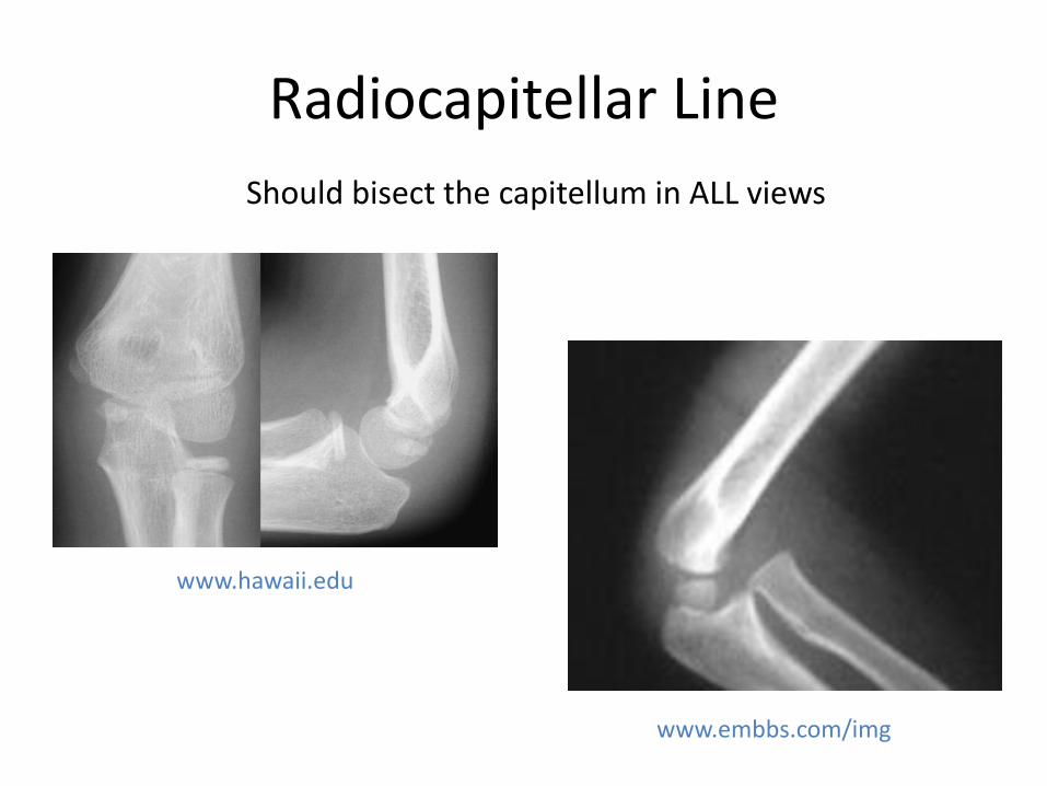
Radiocapitellar Line
Should bisect the capitellum in ALL views
www.embbs.com/img
www.hawaii.edu

2yo fell off stool - What’s this?
www.hawaii.edu
C 1R 3I 5T 7O 9E 11
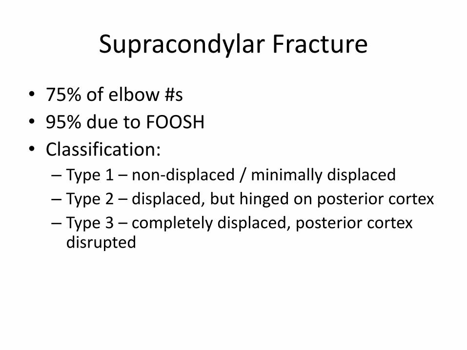
Supracondylar Fracture
• 75% of elbow #s
• 95% due to FOOSH
• Classification:– Type 1 – non-displaced / minimally displaced
– Type 2 – displaced, but hinged on posterior cortex
– Type 3 – completely displaced, posterior cortex disrupted
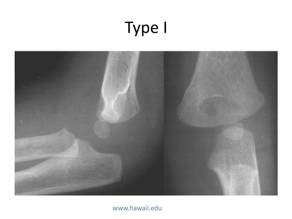
Type I
www.hawaii.edu
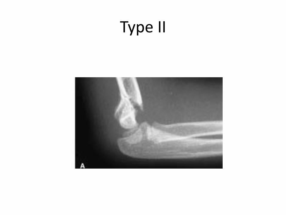
Type II
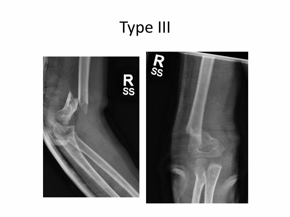
Type III
Supracondylar Fracture Complications
• Very high rate of complications!!
– Neurologic injury (8-15%)
• Ant interosseuous branch of median n
• Radial and ulnar nerves also may be involved
– Radial artery (2% overall, 50% in Type III)
– Compartment Syndrome
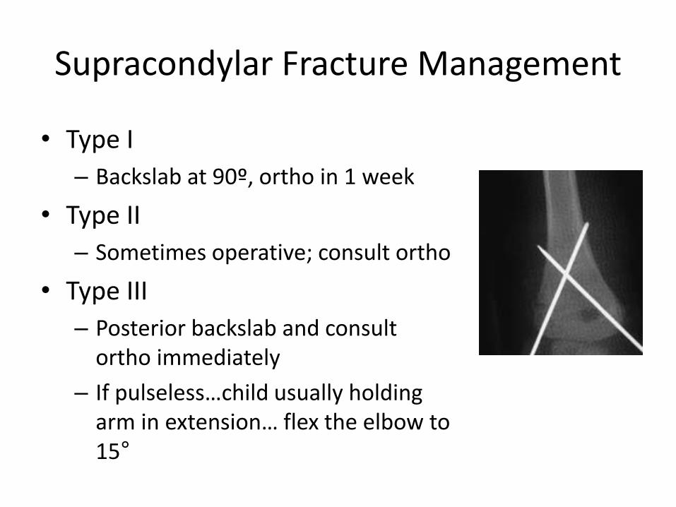
Supracondylar Fracture Management
• Type I
– Backslab at 90º, ortho in 1 week
• Type II
– Sometimes operative; consult ortho
• Type III
– Posterior backslab and consult ortho immediately
– If pulseless…child usually holding arm in extension… flex the elbow to 15°

What are these?www.hawaii.edu
Lateral epicondyle fracture2nd most common elbow fracture
Medial epicondyle fracture
http://www.rch.org.au/clinicalguide/guideline_index/fractures
CRITOE
2 year old left arm
9 year old right arm
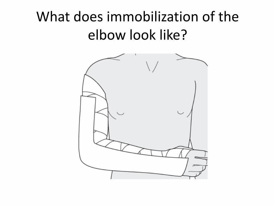
What does immobilization of the elbow look like?
Case
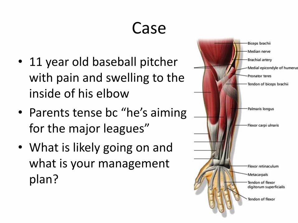
• 11 year old baseball pitcher with pain and swelling to the inside of his elbow
• Parents tense bc “he’s aiming for the major leagues”
• What is likely going on and what is your management plan?
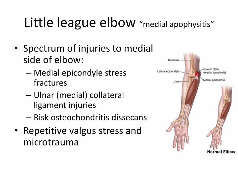
Little league elbow “medial apophysitis”
• Spectrum of injuries to medial side of elbow:– Medial epicondyle stress
fractures
– Ulnar (medial) collateral ligament injuries
– Risk osteochondritis dissecans
• Repetitive valgus stress and microtrauma
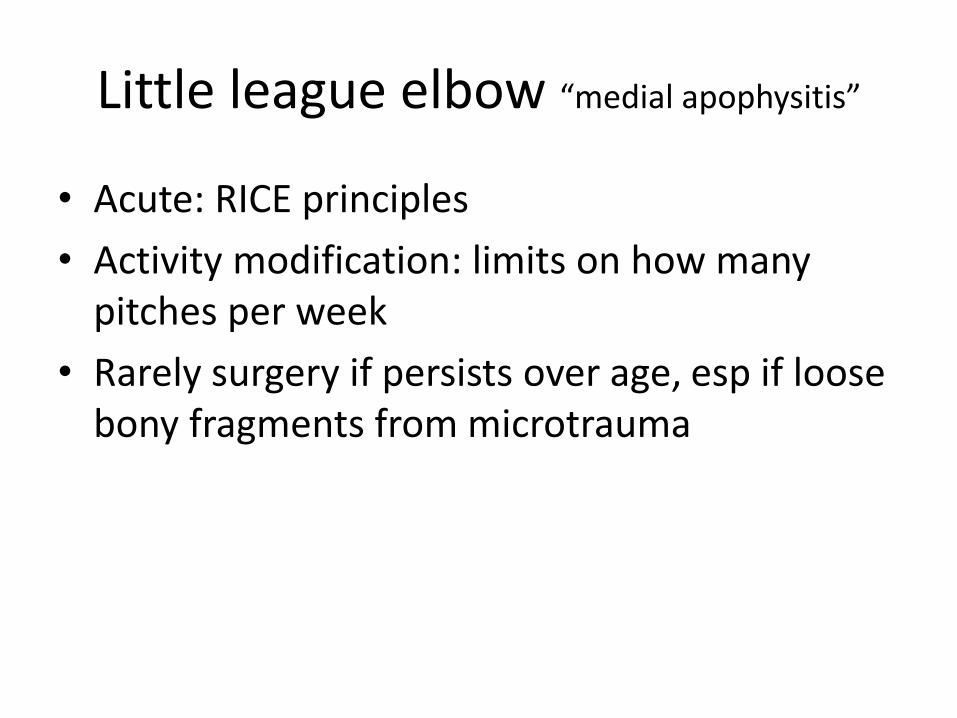
• Acute: RICE principles
• Activity modification: limits on how many pitches per week
• Rarely surgery if persists over age, esp if loose bony fragments from microtrauma
Little league elbow “medial apophysitis”
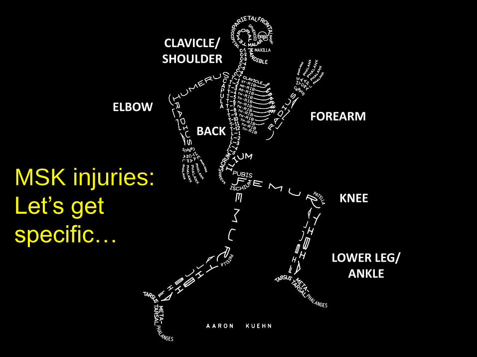
ELBOWFOREARM
CLAVICLE/ SHOULDER
LOWER LEG/ ANKLE
MSK injuries:
Let’s get
specific…
BACK
KNEE
CLAVICLE/ SHOULDER
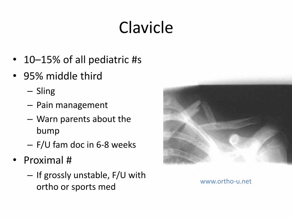
Clavicle
• 10–15% of all pediatric #s
• 95% middle third
– Sling
– Pain management
– Warn parents about the bump
– F/U fam doc in 6-8 weeks
• Proximal #
– If grossly unstable, F/U with ortho or sports med
www.ortho-u.net
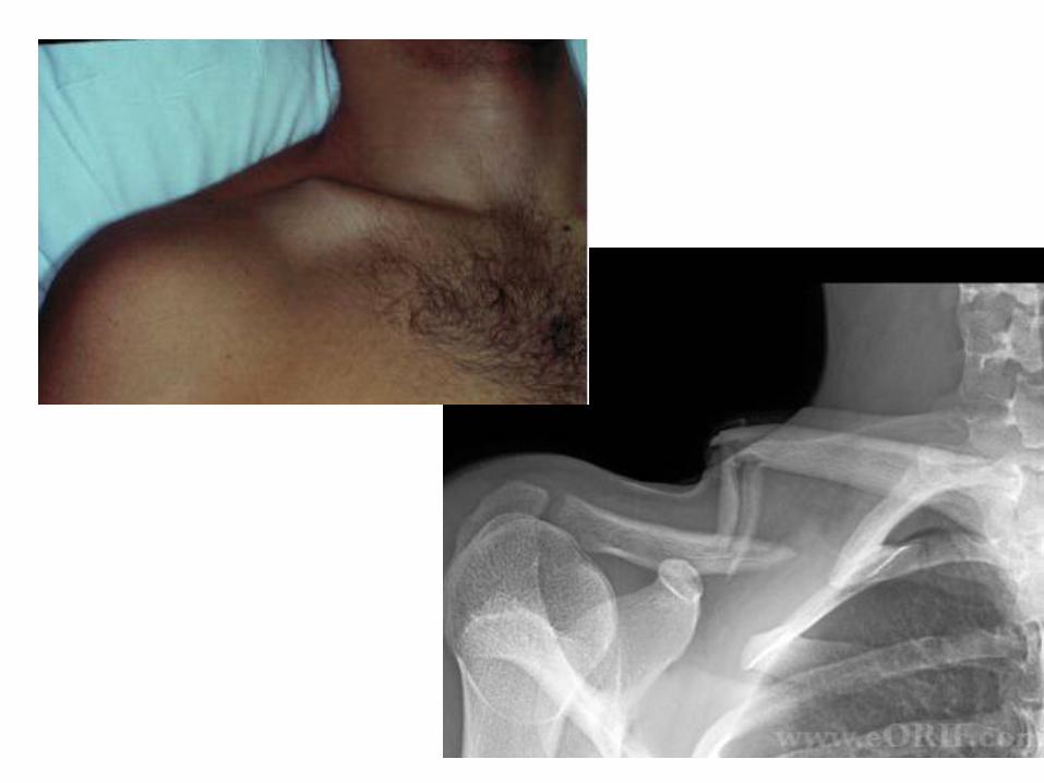
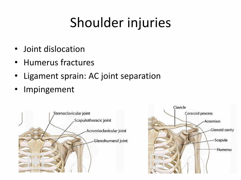
Shoulder injuries
• Joint dislocation
• Humerus fractures
• Ligament sprain: AC joint separation
• Impingement
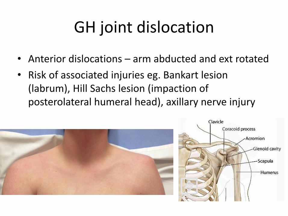
GH joint dislocation
• Anterior dislocations – arm abducted and ext rotated
• Risk of associated injuries eg. Bankart lesion (labrum), Hill Sachs lesion (impaction of posterolateral humeral head), axillary nerve injury
GH joint dislocation
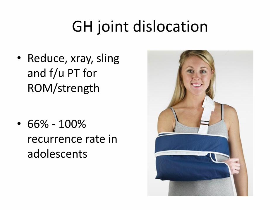
• Reduce, xray, sling and f/u PT for ROM/strength
• 66% - 100% recurrence rate in adolescents
Case 1
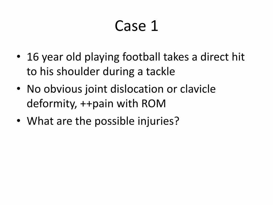
• 16 year old playing football takes a direct hit to his shoulder during a tackle
• No obvious joint dislocation or clavicle deformity, ++pain with ROM
• What are the possible injuries?
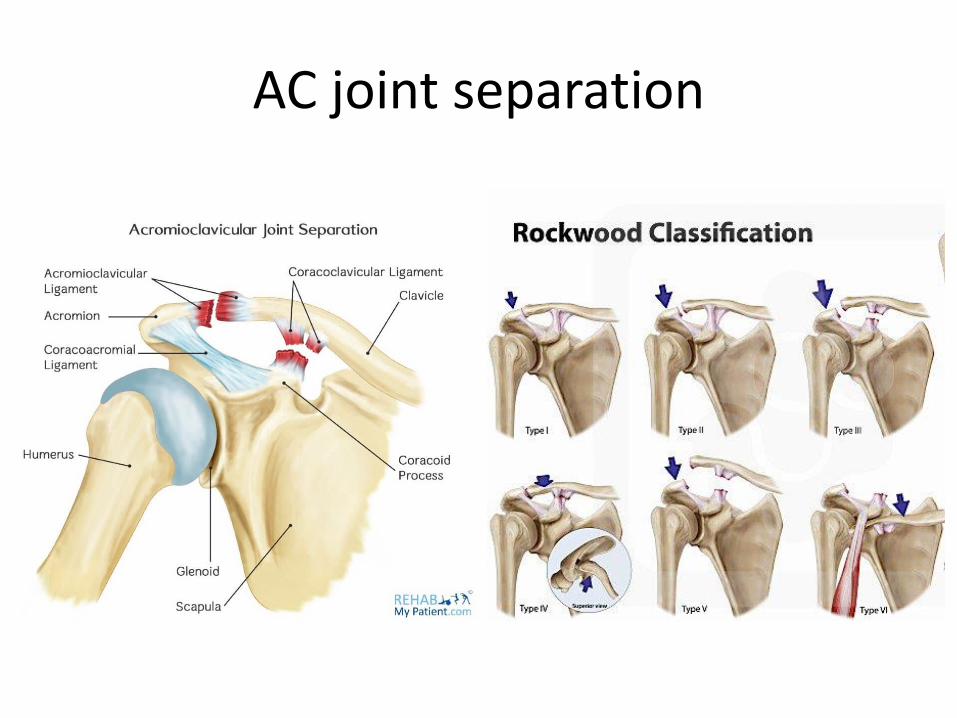
AC joint separation
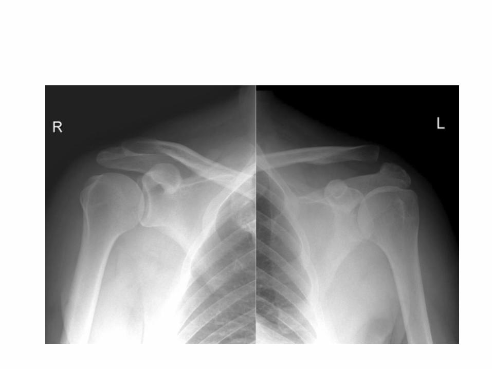
Case 2
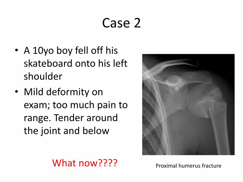
• A 10yo boy fell off his skateboard onto his left shoulder
• Mild deformity on exam; too much pain to range. Tender around the joint and below
What now???? Proximal humerus fracture
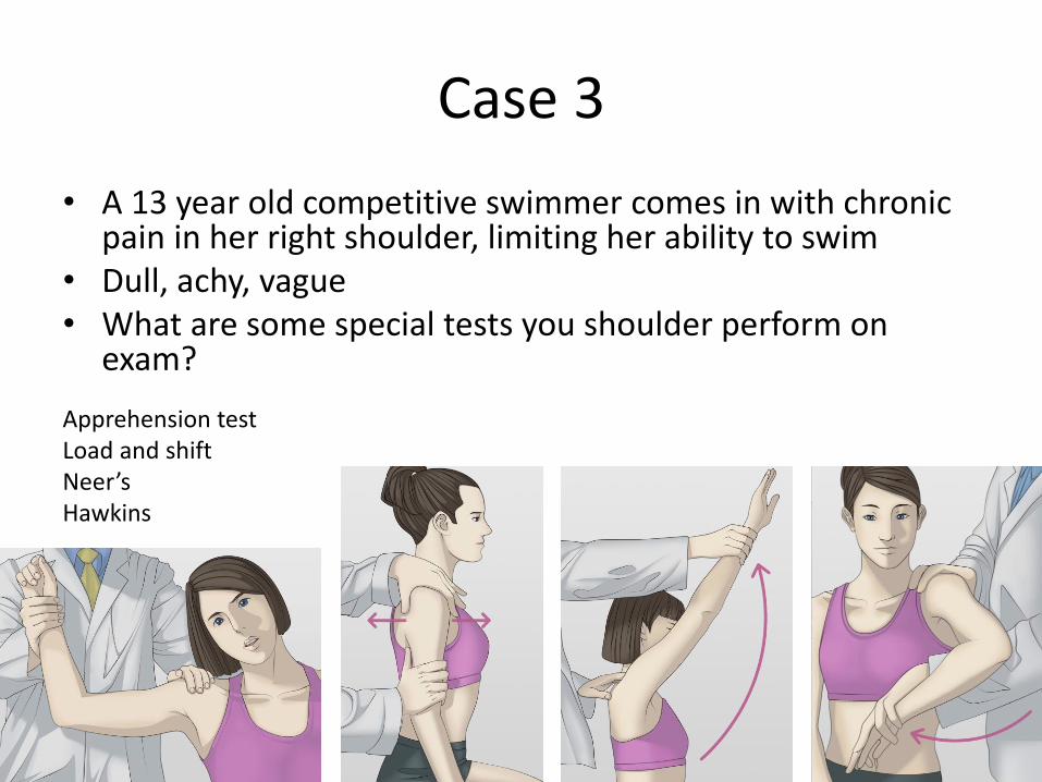
Case 3
• A 13 year old competitive swimmer comes in with chronic pain in her right shoulder, limiting her ability to swim
• Dull, achy, vague• What are some special tests you shoulder perform on
exam?
Apprehension testLoad and shiftNeer’sHawkins

Impingement
• Impingement of rotator cuff, or biceps tendon, or bursa
• Secondary to GH joint instability (lax ligaments, rotator cuff weakness, capsule deformity overhead athletes
• Special tests: – Hawkins and Neer’s
– Usually +veimpingement tests: eg. apprehension
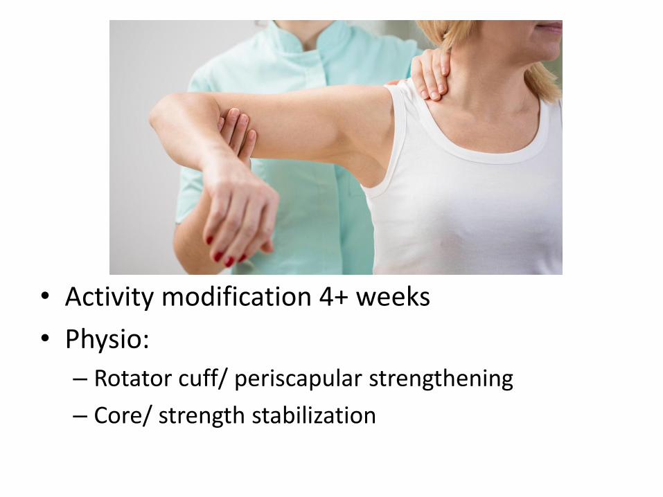
• Activity modification 4+ weeks
• Physio:
– Rotator cuff/ periscapular strengthening
– Core/ strength stabilization

ELBOWFOREARM
CLAVICLE/ SHOULDER
LOWER LEG/ ANKLE
MSK injuries:
Let’s get
specific…
BACK
KNEE
LOWER LEG & ANKLE
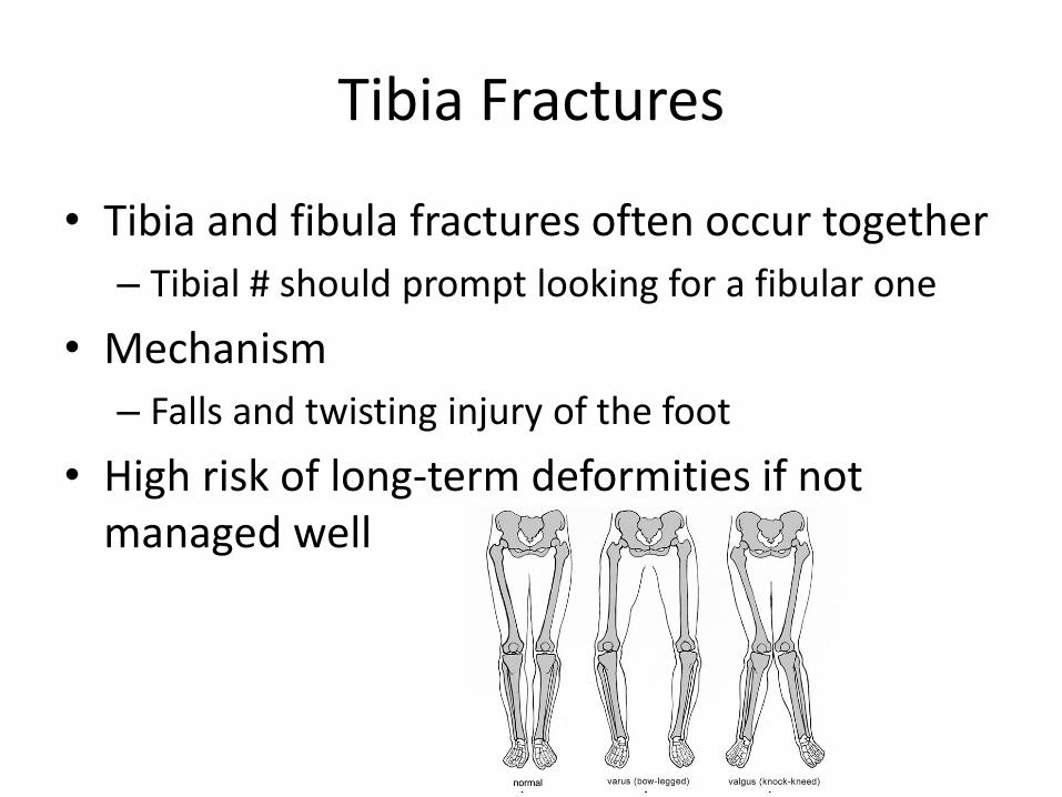
Tibia Fractures
• Tibia and fibula fractures often occur together
– Tibial # should prompt looking for a fibular one
• Mechanism
– Falls and twisting injury of the foot
• High risk of long-term deformities if not managed well
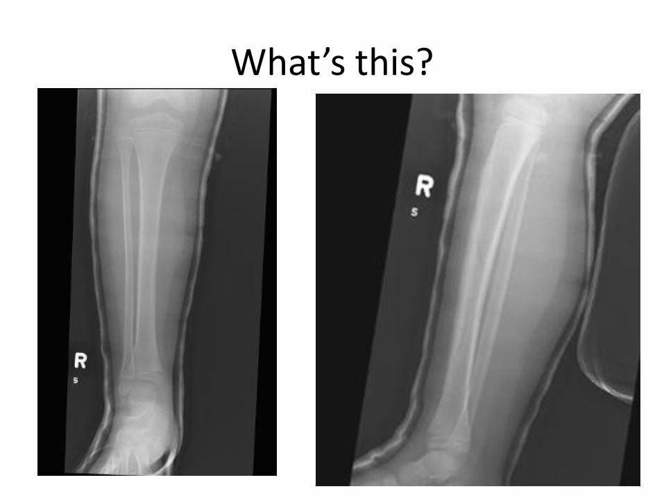
What’s this?
Toddler’s Fracture
• Common!
• Low energy mechanism
• Spiral/oblique fracture of tibia – often subtle
• Above knee walking cast for 3 weeks
Tib/Fib Shaft Fractures
• If undisplaced: above-knee cast, NWB, f/u ortho
• Minimally displaced – often reduce quite easily during casting (use sedation)
• Always do post-cast films and be careful of foot position
• For significant displacement, or involvement of the growth plate – d/w ortho
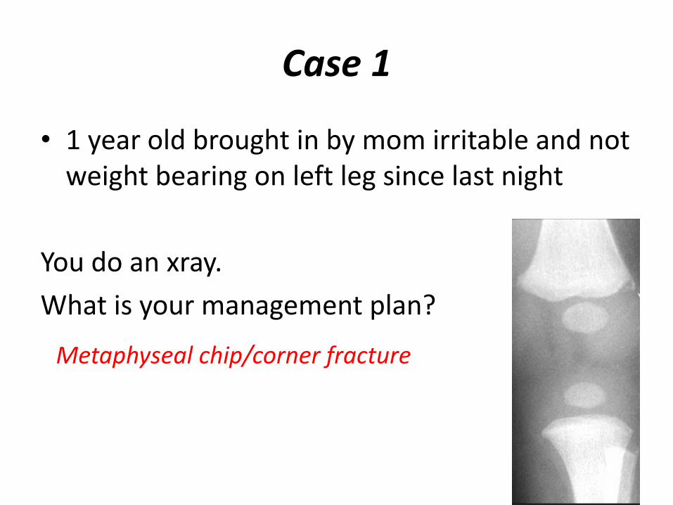
Case 1
• 1 year old brought in by mom irritable and not weight bearing on left leg since last night
You do an xray.
What is your management plan?
Metaphyseal chip/corner fracture
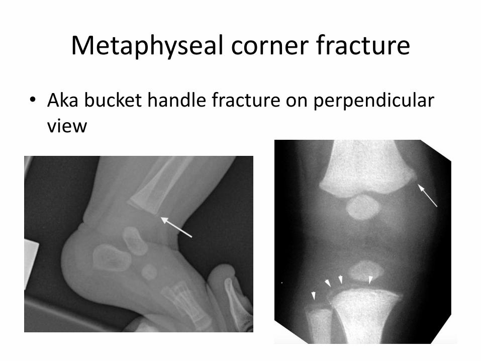
Metaphyseal corner fracture
• Aka bucket handle fracture on perpendicular view
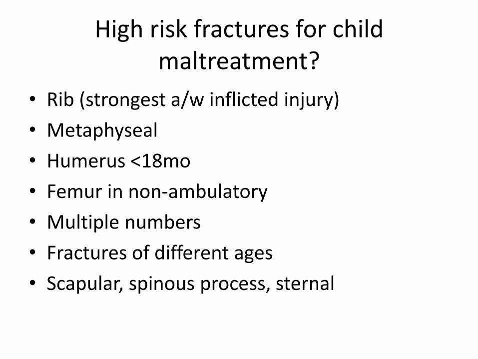
High risk fractures for child maltreatment?
• Rib (strongest a/w inflicted injury)
• Metaphyseal
• Humerus <18mo
• Femur in non-ambulatory
• Multiple numbers
• Fractures of different ages
• Scapular, spinous process, sternal
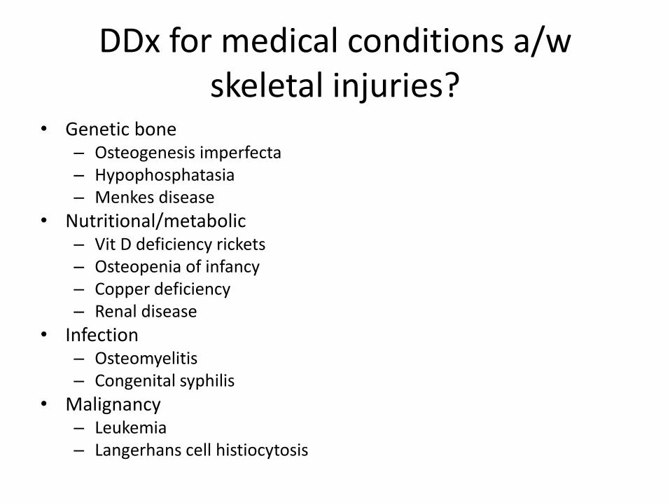
DDx for medical conditions a/wskeletal injuries?
• Genetic bone– Osteogenesis imperfecta– Hypophosphatasia– Menkes disease
• Nutritional/metabolic– Vit D deficiency rickets– Osteopenia of infancy– Copper deficiency– Renal disease
• Infection– Osteomyelitis– Congenital syphilis
• Malignancy– Leukemia– Langerhans cell histiocytosis
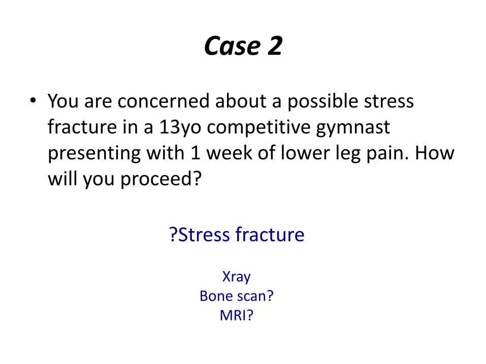
Case 2
• You are concerned about a possible stress fracture in a 13yo competitive gymnast presenting with 1 week of lower leg pain. How will you proceed?
?Stress fracture
XrayBone scan?
MRI?
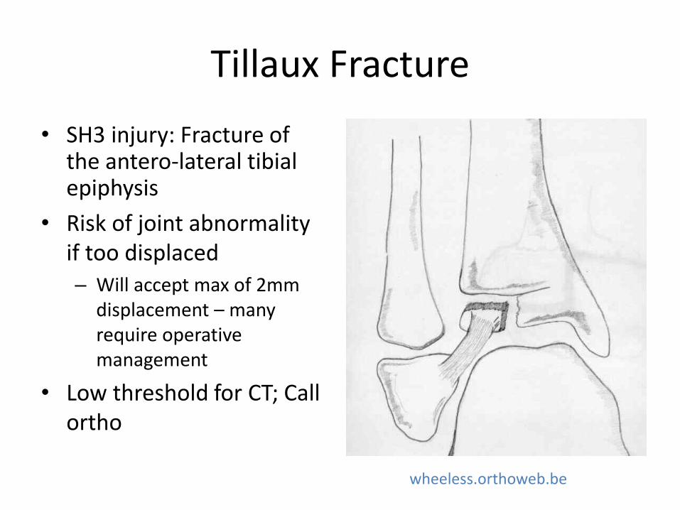
Tillaux Fracture
• SH3 injury: Fracture of the antero-lateral tibialepiphysis
• Risk of joint abnormality if too displaced
– Will accept max of 2mm displacement – many require operative management
• Low threshold for CT; Call ortho
wheeless.orthoweb.be
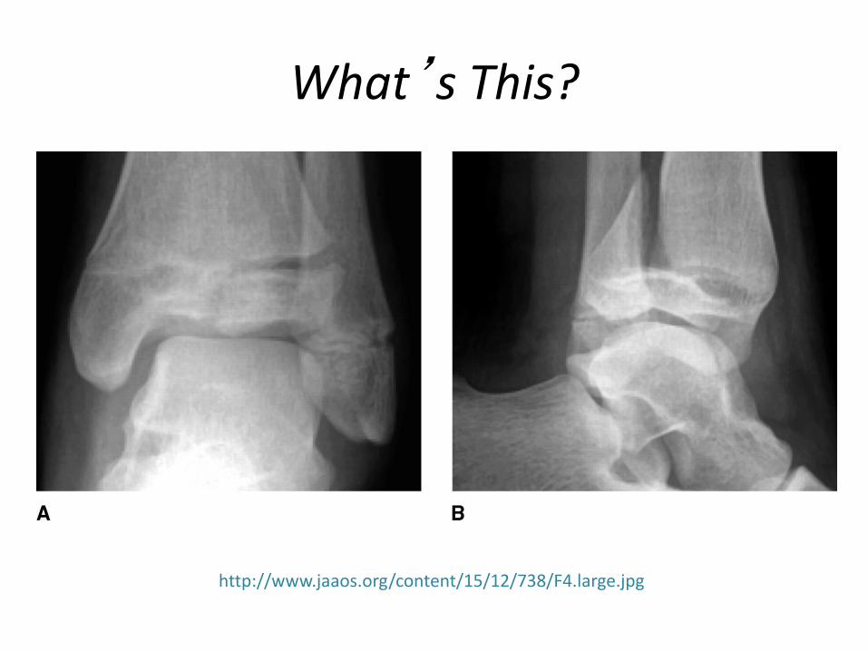
What’s This?
http://www.jaaos.org/content/15/12/738/F4.large.jpg
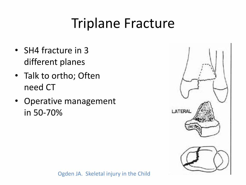
Triplane Fracture
• SH4 fracture in 3 different planes
• Talk to ortho; Often need CT
• Operative management in 50-70%
Ogden JA. Skeletal injury in the Child
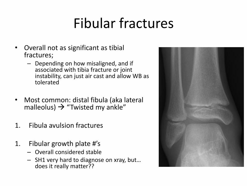
Fibular fractures
• Overall not as significant as tibialfractures;– Depending on how misaligned, and if
associated with tibia fracture or joint instability, can just air cast and allow WB as tolerated
• Most common: distal fibula (aka lateral malleolus) “Twisted my ankle”
1. Fibula avulsion fractures
1. Fibular growth plate #’s– Overall considered stable– SH1 very hard to diagnose on xray, but…
does it really matter??
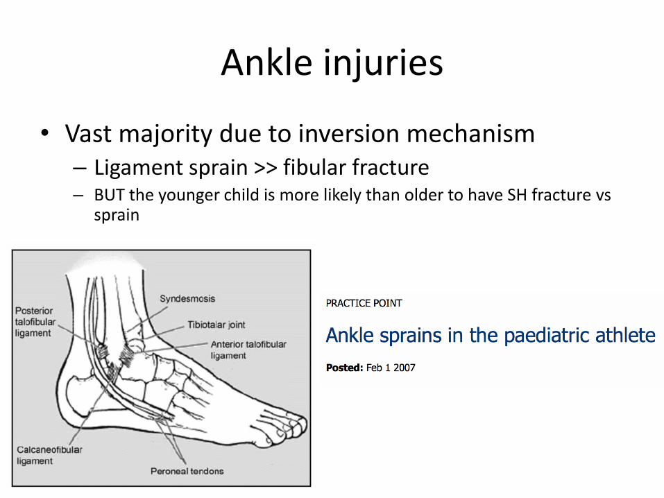
Ankle injuries
• Vast majority due to inversion mechanism– Ligament sprain >> fibular fracture– BUT the younger child is more likely than older to have SH fracture vs
sprain
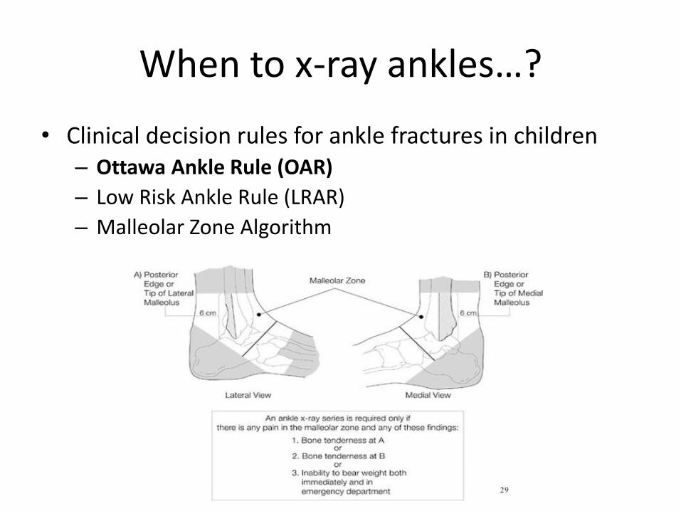
When to x-ray ankles…?
• Clinical decision rules for ankle fractures in children– Ottawa Ankle Rule (OAR)
– Low Risk Ankle Rule (LRAR)
– Malleolar Zone Algorithm
When to x-ray ankles…?
• Clinical decision rules for ankle fractures in children– Ottawa Ankle Rule (OAR)
– Low Risk Ankle Rule (LRAR)
– Malleolar Zone Algorithm
What injuries are not clinically significant?• Lateral ankle sprains• Non-displaced SH I/II of distal fibula• Avulsion fractures of the distal fibula or lateral
talus
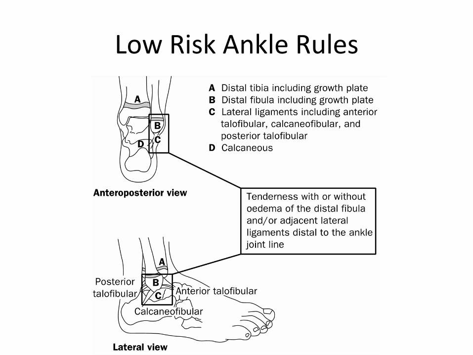
Low Risk Ankle Rules
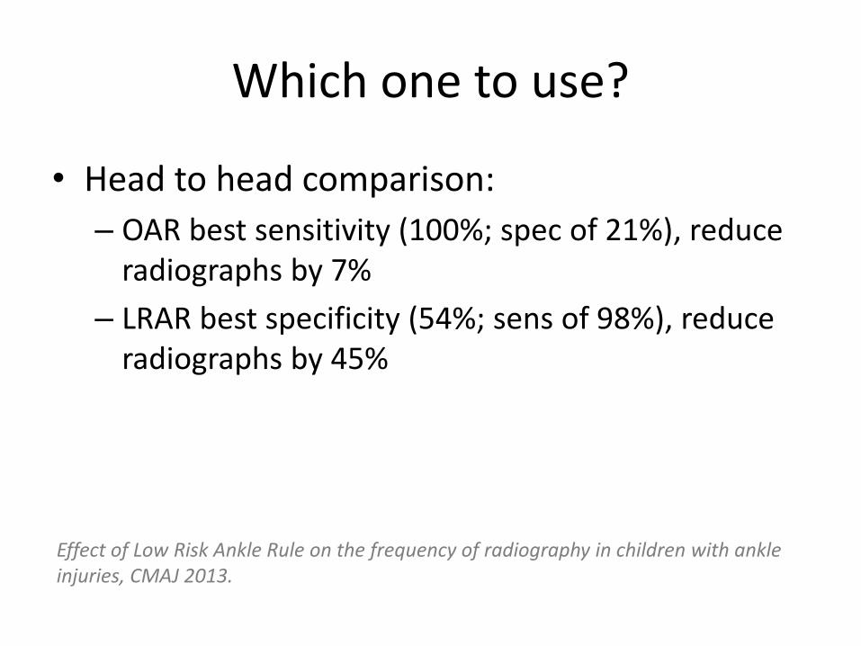
Which one to use?
• Head to head comparison:
– OAR best sensitivity (100%; spec of 21%), reduce radiographs by 7%
– LRAR best specificity (54%; sens of 98%), reduce radiographs by 45%
Effect of Low Risk Ankle Rule on the frequency of radiography in children with ankle injuries, CMAJ 2013.

ELBOWFOREARM
CLAVICLE/ SHOULDER
LOWER LEG/ ANKLE
MSK injuries:
Let’s get
specific…
BACK
KNEE
KNEE
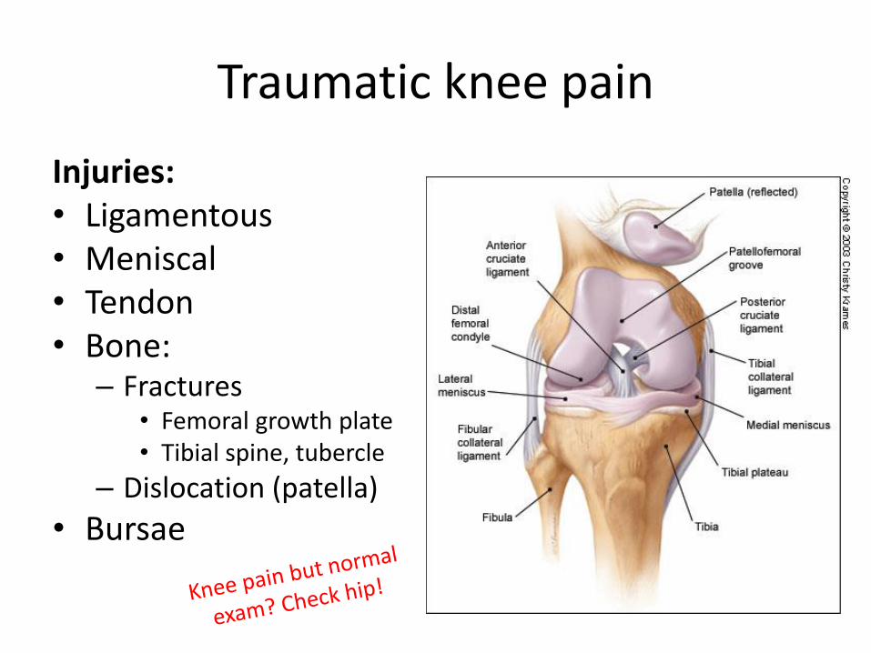
Traumatic knee pain
Injuries:• Ligamentous• Meniscal• Tendon• Bone:
– Fractures• Femoral growth plate• Tibial spine, tubercle
– Dislocation (patella)
• Bursae
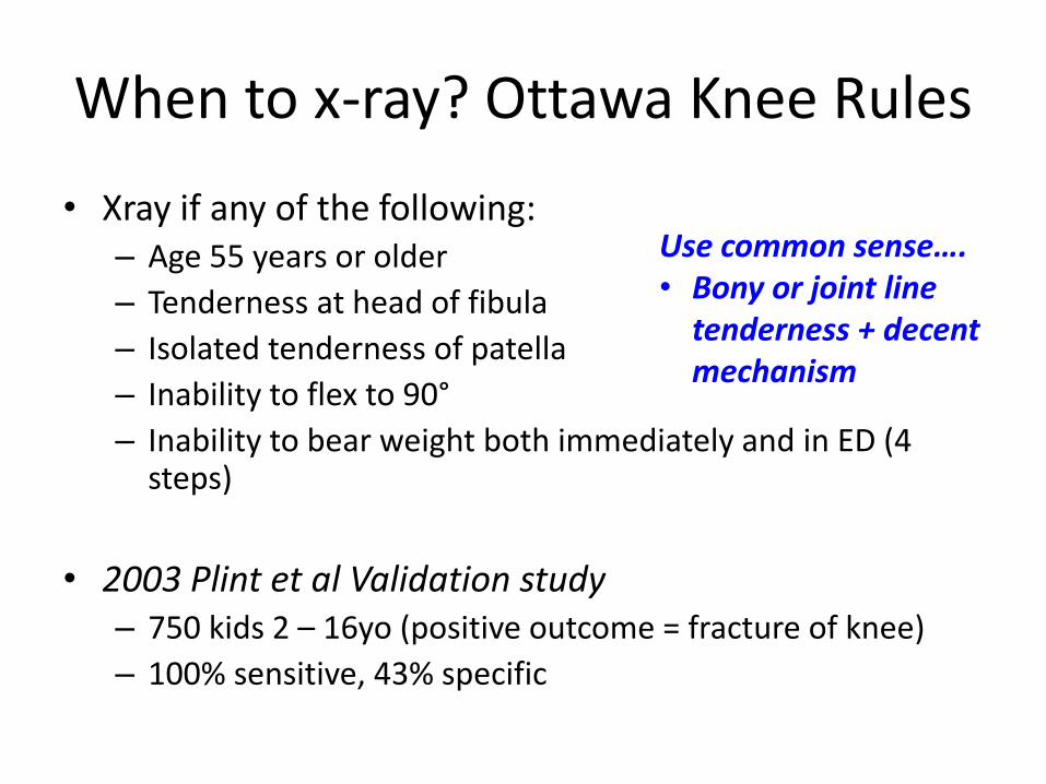
When to x-ray? Ottawa Knee Rules
• Xray if any of the following:– Age 55 years or older
– Tenderness at head of fibula
– Isolated tenderness of patella
– Inability to flex to 90°
– Inability to bear weight both immediately and in ED (4 steps)
• 2003 Plint et al Validation study– 750 kids 2 – 16yo (positive outcome = fracture of knee)
– 100% sensitive, 43% specific
Use common sense….• Bony or joint line
tenderness + decent mechanism
Always image!
• Joint effusion
• “Locked” knee sensation
– Means true intra-articular pathology
– Ie. meniscal tears
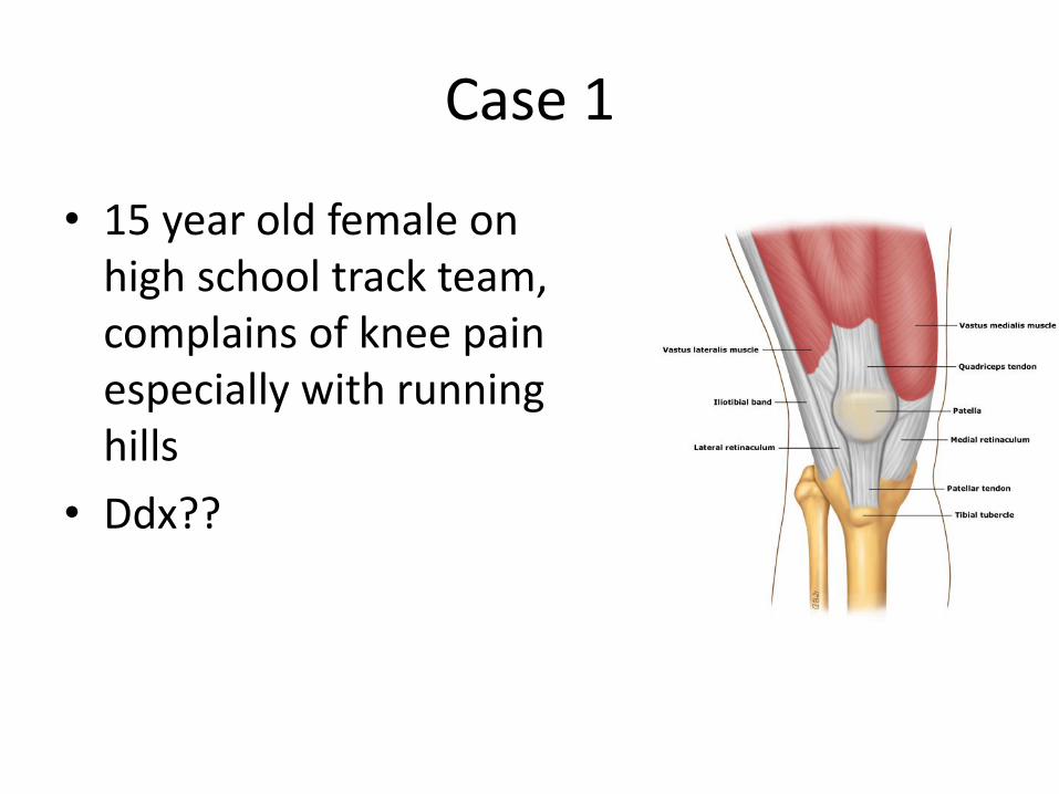
Case 1
• 15 year old female on high school track team, complains of knee pain especially with running hills
• Ddx??
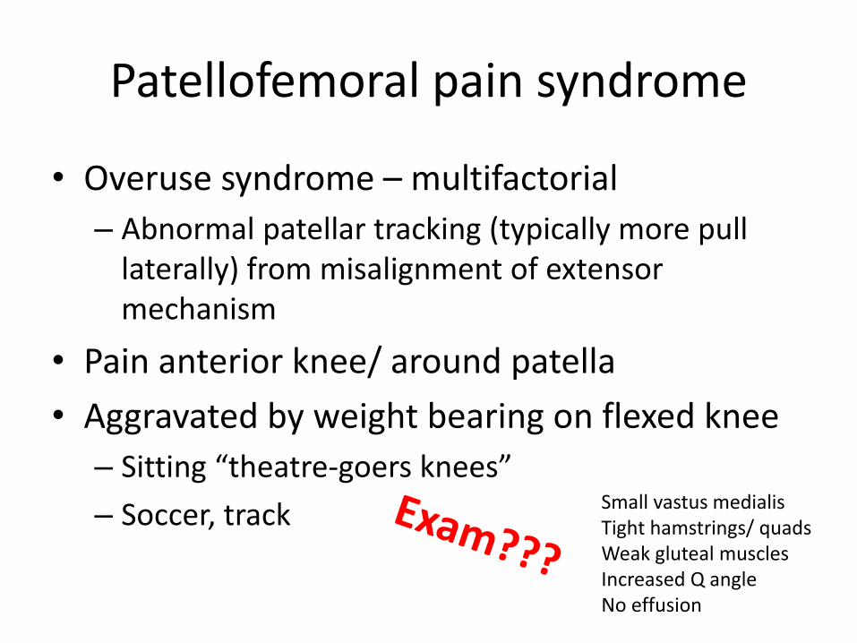
Patellofemoral pain syndrome
• Overuse syndrome – multifactorial
– Abnormal patellar tracking (typically more pull laterally) from misalignment of extensor mechanism
• Pain anterior knee/ around patella
• Aggravated by weight bearing on flexed knee
– Sitting “theatre-goers knees”
– Soccer, track Small vastus medialisTight hamstrings/ quadsWeak gluteal musclesIncreased Q angleNo effusion
Case 2
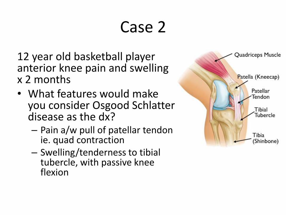
12 year old basketball player anterior knee pain and swelling x 2 months• What features would make
you consider Osgood Schlatterdisease as the dx?– Pain a/w pull of patellar tendon
ie. quad contraction– Swelling/tenderness to tibial
tubercle, with passive knee flexion
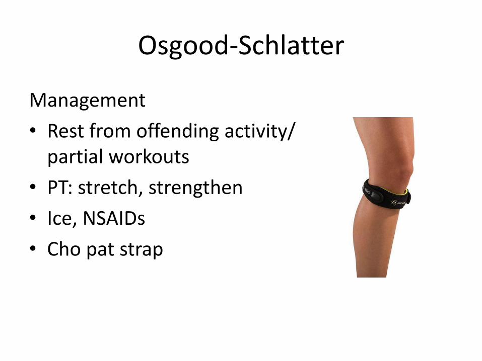
Osgood-Schlatter
Management
• Rest from offending activity/ partial workouts
• PT: stretch, strengthen
• Ice, NSAIDs
• Cho pat strap
Case 3
• 14 year old playing rugby got tackled – heard a pop in his knee. He can’t weight bear and is in a lot of pain….
What is his most likely injury?
ACL injury
• Deceleration injury, heard pop, can’t weight bear, swollen knee = ACL tear until proven otherwise
• Exam: *compare to other side
– Anterior drawer
– Lachman (more sensitive)
– Pivot shift test (specific not sens)
ACL injury – dx/ management
• X-ray?
• MRI?
• Return to activity?
• Surgery?
• Follow-up is the key…. Physio, sports med
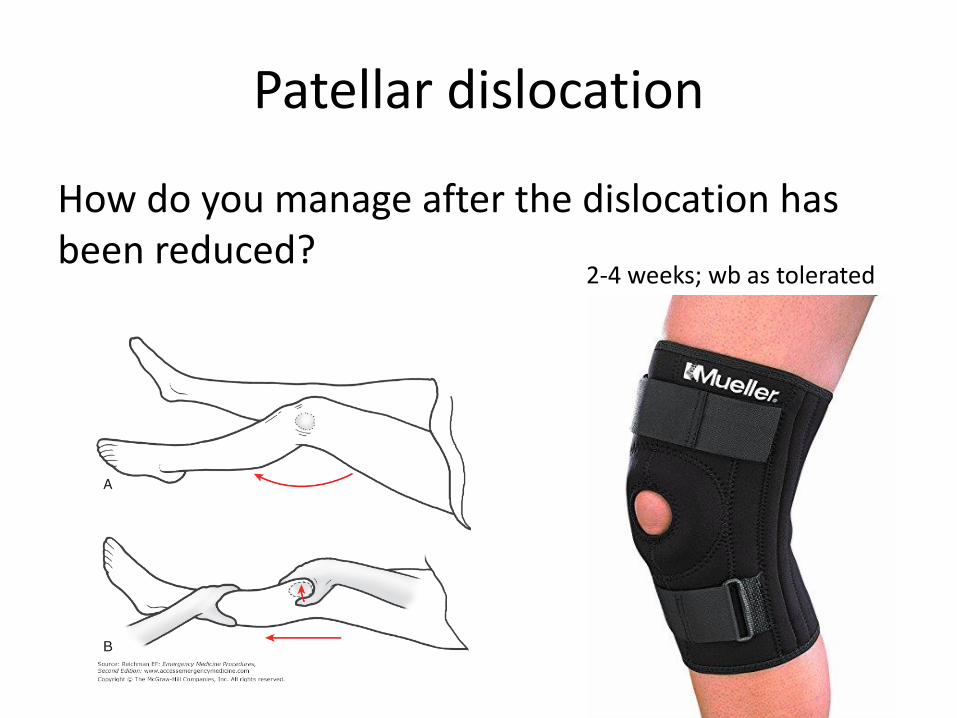
Patellar dislocation
How do you manage after the dislocation has been reduced?
2-4 weeks; wb as tolerated
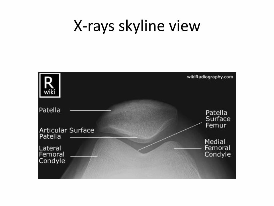
X-rays skyline view

ELBOWFOREARM
CLAVICLE/ SHOULDER
LOWER LEG/ ANKLE
MSK injuries:
Let’s get
specific…
BACK
KNEE

BACK
Case 1
14 year old previously healthy girl who has been suffering from lower back pain for 3 weeks that limits her physical activities
• What could be going on?– Trauma
• ?
– Atraumatic….• Infectious (spinal vs referred)
• Inflammatory eg. transverse myelitis or discitis
• Autoimmune/rheum
• Neoplastic
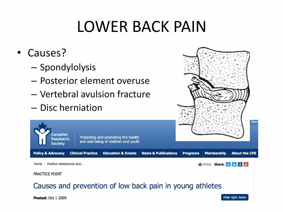
LOWER BACK PAIN
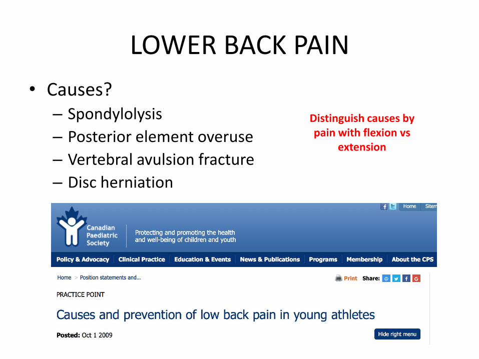
• Causes?– Spondylolysis
– Posterior element overuse
– Vertebral avulsion fracture
– Disc herniation
Distinguish causes by pain with flexion vs
extension
Spondylolysis
• Who is at risk?
– Athletes performing repeated extension/rotation
• How does someone present with it?
• What is spondylolisthesis?
Spondylolysis
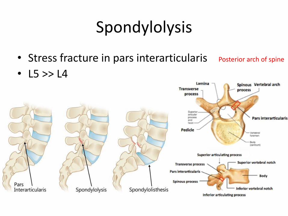
• Stress fracture in pars interarticularis
• L5 >> L4
Posterior arch of spine
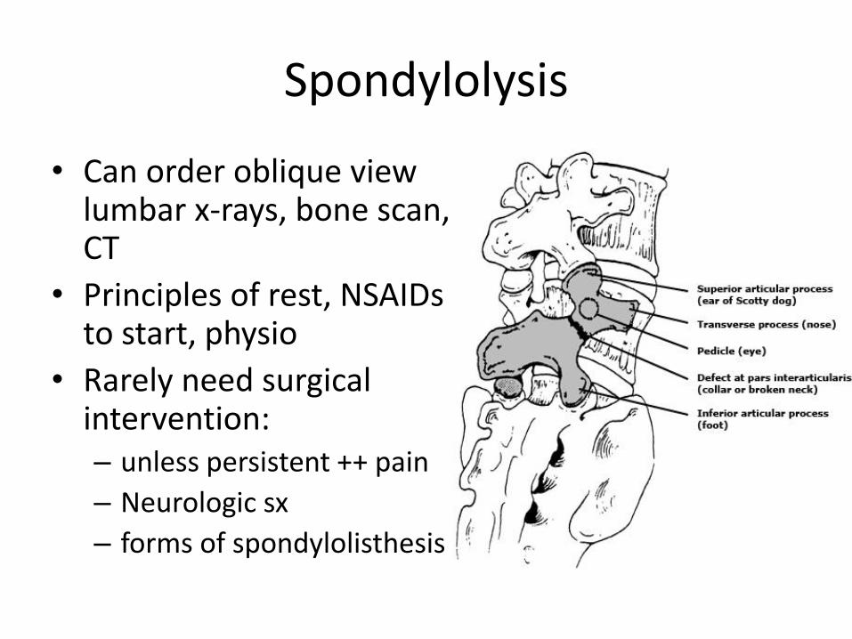
Spondylolysis
• Can order oblique view lumbar x-rays, bone scan, CT
• Principles of rest, NSAIDs to start, physio
• Rarely need surgical intervention: – unless persistent ++ pain
– Neurologic sx
– forms of spondylolisthesis
LOWER BACK PAIN
• Causes?– Spondylolysis
– Posterior element overuse
– Vertebral avulsion fracture
– Disc herniation
Distinguish causes by pain with flexion vs
extension
Case 2
• 7 year old at the playground, falls off the side of a slide, gets up and walks away from it, but complains of persistent mid-back pain
• How do you assess the child’s back pain?
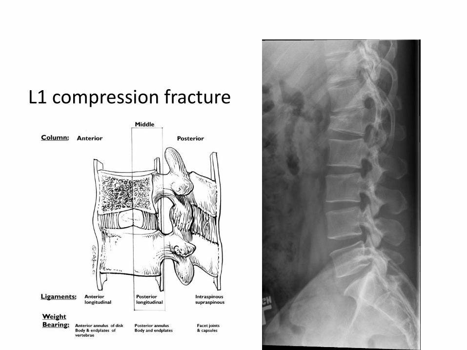
L1 compression fracture
Compression fractures
• Hyperflexion and axial/compressive loading
• Anterior column compressed
• CT used to confirm that vertebral body pedicles and lamina are intact– Consider if more than 50% compression or medial
or posterior columns not intact on x-ray
• Often managed conservatively; orthoconsultation
• Do not return to sport unless no pain

ELBOWFOREARM
CLAVICLE/ SHOULDER
LOWER LEG/ ANKLE
MSK injuries:
Let’s get
specific…
BACK
KNEE
Summary
• Approach injuries as acute vs chronic/overuse
• Create ddx based on the different tissues at the site of pain
• Know your anatomy and MSK exam!
• Remember basic injury management principles
• Some fractures are more common in peds than others, and management is typically conservative
• Emphasize the right time to return to play to avoid longer-term issues/ re-injury

QUESTIONS?