Embed Size (px)
Citation preview

©2018 American Pediatric Surgical Association 1
PEDIATRIC SURGICAL CRITICAL CARE SYLLABUS & STUDY GUIDE
First Edition 2018
On behalf of the American Pediatric Surgical Association Critical Care Committee
Editors-in-Chief:
Mary K. Arbuthnot, DO
Samir Gadepalli, MD Pramod S. Puligandla, MD
©2018 American Pediatric Surgical Association 2
Corresponding Author:
Mary K. Arbuthnot, DO LCDR, MC, USN
Department of General Surgery Pediatric Surgical Critical Care
Naval Medical Center Portsmouth [email protected]
[email protected] Cardiovascular Critical Care
Respiratory Critical Care Neurological Critical Care
Infectious Disease Renal Disease
Gastrointestinal Disease Critical Care Nutrition
Hematology Endocrinology
Analgesia and Sedation Toxicology
Thermal Injuries Obstetrical Critical Care Pediatric Emergencies
The Elderly Emergency/Trauma Surgery
Transplant Statistics
Ethics Principles of Administration
Contributors:
Shahab Abdessalam, MD
Director, Trauma, Burns and Critical Care Associate Professor of Surgery
Children’s Hospital and Medical Center - Omaha Omaha, Nebraska Infectious Disease
Renal Disease
Jennifer H. Aldrink, MD Associate Professor of Clinical Surgery and Pediatrics Department of Surgery, Division of Pediatric Surgery
The Ohio State University College of Medicine

©2018 American Pediatric Surgical Association 3
Nationwide Children’s Hospital Columbus, OH
Emergency/Trauma Surgery Ethics
Kelly Austin, MD, MS, FACS, FAAP
Associate Professor of Surgery University of Pittsburgh School of Medicine
Children’s Hospital of Pittsburg of UPMC Pittsburgh, PA
Gastrointestinal Disease Obstetrical Critical Care
David W. Bliss, MD, FACS, FAAP
Professor of Clinical Surgery Keck USC School of Medicine Cardiovascular Critical Care
Neurological Critical Care Critical Care Nutrition
Alexander Feliz, MD
Assistant Professor of Surgery and Pediatrics University of Tennessee Health Science Center
Le Bonheur Children's Hospital Hematology
Endocrinology
Samir Gadepalli, MD Assistant Professor of Surgery
Division of Pediatric Surgery, Department of Surgery University of Michigan
Ann Arbor, MI Cardiovascular Critical Care
Statistics Principles of Administration
David Juang, MD
Director, Trauma and Surgical Critical Care Director, Pediatric Surgical Critical Care Fellowship
Children’s Mercy Hospital Associate Professor of Surgery
University of Missouri - Kansas City Kansas City, MO Thermal Injuries
Transplant

©2018 American Pediatric Surgical Association 4
Chris Newton, MD
Director, Trauma and Surgical Critical Care Director, Pediatric Neuroscience Center
Department of Surgery UCSF-Benioff Children's Hospital Oakland
Pediatric Emergencies
Pramod S. Puligandla, MD Professor of Pediatric Surgery, Pediatrics and Surgery
Program Director, Pediatric Surgery McGill University, Faculty of Medicine
Attending Staff, Pediatric General and Thoracic Surgery Attending Staff, Pediatric Critical Care Medicine
Montreal Children's Hospital, McGill University Health Centre Montreal, QC, Canada Respiratory Critical Care
Robert Ricca, MD
CAPT, MC, USN Associate Professor of Surgery, USUHS
Director, Surgical Services Naval Medical Center Portsmouth, VA
Toxicology The Elderly
Ana Ruzic, MD
Assistant Professor of Surgery Division of Pediatric Surgery Kentucky Children’s Hospital
Analgesia and Sedation
PEDIATRIC SURGERY NaT www.pedsurglibrary.com/apsa
Disclaimer: The views expressed in this article are those of the authors and do not necessarily reflect the official
policy or position of the Department of the Navy, Department of Defense, or the United States Government
Copyright Statement: I am (a military service member) (an employee of the U.S. Government). This work was prepared as part of my official duties. Title 17 U.S.C. 105 provides that “Copyright protection under this title is
not available for any work of the United States Government.” Title 17 U.S.C. 101 defines a United States Government work as a work prepared by a military service member or employee of the United States
Government as part of that person’s official duties.

©2018 American Pediatric Surgical Association 5
Table of Contents:
Chapter 1: Cardiovascular Critical Care page 6 Chapter 2: Respiratory Critical Care page 18 Chapter 3: Neurological Critical Care page 31 Chapter 4: Infectious Disease page 36 Chapter 5: Renal Disease page 43 Chapter 6: Gastrointestinal Disease page 46 Chapter 7: Critical Care Nutrition page 48 Chapter 8: Hematology page 49 Chapter 9: Endocrinology page 54 Chapter 10: Analgesia and Sedation page 58 Chapter 11: Toxicology page 64 Chapter 12: Thermal Injuries page 66 Chapter 13: Obstetrical Critical Care page 70 Chapter 14: Pediatric Emergencies page 71 Chapter 15: The Elderly page 79 Chapter 16: Emergency/Trauma Surgery page 81 Chapter 17: Transplant page 86 Chapter 18: Statistics page 89 Chapter 19: Ethics page 91 Chapter 20: Principles of Administration page 92 References page 95

©2018 American Pediatric Surgical Association 6
1. Cardiovascular Critical Care i. Physiology
i. Understand the definitions of CO, preload, afterload, compliance, SVR
ii. Understand the relationship between CO, right atrial pressure and venous return (Frank-Starling Relationship) and how it changes with certain conditions i.e cardiac tamponade, congestive heart failure, Persistent Pulmonary Hypertension of the Newborn (PPHN)
Image courtesy of Pediatric Surgery NaT “Cardiophysiology and Shock”

©2018 American Pediatric Surgical Association 7
iii. Understand the phases of the cardiac cycle
iv. Understand the effects of preload and afterload on the pressure-volume loops and the changes with pulmonary hypertension, cardiac tamponade, CHF
ii. Understand fetal circulation and the physiology associated with
persistence of fetal circulation
iii. Know the common congenital heart defects. Images can be found at: http://www.stanfordchildrens.org/en/topic/default?id=congenital-heart-disease-90-P02346) Classification of congenital heart lesions
Noncyanotic heart disease Cyanotic heart disease
left to right shunts decreased pulmonary blood flow
atrial septal defects
ventricular septal defects
atrioventricular septal defects
aortopulmonary window
patent ductus arteriosus
tetrology of Fallot
pulmonary stenosis
pulmonary atresia
tricuspid atresia
Ebstein’s anomaly
left sided obstructive lesions increased pulmonary blood flow
coarctation of the aorta
congenital aortic stenosis
interrupted aortic arch
mitral stenosis
transposition of the great vessels
double outlet right ventricle
total anomalous pulmonary venous
return
truncus arteriosus
single ventricle physiology
hypoplastic left heart
double inlet left ventricle
Image courtesy of Pediatric Surgery NaT “Congenital Heart Disease” Congenital heart disorders
left to right shunt (with
congestive heart failure)
atrial septal defect
patent ductus arteriosus

©2018 American Pediatric Surgical Association 8
ventricular septal defect
atrioventricular canal
right to left shunt tetralogy of Fallot
tricuspid atresia
transposition of the
great vessels
total anomalous
pulmonary venous
return
truncus arteriosus
obstructive lesions (with
ventricular hypertrophy and
failure)
aortic stenosis
coarctation of the aorta
pulmonary stenosis
interrupted aortic arch
total mixing lesions double outlet right
ventricle
hypoplastic
left heart syndrome
Image courtesy of Pediatric Surgery NaT “Congenital Heart Disease”
iv. Hemodynamic Monitoring
i. Invasive and noninvasive arterial blood pressure monitoring 1. Sphygmomanometer measurements give slightly higher
systolic and lower diastolic pressures compared to intra-arterial measurements
2. Ideal MAP 60-90 but varies depending on cause of HD instability, history of hypertension, presence of TBI
3. In neonates, MAP is (gestational age at birth)+(age in weeks), but perfusion more important than MAP
ii. CVP monitoring 1. CVP alone does not reflect volume status 2. Dynamic decreases of CVP > 2 mmHg with spontaneous
respiration can be indicative of patient who are volume responsive
iii. Pulmonary artery pressure and pulmonary artery occlusion pressure and assessment of LV
1. Understand correct placement 2. Pulmonary artery occlusion pressure (Ppao) can be
used to assess PVR, pulmonary edema, intravascular volume, LV preload and performance

©2018 American Pediatric Surgical Association 9
a. Calculate PVR i. PVR = (mean pulmonary artery pressure
–Ppao)/CO b. Preload=LVEDV c. Afterload = LV wall stress = LVEDV x diastolic
arterial pressure iv. Recognition of normal right atrial tracings and pathological
tracings and how they correspond to the QRS complex A - Atrial contraction X - Atrial relaxation C - Ventricular contraction V - venous return/filling Y - opening of tricuspid valve
v. Cardiac output 1. Non-invasive
a. Echo i. Can be used to assess stroke volume,
ejection fraction, fluid resuscitation ii. Can assess RV pathology: pulmonary
embolism (RV overload), cardiac tamponade (pericardial fluid, right ventricular diastolic collapse)
iii. Can assess diastolic dysfunction: acute heart failure, volume overload
b. Transcutaneous/esophageal Doppler ultrasound, bioimpedence, bioreactance, passive leg raise, pulse variation
2. Invasive a. Thermodilution (pulmonary arterial catheter),
pulse waveform analysis vi. Tissue Oxygenation/Perfusion
1. Pulse oximetry a. Tissue light absorption 660 nm (red) and 940
nm (infrared) b. SpO2 <90% = hypoxia c. Understand the oxygen-hemoglobin
disassociation curve and the Bohr effect i. Understand the p50 of Hgb and how PaO2
correlates with SpO2
Oxyhemoglobin Dissociation

©2018 American Pediatric Surgical Association 10
Image courtesy of Pediatric Surgery NaT “Respiratory Care”
2. Understand Oxygen content/delivery/Fick Principle
and must know formulas: i. DO2 = CO x CaO2
ii. VO2 = CO (CaO2 – CvO2) iii. CaO2 = (Hb X 1.34 X SaO2) + (PaO2 X
.003) iv. CvO2 = (Hb X 1.34 X SvO2) + (PvO2 X
0.003) b. Understand the difference between ScvO2 vs.
SvO2 i. ScvO2 from central line in SVC, neglects
drainage from lower body, usually lower than SvO2 by 2-3% because lower body extracts less O2
1. May be altered by left to right cardiac shunts
ii. Not a good surrogate in shock

©2018 American Pediatric Surgical Association 11
1. Can overestimate SvO2 in septic shock and underestimate in cardiogenic or hypovolemic shock
2. ScvO2 <65% likely reflects inadequate DO2
3. NIRS a. Determines tissue O2 saturation (StO2)
i. Approximates saturation in small end vessels
ii. >85% adequate resuscitation v. Shock
i. Classification of Shock 1. Know the differences between Hypovolemic,
Cardiogenic, Extracardiac, Obstructive, Distributive, Mixed Shock
a. Define SIRS and Sepsis b. Physiologic characteristics of shock:
2. Cellular response a. Understand the relationship between cellular
respiration/oxidative phosphorylation/ATP b. Understand anaerobic metabolism and lactate
production c. Prevailing theory is there is a defect in cellular
oxygen utilization, not impaired tissue oxygenation
i. Tissue levels of O2 can be increased in severe sepsis
3. Resuscitation a. Surviving Sepsis Campaign: International
Guidelines for Management of Sepsis and Septic Shock: 2016 Goals:
i. CVP 8-12 mmHg (12 -15 if ventilated) ii. MAP ≥ 65 mmHg
iii. UOP ≥ 0.5 mL/kg/hr iv. ScvO2 >70%
b. Fluid therapy for sepsis-induced hypoperfusion i. 30 mL/kg IV crystalloid in first 3 hours
ii. Additional fluids guided by hemodynamics
c. Vasopressor therapy i. Norepinephrine is the first line
vasopressor ii. Addition of vasopressin or epinephrine to
norepinephrine to reach MAP goals or decrease norepinephrine dosage
d. Antibiotics

©2018 American Pediatric Surgical Association 12
i. Within 1 hour, after blood cultures ii. Know empiric regimens
e. Steroids i. Recommend against steroids if IV fluids
and pressors are able to restore hemodynamics. If not, recommend hydrocortisone 200 mg/day
f. If shock is not resolving, assess hemodynamic (cardiac) function
g. Lactate can help to guide resuscitation vi. Arrhythmias
i. Approach to arrhythmias 1. Fast or slow? HR >100 or <100? QRS wide or narrow?
Regular or irregular? ii. Tachyarrhythmias
1. Regular narrow complex tachycardia a. Sinus tachycardia b. Atrial Tachycardia c. Paroxysmal SVT: AVNRT and AVRT d. Atrial Flutter
2. Irregular Narrow Complex Tachycardia a. Sinus tachycardia with PAC’s b. Multifocal Atrial Tachycardia c. Atrial Fibrillation d. Atrial Flutter with Variable Block
3. Wide Complex Tachycardia a. QRS >120 ms due to 4 mechanisms:
i. VT ii. Antidromic accessory conduction
pathway (WPW) iii. SVT with pre-existing/rate-related BBB iv. Pacemaker mediated tachycardia
b. Regular Wide Complex Tachycardia i. Ventricular Tachycardia
ii. Regular SVT with pre-excitation iii. Regular Supraventricular Tachycardia
with BBB c. Irregular Wide Complex Tachycardia
i. Polymorphic Ventricular Tachycardia 1. Torsade’s de pointes
ii. Ventricular Fibrillation iii. Irregular SVT with pre-excitation iv. Irregular SVT with BBB
iii. Bradyarrhythmias 1. Be able to describe the following types of sinus node
dysfunction/Sick Sinus Syndrome (SSS)

©2018 American Pediatric Surgical Association 13
a. Sinus bradycardia b. Sinus exit block c. Sinus arrest
iv. Nodal Arrhythmias 1. AV Block
a. First-degree b. Second degree
i. Mobitz I/Wenckebach ii. Mobitz II
c. Third degree/Complete heart block 2. AV (Junctional) Escape Rhythm 3. Ventricular Escape Rhythm 4. Accelerated Idioventricular Rhythm
vii. Heart Failure i. NYHA Classification
ii. Pathophysiology of heart failure 1. Systolic vs. Diastolic 2. Right heart vs. Left Heart
iii. Management 1. Preload and afterload reduction 2. Inotropic therapy 3. Pharmacologic therapy
a. Diuretic, glycosides, ACEI, vasodilators, CCB, beta blockers, statins
4. Mechanical Support and Transplantation a. Intra-Aortic Balloon Pump
i. Normal and abnormal waveforms b. Ventricular Assist Devices (bridge to transplant
vs. destination therapy) c. ECMO
viii. Valvular Heart Disease i. Aortic Stenosis
1. Reliance on HR for maintenance of CO a. Thick noncompliant left ventricle b. NSR important (hypertrophied ventricle needs
atrial kick) c. Hypovolemia and hypotension poorly tolerated
2. Angina, syncope, heart failure => end stage disease 3. After onset of symptoms, 5-yr survival drops to zero 4. Classification of AS 5.
Type Valve Area (cm2) Max Aortic Velocity Mean Pressure
Gradient
Mild 1.5-2.0 2.0-2.9 <20
Moderate 1.0-1.5 3.0-3.9 20-39

©2018 American Pediatric Surgical Association 14
Severe 0.6-1.0 4.0-4.9 40-59
Very Severe <0.6 >5.0 >60
Created by Mary Arbuthnot 6. Class I Indications for repair
a. Symptomatic patients with severe AS b. Patients with AS undergoing CABG c. Patients with AS undergoing surgery on aorta or
other valves d. Patients with severe AS and EF < 50%
ii. Mitral Stenosis 1. Almost always a result of rheumatic fever 2. LA dilates => predisposes to thrombi, SVT, a. fib,
reduced LV preload 3. Increased LA pressure->pulmonary edema->elevated
PVR->pulmonary HTN-> Right heart failure 4. Management goals: maintain NSR, avoid tachycardia,
even fluid balance, avoid elevations in PVR 5. Class I Indications for Repair
a. NYHA Class III-IV heart failure and moderate to severe AS when valvotomy is unavailable or not possible
b. Symptomatic patients with moderate to severe MS and moderate to severe MR
iii. Aortic Insufficiency 1. Due to disease of the valve, aortic root or both
a. Heavy calcified valves, Marfan’s/syphilis can dilate root, trauma, endocarditis, dissection
b. LV dilates -> eccentric hypertrophy c. Slow HR allows more time for regurgitation.
Should be maintained in 80-100 range, avoid acute increases in BP, maintain preload
d. Class I indications for repair of chronic AI i. Symptomatic patients with severe AI
ii. Asymptomatic patients with severe AI and EF <50%
iii. Severe AI undergoing CABG or other cardiac surgery
iv. Mitral Regurgitation 1. Chronic MR 2-3% of population
a. Developing world – rheumatic heart disease most common
b. US – degenerative MR most common i. Degenerative MR – leaflets abnormalities,
MVP, dilation of mitral annulus due to ischemia or remodeling
ii. LA has time to dilate

©2018 American Pediatric Surgical Association 15
c. Acute MR – ischemia or rupture of papillary muscles, endocarditis, trauma
i. LA does not have time to dilate -> pulmonary edema
d. LA enlarges, predisposes or arrhythmias e. LV undergoes eccentric hypertrophy f. Avoid bradycardia and acute elevations in BP g. Class I indications for Repair
i. Symptomatic patients with severe MR ii. Patients with severe MR and > NYHA class
II symptoms iii. Asymptomatic patients with severe MR
and EF between 30-60% and/or with an LV and systemic dimension less than or equal to 40 mm
ix. Aortic Dissection i. Rupture of the intima and separation of the layers of the aortic
wall. Blood in the false lumen obstructs flow to branches of the aorta
ii. Know the associated risk factors 1. HTN 2. Congenital (bicuspid aortic valve) 3. Pregnancy 4. Crack/Cocaine use 5. Connective tissue disease
a. Ehlers-Danlos, Marfan, Loeys-Dietz iii. Types of Dissection
1. Acute – Diagnosis made within 2 weeks of symptoms 2. DeBakey Classification
a. Type I – involvement of both the ascending and descending aorta
b. Type II – involves only the ascending aorta c. Type III – involves only the descending aorta
3. Stanford System a. Type A – involves the ascending aorta (more
common, grimmer prognosis.) b. Type B – involves the aorta distal to the left
subclavian artery iv. Diagnosis and treatment of acute dissection
1. Helical CT 80% sensitivity and specificity a. Limitation – ability to detect ascending aortic
dissection 2. MRI – 95% sensitivity and specificity, can evaluate
branch vessels a. Limitation – long study
©2018 American Pediatric Surgical Association 16
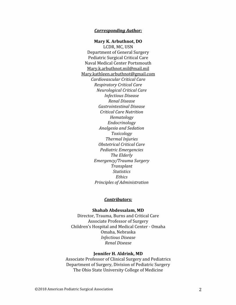
3. TEE – infrequently used as it requires sedation and does not visualize much of the aortic arch
4. Immediate administration of anti-hypertensives – arterial pressure as low as possible to maintain perfusion
a. Beta-blockers first line (esmolol) b. Nitroprusside – can use concurrently, not as
single agent (reflex tachycardia) 5. Uncomplicated distal dissections – medically managed
a. Indications for surgery in distal dissections – expanding aneurysm, impeding rupture, ongoing pain, impairment of blood flow to a limb or organ
b. Endovascular repair an option 6. Proximal dissections – managed surgically
a. Usually median sternotomy – goal is to primarily repair the intimal tear, replacement of the dilated aorta, and aortic valve replacement/repair.
x. Cardiopulmonary Bypass i. Causes systemic reaction that has adverse effects
1. Inflammatory mediators: complement, endotoxin, TNF-alpha, platelet-activating factor, cytokines, free radicals, NO
2. Caused by hypothermia, hemodilution, nonpulsatile flow, exposure to the circuit, ischemia-reperfusion injury, activation of the complement system through the alternative pathway
3. Can lead to acute lung injury, renal failure, altered hepatic function, coagulopathy, multiple organ failure
xi. Acute Coronary Syndromes i. STEMI
1. PCI – understand indications 2. Thrombolytic therapy – understand indications and
contraindications ii. NSTEMI and Unstable Angina (UA)
1. UA – coronary ischemia at rest or with minimal exertion often lasting >20 minutes
2. NSTEMI – unstable symptoms accompanied by elevated troponins without acute ST segment elevation
3. TIMI Risk Score iii. Management
xii. Hypertensive Crises i. Hypertensive Crises
1. Severe elevations in BP 2. Can reduce BP gradually over 24-48 hours
a. Oral meds, no ICU or a-line

©2018 American Pediatric Surgical Association 17
ii. Hypertensive Emergencies 1. Severe elevation in BP with acute end-organ damage
(CVS, CNS, renal) 2. Requires reduction of BP within 1-2 hours
a. IV meds, a-line, ICU b. Lower MAP 15-25% over several minutes/hours.
Can result in hypoperfusion if lowered too fast

©2018 American Pediatric Surgical Association 18
2. Respiratory Critical Care i. Be familiar with airway anatomy ii. Be familiar with the bronchopulmonary segments of the lung
“The bronchopulmonary segments of the lung are shown here”
Image and caption courtesy of Pediatric Surgery NaT “Respiratory Care”
iii. Know the differences in Adult and Pediatric Airways iv. Noninvasive Airway Management
i. Understand the different ways to provide supplemental oxygen 1. Nasal cannula, simple mask, partial rebreather,
nonrebreather, Venturi mask, high flow nasal cannula 2. Both high flow and low flow can deliver a range of FiO2.
High and low describe flow of gas a. Nasal Cannula: 1-6 L/min (21-44% O2) b. Simple Face Mask: 6-10 L/min (35-60% O2) c. Nonrebreather 6-9 L/min (60-90% O2), 10-15
L/min (95-100% O2)

©2018 American Pediatric Surgical Association 19
d. Venturi Mask: 4-8 L/min (24-40% O2), 10-12 L/min (40-50% O2)
v. Noninvasive Positive Pressure Ventilation (NIPPV) i. Understand the advantages and differences between CPAP and
BiPAP ii. CPAP provides continuous positive pressure during in
inhalation and exhalation (does not provide true ventilatory support)
1. Improves oxygenation by: a. Preventing airway collapse b. Expanding end-expiratory lung volume and
increasing FRC c. Improves LV function by reducing pre-load and
afterload. iii. BiPAP provides increased pressure support during inhalation
as well as PEEP, and has the ability to set up a back-up ventilatory rate. Ventilation achieved from pre-set IPAP and will vary based on total thoracic compliance and resistance as well as autopeep.
iv. Benefits: decrease in direct airway trauma, preservation of upper airway defense mechanisms, maintenance of upper airway function and comfort, decrease in sedation, assists spontaneous respirations. Drawbacks: no direct removal of secretions
vi. Understand the clinical indications to intubate vii. Understand how to perform Rapid Sequence Intubation
i. Pre-oxygenation, positioning, monitoring, know equipment including ETT sizing and types of laryngoscopes, know RSI medications, suction, IV access, preparation for resuscitation
viii. Understand how to manage a difficult airway and the ASA difficult airway algorithm (see below)
i. Visit http://airwayeducation.homestead.com/ASA.html ix. Understand ABG interpretation and acid-base status
i. Henderson-Hasselbach equation 1. pH = 6.1 + log(HCO3/(0.03 x PaCO2))
a. [H+] = 24 x paCO2/HC03- b. pH = -log [H+]
ii. Ask the following questions when presented with a case scenario (6):
1. What is the pH? Acidemia or Alkalemia?
Variable Normal Range
pH 7.35-7.45
pCO2 35-45
HCO3- 22-26

©2018 American Pediatric Surgical Association 20
Anion Gap (AG) 8-12
Albumin 4
Created by Mary Arbuthnot
2. What is the primary disorder present?
Acid Base Disorder pH pCO2 [HCO3-] Metabolic Acidosis Low Low Low Respiratory Acidosis Low High High Metabolic Alkalosis High High High Respiratory Alkalosis
High Low Low
Created by Mary Arbuthnot
3. Is there appropriate compensation? 4. Is the compensation acute or chronic?
(memorize tables below, especially underlined numbers)
Compensation
Respiratory Acidosis
Acute: for every 10 increase in pCO2 -> HCO3 increases by 1 and there is a decrease of 0.08 in pH
Chronic: for every 10 increase in pCO2 -> HCO3 increases by 4 and there is a decrease of 0.03 in pH
Respiratory Alkalosis
Acute: for every 10 decrease in pCO2 -> HCO3 decreases by 2 and there is a increase of 0.08 in PH
Chronic: for every 10 decrease in pCO2 -> HCO3 decreases by 5 and there is a increase of 0.03 in PH
Metabolic Acidosis
Winter’s formula: pCO2 = 1.5[HCO3] + 8 ± 2 If serum pCO2 > expected pCO2 -> additional respiratory acidosis
Metabolic Alkalosis
For every 10 increase in HCO3 -> pCO2 increases by 6
Created by Mary Arbuthnot
5. Is there an anion gap? a. AG = Na – Cl – HCO3 (normal 8-12) b. AG corrected = AG + 2.5[4 – albumin]
i. Know causes of anion gap (MUDPILES) and non-anion gap acidosis
6. If there is a AG check the delta gap?

©2018 American Pediatric Surgical Association 21
a. Delta gap = (actual AG – 12) + HCO3 i. If delta gap > 30 -> additional metabolic
alkalosis ii. If delta gap < 18 -> additional non-gap
metabolic acidosis iii. If delta gap 18 – 30 -> no additional
metabolic disorders
x. Respiratory Failure i. Characterize the difference between Primary Ventilatory
Failure (Respiratory muscle fatigue, CNS depression, Respiratory muscle weakness, chest wall defects) leading to hypoventilation and hypercapnia vs. Primary Oxygenation Failure (Cardiac failure or pulmonary hypertension, Alveolar disease) leading to hypoxemia
ii. Know the acute and chronic causes of hypoxemic and hypercapnic respiratory failure.
iii. Be able to describe and give examples of the following pathophysiologic processes:
1. Diffusion abnormalities 2. V/Q inequality 3. Shunt 4. Hypoventilation 5. Reduction in inspired PaO2 6. Increased venous admixture
iv. Know how to calculate an A-a gradient
j. ARDS a. Know the Berlin Definition of ARDS:
i. ARDS Definition Task Force., Ranieri VM, Rubenfeld GD, Thompson BT, Ferguson ND, Caldwell E, Fan E, Camporota L, Slutsky AS. Acute respiratory distress syndrome: the Berlin Definition. JAMA. 2012 Jun 20;307(23):2526-33. doi: 10.1001/jama.2012.5669. PubMed PMID: 22797452
b. Be familiar with the pathology/physiology/biology of ARDS: i. Ware LB, Matthay MA. The acute respiratory distress
syndrome. N Engl J Med. 2000 May 4;342(18):1334-49. Review. PubMed PMID: 10793167.
c. Treatment i. ARDSnet
1. Lung protective ventilation: High Peep, Low tidal volume, permissive hypercapnia: http://www.ardsnet.org/tools.shtml

©2018 American Pediatric Surgical Association 22
d. Be familiar with the indication for or against the use of prone positioning, high frequency oscillation, inhaled nitric oxide and corticosteroids in ARDS – see below link for references:
i. http://pulmccm.org/main/2012/review-articles/mechanical-ventilation-in-ards-2012-update/
e. Be familiar with the Pediatric Acute Lung Injury Consensus Conference recommendations regarding pediatric acute respiratory distress syndrome
i. https://www.ncbi.nlm.nih.gov/pmc/articles/PMC5253180/
ii. Pediatric ARDS (PARDS) 1. Excludes children with perinatal related lung
disease (prematurity related lung disease, perinatal lung injury, congenital abnormalities)
2. Timing is within 7 days of known clinical insult 3. Pulmonary edema and respiratory failure not
explained by cardiac failure of volume overload 4. Imaging with findings of new infiltrate consistent
with acute pulmonary parenchymal disease 5. Risk stratification
a. Noninvasive oxygenation: Full face-mask bi-level ventilation or CPAP >/= 5
i. PF ratio </= 300 (no severity stratification)
b. Invasive mechanical ventilation i. Mild: 4 </= OI < 8
ii. Moderate: 8 </= OI <16 iii. Severe: OI </= 16
1. OI =oxygenation index = (Fio2 × mean airway
pressure × 100)/Pao2 f. Be familiar with indications for extracorporeal life support
(ECLS) from ELSO.org i. Neonatal
1. Severe respiratory failure with potentially reversible cause that if refractory to medical management which may be indicated by the following:
a. OI >40 for 4 hours b. OI >20 for >24 hours despite maximal
therapy or persistent episodes of decompensation
©2018 American Pediatric Surgical Association 23
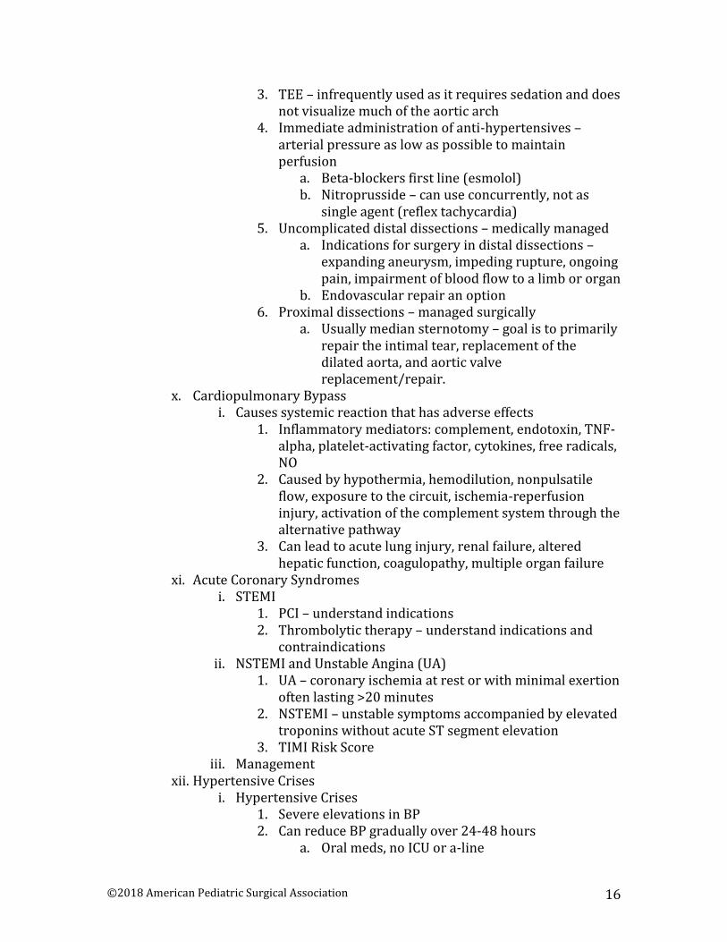
c. Severe hypoxic respiratory failure with acute decompensation (PaO2 <40) unresponsive to intervention
d. Progressive respiratory failure and/or pulmonary hypertension with right ventricular dysfunction or continued high iontropic requirement
e. https://www.elso.org/Portals/0/IGD/Archive/FileManager/8588d1a580cusersshyerdocumentselsoguidelinesforneonatalrespiratoryfailure13.pdf
ii. Pediatric 1. No absolute indications, but consideration is best
within the first 7 days of mechanical ventilation with high levels of support
iii. Adult 1. In hypoxemic failure due to any cause (primary
or secondary), consider when risk of mortality 50%, and is indicated when the risk of mortality is > 80%
a. 50% mortality risk: P/F <150 on > 90% FiO2 and/or Murray score 2-3
b. 80% mortality risk: P/F <100 on >90% FiO2 and/or Murray score 3-4
i. How to calculate the Murray score on MD Calc: https://www.mdcalc.com/murray-score-acute-lung-injury
k. Mechanical Ventilation
a. Know lung volumes and capacities (memorize) b. Know indications for mechanical ventilation c. Know basic concepts of mechanical ventilation
i. Control variables – 4 variables the ventilator adjusts to deliver a breath
1. Pressure, flow, volume, time ii. Phase variables – variables that affect the breath from
beginning to end iii. Trigger – can be triggered by patient effort, determined
pressure, or set volume or flow iv. Limit and cycle variables v. Spontaneous or mandatory breath

©2018 American Pediatric Surgical Association 24
l. “Components of a ventilator breath. 1 - trigger is either initiated by the ventilator or the patient, 2 - limit (what controls the breath) pressure versus volume, 3 - cycle (how the breath is terminated, time versus flow).”
Image and caption courtesy of Pediatric Surgery NaT “Respiratory Care”
a. Understand parameters to set on the ventilator
i. FiO2 ii. Tidal volume (TV)
iii. PEEP iv. Inspiratory flow rate v. Respiratory rate
vi. I:E ratio (Ti = inspiratory time and Te = expiratory time) 1. Ti = TV (L) / Flow rate (1/min) 2. Increasing inspiratory flow rate with constant
RR and TV will shorten I time and lengthen E time
b. Understand the basic modes on the ventilator and pressure volume curves associated with each
i. Volume Control ii. Pressure Control
iii. SIMV iv. Pressure Support

©2018 American Pediatric Surgical Association 25
m. “During volume ventilation, flow is constant during inhalation until the desired volume is reached. Flow then goes to zero as exhalation is a passive process of negative flow. With pressure ventilation, flow is highest in the beginning of the inhalation until the PIP is reached then flow decreases.”
Image and caption courtesy of Pediatric Surgery NaT “Respiratory Care”
i. APRV

©2018 American Pediatric Surgical Association 26
“A high continuous pressure (Phigh) is delivered for a long duration (T high) then falls to a lower pressure (Plow) for a shorter duration (Tlow). The clinician adjusts the high and low pressures with release time. Releasing to the lower pressure allows lung volume to decrease to FRC alllowing ventilation””
Image and caption courtesy of Pediatric Surgery NaT “Respiratory Care”
b. Understand pressure-volume loops and how these change with changes in compliance on both volume controlled and pressure controlled ventilators
i. Understand the inflection points on the pressure-volume loops
I

©2018 American Pediatric Surgical Association 27
“Spontaneous breaths go clockwise. Ventilator breaths go counterclockwise. The bottom of the loop is the PEEP. The area to the right of the line drawn in the middle of the loop represents inspiratory resistance (red) and the area to the left (blue) is the expiratory resistance (blue) . When the red area becomes bigger (hysteresis) it signals higher inspiratory resistance such as a kinked endotracheal tube, patient biting the tube or secretions in the tube. When the blue area gets bigger, it demonstrates higher expiratory resistance such as bronchospasms. The upper point of the curve is the dynamic compliance of the lung.”
Image and caption courtesy of Pediatric Surgery NaT “Respiratory Care”
n. High Frequency Oscillatory Ventilation Pearls a. High rate, short expiratory time b. Preserves end-expiratory volumes and results in intrinsic
PEEP, minimizes cyclic stretch, avoids overdistention c. Alveoli recruitment secondary to mean airway pressure and I:E
ratio d. Frequency 3-15 Hz superimposed on high mean airway
pressures (MAP) (4-5 cm H20 greater than CMV). i. Higher frequency in infants 12-15 Hz
ii. Moderate frequency in young children 8-12 Hz iii. Low levels in older children 3-8 Hz
e. Amplitude (ΔP) is set to achieve good “wiggle” (visible vibrations of trunk to the level of the abdomen) and is

©2018 American Pediatric Surgical Association 28
f. TV correlates with amplitude (ΔP) and is inversely related to frequency
g. Hypoxemia is treating by increasing MAP or decreasing frequency
h. Hypercarbia treated by increasing amplitude, decreasing frequency, or partially deflating ETT cuff
i. Requires frequency monitoring of CXR to evaluate for ovedistention
i. Goal for diaphragm at 8-10th rib o. Obstructive vs. Restrictive Airway Disease
a. Asthma and COPD – understand treatment options, indications for intubation
1. Understand what patients are at risk for auto-PEEP and how to identify this on the ventilator waveform
2. Understand need for prolonged Te
Image courtesy of Pediatric Surgery NaT “Respiratory Care”
b. Be able to recognize and correctly identify flow-volume loops and how they change with obstructive, restrictive or compressive pathology

©2018 American Pediatric Surgical Association 29
Image courtesy of Pediatric Surgery NaT “Respiratory Care”
p. Pulmonary Embolism a. Understand the risk factors for PE b. Understand the hemodynamic alternations in massive PE
i. What are the effects of the RV, RVEDP, MAP that result in subendocardial ischemia of RV
ii. Understand the effects on LV preload that can result in shock
c. Be able to describe the gas-exchange abnormalities and how it affects the A-a gradient
i. V/Q mismatch ii. Low mixed-venous O2 sat
d. Clinically, what are the symptoms of massive PE and what is the most common clinical finding
e. Understands methods to diagnose pre-test probability f. What are diagnostic findings
i. Lab findings ii. On ECG

©2018 American Pediatric Surgical Association 30
1. Tachycardia, S1Q3T3 iii. On Echo
1. RV dilation, hypokinesis, septal flattening, TR, decreased inspiratory collapse of IVC
iv. Chest X-ray 1. Westermark sign 2. Hampton Hump
v. Spiral CT vi. VQ Scan
g. Understand management options i. Limited volume resuscitation
ii. Vasopressors and/or afterload reduction iii. Ventilator support
1. Effect of positive pressure ventilation on RV function
iv. Understand indications and for anticoagulation and thrombolysis
1. Know contraindications to fibrinolysis – see link below
a. http://circ.ahajournals.org/content/123/16/1788.full
h. Understand recommendations for catheter based intervention,
surgical embolectomy and IVC filters

©2018 American Pediatric Surgical Association 31
3. Neurological Critical Care a. Understand cerebral blood flow and autoregulation (Monro-Kellie
Doctrine)
“Initially, the intracranial pressure remains unchanged with increasing volumes due to compensation mechanisms. At elevated ICP, small volume increases cause a significant change in pressure leading to secondary brain injury.” Image and caption courtesy of Pediatric Surgery NaT “Traumatic Brain Injury”
b. Understand the causes of Altered Mental Status and how they present i. Know GCS coma scale
ii. Toxic Metabolic encephalopathy 1. Understand common causes
iii. Delirium 1. Understand causes and risk factors 2. Understand scoring systems in the ICU 3. Understand the term Persistent Vegetative State
iv. Coma 1. Know causes of coma and methods to evaluate brain
function 2. Understand Locked-In Syndrome
c. Seizures/Status Epilepticus (SE)

©2018 American Pediatric Surgical Association 32
i. Know metabolic changes and medications that can provoke seizure
ii. Be able to differentiate between different types of seizures iii. Know the definition of SE
1. Convulsive vs. nonconvulsive SE and difference in outcome
iv. Know first, second and third lines of Anti-epileptic drugs (AEDs)
v. Know indications for seizure prevention in TBI vi. See link for more information:
1. https://www.neurocriticalcare.org/sites/default/files/pdfs/SE%20Guidelines%20NCS%200412.pdf
d. Stroke i. Ischemic Stroke, Subarachnoid Hemorrhage, Intracerebral
Hemorrhage 1. Understand risk factors 2. Know clinical presentation 3. Know components of evaluation and management
a. Diagnosis b. Role for Thrombolysis, recanalization,
reperfusion c. Role for Prevention of infarct expansion and
hemorrhagic conversion d. Prevention and management of complications
4. Know blood pressure parameters 5. Know methods of treatment of cerebral vasospasm
e. Infections i. Review diagnosis, CSF findings, management
ii. Acute Bacterial Meningitis 1. Pathogens
a. Neonates – GBS, GNB (E. Coli), Listeria monocytogebes
b. Toddlers – Strep pneumonia, N. meningitidis (less common, H. influenza type B)
c. Older children - Strep pneumonia, N. meningitides
d. Any age – Mycobacterium tuberculosis 2. Common CSF findings
a. Pleocytosis with neutrophil predominance (typically <1000 WBC/microL, CSF <6 WBC/microL abnormal in infants > 3 months), elevated CSF protein (100-500 mg/dL), decreased CSF glucose (<40mg/dL), and the presence of an organism on CSF Gram stain
iii. Brain Abscess 1. Etiology

©2018 American Pediatric Surgical Association 33
a. Hematogenous spread (usually cyanotic congenital heart disease)
i. MCA distribution ii. S. aureus or strep
b. Direct extension i. Frontal lobes (sinus infections)
1. Anaerobic or strep ii. Temporal lobes or cerebellum (otic
infections) 1. mixed flora
c. Head trauma i. Over fracture area
ii. Skin flora iv. Viral encephalitis
1. Transmission a. Hematogenous
i. Anthropod-borne viruses (following insect bite)
b. Intraneuronal i. Herpes family
2. Treatment a. Acyclovir b. Supportive therapy
f. Posterior Reversible Encephalopathy Syndrome (PRES)
i. Dysfunctional cerebral auto-regulation and compromised cerebral endothelial barrier
ii. Presents with progressive mental status changes, headache, visual disturbances, seizures, hypertension often present, symmetrical white matter edema in posterior cerebral hemispheres (parieto-occipital regions)
iii. Associated conditions 1. HTN, pre/eclampsia, post-transplant, autoimmune,
electrolyte or endocrine disorders, TTP/HUS, sepsis, hepatic failure, massive blood transfusion, EPO therapy or porphyria
iv. Associated medications 1. Immunosuppressives, immunomodulators,
chemotherapeutic agents, high dose steroids v. Treatment: control of HTN and seizure
g. Review the following Neuromyopathies i. Myasthenia Gravis
ii. Guillain-Barré iii. Critical Illness Polymyopathy
h. Traumatic Brain Injury i. Avoid hypotension and hypoxia

©2018 American Pediatric Surgical Association 34
ii. PaO2<60 and SBP > 90 iii. <8 intubate iv. Signs of intracranial HTN prompt HOB elevated to 30 degrees,
hyperventilation (PaCO2 35-45), and treatment with hypertonic agents
v. Know indications for invasive ICP monitoring vi. Know treatments for elevated ICP >20 mmHg
1. CSF drainage 2. Osmotherapy (pros and cons)
a. Hypertonic saline b. Mannitol
3. Metabolic therapy to suppress cerebral metabolic rate (CMRO2)
a. Pharmacological or hypothermia i. Understand controversy regarding
barbiturate use and hypothermia in children
4. ICP monitoring a. Be able to differentiate between a
ventriculostomy and a passive ICP monitor (“bolt”)
b. Describe non-invasive options for ICP monitoring
i. Transcranial Doppler US ii. Tympanic membrane displacement
iii. MRI/CT 5. Know indications for surgical
intervention/decompression i. Brain Death
i. Know the key criteria for brain death 1. Coma, absent brain stem reflexes, apnea 2. Know the confounding factors that preclude the
diagnosis of brain death a. Hypothermia, metabolic derangements,
intoxication ii. Know the components of the brain death exam
1. Know the steps of the apnea test a. Preoxygenate with 100% FiO2 b. Ensure no hypercarbia on ABG c. Disconnect the ventilator but supply
oropharyngeal oxygen d. Monitor for signs of respiration e. Obtain ABG at 8-10 min
2. A positive test is a PaCO2 of ≥ 60 mmHg or an increase
of ≥ 20 mmHg over a normal baseline with no

©2018 American Pediatric Surgical Association 35
respiratory effort, and supports the diagnosis of brain death
a. Test should be terminated for hemodynamic instability or if the patient attempts to breathe
iii. Know guidelines for organ retrieval
1. Who approaches the family? 2. Difference between donation after cardiac death (DCD)
and donation after brain death (DBD) iv. Understand physiologic changes associated with brain death
and appropriate management j. Spinal Cord Injury
i. Early neuroprotective measures ii. Recognize and treat neurogenic shock
iii. Anticipate bradycardia iv. Risk of hyperkalemia, avoid succinylcholine v. Know indications for surgical intervention
vi. Controversy regarding steroids – studies confounded, some say there is no role, others say use only if <8 hours, with neurological deficits, and not in penetrating trauma
1. Know associated risks vii. Secondary prevention: respiratory/secretion management,
VTE prophylaxis, stress ulcer prevention, skin care

©2018 American Pediatric Surgical Association 36
4. Infectious Disease a. Sepsis – New definitions based on the 3rd International Consensus
Definitions for Sepsis and Septic Shock (Sepsis -3) i. You can review the new guidelines at:
https://jamanetwork.com/journals/jama/fullarticle/2492881 b. Sepsis is defined as a life threatening organ dysfunction caused by a
deregulated host response to infection c. Organ dysfunction can be identified as an acute change in total
Sequential (sepsis-related) Organ Failure Assessment (SOFA) score (See below) of >/= 2 points due to the infection
i. SOFA zero in patients without pre-existing organ dysfunction ii. SOFA >/= 2 points reflects a mortality risk of ~10%
iii. Septic shock is sepsis with persisting hypotension requiring vasopressors to maintain MAP ≥65 mmHg and having a serum lactate > 2 mmol/L despite adequate volume resuscitation
1. With these criteria mortality exceeds 40% iv. qSOFA (quick SOFA) can be used to identify patients at
bedside that have an increased chance of proloneged ICU stay or death
1. altered mental status, SBP ≤100, or respiratory rate ≥ 22/min
d. Be familiar with the sequential organ failure assessment (SOFA) score and the qSOFA score which has replaced SIRS criteria (https://www.ncbi.nlm.nih.gov/pubmed/28098591). Q-SOFA : Assessment Score SBP </= 100 1 RR >/= 22 1 AMS (GCS </=13) 1 Presence of 2 or more points is prognostic of increased risk of death or prolonged hospital stay. Used to identify patients outside the ICU that might be septic.
e. Old definitions of sepsis below for historical value: i. Systemic Inflammatory Response Syndrome (SIRS)
1. 2 or more of the following: a. WBC >12K or <4K or bands >10% b. Hyperthermia (>38) or Hypothermia (<35) c. Hypocapnia (PaCO2 <32mmHg) or RR >20 d. HR > 90
ii. Sepsis = SIRS caused by an infection

©2018 American Pediatric Surgical Association 37
iii. Severe Sepsis = sepsis associated with organ hypoperfusion/dysfunction (oliguria, mental status, lactic acidosis), or hypotension
iv. Septic Shock = severe sepsis + hypotension (MAP <60 or <80 if history of hypertension) refractory to fluid administration and signs of organ hypoperfusion/dysfunction
v. Refractory Septic Shock = maintenance of MAP >60 (or >80 if hypertensive) via the administration of high dose vasopressors (dopamine >15 micrograms/kg/min, Epi >0.25 micrograms/kg/min, NE >0.25 micrograms/kg/min) and adequate fluid administration
vi. Mediators 1. Cytokines
Cytokine Effect TNF – alpha Hypotension, Fever,
Cachexia IL - 1 Hypotension, Fever,
Skeletal Muscle Breakdown
IL - 6 Fever
Created by Mary Arbuthnot
2. Platelet-Activating Factor (PAF) a. Stimulates immune mediators and initiates
platelet chemotaxis b. Causes pulmonary hypertension,
bronchoconstriction, profound systemic hypotension
3. Leukotriene B4 (LTB4) a. Potent chemotactic factor for neutrophils,
vascular fluid leakage in the capillaries leading to edema, and gas exchange abnormalities
4. Nitric Oxide and oxygen radicals a. NO substrate for highly reactive free radical
peroxynitrate involved in microbial destruction and causes direct tissue damage referred to as NO-induced reperfusion injury
vii. Goals: Improve tissue delivery (DO2) 1. See shock section above
viii. Understand the systemic effects of sepsis 1. CV, Respiratory, Renal, Neuromuscular, Hematologic
ix. Therapy 1. Antibiotics – empiric 2. Review Supportive therapy

©2018 American Pediatric Surgical Association 38
a. Vasopressors i. Know first and second line in septic shock
b. Respiratory support c. Inotropic support d. Steroids e. Caution with medications (i.e. etomidate and
peds) f. Infections
i. Nosocomial 1. HAP (hospital acquired)/HCAP (healthcare
associated)/VAP (ventilator associated) a. 2nd most common after UTI b. New infiltrate and one clinical feature (fever,
leukocytosis, purulent secretions) c. Dx from culture of lower respiratory tract in
intubated patients d. Know ICU admission criteria e. Know antibiotic regimens and length of therapy
i. Know risk factors for MDR pathogens f. ATS guidelines:
https://www.thoracic.org/statements/resources/mtpi/guide1-29.pdf
g. Know preventions techniques 2. CA-UTI
a. Know complicated vs. uncomplicated, Catheter-related, ICU-related UTI
b. Know common pathogens i. E. coli (75-95%), proteus, klebsiella, staph
saprophyticus 1. 71% ICU acquired are GNR, and
Candidia sp. accounts for ¼-1/3 ICU related UTIs
c. Know antibiotic regimens 3. CLABSI
a. Bloodstream infection in a patient with a catheter with no other source
b. Know definitions of definite, probable and possible CLABSI
i. Suggestions of CLABSI: catheter blood culture colony count is 5-10x greater than peripheral or >100 CFU, or if culture is positive >2 hours before peripheral culture
ii. Know risk of infection based on site iii. Know common organisms

©2018 American Pediatric Surgical Association 39
1. Which are responsible for complicated CLABSI
iv. Know treatment options
4. C. Difficile a. Presence of sx (diarrhea ≥ 3 unformed stools in
24 hours or less b. Dx: + stool with presence of toxigenic C. diff or
toxins or pseudomembranous colitis on colonoscopy
i. Know types of tests 1. Culture + EIA 2. Antigen testing: EIA for glutamate
hydrogenase 3. Cytotoxin assay: tissue culture
assay 4. EIA for toxin A only or toxins A and
B a. Toxin A (enterotoxin) and
toxin B (cytotoxin) b. Toxin B essential for
virulence and 10x more potent than toxin A for mediating mucosal damage
c. Hypervirulent strain: NAP1/BI/027
d. Binary toxin 5. PCR
c. Know risk factors d. Define fulminant C. diff e. Know management for mild-moderate, severe,
severe complicated, and recurrent C. Diff f. Surgical indications
5. Influenza a. Both influenza A (including H1N1) and influenza
B can cause bronchiolitis in infants, as well as viral pneumonia and severe multisystem disease
i. Main cause of viral pneumonia in school aged children
ii. Three serotypes (A,B, C) further divided into subtypes based on hemagglutin and neuraminidase genes
iii. Gene segments for the surface glycoproteins are unstable and mutate resulting in “antigenic shift” regularly
©2018 American Pediatric Surgical Association 40
iv. Virus causes destruction of the ciliated respiratory epithelium, followed by airway edema and inflammatory cell migration which can lead to secondary bacterial infections
b. Be familiar with current CDC recommendations for vaccination against influenza: https://www.cdc.gov/flu/index.htm
6. Sinusitis – keep on your differential, CT scan to evaluate 7. Meningitis – review diagnosis (typical CSF findings) and
treatment
ii. Surgical Site infections 1. Be familiar with SCIP (surgical care improvement
project) measures a. http://www.jointcommission.org/assets/1/6/S
urgical%20Care%20Improvement%20Project.pdf
iii. Necrotizing soft tissue infections 1. High mortality rate 2. Know Fournier’s gangrene and Ludwig’s angina 3. Know three types
a. Type 1 – 55-75%: polymicrobial, know organisms
b. Type 2: mono or bi-microbial (GAS, Staph aureus)
c. Type 3: clostridial infections i. Also vibrio from marine exposure
4. Understand predisposing factors, clinical manifestations (pain out of proportion), lab abnormalities (LINRIC score), imaging options (MRI)
5. Treatment a. Early surgical debridement with 24 hour re-
exploration b. Fluid resuscitation and antibiotics
g. Retropharyngeal abscess i. Rare, usually occurs in children <5 years of age
ii. Characterized by high fever and dysphagia with a lesser degree of airway obstruction
iii. Many times requires drainage h. Lemierre syndrome/disease
i. Thrombophlebitis of the internal jugular vein with associated metastatic emboli in the lung
ii. Rare, caused by Fusobacterium Necrophorum

©2018 American Pediatric Surgical Association 41
iii. Afflicts teenagers and young adults and presents with sepsis and upper airway obstruction
i. Review antimicrobial stewardship. i. Pharmokinetics (PK):
1. Vd = Volume of distribution = amount of drug in body/plasma dug concentration
2. CL = Clearance = rate of drug elimination/plasma drug concentration
3. Half life = 0.7 x Vd/CL a. 50% desired concentration after 1 half life, and
94% after four half-lives 4. Loading dose is a function of desired plasma
concentration (Cp) and the Vd, and the bioavailability (F) and is not reduced in liver/kidney dysfunction and = Cp x Vd/F
5. Maintenance dose is a function of the Cp, F and is reduced in liver/kidney dysfunction and = Cp x CL/F
6. Drug elimination is wither zero order (constant rate ) or first order (proportional to drug concentration and decreases exponentially)
7. Drug metabolism in the liver is either phase I or II a. Phase I: reduction, oxidation, hydrolysis
i. Active metabolites b. Phase II: acetylation, glucuronidation, sulfation
i. Inactive metabolites 8. Cmax = peak plasma level, highest concentration of drug
in the blood 9. Cmin = trough level 10. AUC = area under the serum concentration curve,
measurement of drug absorbed and the persistence of the drug
ii. Pharmodynamics (PD) 1. MIC = minimum inhibitory concentration, lowest
concentration of antibiotic that inhibits the bacterial in vitro growth
iii. PK/PD activity 1. Cmax/MIC ratio = Peak/MIC ratio and predicts the
efficacy of concentration dependent antibiotics 2. T>MIC = time above MIC, percentage of time >24 hours
that a drug concentration exceeds MIC 3. AUC24/MIC ratio = AUC over 24 hours/MIC, predicts
efficacy of concentration dependent antibiotics iv. Concentration dependent antibiotics (aminoglycosides,
tobramycin) have increased killing rates as concentration rises 1. Higher dose maximizes the Cmax/MIC ratio

©2018 American Pediatric Surgical Association 42
2. Optimization of PD reduces toxicity v. Time dependent antibiotics (Beta-lactams, carbapenems) have
slow and continuous killing characterisitics associated with T>MIC
1. Limited postantibiotic effect and may require alternative dosing strategies (extended IV or continuous IV infusion)
vi. More information at 1. https://www.medscape.org/viewarticle/767071
vii. Review pharmokinetics, pharmodymanics, indications for, and patterns of resistance for the following antimicrobials:
1. Antibiotics a. Beta-lactams b. Glycopeptides (Vancomycin) c. Fluoroquinolones d. Aminoglycosides e. Macrolides f. Daptomycin g. Tigecycline h. Linezolid
2. Antifungals a. Polyenes
i. Amphotericin b. Azoles
i. Voriconazole, posaconazole c. Echinocandins
i. Capsofungin ii. Anidulafungin
iii. Micafungin 3. Antivirals
a. Antiherpes i. Acyclovir
ii. Foscarnet iii. ganciclovir
b. Anti-influenza i. M2 channel inhibitors
ii. Neuramidase inhibitors (timing of administration)
viii. Practice guidelines for antimicrobial, antifungal and antivirals from Infectious Disease Society for America below
1. http://www.idsociety.org/IDSA_Practice_Guidelines/

©2018 American Pediatric Surgical Association 43
5. Renal Disease a. Acute Kidney Injury
i. RIFLE and KDIGO CRITERIA 1. http://ckj.oxfordjournals.org/content/6/1/8.full 2. https://www.ncbi.nlm.nih.gov/pubmed/?term=233942
11[uid]
RIFLE CRITERIA R = Risk of Renal Dysfunction
= Crx1.5 or GFR decrease >25% or UOP <0.5MKH x 6 hr I=Injury to Kidney
=Crx2 or GFR decrease >50% or UOP <0.5 MKH x 12 hr F=Failure of Kidney Function
=Crx3 or GFR decrease >75% or Cr >4 or UOP <0.3 MKH o 24 hr or anuria x 12 hr
L=Loss of Kidney Function =Persistent acute renal failure for > 4 weeks
E=End Stage Renal Disease =ESRD KDIGOAKI Work Group International Stages of AKI in Adults and Children
Stage I Stage II Stage III
SCr increase ≥0.3 mg/dL in 48 h
or
SCr increase 2.0–2.9
times
SCr = 3x baseline or
SCr > 4.0 mg/dL or
RRT initiation or
If < 18 y of age then eGFR < 35
mL/min/1.73
UO< 0.5 mL/kg/h for 6–12 h UO < 0.5 mL/kg/h for 12
h
UO < 0.5 mL/kg/h for 24 h or
UO < 0.3 mL/kg/h for 12 h
SCr - cerum creatinine, UO - urine output, RRT - renal replacement therapy, eGFR - estimated glomerular
filtration rate
“Chronic kidney disease is defined as the persistence of renal dysfunction beyond the
period of resolution of the causative injury and is associated with a progressive decline
in the glomerular filtration rate (GFR).
End stage kidney disease refers to chronic kidney disease requiring dialysis or kidney
transplant.”
Image courtesy of Pediatric Surgery NaT “Fluid and Electrolytes”
ii. Review Renin-angiotensin-Aldosterone axis and key pathophysiology
iii. Know risk factors for acute kidney injury

©2018 American Pediatric Surgical Association 44
iv. Labs 1. BUN out of proportion to Cr
a. Pre-renal, UGIB, sepsis, steroids, tube feeds 2. Cr out of proportion to BUN
a. Rhabdomyolysis 3. FENa 4. FEUrea 5. Dipstick: protein (consider nephrotic syndrome),
blood/no RBCs (consider myoglobinuria) 6. Sediment
a. Muddy brown casts/epi's – ATN b. RBC casts – GN c. WBC casts – AIN or pyelonephritis
v. Optimize volume status, support HD, avoid nephrotoxins, renally dose medications
1. If Cr rises >1.5 in 24 hours, assume eGFR <15 vi. Know treatment of special cases
1. GN, TTP, Rhabdomyolysis, AIN, drug crystals, obstruction
b. Chronic Renal Failure i. Review Definition
ii. Renal Replacement Therapy 1. CRRT
a. 24 h/d, slower solute clearance, large total clearance, requires anticoagulation (regional citrate vs. heparin)
b. Subtypes i. Continuous venovenous (VV)
hemofiltration/CVVHF or Continuous arteriovenous (AV) hemofiltration
1. UF is replaced with solution, can add or remove volume
2. Some solute removal ii. Continuous VV/VA Hemodialysis /CVVHD
1. Blood flow countercurrent to dialysate flow and removes solute by diffusion
2. Fluid removal iii. Continuous VV/VA hemodiafiltration
CVVHDF 1. Dialysate flow countercurrent to
blood flow 2. Solute removal and UF 3. May enable higher dose of RRT
iv. Slow continuous ultrafiltration (SCUF) 1. Low volume UF for fluid balance
©2018 American Pediatric Surgical Association 45
2. No impact on solutes 2. IHD
a. Intermittent, titratable b. Shorter periods of anticoagulation c. Increased fluid shifts d. Need HD stability, need adequate MAP to
maintain flow to the circuit i. IHD – solute removal by diffusion, volume
removed by UF ii. Sustained low efficiency dialysis (SLED)
or extended daily dialysis (EDD) – low blood flow rates, high solute clearance, with HD stability
iii. PD – more common in children c. Acid-Base Disorders
i. Respiratory/Metabolic Acidosis/Alkalosis 1. See ABG section above for discussion on acid base
d. Electrolyte/Metabolic Disturbances – Review causes, differential dx, and treatment
i. Sodium 1. Hyponatremia 2. Hypernatremia
ii. Potassium 1. Hypokalemia 2. Hyperkalemia
iii. Review disorders of Calcium, Magnesium, and Phosphate –
causes and treatments, physical findings
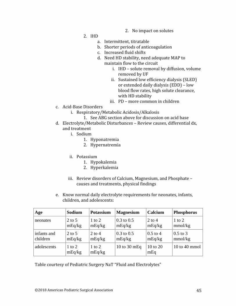
e. Know normal daily electrolyte requirements for neonates, infants, children, and adolescents:
Age Sodium Potassium Magnesium Calcium Phosphorus
neonates 2 to 5
mEq/kg
1 to 2
mEq/kg
0.3 to 0.5
mEq/kg
2 to 4
mEq/kg
1 to 2
mmol/kg
infants and
children
2 to 5
mEq/kg
2 to 4
mEq/kg
0.3 to 0.5
mEq/kg
0.5 to 4
mEq/kg
0.5 to 3
mmol/kg
adolescents 1 to 2
mEq/kg
1 to 2
mEq/kg
10 to 30 mEq 10 to 20
mEq
10 to 40 mmol
Table courtesy of Pediatric Surgery NaT “Fluid and Electrolytes”
©2018 American Pediatric Surgical Association 46
6. Gastrointestinal Disease a. Acute Liver Dysfunction
i. Review causes 1. Extrahepatic biliary duct obstruction 2. Increased bilirubin production 3. Impaired bilirubin excretion (dysfunction, hepatitis,
cholestasis) 4. Define Fulminant hepatic failure, know causes and
management ii. Treatment: Largely supportive management
iii. Classification of hepatic encephalopathy (HE) 1. Type A: associated with acute liver failure 2. Type B: associated with portosystemic bypass without
liver disease 3. Type C: associated with chronic liver disease, and
divided in to episodic, persistent, and minimal iv. Review precipitating factors: nitrogen load, metabolic
disorders, medications, infection, surgery, TIPS, etc. b. Hepatorenal syndrome – impaired renal function in the setting of
advanced liver disease and portal HTN, leads to splanchnic vasodilation, resulting in renal vasoconstriction
i. Review types (1 &2) and treatment 1. Type 1 – most severe
a. Rapid decline of CrCl or Cr increase >2.5 in 2 weeks
2. Type 2 a. Insidious onset, modest decline in renal fxn;
ascites resistant to diuretics c. Acute GI Bleed
i. UGI (80%) vs. LGI 1. Review causes of both
ii. Treatment: large bore access, resuscitation, localize source and control – clipping, epinephrine injection, coagulation, sclerotherapy/banding for varices
1. Endoscopy a. Forrest classification for rebleeding risk in PUD
based on endoscopy b. High – active bleeding, nonbleeding visible vessel c. Intermediate – adherent clot d. Low – ulcer with eschar, clean nonbleeding ulcer
bed 2. Angioembolization 3. TIPS 4. Surgical (5-10%)
©2018 American Pediatric Surgical Association 47
5. Medication: h. pylori tx, PPI, octreotide for variceal hemorrhage
d. Acute Pancreatitis i. Review causes:
1. Common: alcohol, gallstones, ERCP, idiopathic, microlithiasis, Sphincter of Oddi dysfunction
2. Rare: HyperCa++, HyperTGL, medications (antiretrovirals, diuretics, sulfa, flagyl, valproic acid, 6-MP/L-asparaginase in kids with ALL/chemo, infection, perforated PUD, ischemic, obstruction
ii. Understand scoring systems: Apache II, Ranson, BISAP, HAPS, CT severity index, CRP, procalcitonin
1. No system is superior iii. Treatment
1. Fluid resuscitation is key a. LR superior to NS (avoid in hyper Ca++ induced
cases) 2. Nutritional support- enteral preferred 3. ABX in infected necrosis only
a. If suspected, CT with IV contrast b. Treat necrosis with abx (carbapenem) and
percutaneous drainage or endoscopic drainage c. Surgical drainage when less invasive measures
fail: “step-up” approach e. Abdominal compartment syndrome
i. Abdominal perfusion pressure = MAP – intra-abdominal pressure
ii. Common causes: massive resuscitation and massive transfusions, intraperitoneal or retroperitoneal hemorrhage, ascites, obstruction, loss of domain (after complex VHR)
iii. Define Primary vs. secondary and acute vs. chronic iv. Clinical manifestations
1. Distended tense abdomen, hypoxia, hypercapnia, elevated peak inspiratory pressures with decreased compliance on vent OR shortness of breath, elevated CVP/PCWP, oliguria/anuria, elevated bladder pressures, decreased CO, hypotension, vasopressor requirement, ICP
v. Treatment 1. Surgical decompression - definitive 2. Paralytics/diuretics - IAH 3. Paracentesis - ascites 4. Escharotomy (burns)
©2018 American Pediatric Surgical Association 48
7. Critical Care Nutrition a. Determine who will benefit from parenteral or enteral nutrition b. Be able to determine nutritional requirements
i. Calculate energy expenditure a. CHO 3.4 kcal/g, lipids 9 kcal/g, pro 4 kcal/g
2. Harrison-Benedict equation (BEE) a. Men = 66.5 + (13.7 × wt) + (5.0 × ht) − (6.76 ×
age) b. Women = 65.5 + (9.6 × wt) + (1.85 × ht) − (4.68 ×
age) Berg, Sheri; Bittner, Edward (2013-11-14). The MGH Review of Critical Care Medicine
3. Indirect calorimetry (REE) a. REE = (3.9 × VO2) + (1.1 × VCO2) − 61 Berg, Sheri; Bittner, Edward (2013-11-14). The MGH Review of Critical Care
4. Stress factor a. 20% for mild stress (BEE X 1.2) = caloric needs b. 100% for severe stress (severe burns) (BEE X 2)
5. BEE = 25 kcal x wt (kg) is a quick way to calculate ii. Protein
1. 0.8-1 gm/kg/d 2. Also has stress factor
a. Measure nitrogen balance (N2 intake-N2 output) i. (Pro intake (g)/6.25) – (Urine Urea
Nitrogen +4) b. Want balance to be positive 4-6 g/d
iii. Lipids 1. 30% or less, higher if CO2 retention
iv. CHO 1. Remaining calories (40-50%)
v. Vitamins/immunonutrition 1. Review conditions associated with nutrient deficiencies
vi. Know refeeding syndrome and associated electrolyte disturbances
vii. Know how to calculate and interpret the Respiratory Quotient (RQ)
1. RQ = CO2 eliminated/O2 consumed a. Fatty acid 0.7 b. Protein 0.8 c. Glucose 1.0

©2018 American Pediatric Surgical Association 49
8. Hematology a. Review the intrinsic and extrinsic coagulation pathways:
Image courtesy of Pediatric Surgery NaT “Transfusion and Coagulation Therapy”
b. Evaluation i. History
1. Localized or diffuse bleeding, history of liver dysfunction or malnutrition (vit K deficiency), medications
ii. Physical 2. Signs of portal HTN, splenomegaly, signs of
retroperitoneal bleeding i. Review the components and interpretation of
Thromboelastofraphy (TEG) a. R = reaction time (initial fibrin plug) b. K = time from plug to clot formation c. Alpha angle = rate of clot formation d. MA = clot strength/platelet function e. LY30 = percentage of clot lysis at 30 minutes

©2018 American Pediatric Surgical Association 50
Image courtesy of Pediatric Surgery NaT “Transfusion and Coagulation Therapy”
i. Labs 1. PT/PTT/INR/platelets/fibrinogen (caution with PTT
from heparin infused lines -> may heparin adjusted PTT)
2. Isolated PT-INR elevation may be due to factor VII deficiency
3. Elevated PT-INR and PTT suggests multiple defects of II, V, X and can be seen in extremely low fibrinogen levels
DIC PT-INR, aPTT Fibrinogen, platelets
Massive transfusion PT-INR, aPTT Fibrinogen, platelets
TTP Platelets, PT-INR, aPTT, normal fibrinogen, mild anemia
HUS Hemolytic anemia, +/-Platelets; PT-INR, aPTT, fibrinogen normal
Heparin aPTT, +/- PT-INR Warfarin PT-INR; aPTT Created by Mary Arbuthnot
c. Thrombotic disorders
i. Know VTE prophylaxis recommendations ii. Risk factors in children: inherited prothrombotic disorder,
neoplasm (especially ALL), congenital heart disease, SLE,

©2018 American Pediatric Surgical Association 51
renal disease, infection, OCP use, surgery, asparaginase therapy
1. Greatest risk factor in children is presence of central venous catheter (CVC) devices, mechanical ventilation, malignancy, systemic infection, and prolonged hospital stay (> 5 days)
2. Review inherited prothrombotic conditions (% general population affected):
a. Factor V Leiden (4-5%) b. Prothrombin G20210A polymorphism (2%) c. Antithrombin (AT) deficiency (0.02-0.2%) d. Protein C deficiency (0.2-0.5%) e. Protein S deficiency (0.2-0.5%)
d. Platelet disorders i. Platelet destruction
1. Neonatal a. < 72 hours: placental insufficiency; neonatal
alloimmune thrombocytopenia; birth asphyxia; perinatal infection; congenital infections; and maternal autoimmune disorders, including idiopathic thrombocytopenic purpura
b. > 72 hours: sepsis, necrotizing enterocolitis, congenital infection, maternal autoimmune disorders, and congenital syndromes, including thrombocytopenia with absent radius
2. Immune a. ITP, drug induced (heparin, quinidine), infection
(HIV, H. pylori, hep C, sepsis related), autoimmune (SLE)
3. Nonimmune a. DIC (sepsis, trauma, obstetric emergencies,
hypoxia or severe acidosis) b. TTP, dilutional (CABG, prolonged surgery,
sepsis), microangiopathy (malignant HTN, cardiac valve dysfunction)
ii. Qualitative platelet disorders: Phenotype/Genes involved (inheritance). Review diagnostic evaluation and presentation.
1. Platelet adhesion – GPIb/GPIX/V (AR) a. Bernard-Soulier b. VonWillebrand’s disease – platelet type
2. Platelet activation – ADP receptors – mostly recessive 3. Platelet aggregation – GPIIb/IIIa
a. Glanzmann’s thrombasthenia (AR) 4. Platelet secretion – GATA 1, HPS1, HPS2, HPS3, HPS4,
HPS5, HPS6, HPS7, CHS1/LYST (mostly recessive)

©2018 American Pediatric Surgical Association 52
a. Gray platelet syndrome b. Chediak-Higashi syndrome
5. Platelet procoagulant activity – ABCA1 (unknown inheritance)
a. Scott Syndrome iii. Acquired platelet dysfunction: know duration of dysfunction
and treatment 1. Aspirin – 3-7 day recovery 2. NSAIDs – 24 hour recovery 3. Clopidigrel – 7 days recovery 4. Glycoprotein IIb/IIIa inhibitors – 12 hour or less
recovery 5. Uremia – Tx: dialysis, DDAVP, conjugated estrogens,
RBCs and epo e. Hereditary coagulation disorders
i. Hemophilia A (VIII) and B (IX) deficiency – know diagnosis and treatment
1. Review factor replacement in trauma and surgery f. Differential diagnosis for Neonatal Anemia
i. acute blood loss from multiple lab draws ii. anemia of prematurity.
iii. blood loss during delivery from placental abruption, placenta previa, umbilical cord rupture, and twin-twin transfusion
iv. Iron deficiency g. Know indications for and risks of blood component therapy
i. RBCs – 1 unit increases Hgb by 1 or Hct by 3% 2. Blood volume:
a. Premature infant 89 - 105 mL/kg b. Term infant: 80 mL/kg then drops to 73 to 77 mL/kg c. Child: 65 - 70 mL/kg d. Adolescent: 60 mL/kg.
ii. Platelets – 1 unit increases by 5,000; 1 unit/10 kg raises by 25,000/µL
iii. Plasma (FFP, thawed plasma) – INR > 1.5 or PTT >1.5x midpoint of normal, give 2-4 units q6 and expect 20% increase in factors. Dosed 10 mL/kg fresh frozen plasma (FFP) Contains all factors, most importantly V and VIII.
iv. Cryopercipitate – Transfuse for fibrinogen <100 mg/dL. Typically pool of 10 units transfused, each increasing fibrinogen by 5 mg/dL. Dosed 1 unit/5 kg cryoprecipitate. Contains fibrinogen, Factor VIII, XIII, vWF, fibronectin
v. Tranexamic acid (TXA) 1. Synthetic lysine analog, inhibits plasminogen activation
and the activity of plasmin, decreasing fibrinolysis and promoting stable clot formation.
©2018 American Pediatric Surgical Association 53
2. CRASH-2 trial positive effects of TXA on acute coagulopathy of trauma patients and effect greatest within the first 3 hours after injury. There is no universally accepted dose for pediatrics.
vi. Vitamin K 1. No studies to date evaluating efficacy in immediate
preoperative period 2. Time dependent; coagulation factors must be produced
in the liver 3. Best managed with FFP for immediate replacement
vii. Factor VII 1. Binds to exposed tissue factor at site of endothelial
injury 2. Off-label indications (cardiopulmonary surgery, trauma,
underlying malignancy, ECMO) viii. Erythropoeitin (EPO) - Anemia of prematurity
1. administration in neonates shown effective in anemia prevention, especially LBW infants
ix. Risk of infection: 1. Hep B: 1:200,000 to 360,000 2. Hep C: 1:1,000,000 to 2,000,000 3. HIV: 1:1,500,000 – 2,000,000 4. HTLV: 1:2,000,000
x. Other risks: 1. TRALI – Transfusion-associated acute lung injury -
Shortness of breath (SOB) from immune/inflammatory reactions
2. TACO – Transfusion associated circulatory overload – SOB due to large fluid volume
3. Allergic reactions to proteins or cells 4. ta-GVHD – transfusion associated graft-versus-host
disease from immune attack of the recipient by transfused cells
5. ABO incompatibility leading to destruction of transfused cells
6. FNHTR – Febrile nonhemolytic transfusion reaction – fever due to cytokines in the transfusion

©2018 American Pediatric Surgical Association 54
9. Endocrinology a. Acute Diabetic Emergencies
i. DKA 1. Type 1 and some insulin dependent Type 2 diabetics 2. Severe anion gap acidosis, hyperglycemia,
hyperosmolality, serum and urine ketones, glucosuria, polyuria resulting in hypovolemia and pre-renal azotemia
3. Glucose generally 300 - 800 mg/dL and often 350 - 500 mg/dL a. > 900 mg/dL DKA who are comatose. b. pH generally >7.2 c. serum osm usually >300 d. Low total body K+ stores but serum levels appear
elevated but to extracellular shifts e. Initial apparent hyponatremia but this is false and
actually usually hypernatremia when corrected for hyperglycemia
f. Hypophosphatemia and hypomagnesemia 4. Know treatment and management and identify
precipitating causes ii. Hyperosmolar Hyperglycemic State (HHS)
1. Usually type 2 diabetics 2. Sx: Tachycardia, hypotension, altered mental status, up
to 10 L fluid deficient, hyperosmolar, K+ deficient g. Glucose >600, can be > 1000 mg/dL h. Serum osm >330 i. No serum ketones
3. Know management and treatment. Risk of cerebral edema.
b. Thyroid Disorders i. Myxedema coma
1. Manifestation of long term hypothyroidism 2. Caused by precipitating event in partially treated
hypothyroid patient 3. 60% mortality rate, requires prompt intervention 4. Sx: Hypothermia, altered mental status, cardiac
depression, ileus, urinary retention 5. Know lab abnormalities, ECG findings, and treatment
a. 5-10% concurrent AI – treat with hydrocortisone before thyroid hormone
i. 100 mg hydrocortisone every 8 hours, followed by 200-300 μg IV load of Thyroxine
ii. Euthyroid sick syndrome
©2018 American Pediatric Surgical Association 55
1. Low T4, Low T3, Low TSH a. High endogenous cortisol and exogenous
glucocorticoid therapy b. Circulating inhibitors of deiodinase activity c. Treatment with amiodarone and high doses of
propranolol d. Cytokines (such as TNH, I-alfa, NF-kB, and IL-6)
2. IF no signs of hypothyroidism, suggest not treating iii. Thyroid Storm
1. Inhibition of binding of thyroid hormone to thyroxin-binding globulin causing acute rise in free hormone
2. Usually precipitated by medical problem (infections, surgery, stress, trauma, DKA, labor, cardiac disease, iodinated contrast dye) in partially treated hyperthyroid patient
3. Sx: Fever, tachycardia, a. fib, mental status changes, n/v, diarrhea. An lead to vascular collapse and MODS, coma and death
4. Know lab abnormalities and treatment (thyroid directed therapy and supportive management) a. Fluids, beta-blockers, thionamide to block synthesis,
iodine to block release, corticosteroids to block T4->T3 conversion
c. Adrenal Disorders i. Adrenocortical Insufficiency (AI)
1. Reversible dysfunction a. CIRCI: critical illness-related corticosteroid
insufficiency i. Sepsis/sirs/pancreatitis/hepatorenal
syndrome/hypothermia b. Drugs:
i. Primary: ketoconazole, etomidate, metyrapone, mitotaine
ii. Secondary: corticosteroids, megestrol acetate
iii. Increased production: rifampin, phenytonin
2. Primary AI – rare <0.015% of population a. Autoimmune adrenalitis (Addison’s) b. HIV infection: antiretrovirals, HIV, CMV c. Metastatic carcinoma: lung, breast, kidney d. Systemic fungal infection: Histoplasmosis,
Cryptococcus, Blastomycosis e. Tuberculosis

©2018 American Pediatric Surgical Association 56
f. Adrenal hemorrhage/infarction: DIC, meningococcemia, anticoagulation, antiphospholipid syndrome, HIT, trauma
3. Secondary AI a. Primary tumors (craniopharyngioma, ependymoma,
metastatic), irradiation, infiltration, Wegener autoimmune hypophysitis, chronic glucocorticoid use, congenital absent ATCH, proopiomelanocortin deficiency, combined pituitary hormone deficiency
4. Glucocorticoid tissue resistance a. Sepsis, SIRS (ARDS, trauma, burns, pancreatitis, liver
failure, post-cardiac surgery, HELLP syndrome) 5. Diagnosis (these are specific but not sensitive):
a. Random cortisol < 10μg/dL or b. Delta cortisol < 9 μg/dL after 250- μg corticotropin
stimulation test i. CRICI type I: random cortisol <10
ii. CRICI type II: random cortisol >10 but delta cortisol <9. Associated with high proinflammatory mediators and higher severity of illness and mortality.
6. Treatment in vasopressor dependent patients (norepinephrine or equivalent >0.1 μg/kg/min) within 12 hours of onset of septic shock. a. Controversial but consider 7-10 course of low dose
hydrocortisone (200 mg/d) b. Stop if vasopressor dependency still present after 2
days c. Strict infection surveillance
7. Treatment of acute AI/CIRCI a. Hydrocortisone 50 mg IV every 6 hours or 100 mg
bolus then 10 mg/hr continuous infusion for at least 7 days, can continue up to 10-14 days. Patients should be vasopressor and ventilator free before taper. Taper should be 50 mg every 8 hours for 3-4 days, then every 12 hours (IV or PO) for 3-4 days then daily for 2-4 days.
b. Optional Fludrocortisone 50 μg orally daily c. Methylprednisolone and hydrocortisone
interchangeable d. Do not use dexamethasone. It lacks
mineralocorticoid activity, suppresses HPA axis, has long half-life, and should be avoided prior to a cosyntropin test.
e. Critically ill patient received etomidate? (Inhibits 11 β-hydroxylase enzyme)
©2018 American Pediatric Surgical Association 57
i. Should probably be treated within 6 hours with stress dose hydrocortisone for 24 hours (200 mg day 1, 100 mg day 2)
d. Syndrome of Inappropriate Antidiuretic Hormone Secretion (SIADH) i. Excessive ADH secretion leading to impaired water excretion,
decreased UOP, hyponatremia; isovolemic or hypervolemic volume status
ii. Si/sx of hyponatremia iii. Diagnosis: decreased serum osm, increased urine osm and
urine sodium, hyponatremia, euvolemia, normal thyroid/adrenal function, no recent diuretics
iv. Treat underlying cause, fluid restriction, demeclocycline for acute severe hyponatremia
v. Calculate sodium deficit and slowly resolve 1. Sodium deficit = TBW x ([Na+]normal – [Na+] current)
a. TBW = 0.6 x lean body mass (males) b. TBW = 0.5 x lean body mass (females)
e. Diabetes Insipidus (DI) i. Central vs. nephrogenic, causes (reversible or not) and
treatment options 1. Central
a. AKA neurogenic DI b. Deficient secretion of ADH
i. 25% idiopathic, also due to trauma, intracranial lesion, pituitary resection
2. Nephrogenic a. ADH resistance at the distal tubules and collecting
ducts b. Can be due to chronic lithium use, hypercalcemia,
persistent hypokalemia, renal disease, pregnancy, hereditary
c. Diagnosis: fluid deprivation test, serum sodium usually high (low in primary polydipsia), desmopression (nephrogenic nonresponsive, central DI urine osm will increase by 50%)
f. Cerebral salt wasting i. Hypovolemia hyponatremia from inappropriate urine
sodium excretion thought to be due to impairment of Na+ and H2O regulation in the CNS
1. Brain injury (SAH most common), encephalitis, CNS tumors, CNS surgery
ii. Diagnosis 1. Hyponatremia and volume depletion (Na+ <135) urine
osm >100, urine Ba+ >40, hypovolemic, low plasma osm iii. Management: sodium and water repletion, salt tablets or
mineralocorticoids may be needed.

©2018 American Pediatric Surgical Association 58
10. Analgesia and Sedation a. Neuromuscular blockade
i. Depolarizing neuromuscular blockade 1. Succinylcholine
a. Rapid onset, short duration of action, ideal for RSI
b. Potassium release can be dangerous in stroke, paralysis, burn, crush, and spinal trauma
c. Also risk of arrhythmia (bradycardia in children), myalgias, myoglobinemia, increased ICP/intraocular/gastric pressure, prolonged paralysis in patients with plasma-cholinesterase deficiencies, masseter muscle rigidity. Is triggering agent for malignant hyperthermia. Caution use in children unless needed for emergent RSI due to reports of hyperkalemic cardiac arrest in patients with undiagnosed muscular dystrophy
ii. Nondepolarizing neuromuscular blockade 1. Pancuronium
a. Long acting, low cost, long duration of action, metabolized in the liver and excreted in the kidneys. Vagolytic effect can lead to tachycardia and hypertension
2. Doxacurium a. Little or no histamine release or CV side effects,
elimination independent of renal function 3. Vecuronium
a. Structurally similar to pancuronium, less vagolytic effect, liver metabolism, excreted by kidneys
4. Atracurium a. Hoffman degeneration independent of renal or
hepatic function b. Cisatracurium – cis-isomer, lacks histamine
release 5. Rocuronium
a. Rapid onset, 6- sec, eliminated through hepatic metabolism

©2018 American Pediatric Surgical Association 59
Drug Intubating dose (mg/kg) Continuous infusion (mcg/kg/min)
pancuronium 0.1 NA
vecuronium 0.1 1
rocuronium 0.6 to 1.2 3 to 10
isatracurium 0.1 0.4 to 4
Table courtesy of Pediatric Surgery NaT “Sedation and Analgesia”
b. Sedatives i. Benzodiazepines (BNZ): modulate GABA (main inhibitory
neurotransmitter) 1. Midazolam
a. Rapid onset, short duration. b. Can be given oral, nasal, sublingual, rectal, IM or
IV. c. Metabolized by CYP 450 system. Active
metabolite renally excreted and may accumulate in renal insufficiency. Liver disease and heparin use can increase fraction of free drug.
2. Diazepam a. Rapid onset, 1-2 hour duration of action b. Metabolized in the liver to active metabolites
3. Lorazepam a. Less chance for drug-drug interactions due to
metabolism by phase II glucuronidation, and can use with mild liver dysfunction. No renal excretion of active metabolites and can be used in renal disease.
4. BNZ Antagonist: Flumazenil – GABA antagonist a. Has a short half life and may need redosing
5. Side effects: a. Blunts CNS response to hypercarbia and hypoxia b. Decreases sympathetic outflow which can result
in hemodynamic compromise in septic shock, hypovolemia, and cardiovascular disease
c. Toxicity due to excipients (inactive compounds added to preserve drug or facilitate drug delivery) -> lead to new onset acidosis, hyperosmolality, seizures, arrhythmias, and hemodynamic instability
i. Propylene glycol (diazepam and lorazepam)
ii. Benzyl alcohol (diazepam, midazolam, and lorazepam)

©2018 American Pediatric Surgical Association 60
d. “gasping” respirations in neonates ii. Non-benzodiazepines
1. Propofol a. Alkylphenol family, excipients include 10%
soybean oil and 1.2% egg phosphatide b. GABA mediated inhibition of neurotransmission c. High lipid solubility and large volume of
distribution, rapid onset and clearance d. Three compartment pharmokinetics: 1) central
vascular compartment 2) SNC 3) peripheral tissues
e. Rapid hepatic metabolism (phase II glucuronidation) and also metabolism in lung and kidney
f. NO analgesic properties, only sedative, amnestic and hypnotic properties
g. Side effects: respiratory depression/apnea, decreased cough and gag, decreased tidal volumes, negative inotrope and decreased PVR. Decreased ICP and cerebral metabolic rate. Antiemetic, anticonvulsant. Can cause pain at injection site. Can cause hypertriglyceridemia, caution with egg/peanut/soy allergy, PRIS
i. Propofol infusion syndrome (PRIS): lactic acidosis, rhabdomyolysis, arrhythmias (refractory bradycardia), myocardial failure, renal failure, hyperlipidemia, hepatomegaly, muscle necrosis
1. Associated with high doses, thought to be due to impairment of fatty acid chain oxygenation and inhibition of oxidative phosphorylation in the mitochondria, ATP depletion and cellular hypoxia
2. Treatment: stop drug, supportive measures, may require temporary cardiac pacing or dialysis
2. Barbiturates a. Low doses – GABA receptor agonist enhancing
inhibitory neurotransmission. High doses – direct cell hyperpolarization. Also inhibits synaptic transmission of glutamate and Ach (excitatory neurotransmitters).
b. Hepatic metabolism and renal excretion

©2018 American Pediatric Surgical Association 61
c. Sedative and hypnotic effects, no analgesia at low doses
d. Potent anticonvulsants, reduces cerebral blood flow and metabolism
i. Pentobarbital – short onset of action, long duration of action
ii. Thiopental and methohexital – short acting. Thiopental has rapid onset of action (RSI)
e. Side effects: dose dependent respiratory depression, negative inotropes, arterial vasodilation (with reflex tachycardia to maintain CO) and can cause HD instability in pre-existing cardiac disease. Thiopental can cause bronchospasm. Multiple doses or continuous infusion can result in immunosuppression and are at increased risk of infection. Need to follow levels.
3. Etomidate a. Is an imidazole, modulates GABA receptors,
undergoes 3 compartment pharmokinetics (see above), undergoes hepatic esterase metabolism, renal and bile (to a lesser degree) excretion, inhibits adrenal steroid synthesis
b. Does not cause hemodynamic perturbations, does not block sympathetic responses to laryngoscopy and intubation. Causes decreased cerebral blood flow, metabolic rate and ICP while maintaining CPP.
c. Side effects: PONV, adrenal toxicity 4. Ketamine
a. PCP derivative, NMDA receptor antagonist, inhibits glutamate meditated neurotransmission
b. Hepatic n-methylation to active metabolite, norketamine, then to inactive metabolite that is renally excreted
c. Can give oral, nasal, rectal, IM, IV d. Produces sedation, dissociative amnesia,
anxiolysis and analgesia e. Maintains spontaneous respirations and
respiratory tone, increase in catecholamine release, increase in cholinergic receptor stimulation and bronchodilation (good for asthmatic children)
f. Side Effects: oropharyngeal secretions, laryngospasm, direct negative inotropic effects

©2018 American Pediatric Surgical Association 62
counteracted by indirect sympathomimetic activity resulting in increased HR and CO. Risk for HD instability in patients with poor cardiac function, hypovolemia, or diminished endogenous catecholamines. Use with caution in pulmonary hypertension because can increase pulmonary vascular resistance. No increase in ICP demonstrated in recent data, and may actually lower ICP.
5. Dexmedetomidine a. Alpha-2 receptor agonist in brain and spinal
cord, decreases norepinephrine production and leads to decrease in sympathetic outflow and increase in GABA activity, releases substance P producing analgesia
b. Phase I CYP 450 oxidation and phase II glucuronidation in liver, caution in liver dysfunction
c. Preserves spontaneous breathing and has favorable HD profile
d. Side effects: dose dependent bradycardia c. Analgesics
i. Opioids 1. Morphine
a. Acts on μ-receptors. High bioavailability if orally given but undergoes extensive first-pass metabolism in the liver, and metabolites renally excreted. 1:3 IV to oral ratio. Can cause histamine release resulting in asthma exacerbations.
2. Fentanyl a. Synthetic derivative of meperidine, 100 x more
potent than morphine, less hypnotic and sedative effects, μ receptor agonist, rapid onset, short duration due to rapid CNS penetration and redistribution into fat. Hepatically metabolized and renally excreted, but well tolerated in renal and hepatic dysfunction. Caution: rapid blousing can cause chest wall rigidity and can affect ability to oxygenate and ventilate. Can also cause bradycardia.
3. Remifentanil a. Very short acting, μ-receptor agonist, only IV,
metabolism via tissue and plasma esterases and then renally cleared. 250x more potent than
©2018 American Pediatric Surgical Association 63
morphine, 5-10 min half life, potent respiratory depressant
4. Hydromorphone a. μ-receptor agonist, 5x more potent than
morphine with longer duration of action. Hepatic metabolism and renal excretion and requires reduced dosing with dysfunction. Caution with dose-dependent respiratory depression and with children with myocardial dysfunction.
5. Methadone a. μ -receptor agonist and weakly binds NMDA
receptor. Used traditionally to wean opioid dependent patients. Undergoes CYP metabolism and prone to drug-drug interactions. Can cause QT prolongation and requires ECG monitoring. Metabolites renally excreted. High bioavailability with oral administration
ii. Non-opioids 1. Acetaminophen
a. Caution with overdosing – associated with acute liver failure due to secondary metabolite NAPQI that exhausts glutathione reserves and causes hepatic cellular damage.
2. NSAIDS a. Aspirin – caution with Reye syndrome b. Other NSAIDS – caution with renal insufficiency

©2018 American Pediatric Surgical Association 64
11. Toxicology d. Evaluation should include history, physical, and the following
adjuncts: i. Blood glucose determination and treatment
1. Insulin, ethanol, salicylates, beta-blockers and sulfonylureas can cause hypoglycemia
ii. Pulse oximetry and hgb co-oximetry 1. Evaluate for hypoxemia, hypercarbia, acid base
disturbances, methemoglobinemia iii. ECG
1. Medications associated with arrhythmia: TCAs, anticholinergics, cholinergic, ethylene glycol, beta-blockers, CCB, digitalis, sympathomimetics
2. Prolonged QT syndrome: Antipsychotics (haloperidol, ziprasidone), Antidepressants (citalopram)
iv. Chemistry 1. Calculate anion gap [(Na) – (Cl + HCO3)] (Normal 8-16
mEq/L) 2. MUDPILES
a. Methanol b. Uremia c. Diabetic Ketoacidosis d. Propolene Glycol e. Infection, Isoniazid, Iron, Inborn errors of
metabolism f. Lactic Acidosis g. Ethylene Glycol h. Salicylates
v. Osmolar gap 1. 2xNa + BUN/2.8 + glucose/18 (Normal < 10) 2. Ketones, methanol, ethanol, ethylene glycol, isopropyl
alcohol increase osmolar gap 3. Only alcohol to increase osmolar gap but not anion gap
is isopropyl alcohol vi. Drug assay
1. Quantitative Drug Assays: Aspirin, acetaminophen, anti-epiletics, digoxin, alcohols, iron, lithium, methemoglobin, theophylline
2. Illicit drug assays: caution OTC (Dextromethorphan (DM)) cold remedies may result in false positive PCP screen, and poppy seeds may result in false positive opioid screen
vii. UA

©2018 American Pediatric Surgical Association 65
1. Calcium oxalate crystals may be seen after ethylene glycol ingestion
viii. Pregnancy screen if childbearing age e. Review common toxidromes and common antidotes
i. Know who requires NAC treatment for acetaminophen toxicity 1. Review the Rumack-Matthew nomogram (the lower
sold line typically defines who requires treatment)
©2018 American Pediatric Surgical Association 66
12. Thermal injuries a. Burns
i. Partial thickness burns 1. 1st degree
a. Sun or minor flash burn, bright red, dry without bullae, hyperesthetic, heals in 3-6 days
2. 2nd degree a. Higher intensity or longer exposure time to flash,
brief exposure to flames or hot liquid, mottled red, moist with bullae, pain as well as time to healing are inversely proportional to depth of burn (10-35 days)
ii. Full thickness burns 1. 3rd degree
a. Higher intensity or longer exposure time to flash, longer exposure to flames or hot liquid, contact with steam or hot metal, chemical burns, high voltage electrical burns. Appears pearly white, translucent, parchment, or charred, dry leathery and stiff. Tissue liquefaction. The surface is insensate, sense deep pressure only, treatment requires grafting
iii. Know formula for fluid resuscitation based on TBSA 1. Know Lund-Browder chart 2. Parkland formula
iv. Be familiar with zones of damage in burns 1. Zone of coagulation, zone of stasis, zone of hyperemia
v. Initial would care 1. Initial debridement 2. Topical anti-microbials
a. Be familiar with indication and side effects i. Silver sulfadiazine
1. Covers s. aureus, e. coli, many but not all pseudomonas, proteus and candida
2. SE: transient leukopenia in 15% patients
ii. Mafenide acetate 1. Gram positive and gram negative
coverage but little fungal coverage, limited MRSA coverage
2. Penetrates eschar and cartilage (ears)
3. Pain with application to partial thickness burns

©2018 American Pediatric Surgical Association 67
4. Inhibits carbonic anhydrase and leads to hyperchloremic acidosis
iii. Silver nitrate 1. Gram positive and gram negative
coverage but does not penetrate eschar
2. Leaches Na+, K+, Cl- and water and can result in alkalosis and water loading
vi. Know systemic changes with severe burns 1. ebb” and “flow” phase
vii. Know burn center referral criteria 1. Partial thickness burns involving more than 10% TBSA 2. Full thickness in any age group 3. Burns involving face, hands, feet, genitalia, perineum, or
major joints 4. Significant electrical burns including lighting 5. Chemical burns 6. Inhalational injury 7. Burns in patients with pre-existing medical conditions
that can complicate management, prolong recovery, or affect mortality
8. Lesser burns in patients with concomitant trauma sufficient to influence outcome
9. Any size burn in a child in a hospital without qualified personnel or the equipment needed for the care of children
10. Any size burn in a patient who will require special social or psychiatric intervention or long term rehabilitation
b. Inhalational Injuries i. Smoke exposure in a closed space, 1/3 of all flame burns
ii. Suspect in setting of facial burns, singed nasal hairs, carbonaceous sputum, tachypnea and hoarseness
iii. Due to rapidly progressing airway edema, intubation warranted, and bronchoscopy to definitively diagnose
iv. 50% develop pneumonia and 20% develop ARDS v. Consider Carbon Monoxide (CO) toxicity
1. CO > 10%, severe if <20-25%, confusion, cardiac ischemia or both
2. 1/3 patients with CO toxicity also have cyanide toxicity – suspect if lactates elevated, treatment is hydroxycobalamin
c. Electrical Injures i. High voltage >1000 volts

©2018 American Pediatric Surgical Association 68
ii. Increased incidence of acute renal failure due to and require significant fluid resuscitation (especially if hemochromogens present in the urine):
1. Extensive unapparent subcutaneous injury in limb with unburned skin and gross underestimation of resuscitation fluid needs
2. Mass of muscle injured may cause rhabdomyolysis resulting in direct renal tubule damage
iii. Extensive fasciotomy needed in setting of deep tissue necrosis, compartment syndrome, or progressively severe hyperkalemia
d. Chemical injuries i. Remove clothing and remove (brush off if dry) then dilute the
offending agent. Do not try to neutralize (causes heat generation)
ii. Common alkalis 1. Ammonia, bleach, lye, sodium and potassium hydroxide
and cement 2. Liquefaction necrosis and saponification, more
extensive than they appear, TBSA calculations may underestimate needs
iii. Common acids 1. Sulfuric avid, nitric acid, hydrochloric acid, phosphoric
acid, HYDROFLUORIC ACID (HFA) 2. Coagulation necrosis and thick eschar, usually less deep 3. HFA burns: Fluoride ion is a calcium chelator causing
severe hypocalcemia and cardiac arrhythmia a. Treatment includes irrigation followed by 2.5%
calcium gluconate gel to the affected area e. Hypothermia
i. Core temperature < 35° C 1. Mild 32-35° C, moderate 28-32° C, severe<28° C 2. Be familiar with musculoskeletal and neurologic signs
for each classification 3. Know initial management and rewarming techniques
a. Rewarm to 32° C 4. Recognize physiologic changes and lab/ECG findings
a. J (Osborn) waves at junction of QRS and ST segment
ii. Frostbite – crystal formation in the intracellular and extracellular fluid
1. Rapid rewarming at 40-42° C in 15-30 min 2. >1 week before true determination of depth of damage 3. Elevate limb to avoid pressure ischemia 4. Tetanus prone wounds, tetanus toxoid should be given
if needed. f. Hyperthermia

©2018 American Pediatric Surgical Association 69
i. Core temperature > 41° C 1. Heatstroke includes CNS dysfunction and Temp >40° C
(no CNS abnormalities in heat exhaustion) 2. Know clinical findings and treatment
a. Most common finding is tachycardia b. Sweating may be present or absent c. Conductive and evaporative cooling, cool to 38°C
ii. Malignant Hyperthermia (MH) 1. Autosomal dominant, occurs in 1:50 to 1: 150,000
patients who receive anesthesia a. Occurs within 30 minutes of anesthesia in 90%
of patients 2. Can be drug or stressed induced
a. Halothane and succinylcholine account for most cases
b. Less commonly muscle relaxants, inhalational anesthetic agents, ethanol, caffeine, sympathomimetics, parasympathomimetics, cardiac glycosides, quinidine analogs, infection, physical or emotional stress, anoxia, or high ambient temperature
3. Related to impaired calcium transport in skeletal muscle
a. Impaired reuptake of calcium into the sarcoplasmic reticulum (SR) and a defect in the calcium-mediated coupling contraction mechanism
4. Signs a. Increase in end tidal CO2, muscle rigidity
(extremities, chest or jaw), tachycardia, hypertension, mottling
b. Elevated temperature occurs later c. Labs: elevated Na+, Ca+, Mag, K+, phosphate,
CPK, LDH 5. Treatment
a. Dantrolene: prevents release of calcium into cell by the SR
i. Rapid IV push 2.5 mg/kg and repeated every 5 minutes until symptoms subside or max 10 mg/kg administered. Then 1 mg/kg every 4-6 hours for 36-48 hours
ii. Evaporative cooling if needed b. Know definitive work-up recommendations
©2018 American Pediatric Surgical Association 70
13. Obstetrical Critical Care a. Be familiar with normal physiologic changes in pregnancy
b. Hemorrhage i. Most common cause of ICU admission and death in pregnancy
ii. Pregnant patients are more likely to go into DIC iii. Common causes:
1. Prepartum: ectopic pregnancy, uterine rupture during VBAC (vaginal birth after cesarean section), abortion, placental previa or abruption, trauma
2. Postpartum: uterine atony, uterine rupture, uterine inversion, retained placenta, surgical trauma
c. Pre-eclampsia i. New-onset HTN in pregnancy without history of chronic HTN
and with proteinuria 1. Usually after 32 weeks gestation 2. BP > 140/90 on 2 occasions 6 hours apart and 300 mg
protein in 24 hour sample 3. Severe pre-eclampsia: SBP of 160 or DBP 110 or
greater on 2 occasions 6 hours apart and 5 gm protein in 24 hour sample, oliguria, cerebral or visual disturbances, pulmonary edema or cyanosis, epigastric or RUQ pain, thrombocytopenia, fetal growth restriction
d. Eclampsia: severe pre-eclampsia with seizures i. Treatment: delivery (consult OBGYN), HTN control (IV
labetalol and hydralazine), IV magnesium, monitor for DIC, consider Head imaging (risk of ICH)
e. HELLP i. H – hemolysis, bilirubin < 1.2 mg/dL
ii. EL – elevated liver enzymes LDH >600, ALT or AST >70 iii. LP – low platelets <150,000 iv. Occurs in up to 20% women with severe pre-eclampsia, need
to consider HUS-TTP or acute fatty liver of pregnancy on differential
v. Treatment: delivery, close monitoring for HTN, DIC, hemolysis, supportive care
f. Embolism: know that various types, presentation, and management i. Amniotic fluid embolism
ii. Venous air embolism iii. Venous thromboembolism
©2018 American Pediatric Surgical Association 71
14. Pediatric Emergencies g. Know how to diagnose and manage pediatric surgical emergencies:
i. Neonatal and congenital anomalies: 1. Omphalocele
a. Pathology: i. Abdominal contents herniate through
umbilicus, covered by amniotic sac ii. Defect in embryonic disc
iii. Associated cardiac and genetic defects b. History:
i. Often diagnosed with pre-natal ultrasound.
ii. Often carried close to term iii. Often planned delivery: may be vaginal iv. If transported: cover sac with bag, keep
warm, NG tube, IV fluids. c. Diagnosis: Physical exam is diagnostic.
Associated anomalies may be evaluated with: i. Echocardiogram
ii. Chromosomes iii. Renal and head ultrasound
d. Treatment: Repair if small, “paint” and wait if very large
i. NGT decompression ii. Sac covering options: Silvadene, Newer
generation wound products (mepilex), iii. Antibiotics iv. If large: evaluate liver function and
hepatic vasculature. 2. Gastroschisis
a. Pathology: Herniation through lateral abdominal wall (usually right-sided), no sac covering contents
b. History: i. Fewer associated anomalies (most
common is intestinal atresia) ii. Often diagnosed prenatally
iii. Often deliver slightly premature. May be vaginal.
iv. If transported: cover sac with bag, keep warm, NG tube, IV fluids
c. Diagnosis: Physical exam d. Treatment:
i. NGT, and antibiotics. Usually requires intubation for bowel reduction.

©2018 American Pediatric Surgical Association 72
ii. Option of Silo and staged reduction iii. Surgical repair
1. Traditional fascial and skin closure 2. “Sutureless repair” with dressings
over intact umbilical stalk 3. Congenital diaphragmatic hernia
a. Pathology: Herniation of abdominal contents through defect in the diaphragm and into the chest
i. Types 1. Morgagni 2. Bochdalek (most common)
ii. 90% left sided b. Associated with pulmonary hypoplasia and
pulmonary hypertension c. Diagnosis:
i. Often noted prenatally on US ii. Plain films with Bowel and/or NG tube in
chest. iii. Evaluate heart with echocardiogram
d. Treatment: i. Intubation, often HFOV and/or ECMO,
ii. NGT decompression iii. Medical management of pulmonary HTN iv. Operative repair (when stable)
1. Patch vs. primary closure 4. Tracheoesophageal fistula (TEF)
a. Know the types i. Most common is esophageal atresia with
distal TEF b. Associated abnormalities: VACTERL syndrome
(vertebral, anal, cardiac, tracheal, esophageal, renal, limb defects), CHARGE
c. History – May be known from prenatal US or unknown at the time of birth. Classic story of neonate unable to feed and unable to pass NG tube
d. Diagnosis: i. Plain film with NG tube advanced as far as
possible. Consider including lateral CXR ii. Workup associated anomalies (cardiac
echo, spine films, anal exam) e. Treatment:
i. NPO and suctioning of upper pouch ii. Planned surgical ligation of fistula
iii. Repair atresia if possible at same surgery

©2018 American Pediatric Surgical Association 73
iv. After intubation: 1. May need bronchoscopy and
control of fistula 2. May need Gastrostomy tube
urgently if stomach becomes distended (may place to water seal if losing tidal volume)
5. Necrotizing enterocolitis a. Ischemia of lower gastrointestinal tract b. Most common in pre-term infants c. Risk factors: prematurity, umbilical lines, blood
transfusion, fortification of feeds, PDA with reversal of flow, treatment with NSAIDS for PDA, sepsis, hypotension, ischemia
d. History: Abdominal distention, feeding intolerance, increased residuals, bloody stools, pneumatosis intestinalis or portal venous gas on plain film, pneumoperitoneum
e. Diagnosis: Plain films and consistent history f. Treatment:
i. If no free air: NPO, gastric decompression, pediatric surgery consult, antibiotics
ii. If free air, sentinel loops, or clinical deterioration: surgery for laparotomy or drain placement (based on weight)
6. Biliary atresia a. Most common need for pediatric liver
transplantation i. Type I: obliteration of common bile duct
ii. Type II: obliteration of the common hepatic duct
iii. Type III: obliteration at the level of the porta hepatis (left and right main hepatic ducts - most common)
b. Progresses to cirrhosis, portal HTN, liver failure c. History: neonate with persistent direct
hyperbilirubinemia d. Diagnosis:
i. Fractionated bilirubin levels ii. HIDA Scan
iii. May require operative exploration for cholangiogram
iv. May need liver biopsy e. Treatment: Kasai procedure
(hepatoportoenterostomy), most successful if before 8 weeks

©2018 American Pediatric Surgical Association 74
ii. Pediatric abdominal pain, emesis or bloody stool: 1. Malrotation with volvulus
a. Incomplete fixation of bowel b. History: Bilious emesis c. Diagnosis: Get plain film then upper GI (stat) –
upper GI shows C-loop to the right of spine d. Treatment: Requires emergent laparotomy.
i. Ladd’s procedure: 1. Detorsion and evaluate bowel
viability. Divide Ladd’s bands. Perform Appendectomy. Widen mesenteric base. Colon to the left, small bowel to the right.
2. Pyloric stenosis a. Hypertrophic pyloric sphincter leading to gastric
outlet obstruction b. History:
i. 2-8 weeks of life, most common in first born males
ii. Nonbilious, projectile emesis, associated with hypokalemic, hypochloremic metabolic alkalosis
c. Diagnosis: US or upper GI contrast study to confirm
d. Treatment: NOT a true surgical emergency. Must correct electrolyte abnormalities prior to surgery; laparoscopic or open pyloromyotomy
3. Meckel’s diverticulum a. Remnant of vitelline (omphalomesenteric) duct
containing gastric or pancreatic tissue b. History: Usually painless rectal bleeding.
Possibly can get inflamed or perforated diverticula.
c. Know the rule of 2’s i. 2% of population
ii. Often age 2 or less iii. 2 in long, 2 cm diameter, 2 feet from the
illeal-cecal valve iv. 2 types of mucosa (gastric or pancreatic)
d. Diagnosis: With meckel scan (technetium-99 pertechnetate)
e. Treatment: Surgically resect. Diverticulectomy vs. segmental bowel resection based on anatomy of diverticula. Identify feeding vessel.
4. Intussusception

©2018 American Pediatric Surgical Association 75
a. Telescoping of one segment of bowel into another
b. History: Intermittent colicky pain, knees to chest, current jelly stools (late finding, mucosal ischemia and sloughing), lethargy may be only presenting sign. May have emesis.
c. Diagnosis: Radiology i. Paucity of gas in RLQ on KUB
ii. Ultrasound is diagnostic test of choice d. Treatment:
i. Primary treatment is air-contrast enema ii. Surgery if multiple enema attempts fail or
if signs of perforation iii. Consider presence the lead point if an
unusual or recurrent presentation 5. Appendicitis
a. Inflammation, infection or perforation of the appendix. Often associated with obstruction or fecalith.
b. History: Usually with 1-3 days of progressively worsening abdominal pain. Usually diffuse initially then localizing in the right lower quadrant. Fever, emesis and anorexia are common.
c. Diagnosis: i. History and physical: Know the basics of
“appendicitis” scores (PAS) ii. Labs – white count
iii. Films – US and CT if needed d. Treatment:
i. ABX and fluids: treat sepsis ii. Appendectomy
iii. Know indications for drainage and/or interval appendectomy.
iii. Inguinal and scrotal pathology 1. Incarcerated hernia
a. Indirect inguinal hernia sac. May or may not have been known since birth. More common in premature infants.
b. History: inguinal bulge with new onset pain, swelling, erythema, and emesis
c. Diagnosis: distinguish from hydrocele i. Know key physical exam findings
ii. Ultrasound if unclear d. Treatment:
©2018 American Pediatric Surgical Association 76
i. Non-incarcerated hernias may be scheduled electively. Consider options of early repair vs. repair after 55 weeks corrected gestational age.
ii. Incarcerated but able to manually reduce, followed by urgent surgical repair within days.
iii. If unable to reduce, emergency surgical reduction, repair and evaluation of bowel viability.
2. Testicular torsion a. Bell-clapper anatomy with torsion of testicle on
its pedicle. b. Typical age range c. History: Sudden onset of pain, may be
undescended testes, may have had minor trauma, often with emesis.
d. Diagnosis: Physical exam findings can be diagnostic. U/S is an option for unclear findings or to asses vascular flow.
e. Treatment: Emergent surgery with detorsion and orchidopexy of bilateral testes.
iv. Airway emergencies 1. Epiglottitis
a. Bacterial infection involving the epiglottis b. Used to be due to HiB, since vaccinations mostly
due to group A strep (10% HiB in vaccinated children)
c. History: Stridor, drooling, muffled voice, no cough, tripoding
d. Diagnosis: Thumbprint on lateral neck x-ray e. Treatment:
i. Keep patient calm (maintain “position of comfort)
ii. Supplemental O2 as needed iii. Antibiotics iv. Avoid intubation if possible, in controlled
setting if needed. 2. Croup
a. Subglottic edema secondary to viral infection, often parainfluenza
b. History: barking cough, stridor, nontoxic c. Diagnosis: History and viral swabs d. Treatment:
i. Dexamethasone, racemic epinephrine, heliox

©2018 American Pediatric Surgical Association 77
ii. If desaturation present, consider intubation with a smaller than usual ETT
3. Bronchiolitis a. RSV in the wider, upper airway with secretions
and lower airway inflammation b. Diagnosis: History, chest film and RSV swab. c. Treatment: Supportive care
4. Bacterial tracheitis a. Superinfection in setting of viral illness b. S. aureus or HiB c. History: Stridor, cough, tenacious sputum, toxic,
fever, not responsive to conservative management or airway steroids
d. Diagnosis: Sputum culture and Chest films e. Treatment:
i. Intubation if needed, ii. Antibiotics and supportive care,
iii. Consider bronchoscopy 5. PTA (peritonsillar abscess)
a. Group A strep, S. aureus b. History: Trismus, drooling, uvular deviation,
tonsillar asymmetry c. Diagnosis: Physical exam. Occasionally can
benefit from CT/MRI (distinguish between PTA and Retropharyngeal abscess)
d. Treatment: Incision and drainage and antibiotics 6. Retropharyngeal or lateral pharyngeal abscess
a. Group A strep, S. aureus, anaerobes b. Bacterial infection in the potential space
posterior to the esophagus and anterior to spine c. History
i. Usually < 5 years old ii. Stridor, drooling, neck pain with
movement, torticollis, fever, toxic, voice changes
d. Diagnosis: X-ray for diagnosis (may need CT for lateral pharyngeal abscess, less common)
e. Treatment: i. Culture and IV antibiotics
ii. OR - Incision and drainage if not already draining adequately
7. Foreign body aspiration a. History:
i. Drooling, dysphagia, stridor in the absence of fever, cough or wheezing
ii. Peak 6 months to 4 years of age

©2018 American Pediatric Surgical Association 78
b. Diagnosis: Look for FB on x-ray, unilateral air trapping, decreased aeration, or atelectasis
c. Treatment: i. If unclear on films, may need diagnostic
bronchoscopy. ii. OR for removal of object, rigid or flexible
with wash.

©2018 American Pediatric Surgical Association 79
15. The Elderly h. Comprise approximately 1/10 of population but account for 1/3
health care resources i. Physical factors predispose to injury
i. Understand the physiologic changes in the elderly (Disease is compounded by a decreased physiologic reserve)
1. Cardiovascular: a. Stiffening of the myocardium decreased
diastolic relaxation and stroke volume increased reliance on atrial contribution to EDV.
b. Increased sensitivity to hyper or hypovolemia. c. Decreased response to beta-adrenergic
stimulation and decreased ability to increase HR to adequately increase CO in times of stress
d. Atherosclerosis leads to arterial wall stiffening, reduced diastolic pressure despite systolic HTN limiting coronary blood flow, especially during times of stress
e. Prescription antihypertensives/beta-blockers may blunt physiologic response to stress. A good medical history is imperative.
2. Pulmonary: a. Chest wall more rigid, decreased compliance,
increased reliance on diaphragm as accessory muscles atrophy
b. FVC and FEV1 decreased c. Thickening of alveolar basement membrane and
decreases gas diffusion leading to V/Q mismatch and higher A-a gradients
d. Decreased mucociliary clearance, increase importance of pulmonary toilet
3. Kidney a. Cortical mass loss due to glomerulosclerosis b. Reduced GFR c. Decreased ability to reabsorb sodium and
secrete potassium and hydrogen ions d. Decreased renin production and attenuated
response to ADH 4. CNS
a. Cortical atrophy, increased subdural space, resulting in more frequent SDH and SAH due to shearing forces on parasagittal bridging veins, compounded by use of anticoagulants/antiplatelets in this population

©2018 American Pediatric Surgical Association 80
b. Decreased visual, auditory function, pain perception, and reflexes
5. MSK a. Loss of muscle mass and strength b. Osteoporosis leads to increased fracture risk
6. Decreased thermoregulation (multifactorial due to loss of muscle mass, thinning of the skin, endocrine/metabolic changes.)
7. Decreased immune function j. Special cases
i. Splenic injury – more likely to fail conservative management ii. Rib fracture – 3x risk of pneumonia, and mortality increases
linearly with number of ribs fractured. Analgesia (often epidural) and pulmonary toilet are mainstays of therapy.
1. 1-2 ribs: 10% mortality 2. 7+ ribs: 40% mortality
iii. ICH in setting of anticoagulation 1. Consider FFP/vitamin K/rVIIa for Coumadin
iv. Platelets and desmopressin for clopidogrel v. Increased risk of central cord syndrome (fall forward, chin
strikes floor, neck hyperextends, and acute pinching of spinal cord and bleeding in central regions)
1. Easily missed if not suspected clinically 2. CT scan can miss diagnosis, need MRI
vi. Upper c-spine injury predominant, odontoid fracture most common

©2018 American Pediatric Surgical Association 81
16. Emergency/Trauma Surgery a. Head trauma
i. Be able to describe SDH, SAH, Epidural hematoma, diffuse axonal injury ischemia, elevated ICP
ii. Know GCS scale iii. ABC’s of TBI care
1. Protect airway, intubate if needed 2. Goal paO2 80-100 mmHg, avoid hyperventilation due to
cerebral vasoconstriction, target PaCo2 35 mmHg, except in acute decompensation
3. Target normal blood pressure, consider direct monitoring of brain tissue oxygen tension (PbtO2) (<15-20 mmHg is low)
b. Neck trauma i. Know the 3 zones of the neck
Image courtesy of Pediatric Surgery NaT “Neck Exploration for Penetrating Trauma”
ii. Hard signs of neck injury warrant surgical intervention,
AIRWAY always first priority

©2018 American Pediatric Surgical Association 82
1. Airway compromise, shock, active bleeding, pulsatile/expanding hematoma, subcutaneous emphysema or air bubbling from wound, unilateral pulse deficit, thrill or bruit, stridor or hoarseness, signs of stroke
iii. Soft signs: Dysphagia, voice change, hemoptysis, wide mediastinum
iv. Always do complete neurological exam v. Review treatment algorithm:
http://gsresidency.surgery.med.ubc.ca/files/2013/09/Shiroff_etal_HeadandNeck.pdf
1. Penetrating injury (violates platysma) a. Hard signs -> OR b. Soft signs
i. Zone I – Angiogram: Arch and Great Vessels; Bronchoscopy, Esophagography/Esophagoscopy
ii. Zone II – Immediate neck exploration or Angiography, Bronchoscopy, Esophagography/Esophagoscopy
iii. Zone III - Angiography, Bronchoscopy, Esophagography/Esophagoscopy
vi. Know surgical approach for zone II injuries and how to gain access to distal and proximal carotid
vii. Know grading scale for blunt cerebrovascular injuries, associated stroke risk, and treatment (Review at: http://sajs.redbricklibrary.com/index.php/sajs/article/viewFile/1798/542/991)
1. Grade I: irregularity of vessel wall or dissection/intramural hematoma with <25% luminal stenosis
a. Treatment: anticoagulation or stenting if progression to the next grade of injury or enlarging pseudoaneurysm
2. Grade II: intraluminal thrombus or raised intimal flap or dissection/intramural hematoma with >25% luminal stenosis
a. Treatment: as for grade I 3. Grade III: Pseudoaneurysm
a. Treatment: as for grade I 4. Grade IV: Vessel occlusion
a. Treatment: anticoagulation 5. Grade V: vessel transection
a. Treatment: open repair viii. Hanging or strangulation

©2018 American Pediatric Surgical Association 83
1. Hangman fracture – bilateral fracture through pedicles of C2 and associated spinal cord injury
2. Consider CTA in the setting of garroting to evaluate for vascular injury
c. Cardiac Trauma i. Be familiar with Beck’s triad and treatment of pericardial
tamponade ii. Know indications for ED thoracotomy for blunt and
penetrating trauma 1. http://westerntrauma.org/documents/PublishedAlgori
thms/WTACriticalDecisionsResuscitativeThoracotomy.pdf
iii. Know the evaluation and management of blunt cardiac injury (BCI)
1. Admission ECG if BCI suspected a. If normal, no BCI b. If abnormal, admit for continuous monitoring for
24-48 hours, 2. If HD unstable, obtain TTE (TEE if needed) 3. Sternal fracture does not predict BCI 4. Neither CPK nor troponins are useful predictors for who
has BCI or who will have complications d. Vascular Trauma
i. Be able to calculate an Ankle-Brachial Index (ABI) ii. Hard signs of vascular trauma OR
1. Active arterial bleeding, pulselessness/evidence of ischemia, expanding pulsatile hematoma, bruit or thrill, ABI <0.9
iii. Soft signs of vascular trauma observation 1. Neurologic injury in proximity to vessel, small to
moderate sized hematoma, unexplained hypotension, large blood loss at scene, injury (fracture, dislocation, penetrating injury) proximal to major vessel
iv. Vascular screening indicated for knee dislocations and supracondylar and proximal tibial fractures
v. Groin, thoracic outlet, and neck injuries (in absence of hard vascular signs) warrant vascular screening
e. Emergency Abdominal Surgery i. Initial exploration, packing, exposure
ii. Pringle and Heaney maneuvers iii. Know the 3 zones of the retroperitoneum and which zones
should be explored. 1. Penetrating – explore all 3 2. Blunt – explore zone 1 only unless expanding, ruptured
or pulsatile hematoma
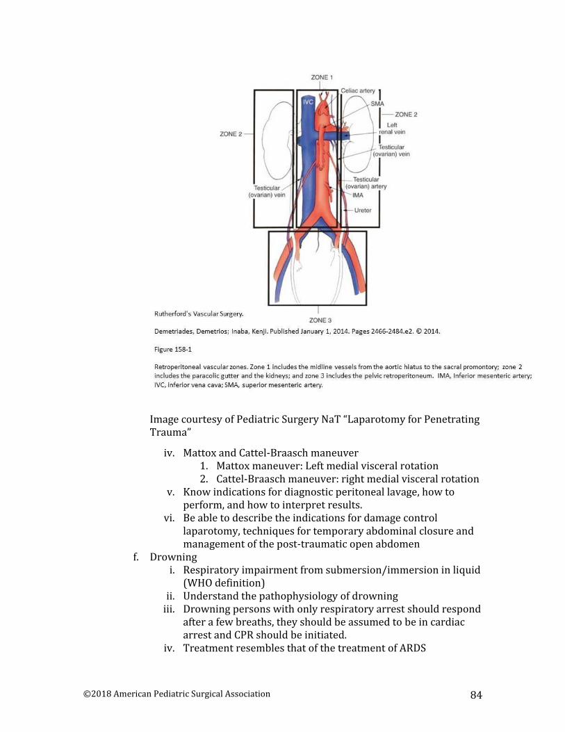
©2018 American Pediatric Surgical Association 84
Image courtesy of Pediatric Surgery NaT “Laparotomy for Penetrating Trauma”
iv. Mattox and Cattel-Braasch maneuver 1. Mattox maneuver: Left medial visceral rotation 2. Cattel-Braasch maneuver: right medial visceral rotation
v. Know indications for diagnostic peritoneal lavage, how to perform, and how to interpret results.
vi. Be able to describe the indications for damage control laparotomy, techniques for temporary abdominal closure and management of the post-traumatic open abdomen
f. Drowning i. Respiratory impairment from submersion/immersion in liquid
(WHO definition) ii. Understand the pathophysiology of drowning
iii. Drowning persons with only respiratory arrest should respond after a few breaths, they should be assumed to be in cardiac arrest and CPR should be initiated.
iv. Treatment resembles that of the treatment of ARDS

©2018 American Pediatric Surgical Association 85
v. Induced hypothermia (32-34 C) may be neuroprotective vi. Duration of submersion related to risk of death/severe
neurologic impairment 1. 0-5 min – 10% 2. 6-10 min – 56% 3. 11-25 min – 88% 4. >25 min - ~100%

©2018 American Pediatric Surgical Association 86
17. Transplant a. Lung
i. Common causes: 1. COPD, idiopathic pulmonary fibrosis (IPF), cystic
fibrosis (CF), alpha-1 antitrypsin deficiency, pulmonary HTN (PHTN)
ii. Post-operative pathophysiology 1. Pulmonary denervation – results in bronchial
hyperresponsiveness and decreased inhalation cough response
2. Atelectatsis due to decreased surfactant and pulmonary edema from loss of lymphatic drainage
3. Pulmonary reimplantation response (PRR) or ischemia reperfusion injury
a. Associated with long ischemia times and results in vascular injury and noncardiac pulmonary edema
iii. Outcomes 1. Mortality:
a. 30 day mortality: 10-15%; related to immediate post-operative complications/graft failure
b. 1 year mortality: 30%; related to infection from immunosuppression. Small risk of lymphoma
c. CF – higher risk for infection (due to pre-operative infection)
d. Congenital heart disease – higher risk for bleeding
e. Severe PHTN – higher risk of ischemia-reperfusion injury
f. Obliterative bronchiolitis – most limiting factor to long term survival
b. Heart i. Common causes:
1. Ischemic cardiomyopathy, congenital heart disease, valvular disease, autoimmune/metabolic disease, pregnancy, alcoholism, viral carditis
ii. Post-operative pathophysiology 1. Cardiac denervation – HR changes dependent on
catecholamines; reinnervation at approximately 1 year 2. PHTN is a risk factor for post-transplant acute RV
failure 3. Oliguria/renal failure secondary to cardiopulmonary
bypass (CPB) and cyclosporine A 4. Post-operative hemodynamic instability may be due to:
a. Ischemia-reperfusion injury

©2018 American Pediatric Surgical Association 87
b. RV failure c. Technical error (SVC stenosis, ruptured chordae) d. Acute rejection
iii. Outcomes 1. 1 year mortality: 20% 2. 5 year mortality: 30% 3. Rejection – up to 2/3 do not experience, surveillance
consists of endomyocardial biopsy 4. Post-transplant coronary artery disease (CAD) is a
major cause of late mortality 5. 80% experience hypertension due to cyclosporine A
c. Liver i. Common Causes:
1. Hep C, Hep B, alcohol, nonalcoholic steatohepatitis (NASH), autoimmune hepatitis, primary sclerosing cholangitis (PSC), primary biliary cirrhosis, cancer, fulminant liver failure
ii. Post-operative pathophysiology 1. End-stage liver disease (ESLD) affects all organ systems
and most disturbances are resolved with transplantation
a. Hepatic encephalopathy, cirrhotic and alcoholic cardiomyopathy, hepatopulmonary syndrome, hepatorenal syndrome, coagulopathy
2. First month: increase risk of bacterial and fungal infection
a. HSV reactivation is most common opportunistic infection in immediate period
3. Second to sixth month: opportunistic infections a. CMV, aspergillus, pneumocyctis, fungal
4. After 6 months: community acquired infection 5. Other complications: graft dysfunction, hepatic artery
thrombosis (3%), portal vein thrombosis (100% mortality if untreated), small for size syndrome, primary nonfunction, steatosis in donor liver, ischemia-reperfusion, rejection (need biopsy)
iii. Outcomes 1. 1 year patient survival 87.6% 2. 1 year graft survival 82.4% 3. Worst outcomes for malignancy
d. Kidney i. Common Causes:
1. HTN and diabetes ii. Outcomes
1. CAD – 35-50% risk in dialysis patients and 50-85% in diabetics
©2018 American Pediatric Surgical Association 88
2. Most common complication related to CAD 3. Elderly at higher risk for infectious complication
e. Pancreas i. Common cause
1. Often performed in combination with kidney transplant for type I diabetics, or isolated transplant for type 1 and 2 diabetic without ESRD
ii. Outcomes 1. High complication rate requiring reoperation due to
graft thrombosis, pancreatitis, and duodenal leaks f. Other
i. All post-transplant patients at risk for graft-vs-host disease ii. Neurologic dysfunction is common (stroke, seizure) and
related to calcineurin inhibitors, infections, steroids, encephalopathy
iii. Immunosuppressive agents have significant side effects: renal failure, glucose metabolism, neuro, anaphylaxis, drug interactions
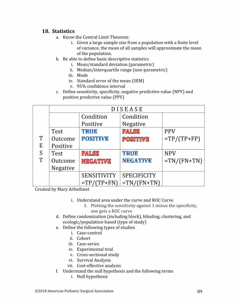
©2018 American Pediatric Surgical Association 89
18. Statistics a. Know the Central Limit Theorem:
i. Given a large sample size from a population with a finite level of variance, the mean of all samples will approximate the mean of the population.
b. Be able to define basic descriptive statistics i. Mean/standard deviation (parametric)
ii. Median/interquartile range (non-parametric) iii. Mode iv. Standard error of the mean (SEM) v. 95% confidence interval
c. Define sensitivity, specificity, negative predictive value (NPV) and positive predictive value (PPV)
T E S T
D I S E A S E Condition
Positive Condition Negative
Test Outcome Positive
PPV =TP/(TP+FP)
Test Outcome Negative
NPV =TN/(FN+TN)
SENSITIVITY =TP/(TP+FN)
SPECIFICITY =TN/(FN+TN)
Created by Mary Arbuthnot
i. Understand area under the curve and ROC Curve 1. Plotting the sensitivity against 1 minus the specificity,
one gets a ROC curve d. Define randomization (including block), blinding, clustering, and
ecologic/population-based (type of study) e. Define the following types of studies
i. Case-control ii. Cohort
iii. Case-series iv. Experimental trial v. Cross-sectional study
vi. Survival Analysis vii. Cost-effective analysis
f. Understand the null hypothesis and the following terms i. Null hypothesis

©2018 American Pediatric Surgical Association 90
1. There is no difference between 2 estimates 2. There is no disease
ii. Alpha – the probability of making a type I error iii. Beta – the probability f making a type II error iv. Power = (1-Beta) v. Type I error – rejecting the null when the null is true (false
positive) vi. Type II error – accepting the null when the null is false (false
negative) vii. Control Event Rate (CER) = number developing condition in a
control group/total number in control group viii. Experimental Event Rate (EER) = number developing condition
in experimental group/total number in experimental group 1. Relative Risk Reduction (RRR) = (CER-EER)/CER 2. Absolute Risk Reduction (ARR) = CER-EER 3. Number Needed to Treat (NNT) = 1/(CER-EER) or
1/ARR g. Be able to recognize and define variables in quantitative data
i. Continuous, Ordinal, Categorical, Dichotomous h. Understand the types of data distribution and be able to recognize a
graphic representation i. Normal distribution
ii. t-distribution iii. chi-squared distribution iv. F-distribution v. Binomial distribution
vi. Poisson distribution i. Understand ways to compare variables
i. Chi-squared, anova, t-test (student and paired), kruskal-wallis ii. Relative risk versus odds and odds ratio
iii. Define number needed to treat iv. Interpret Kaplan Meier or Cox-Hazard ratio v. Matching versus propensity analysis
Central Tendency Comparing 2
Groups Comparing >2
groups
Interval or Continuous Data
Normal (parametric)
Mean t-test (groups not related) or paired
t-test (same group, different point in time0
ANOVA
Not Normal (non-parametric)
Median Wilcoxon-Mann-Whitney
Kruskal-Wallis
Categorical or Ordinal or Binary
Proportion Chi-squared
Created by Mary Arbuthnot
©2018 American Pediatric Surgical Association 91
19. Ethics a. Key tenants:
i. Autonomy – right to self-govern ii. Beneficence – obligation to promote good and prevent or
remove harm iii. Justice – promote the greatest benefit to the largest number of
people iv. Nonmaleficence – need to refrain from inflicting harm
b. Surrogates (health care proxy, family member) represent substituted judgment, and should make the decision the patient would make given the situation or what is in the best interest of the patient if they do not know.
c. Understand the following terms i. Informed consent
ii. Health care proxy or durable power of attorney iii. Living will iv. Do Not Resuscitate v. Withdrawal of care
d. Special populations i. Pediatrics
1. Children may participate in decisions, and may provide assent and/or for procedures if older
ii. Jehovah’s witness 1. May refuse to receive blood or blood products 2. Some may agree to autotransfused blood 3. Practices vary state to state, recommend seeking advice
from hospital legal counsel

©2018 American Pediatric Surgical Association 92
20. Principles of Administration a. Administration and billing codes are more frequent on the exam, so be
familiar with coding and billing. b. American Medical Association established current procedural
terminology (CPT) to describe services performed by clinicians c. CPT mandated for reimbursement
i. For certain conditions, the ICU codes are covered in the surgery billing – for most pediatric surgeons these include Congenital Diaphragmatic Hernia and Abdominal wall defects (gastroschisis and omphalocele)
ii. In order to bill additional ICU coverage, must be an exception to the course outlined in the RUC
d. Relative Value Units (RVU’s) are assigned to three components i. Physician work
ii. Practice expense (overhead) iii. Malpractice (physician liability insurance)
e. International Classification of Diseases, 10th revision (ICD-10) – specific diagnoses or the reason patient is seen
i. Must be linked to CPT code to determine medical necessity for service and whether it is reimbursable
ii. TIME is important 1. Can dictate a separate note and bill for family meetings,
which can be common in an ICU. 2. When providing sedation, a separate note is important
and can be billed as well. i. Critical care codes – 2 primary adult codes are:
3. 99291: critical care, evaluation, and management of the critically ill or critically injured patient, first 30-74 minutes
4. 99292: critical care, evaluation, and management of the critically ill or critically injured patient, each additional 30 minutes (list separately in addition to the code for the primary service)
ii. Other 5. 92950: CPR 6. 31500: endotracheal intubation 7. 93503: Swan-Ganz catheter insertion or placement 8. 36556: CVL placement 9. 36620: A-line placement
iii. Neonatal and pediatric codes 10. 6 inpatient neonatal ICU codes
a. 99468: initial neonatal care, per day, for the evaluation and management of a critically ill neonate 28 days of age or younger

©2018 American Pediatric Surgical Association 93
b. 99469: subsequent neonatal critical care, per day, for the evaluation and management of a critically ill neonate 28 days or younger
c. 99477: initial hospital care, per day, for the evaluation and management of a neonate, 28 days or younger, who requires intensive observation, frequent interventions, and other intensive care services
d. 99478: subsequent intensive care, per day, for the evaluation and management of a recovering, VLBW (<1,500 gm) infant
e. 99479: subsequent intensive care, per day, for the evaluation and management of a recovering infant (1,501-2,500 gm)
f. 99480: subsequent intensive care, per day, for
the evaluation and management of a recovering infant (2,501 – 5,000 gm)
11. 4 pediatric ICU codes a. 99471: initial pediatric critical care, per day, for
the evaluation and management of a critically ill infant for young child, 29 days through 24 months of age
b. 99472: subsequent pediatric critical care, per day, for the evaluation and management of a critically ill infant or young child, 29 days through 24 months of age
c. 99475: initial pediatric critical care, per day, for the evaluation and management of a critically ill infant or young child, 2 through 5 years
d. 99476: subsequent pediatric critical care, per day, for the evaluation and management of a critically ill infant or young child, 2 through 5 years of age
f. Principles of documentation i. Medical record must be complete, legible, dated and signed and
include credentials ii. Rationale for all tests included
iii. Past and present diagnoses readily available iv. Risk factors identified v. Patient progress, treatment response and revision to treatment
plan documented vi. ICD-10 codes should justify services
vii. Cannot bill for what you perform, only for what you document viii. Must include a modifier for surgery resident but NOT for
advanced practitioner (NP or PA)

©2018 American Pediatric Surgical Association 94
g. History i. History includes: HPI, ROS, and past, family and social history
(PFSH) ii. 4 levels:
1. 1) Problem focused: chief complaint (CC), brief HPI (no ROS/PFSH)
2. 2) Expanded problem focused: CC, brief HPI, problem pertinent ROS (no PFSH)
3. 3) Detailed: CC, extended HPI, extended ROS, pertinent PSFH
4. 4) Comprehensive: CC, extended HPI, complete ROS, complete PFSH
h. Physical exam i. 4 levels:
1. Problem focused: 1 body area or organ system 2. Expanded problem focused: 2-7 body areas or organ
systems 3. Detailed: 2-7 body areas or organ systems with 1
system described in detail 4. Comprehensive: 8 or more organ systems or complete,
single-specialty examination i. Decision making
i. Decision making for surgery is usually considered level 4
Type of decision
# of diagnoses of management options
Amount/Complexity of data to be reviewed
Risk of complications or morbidity/mortality
Straight-forward
Minimal Minimal or none Minimal
Low complexity
Limited Limited Low
Moderate complexity
Multiple Moderate Moderate
High complexity
Extensive Extensive High
Created by Mary Arbuthnot
GOOD LUCK!

©2018 American Pediatric Surgical Association 95
References Asensio, J., A., & Trunkey, D. (Eds). (2008). Current Therapy of Trauma and Surgical Critical Care. Philadelphia, PA. Mosby Elsevier. Berg, S, & Bittner, E. (Eds.). (2014). The Massachusetts General Hospital Review of Critical Care Medicine. Philadelphia, PA: Lippincott Williams & Wilkens.
Dorman, T., Britton, F., Brown, D., & Munro, N. (Eds.). (2014). Coding and Billing for Critical Care: A Practice Tool. Prospect, IL: Society of Critical Care Medicine. Lucking, S., Maffei, F., Tamburro, R., & Thomas, N. (Eds.). (2012). Pediatric Critical Care Study Guide: Text and Review. London, UK. Springer-Verlag. Marino, P. (2007). The ICU Book (3rd ed.). Philadelphia, PA: Lippincott Williams & Wilkens. Moore, F., Rhee, P., Tisherman, S., & Fulda, G. (Eds.). (2012). Surgical Critical Care and Emergency Surgery: Clinical Questions and Answers. Hoboken, NJ. John Wiley & Sons. O’Donnell, J., & Nácul, F (Eds.). (2010). Surgical Intensive Care Medicine (Kindle Location 37844). New York, NY. Springer-Verlag. Roberts, P., & Todd, S. (Eds.). (2012). Comprehensive Critical Care: Adult. Mount Prospect, IL. Society of Critical Care Medicine.
A SPECIAL THANK YOU TO THE PEDIATRIC SURGERY NaT FOR DONATING IMAGES AND TABLES TO THE SYLLABUS
www.pedsurglibrary.com/apsa