Embed Size (px)
Citation preview

Pediatric Visual
Diagnosis
Lisa Warren, DO
Department of Pediatrics
Western University of Health Sciences
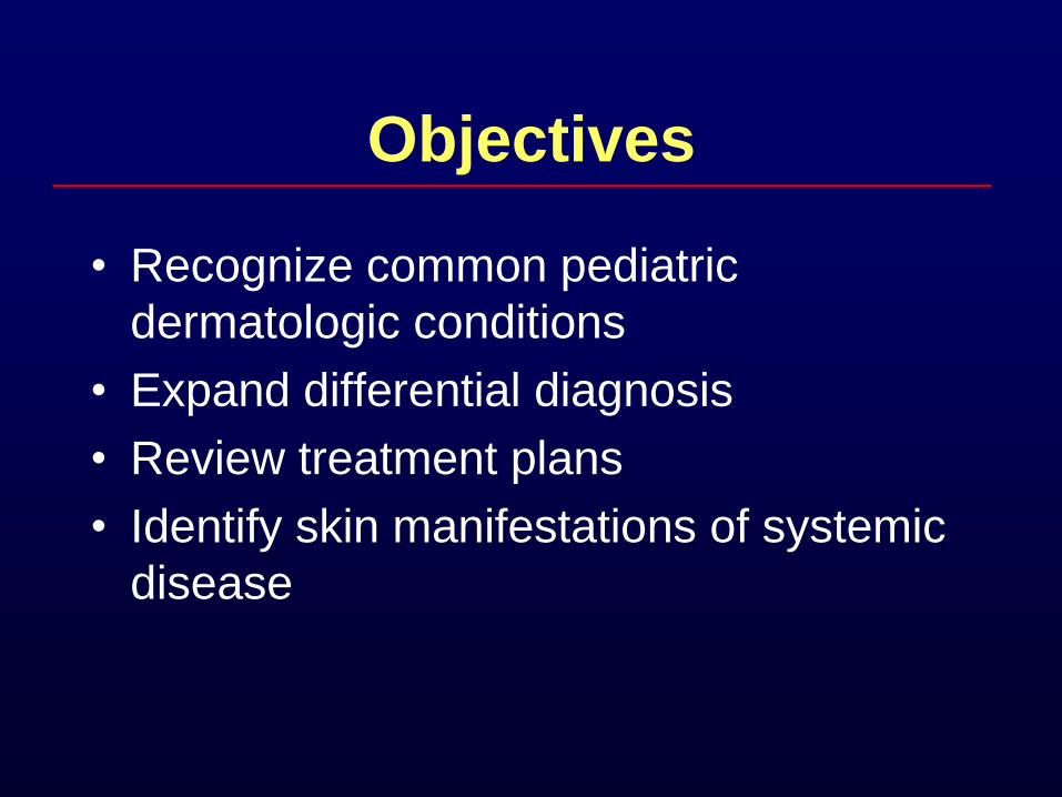
Objectives
• Recognize common pediatric
dermatologic conditions
• Expand differential diagnosis
• Review treatment plans
• Identify skin manifestations of systemic
disease
Question 1
A one week old child’s mother presents to
your office with concerns that the child
has a rash that has progressed since
being discharged from the hospital.
Birth history is unremarkable.
Physical examine shows a splotchy
erythema with a central clear pustule.
Your likely diagnosis is?
a. Erythema toxicum
b. Neonatal pustular melanosis
c. Staph folliculitis
d. Milia
e. Neonatal acne
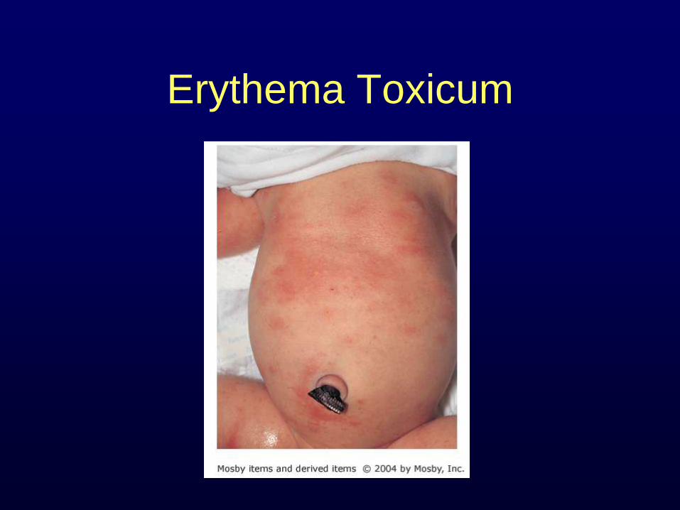
Erythema Toxicum
Erythema Toxicum
• Also described as a “flea bite”
• Intense erythema with / a central papule or pustule.
• 2-3 cm in diameter, on back, face, chest and extremities
• Usually in full term infants, appears usually beginning 24- 48 hours
Erythema Toxicum
• Benign self limited with unknown etiology
• No treatment, it fades within 5-7 days.
• A smear of the pustule reveals numerous Eosinophils
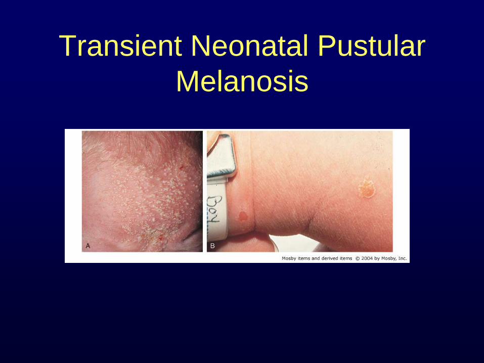
Transient Neonatal Pustular
Melanosis
Neonatal Pustular Melanosis
• 1-2 mm of vesiculopustules or ruptured pustules
• Usually present at birth, but disappear quickly within 24-48 hours
• Leaves pigmented macules with a collaret of scale
• Fades within 3 weeks to 3 month
• Occurs anywhere on the body, but more common on neck, forehead, lower back, and the legs
Neonatal Pustular Melanosis
• Unknown etiology, self limited.
• Gram stain shows neutrophils.
• Common concern is Staph aureus
infection
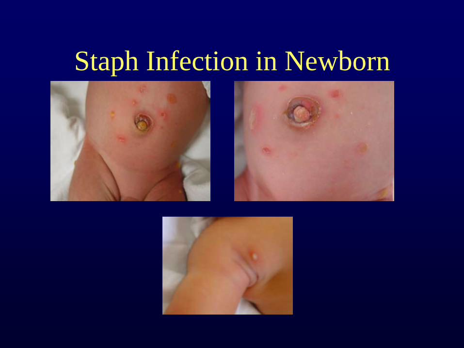
Staph Infection in Newborn
Staph Infection in Newborn
• Lesions tend to appear in the later part of
the first week of life or into the second
week.
• Any body site may be involved with
predilection to the diaper area.
• The bullae are flaccid, containing straw
colored or turbid fluid, rupture easily
leaving a moist denuded area.
Management
• CBC, Blood cultures
• Gram stain and culture wound sites
• Hospitalization and treatment with a
systemic appropriate antibiotic should be
instituted particularly for lesions around the
umbilicus.
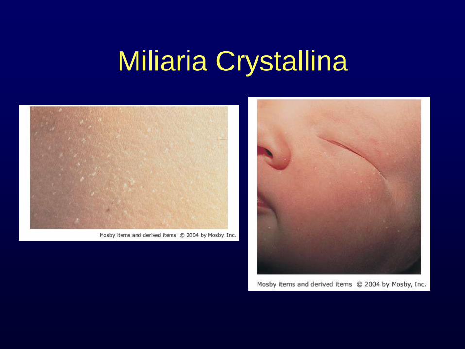
Miliaria Crystallina
Milia
• Small, firm 1-2 mm in diameter
• Tiny thin-walled sweat-retention vesicles rupture readily, then quickly desquamate
• Commonly seen on the face on neonates
• Consist of epithelial lined cysts arising from hair follicles
Milia
• Usually persistent, but may resolved within month to years.
• Usually appear with no apparent cause, but may also appear after skin injury.
• In the mouth it is called Epstein's pearls
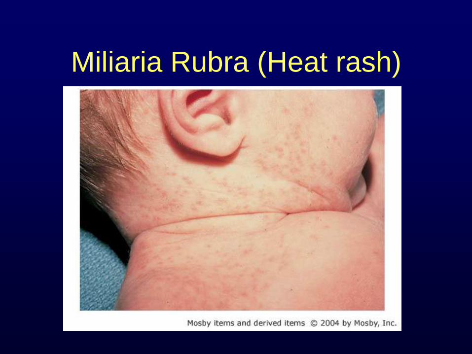
Miliaria Rubra (Heat rash)
Milia Rubra
• Sweat duct obstruction in deeper epidermal or dermal layers.
• Erythematous papulopustular eruption
• Usually over the face, upper trunk, and Intertiginous area of the neck.
• It is usually as a result of tight fitting cloth or use of occlusive lubricants during hot, humid weather.

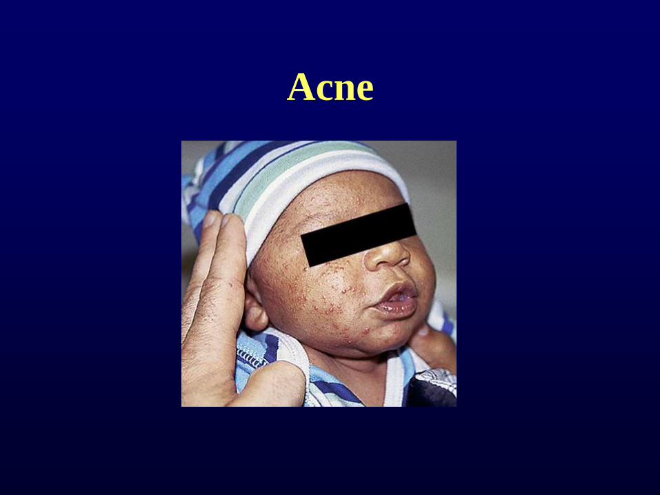
Neonatal acne
Neonatal Acne
• Occurs in 20% neonates
• Thought related to be caused by
stimulation of sebaceous glands by
maternal and endogenous androgens
• Proposed cause - inflammatory reaction
to skin colonization with Malassezia
species

Neonatal acne
• Mean age 3 weeks
• Presence of inflammatory papules and
pustules – no comedones
• Distribution limited to face
• Treat with daily cleansing with soap and
water
• Application 2% ketaconazole or 1%
hydrocortisone
Acne
Infantile acne
• 3 to 4 months of age
• Hyperplasia of sebaceous glands due to
androgenic stimulation
• Inflammatory papules, pustules, and
comedones
• Can treat with benzoyl peroxide, topical
antibiotics, or topical retinoids
• No improvement – consider endocrinopathy
Question 2
A 7-year-old boy is brought to the clinic for an itchy
rash that has been present for 2 weeks. He has
been healthy except for intermittent asthma, and
his mother reports that he frequently has very dry
patches of skin. He has numerous linear vesicles
and blisters on his arms, with surrounding
erythema and mild edema. He has a few similar
lesions on his anterior legs. He scratches the
lesions frequently during your examination.
Your likely diagnosis is:
A. Atopic dermatitis
B. Scabies
C. Tinea Corporis
D. Pityrasis rosea
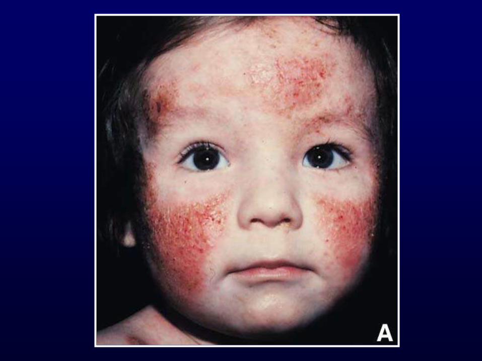
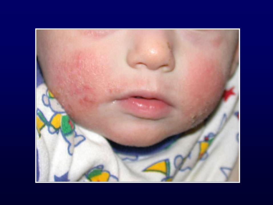
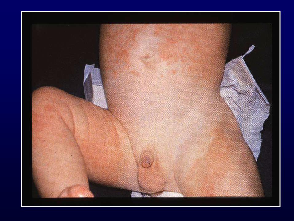

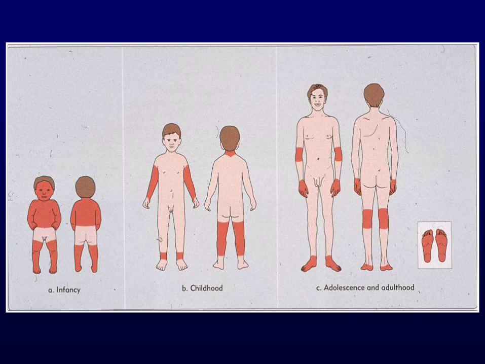
Atopic Dermatitis
• 3-5% of children 6 mo to 10 yr
• Described in 1935
• Ill-defined, red, pruritic, papules/plaques
• Diaper area spared
• Acute: erythema, scaly, vesicles, crusts
• Chronic: scaly, lichenified, pigment
changes
Atopic Dermatitis
Hints to diagnosis
• Generalized dry skin
• Accentuation of skin markings on palms
and soles
• Dennie-Morgan lines
• Fissures at base of earlobe
• Allergic history
Diagnostic Criteria
Major Criteria: At least 3 of the following:
1. Pruritus
2. Personal or family history of atopy
(asthma, allergic rhinitis, atopic dermatitis)
3. Chronic or chronically relapsing dermatitis
4 Typical morphology and distribution

Treatment
• Moisturize
• Baths only
• Anti-histamine
• Topical steroids to red and rough areas
• Immune modulators
Avoidance of Triggers
• Perspiration and overheating
• Irritating clothes – soft cotton
• Avoid harsh soaps, detergents, fabric
softeners, products with fragrance, and
bubble baths
• Exposure to tobacco
• IgE mediated food allergy – Milk, eggs, soy, wheat, peanuts
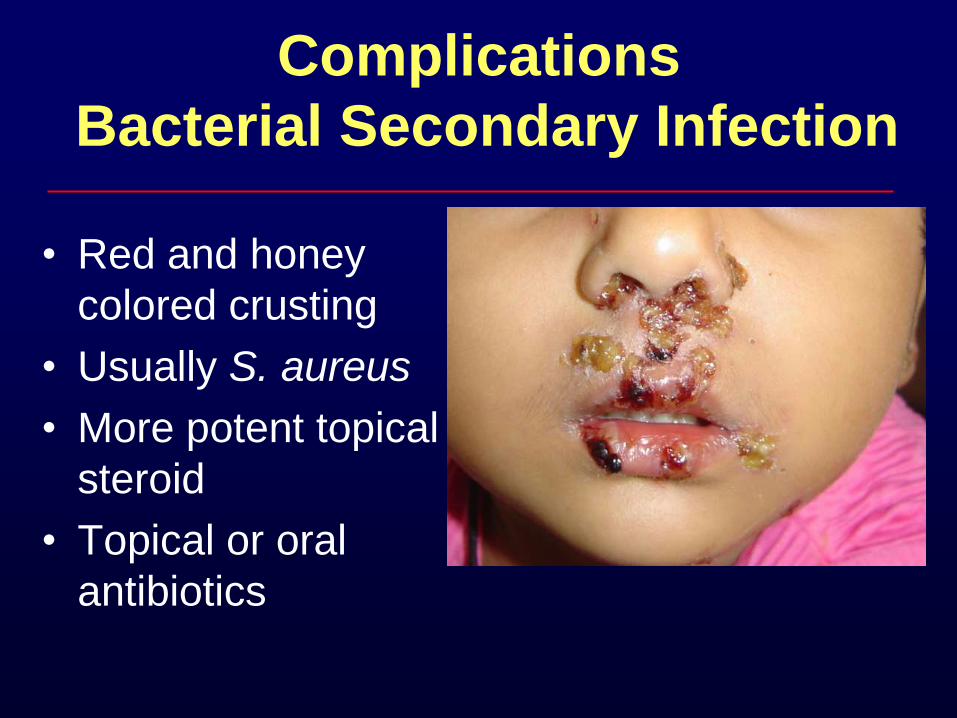
Complications
Bacterial Secondary Infection
• Red and honey
colored crusting
• Usually S. aureus
• More potent topical
steroid
• Topical or oral
antibiotics
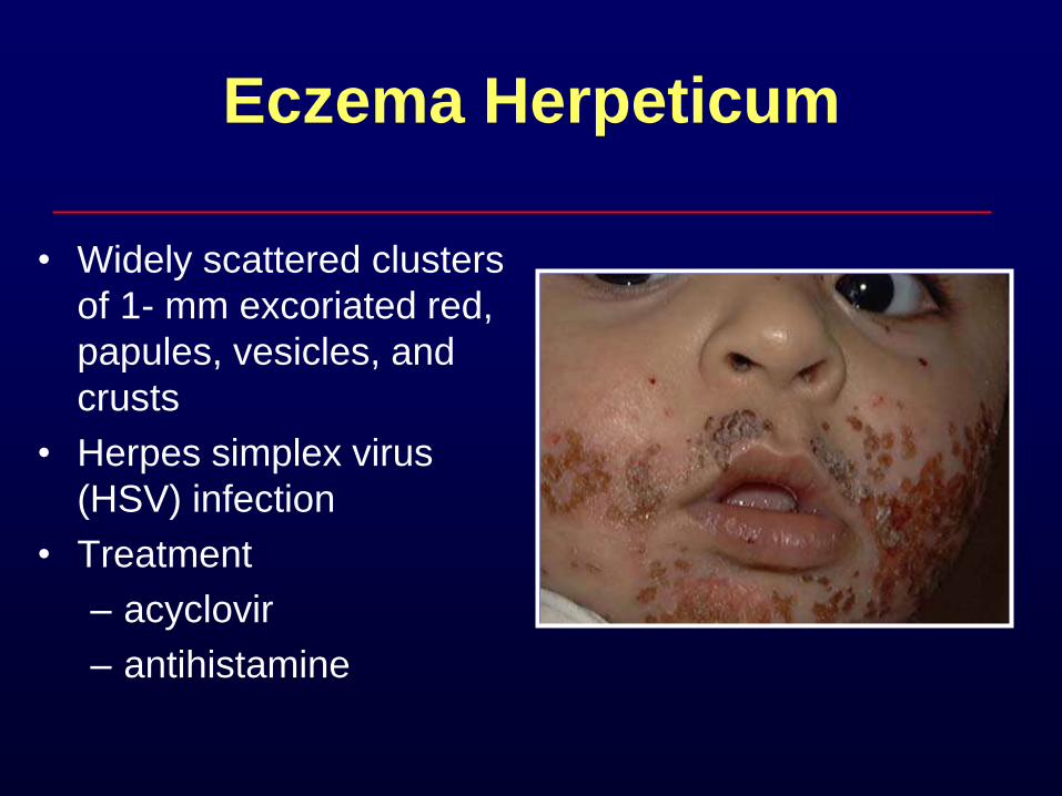
Eczema Herpeticum
• Widely scattered clusters
of 1- mm excoriated red,
papules, vesicles, and
crusts
• Herpes simplex virus
(HSV) infection
• Treatment
– acyclovir
– antihistamine
Scabies
• Intense pruritus
• Diffuse, papular rash
– Between fingers, flexor aspects of wrists, anterior axillary folds, waist, navel
• May be vesicular in children < 2 years
– Head, neck, palms, soles
– Hypersensitivity reaction to protein of parasite
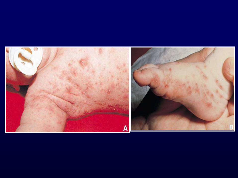
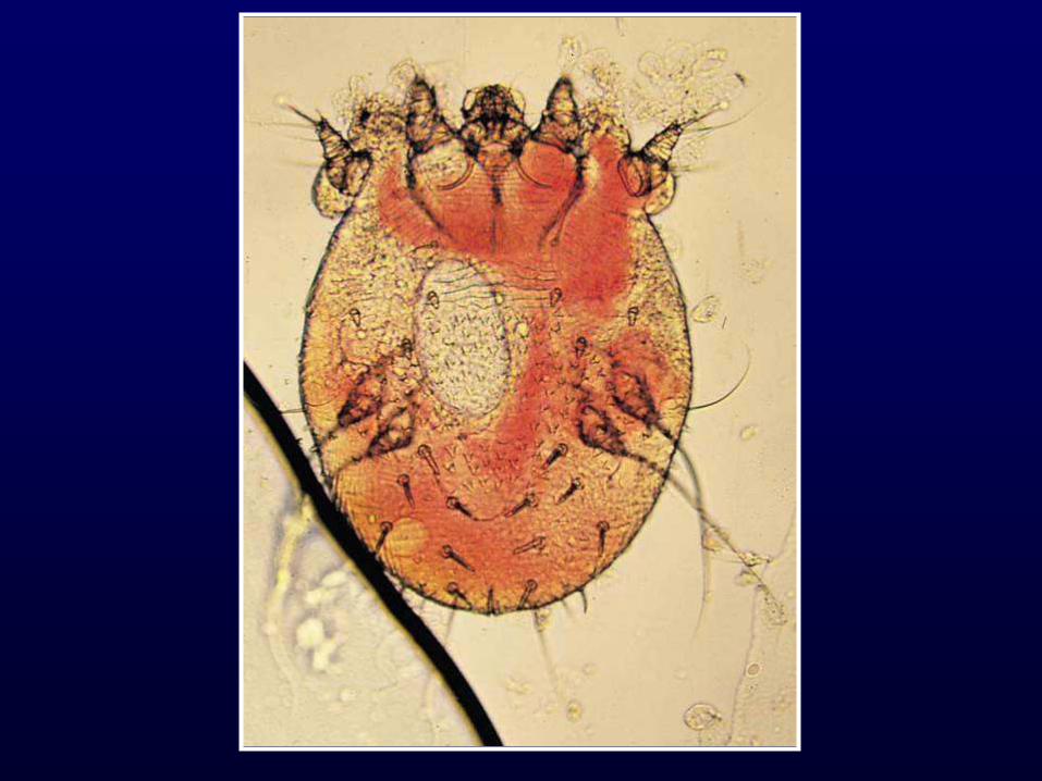
Scabies
• Sarcoptes scabiei mite
• Pruritic burrows pathognomic (irregular, tortuous, and slightly scaly)
• In infants, burrows are widespread with involvement of trunk, scalp, extremities, palms and soles
• Consider in infants with widespread dermatosis that involves the palms and soles

Scabies - Treatment
• 5% permethrin cream for infants, young
children, pregnant and nursing mother
– Kwellada-P or Nix
– Cover entire body from neck down
– Include head and neck for infants
– Wash after 8-14 hours
• Can use Lindane for older children
Scabies - Treatment
• Not after a hot bath
• All family members at same time
• Whole body treatment inc, scalp, neck,
face, ears and under nails
• Repeat week later
• Pruritis can last for weeks
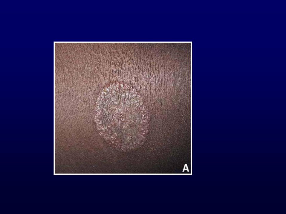
Tinea Corporis
• Contact with other individuals, domestic animals including young kittens and puppies
• Most common group of dermatophytes – Trichophyton
– Microsporum
– Epidermophyton
• Invade keratin, the protein that forms the outermost epidermis, nails, and hair
Tinea Corporis
• Face, trunk or limbs
• Pruritic, circular, slightly erythematous
• Well-demarcated with scaly, vesicular or pustular border
• Mistaken for atopic, seborrheic or contact dermatitis
• Treatment: Topical or oral antifungal
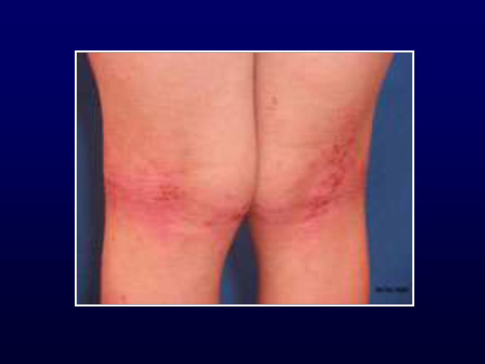
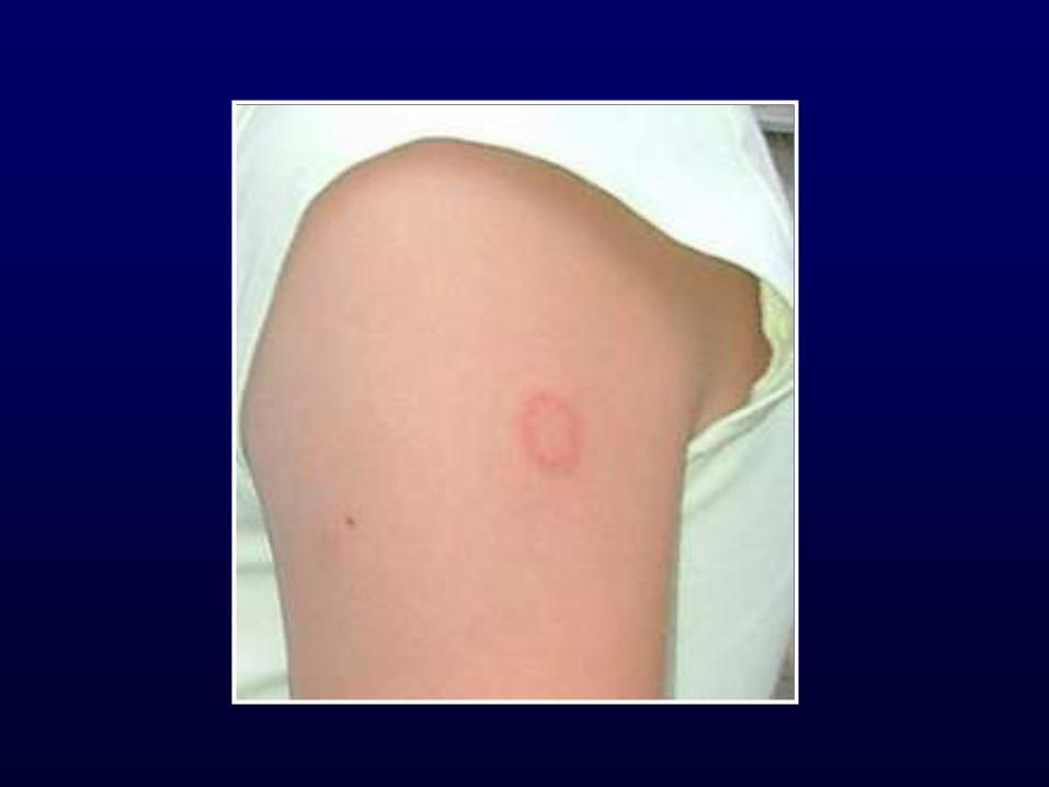
Pityriasis Rosea
• Begins with herald patch
– Large, isolated oval lesion with
central clearing
• More lesions 5-10 days later
• Christmas tree distribution
• Treatment: anti-histamines
Question 3
A 2 year old female presents to your clinic
with a 3 day history of high fever up to 103
and irritability. Mother reports that her
throat seems to bother her because she
refuses to eat and cries when she drinks her
milk. She started developing a rash on her
buttock and hands. Her immunizations are
up to date.
Your likely diagnosis is:
a. Hand foot mouth disease
b. Scarletina
c. Erythema Infectiosum
d. Roseola
e. Herpes Simplex
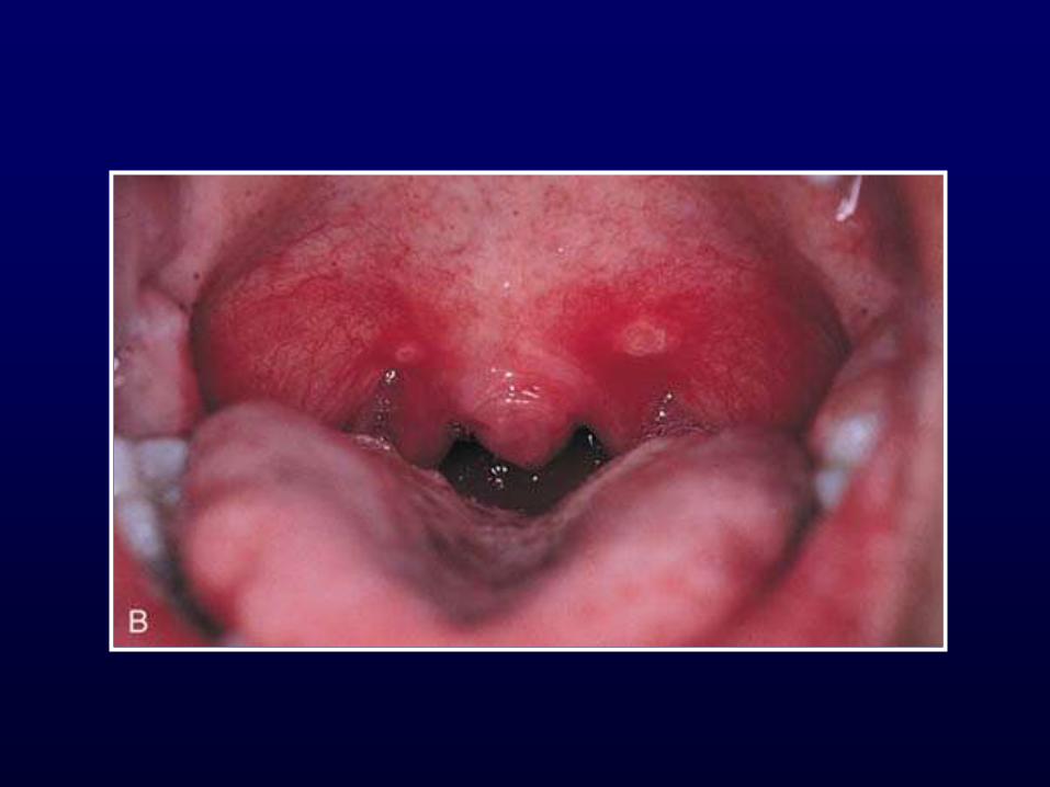

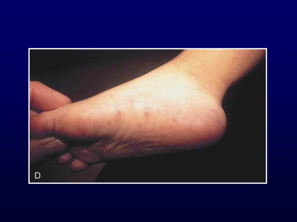

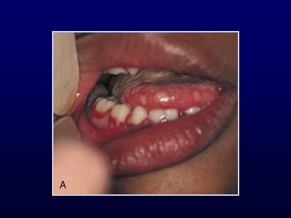
Hand-Foot-Mouth Disease
• Painful, shallow, yellow ulcers surrounded by red halos
• Found on buccal mucosa, tongue, soft palate, uvula and anterior tonsillar pillars
• Oral lesions without the exanthem = herpangina
• Exanthem involves palmar, plantar and interdigital surfaces of the hands and feet +/- buttocks
• Due to Coxsackie A virus
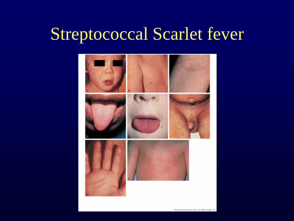
Streptococcal Scarlet fever
Streptococcal Scarlet Fever
• Flushed face,
• Perioral pallor, and a
• Diffuse, blanching, erythematous rash that has a sandpapery consistency on palpation.
• Pastia lines
• Confirm with culture
• Treat with penicillin
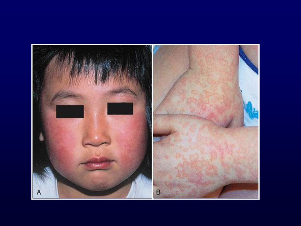
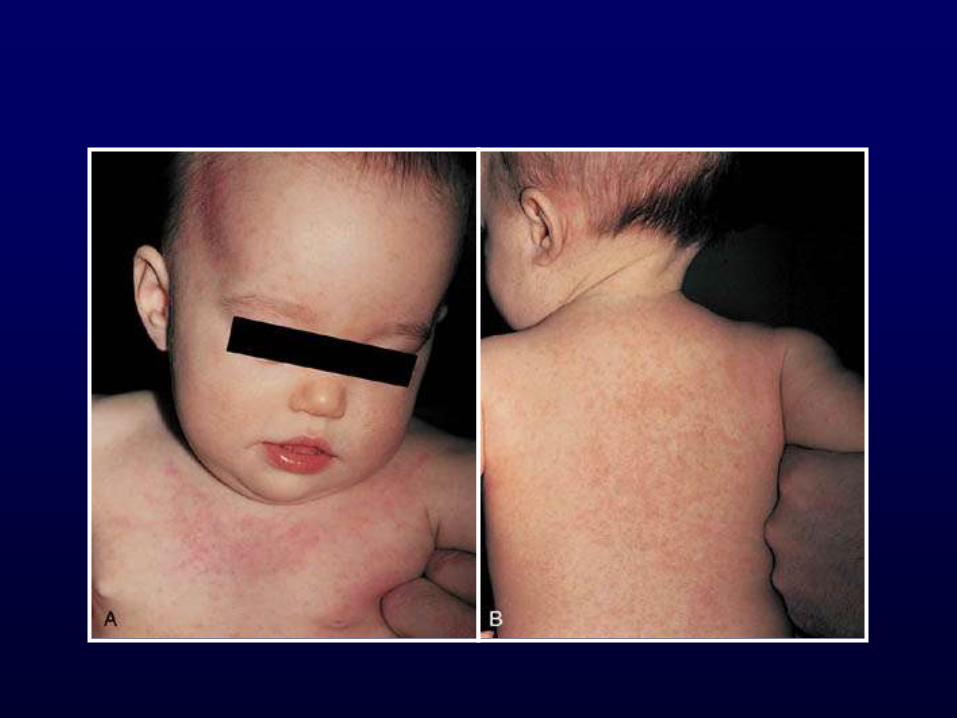
Erythema Infectiosum Fifth Disease
• Parvovirus B19
• Mostly preschool age
• Recognized by exanthem
• Contagious before rash
• Resolution between 3 and 7 days
Erythema Infectiosum
(Fifth Disease)
• On day 1, warm, erythematous, nontender,
circumscribed patches appear over the
cheeks.
• These fade on the following day, as an
erythematous, lacy rash develops on the
extensor surfaces of the extremities.
• No preceding symptoms
• No treatment needed
Roseola
• 6 to 36 months
• Human herpesvirus 6
• High fever without source and irritability
for 3 days
• Rash develops as fever decreases
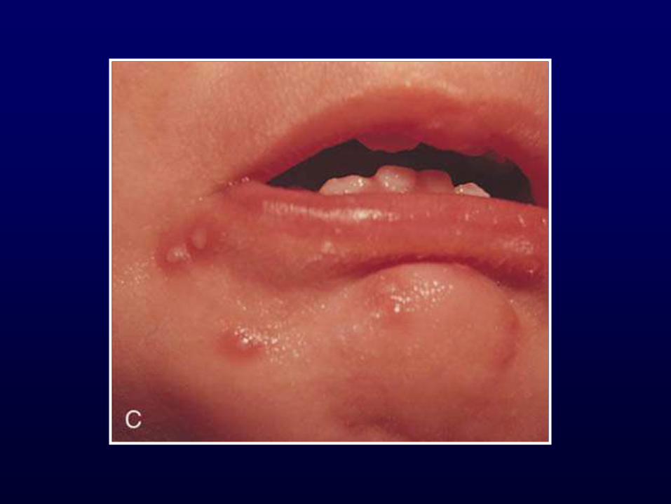
Herpes Simplex
• Gingivostomatitis most common 1º infection
in children
– Fever, irritability, cervical nodes
– Small yellow ulcerations with red halos on mucous
membranes
• Involvement more diffuse – easy to
differentiate from herpangina and exudative
tonsillitis
• Treatment: supportive
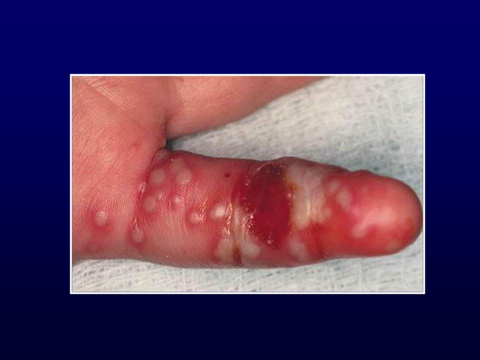
Herpetic Whitlow
• Lesions on thumb usually 2° to autoinoculation
• Group, thick-walled vesicles on erythematous base
• Painful
• Tend to coalesce, ulcerate and then crust
• May require topical or oral acyclovir

Conclusions
• Not all that itches is eczema
• Treatment is often supportive for viral
exanthems
• Remember rashes as a sign of systemic
illness