Embed Size (px)
Citation preview
PEER-REVIEWED ARTICLE bioresources.com
EL-Hefny et al. (2017). “Antibacterial extracts,” BioResources 12(1), 1835-1849. 1835
Chemical Composition and Bioactivity of Salvadora persica Extracts against Some Potato Bacterial Pathogens
Mervat EL-Hefny,a Hayssam M. Ali,b,c Nader A. Ashmawy,d and
Mohamed Z. M. Salem*,e
Potent antibacterial activities of solvent extracts (methanol:n-hexane) from the branch, leaf, and root-wood of Salvadora persica were examined against potato phytopathogenic bacteria, namely Pectobacterium carotovorum subsp. carotovorum, Dickeya solani, Ralostonia solanacerum, Enterobacter cloacae, and Bacillus pumilus. The main chemical constituents analyzed by gas chromatography–mass spectrometry (GC/MS) in the branch extracts were N-benzylbenzamide (71.08%), decane (3.17%), stigmasterol (3.17%), 9-desoxo-9-x-acetoxy-3,8,12-tri-O-acetylingol (2.33%), and β-sitosterol (2.15%). The main components in the leaf extracts were 2,6-dimethyl-N-(2-methyl-α-phenylbenzyl)aniline (28.65%), spiculesporic acid (13.60%), homo-γ-linolenic acid (12.63%), and methyl hexadecanoate (11.01%). The root-wood extracts contained, as primary parts, benzeneacetonitrile (71.47%), 4-aminocarbonyl-5-fluoro-1-α-D-ribofuranosyl-imidazole (10.99%), and benzylisothiocyanate (5.05%). The extracts from the root-wood showed moderate antibacterial activity against the potato bacterial pathogens, which was followed by leaf and branch extracts. The results suggested that S. persica plant extracts could be used as bioagents against potato soft and brown rot bacterial pathogens.
Keywords: Salvadora persica; Leaf; Root-Wood; Branch; Antibacterial activity; Chemical composition
Contact information: a: Department of Floriculture, Ornamental Horticulture and Garden Design, Faculty
of Agriculture (El-Shatby), Alexandria University, Alexandria, Egypt; b: Botany and Microbiology
Department, College of Science, King Saud University, P.O. Box 2455, Riyadh 11451, Saudi Arabia;
c: Timber Trees Research Department, Sabahia Horticulture Research Station, Horticulture Research
Institute, Agriculture Research Center, Alexandria, Egypt; d: Department of Plant Pathology, Faculty of
Agriculture (El-Shatby), Alexandria University, Alexandria, Egypt; e: Forestry and Wood Technology
Department, Faculty of Agriculture (EL-Shatby), Alexandria University, Alexandria, Egypt;
* Corresponding author: [email protected]
INTRODUCTION
Potato is an important vegetable crop in Egypt. Annually, approximately
4,800,000 tons are produced from approximately 178,000 hectares, which makes Egypt
the top potato producer in Africa (FAO STAT 2013). Potato plants are subject to
numerous pathogens and pests, which cause considerable quantitative and qualitative
potato yield losses in Egypt. Such pathogenic problems are caused by bacterial diseases,
especially brown rot caused by Ralostonia solanacerum (Yabuuchi et al. 1995) and soft
rot and blackleg caused by Pectobacterium carotovorum, Dickeya, Enterobacter, and
Bacillus species (Behiry 2013; Salem 2013; Ashmawy et al. 2014).
The first authenticated report of Brown rot disease in Egypt was in the last
century (Sabet 1961), and Mickail et al. (1974) made the first survey on the organism. In
PEER-REVIEWED ARTICLE bioresources.com
EL-Hefny et al. (2017). “Antibacterial extracts,” BioResources 12(1), 1835-1849. 1836
seed potato production, the contamination of seed tubers with soft rot bacteria
(Pérombelon 2002; Toth et al. 2003), is one of the biggest problems, which causes
blackleg, rotting of potato stems in the field, and soft rot of tubers during storage (Gardan
et al. 2003; Laurila et al. 2008).
Salvadora persica (Miswak), which belongs to family Salvadoraceae, has been
used in toothbrushes for the prevention of tooth decay (Arora and Kalia 2013). The leaf
extracts act as an antibacterial agent to various oral bacteria (aerobic) with results
comparable to known antibiotics (Alali and Al-Lafi 2003).
Several studies have reported that S. persica extracts and seed oil have great
medicinal uses in the treatment of nose troubles, gonorrhea, leucoderma, scabies, scurvy,
some skin diseases, joint pain and toothaches; it is also used as a laxative and as a general
body tonic (Elvin-Lewis et al. 1980; Alali and Al-Lafi 2003; Darmani et al. 2003;
Khalessi et al. 2004; Ahmed et al. 2008).
Leaf extracts of S. persica exhibit several pharmacological properties including
carminative, antiseptic, antifungal, antibacterial, diuretic, analgesic, anthelmintic,
astringent, hypoglycaemic, antiplasmodial, anticaries, antispasmodial, antiscorbutic, and
anticonvulsant properties, as well as action against hepatic disorders (Al-Bagieh et al.
1994; Al-Bagieh and Almas 1997; Ali et al. 2002; Almas et al. 2005; Saini et al. 2006;
Paliwal et al. 2007). Extracts of stems have antiplaque (Chawla 1983) and antimicrobial
activities (Almas 2001). Aqueous extracts are more effective than methanol extracts
against some pathogenic bacteria (Al-Bayati and Sulaiman 2008); however, Al-Bagieh
and Almas (1997) showed that alcoholic extracts have more potent antimicrobial activity
than aqueous extracts.
The heterogeneous components extracted from S. persica have been reported to
have antimicrobial activities (Akhtar et al. 2011). Pulp and bark extracts show significant
differences in their antimicrobial activities (Almas and Al-Bagieh 1999). S. persica
extract (20%) is effective as an antifungal and antibacterial agent against Candida
albicans and Enterococcus faecalis (Al-Obaida et al. 2010). The diluted acetone extract
of dry stems (300 mg/mL) shows good inhibitory activity against C. albicans, C.
glabrata, and C. parapsilosis strains with inhibition zones (IZs) that range from 10.33
mm to 15 mm (Noumi et al. 2010).
Volatile oils extracted from the roots and stems of S. persica contain fatty and
other organic acid ethyl esters (Abdelrahman et al. 2003). Aqueous extracts of the roots
contain chlorine, trimethylamine, and sulphur compounds with antimycotic activity (Al-
Otaibi and Angmar 2004). Benzylisothiocyanate is the main component in root oil (Bader
et al. 2002), which has good activity against Herpes simplex virus, Streptococcus mutans,
and Candida albicans (Al-Bagieh 1992, 1998; Al-Bagieh and Weinberg 1988). β-
Sitosterol has been found in the roots of S. persica (Ezmirly et al. 1978). The zone of
inhibition against the growth of Staphylococcus aureus ranges from 10.5 mm to 31.5 mm
for the leaf extract of S. persica and the combination of tetracycline with the stem extract
of S. persica, respectively (Ahmed et al. 2010).
Salvadoricine, an indole alkaloid, has been isolated from S. persica leaves (Malik
et al. 1987). Volatile oils from the leaves contain benzyl nitrile, eugenol, thymol,
isothymol, eucalyptol, isoterpinolene, and β-caryophyllene (Alali and AL-Lafi 2003).
Identified flavanoids and flavanoid glycosides include kaempferol 3-α-L-rhamnosyl-7-β-
xylopyranoside, quercetin, and kaempferol (Kamil et al. 2000).
Ethanolic extracts of the stems contain β-sitosterol, stigmasterol, and β-sitosterol-
D-glucoside (Arora and Kalia 2013). A sulfated glycoside, salvadoside (sodium 1-O-
PEER-REVIEWED ARTICLE bioresources.com
EL-Hefny et al. (2017). “Antibacterial extracts,” BioResources 12(1), 1835-1849. 1837
benzyl-β-D-glucopyranoside-2-sulfate), was isolated from S. persica (Ohtani et al. 1992).
Pyrrolidine, pyrrole, and piperidine derivatives have been identified in S. persica sticks
(Galletti et al. 1993). Salvadoside and salvadoraside, which are glycoside compounds,
have been reported in stem extracts (Kamel et al. 1992). Benzylisothiocyanate, saponins,
tannins, resin, trimethylamine, and alkaloid have been isolated from the roots (El-
Mostehy et al. 1983). β-Sitosterol, manisic acid, and salvadourea [1,3-bis-(3-methoxy-
benzyl)-urea] were isolated from the root by Ray et al. (1975). 2-Furancarboxaldehyde-5-
(hydroxymethyl), furan-2-carboxylic acid-3-methyl- trimethylsilyl ester, and D-erythro-
pentofuranose-2-deoxy-1,3,5-tris-O-(trimethylsilyl) were identified in root methanol
extracts; these components exhibit antioxidant activities (Mohamed and Khan 2013).
Stem essential oils include 1,8-cineole (eucalyptol), α-caryophellene, β-pinene, and 9-epi-
(E)-caryophellene as the major components (Alali et al. 2004).
Most of the studies related to the bioactivity of extracts from S. persica have
focused on the extractives’ effectiveness as a natural tool for dental cleaning and as a
natural analgesic for toothache, as well as their effect on various aspects of oral health
(Alali and Al-Lafi 2003; Balto et al. 2012; Halawany 2012; Chaurasia et al. 2013).
The antibacterial activities of extracts from several plants against bacterial potato
pathogens have been assessed, and quite satisfactory results have been observed (Salem
2013; Ashmawy et al. 2014). The agricultural companies in the Mediterranean countries
are focused on the commercial production of known aromatic herbs such as mint and
basil (Edris et al. 2003) and neglecting the utilization of trees and shrubs, which may
provide new sources of medical and agricultural applications (Bakkali et al. 2008; Abdel-
Megeed et al. 2013; Salem et al. 2013, 2014a,b,c). So there is motivation to search for
new and renewable sources for natural products that are useful against phytopathogenic
bacteria and fungi (Salem et al. 2016a,b).
To date, there are no reports on the bioactivity of extracts from S. persica against
the growth of pathogenic bacteria that attack plants. This study evaluated the antibacterial
activity of extracts that analyzed by gas chromatography–mass spectrometry (GC/MS)
from the leaves, branches, and root-wood of S. persica against the growth of some
pathogenic bacteria.
EXPERIMENTAL
Plant Materials and Reagents Leaves, branches, and root-wood of Salvadora persica were collected in May
2016 from the Jazan Region located on the southwestern part of the Kingdom of Saudi
Arabia. The plant was identified by the Botany and Microbiology Department of the
College of Science at King Saud University. The samples were delivered to the Faculty
of Agriculture at Alexandria University by Dr. Hayssam M. Ali on June 2016.
Extractions were performed at Alexandria University on the various S. persica
components, and the antibacterial activity of extractives was assessed. The plant was
authenticated with the voucher number Zidan0043. Methanol, dimethylsulfoxide
(DMSO) and n-hexane solvents were bought from Sigma Aldrich (Cairo, Egypt).
Extraction About 100 air-dried g of powdered leaf, branch, and root-wood were separately
extracted by soaking in a mixture of methanol:n-hexane (1:1 v/v) for one week. The
PEER-REVIEWED ARTICLE bioresources.com
EL-Hefny et al. (2017). “Antibacterial extracts,” BioResources 12(1), 1835-1849. 1838
extraction process was repeated three times in the week until exhaustion, where every
filtration was done after two days. The combined extract from each plant part was
concentrated using a rotary-evaporator at 45 °C. The concentrated extracts were stored
for one week at 4 °C until further analysis. The extract weights from leaf, branch, and
root-wood components were 6.24, 5.17, and 8.55 g, respectively. Each extract was
prepared in the concentrations of (1000, 500, 250, 125, 64, and 32 µg/mL), by diluting
the extract in 10% DMSO.
Antibacterial Activity Assay The antibacterial activities of leaf, branch, and root-wood extracts from S. persica
were evaluated using the disc diffusion method of Bauer et al. (1966) against the growth
of selected phytopathogenic bacteria: Pectobacterium carotovorum subsp. carotovorum
ippbc038, Dickeya solani, Ralostonia solanacerum, Enterobacter cloacae, and Bacillus
pumilus. These bacterial strains have been associated with blackleg and soft rot disease of
potatoes; also, these bacteria can completely destroy potato plantations, as well as cause
brown rot in potatoes after post-harvest. The discs were impregnated with 20 µL of each
of the concentrated extract (leaf, branch, and root-wood extracts). Mueller Hinton Agar
(MHA) media in sterile Petri dishes were spread with a fresh 24-h-old bacterial
suspension (1.0 x 10⁵ CFU/mL) and sterile discs (Whatman filter paper no. 1) with 4 mm
diameter and were stacked over the inoculated media surface. Three measurements of the
inhibition zones around the discs were recorded in millimeters using a ruler.
The bacterial strains were supplied by the Department of Plant Pathology of the
Faculty of Agriculture (El-Shatby) at Alexandria University (Alexandria, Egypt). Control
discs with negative (DMSO) and positive (gentamicin 20 μg/disc) were performed, and
all tests were performed in triplicate.
GC/MS Analyses of Extracts The chemical compositions of the extracts were analyzed using a Trace GC Ultra-
ISQ Mass Spectrometer (Thermo Scientific, Austin, TX, USA) with a direct capillary
column TG-5MS (30 m × 0.25 mm × 0.25 µm film thickness) apparatus. The GC/MS
was located at the Atomic and Molecular Physics Unit of the Experimental Nuclear
Physics Department at the Nuclear Research Centre of the Egyptian Atomic Energy
Authority (Inshas, Cairo, Egypt). The column oven temperature was initially held at 120
°C and then increased by 5 °C∙min-1 to 200 °C, which was held for 2 min, then increased
to 280 °C (10 °C∙min-1). Temperatures of the injector and detector (MS transfer line)
were kept at 250 °C. Helium, which was the carrier gas, was kept in constant flow rate of
1 mL∙min-1. The solvent delay was 2 min, and diluted samples of 1 µL were injected
automatically using an Auto-sampler AS3000 coupled with the GC unit in the split mode.
EI mass spectra were collected at 70 eV ionization voltages over the m/z range of 40 to
550 in full scan mode. The ion source and transfer line temperatures were set at 200 and
250 °C, respectively. The components were identified by comparison of their retention
times and mass spectra with those of the WILEY 09 and NIST 11 mass spectral database
(Davies 1990).
Statistical Analysis The values of the antibacterial activity are presented as mean of three replicates.
Analysis of variance (ANOVA) was used to evaluate the significant difference among
various treatments with the criterion of p = 0.05. The statistical analysis was performed
PEER-REVIEWED ARTICLE bioresources.com
EL-Hefny et al. (2017). “Antibacterial extracts,” BioResources 12(1), 1835-1849. 1839
using SAS software version 8.2 (2001).
RESULTS AND DISCUSSION
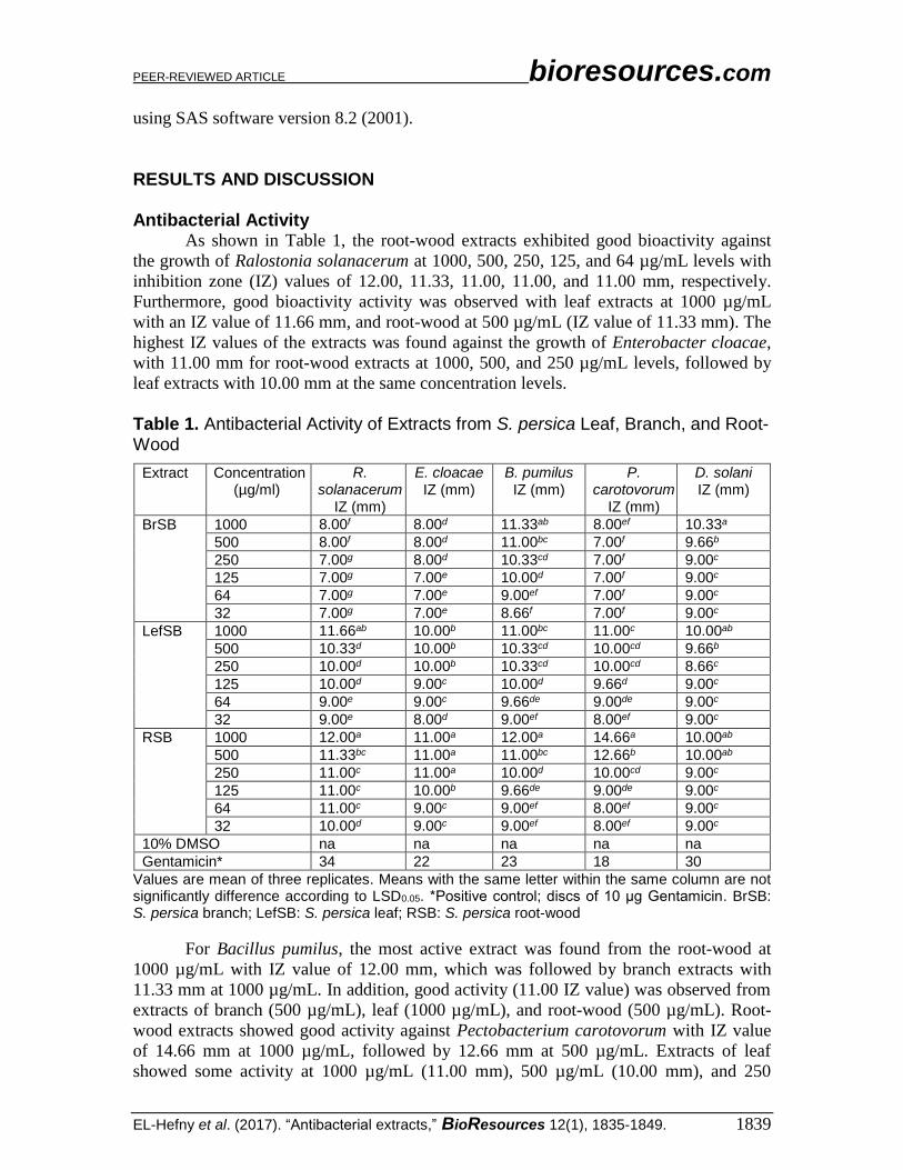
Antibacterial Activity As shown in Table 1, the root-wood extracts exhibited good bioactivity against
the growth of Ralostonia solanacerum at 1000, 500, 250, 125, and 64 µg/mL levels with
inhibition zone (IZ) values of 12.00, 11.33, 11.00, 11.00, and 11.00 mm, respectively.
Furthermore, good bioactivity activity was observed with leaf extracts at 1000 µg/mL
with an IZ value of 11.66 mm, and root-wood at 500 µg/mL (IZ value of 11.33 mm). The
highest IZ values of the extracts was found against the growth of Enterobacter cloacae,
with 11.00 mm for root-wood extracts at 1000, 500, and 250 µg/mL levels, followed by
leaf extracts with 10.00 mm at the same concentration levels.
Table 1. Antibacterial Activity of Extracts from S. persica Leaf, Branch, and Root-Wood
Extract Concentration (µg/ml)
R. solanacerum
IZ (mm)
E. cloacae IZ (mm)
B. pumilus IZ (mm)
P. carotovorum
IZ (mm)
D. solani IZ (mm)
BrSB 1000 8.00f 8.00d 11.33ab 8.00ef 10.33a
500 8.00f 8.00d 11.00bc 7.00f 9.66b
250 7.00g 8.00d 10.33cd 7.00f 9.00c
125 7.00g 7.00e 10.00d 7.00f 9.00c
64 7.00g 7.00e 9.00ef 7.00f 9.00c
32 7.00g 7.00e 8.66f 7.00f 9.00c
LefSB 1000 11.66ab 10.00b 11.00bc 11.00c 10.00ab
500 10.33d 10.00b 10.33cd 10.00cd 9.66b
250 10.00d 10.00b 10.33cd 10.00cd 8.66c
125 10.00d 9.00c 10.00d 9.66d 9.00c
64 9.00e 9.00c 9.66de 9.00de 9.00c
32 9.00e 8.00d 9.00ef 8.00ef 9.00c
RSB 1000 12.00a 11.00a 12.00a 14.66a 10.00ab
500 11.33bc 11.00a 11.00bc 12.66b 10.00ab
250 11.00c 11.00a 10.00d 10.00cd 9.00c
125 11.00c 10.00b 9.66de 9.00de 9.00c
64 11.00c 9.00c 9.00ef 8.00ef 9.00c
32 10.00d 9.00c 9.00ef 8.00ef 9.00c
10% DMSO na na na na na
Gentamicin* 34 22 23 18 30
Values are mean of three replicates. Means with the same letter within the same column are not significantly difference according to LSD0.05. *Positive control; discs of 10 μg Gentamicin. BrSB: S. persica branch; LefSB: S. persica leaf; RSB: S. persica root-wood
For Bacillus pumilus, the most active extract was found from the root-wood at
1000 µg/mL with IZ value of 12.00 mm, which was followed by branch extracts with
11.33 mm at 1000 µg/mL. In addition, good activity (11.00 IZ value) was observed from
extracts of branch (500 µg/mL), leaf (1000 µg/mL), and root-wood (500 µg/mL). Root-
wood extracts showed good activity against Pectobacterium carotovorum with IZ value
of 14.66 mm at 1000 µg/mL, followed by 12.66 mm at 500 µg/mL. Extracts of leaf
showed some activity at 1000 µg/mL (11.00 mm), 500 µg/mL (10.00 mm), and 250
PEER-REVIEWED ARTICLE bioresources.com
EL-Hefny et al. (2017). “Antibacterial extracts,” BioResources 12(1), 1835-1849. 1840
µg/mL (10.00 mm). Branch extracts showed activity against the growth of Dickeya solani
at 1000 µg/mL with an IZ value of 10.33 mm, followed by leaf extracts at 1000 µg/mL
(IZ 10.00 mm), and root-wood extracts at 1000 µg/mL (IZ 10.00 mm) and at 500 µg/mL
(IZ 10.00 mm). Based on the these results, the root-wood extracts from S. persica had
better antibacterial activity against the growth of the studied bacteria compared to leaf
and branch extracts. Overall, the IZ values presented from the extracts are lower than
those reported from the antibiotic used (Gentamicin).
All over the world, many trials have been done to control the diseases of potatoes
without promising control. Some success has been reported with chemical control of
brown rot (Murakoshi and Takahashi 1984), soil fumigants (Weingartner and Shumaker
1988), resistant varieties (Fock et al. 2001; Lopez and Biosca 2004), and antibiotics
(Habashy et al. 1993). Additionally, chemical control (pesticides) with its residues has
been reported to have hazardous effects in Europe and Egypt (Sylvander and Le Floc’h-
Wadel 2000; Parrott and Kalibwani 2004).
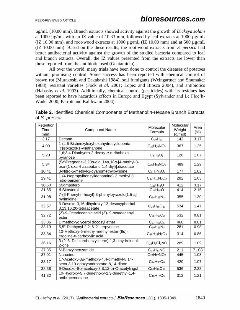
Table 2. Identified Chemical Components of Methanol:n-Hexane Branch Extracts of S. persica
Retention Time (min)
Compound Name Molecular Formula
Molecular Weight (g/mol)
Area (%)
3.17 Decane C10H22 142 3.17
4.09 1-(4,6-Bisbenzyloxyhexahydrocyclopenta [c]isoxazol-1-yl)ethanone
C22H25NO4 367 1.25
5.20 1,6;3,4-Dianhydro-2-deoxy-α-D-ribohexo-pyranose
C6H8O3 128 1.07
5.34 (5α)Pregnane-3,20α-diol,14α,18α-[4-methyl-3-oxo-(1-oxa-4-azabutane-1,4-diyl)],diacetate
C28H43NO6 489 1.29
10.41 3-Nitro-5-methyl-2-cyanomethylpyridine C8H7N3O2 177 1.82
29.41 1-(4-Isopropylbenzylidenamino)-2-methyl-3-nitro-benzene
C17H18N2O2 282 1.03
30.60 Stigmasterol C28H48O 412 3.17
31.65 β-Sitosterol C29H50O 414 2.15
31.98 7-(6-Phenyl-n-hexyl)-3-phenylpyrazolo[1,5-a] pyrimidine
C24H25N3 355 1.30
32.57 3-Desoxo-3,16-dihydroxy-12-desoxyphorbol-3,13,16,20-tetraacetate
C28H38O10 534 1.47
32.72 (Z)-9-Octadecenoic acid (Z)-,9-octadecenyl ester
C36H68O2 532 0.81
33.06 Dimethoxyglycerol docosyl ether C27H56O5 460 0.81
33.19 5,5"-Diethynyl-2,2':6',2"-terpyridine C19H11N3 281 0.98
33.34 10-Methoxy-6-methyl-methyl ester-(8α)-ergoline-8-carboxylic acid
C18H22N2O3 314 0.86
36.16 3-(2',6'-Dichlorobenzylidene)-1,3-dihydroindol-2-one
C15H9Cl2NO 289 1.09
37.35 N-Benzylbenzamide C14H13NO 211 71.08
37.91 Narceine C23H27NO8 445 1.08
38.17 17-Acetoxy-3α-methoxy-4,4-dimethyl-8,14-seco-3,19-epoxyandrostane-8,14-dione
C24H36O6 420 1.07
38.38 9-Desoxo-9-x-acetoxy-3,8,12-tri-O-acetylingol C28H40O10 536 2.33
41.32 10-Hydroxy-5,7-dimethoxy-2,3-dimethyl-1,4-anthracenedione
C18H16O5 312 1.21
PEER-REVIEWED ARTICLE bioresources.com
EL-Hefny et al. (2017). “Antibacterial extracts,” BioResources 12(1), 1835-1849. 1841
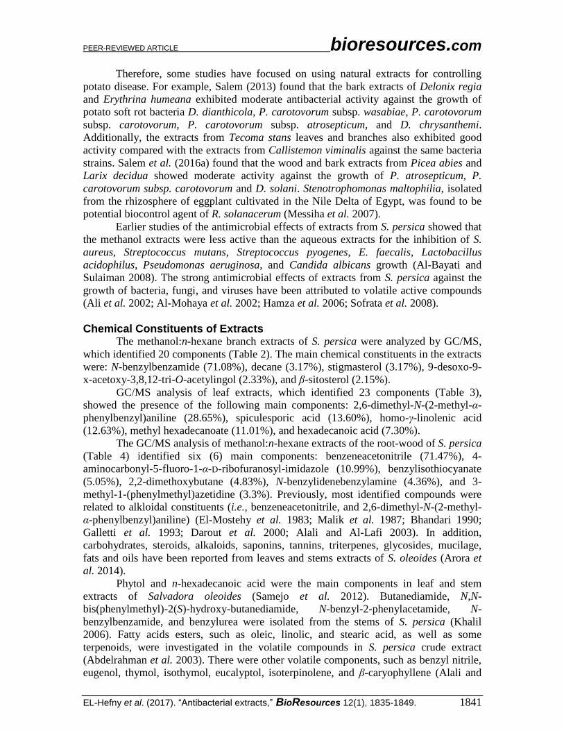
Therefore, some studies have focused on using natural extracts for controlling
potato disease. For example, Salem (2013) found that the bark extracts of Delonix regia
and Erythrina humeana exhibited moderate antibacterial activity against the growth of
potato soft rot bacteria D. dianthicola, P. carotovorum subsp. wasabiae, P. carotovorum
subsp. carotovorum, P. carotovorum subsp. atrosepticum, and D. chrysanthemi.
Additionally, the extracts from Tecoma stans leaves and branches also exhibited good
activity compared with the extracts from Callistemon viminalis against the same bacteria
strains. Salem et al. (2016a) found that the wood and bark extracts from Picea abies and
Larix decidua showed moderate activity against the growth of P. atrosepticum, P.
carotovorum subsp. carotovorum and D. solani. Stenotrophomonas maltophilia, isolated
from the rhizosphere of eggplant cultivated in the Nile Delta of Egypt, was found to be
potential biocontrol agent of R. solanacerum (Messiha et al. 2007).
Earlier studies of the antimicrobial effects of extracts from S. persica showed that
the methanol extracts were less active than the aqueous extracts for the inhibition of S.
aureus, Streptococcus mutans, Streptococcus pyogenes, E. faecalis, Lactobacillus
acidophilus, Pseudomonas aeruginosa, and Candida albicans growth (Al-Bayati and
Sulaiman 2008). The strong antimicrobial effects of extracts from S. persica against the
growth of bacteria, fungi, and viruses have been attributed to volatile active compounds
(Ali et al. 2002; Al-Mohaya et al. 2002; Hamza et al. 2006; Sofrata et al. 2008).
Chemical Constituents of Extracts
The methanol:n-hexane branch extracts of S. persica were analyzed by GC/MS,
which identified 20 components (Table 2). The main chemical constituents in the extracts
were: N-benzylbenzamide (71.08%), decane (3.17%), stigmasterol (3.17%), 9-desoxo-9-
x-acetoxy-3,8,12-tri-O-acetylingol (2.33%), and β-sitosterol (2.15%).
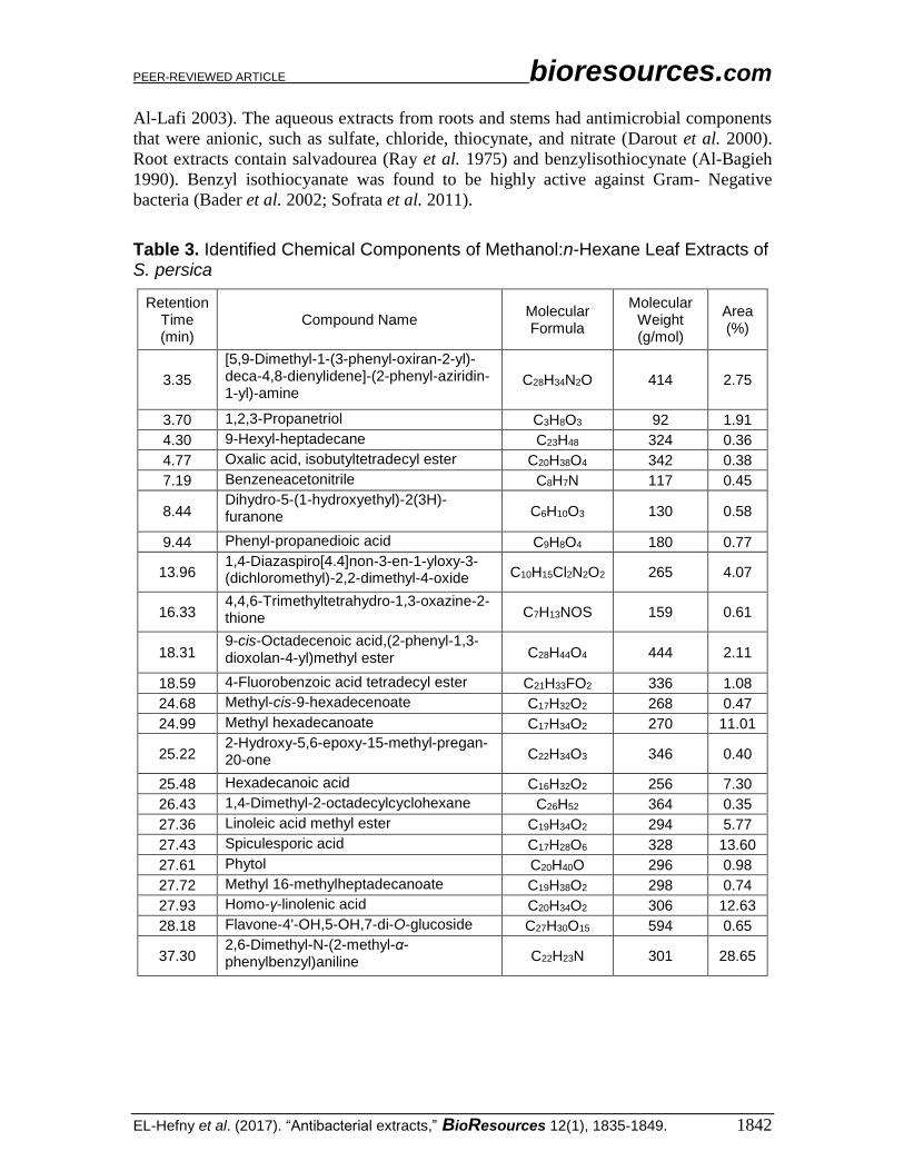
GC/MS analysis of leaf extracts, which identified 23 components (Table 3),
showed the presence of the following main components: 2,6-dimethyl-N-(2-methyl-α-
phenylbenzyl)aniline (28.65%), spiculesporic acid (13.60%), homo-γ-linolenic acid
(12.63%), methyl hexadecanoate (11.01%), and hexadecanoic acid (7.30%).
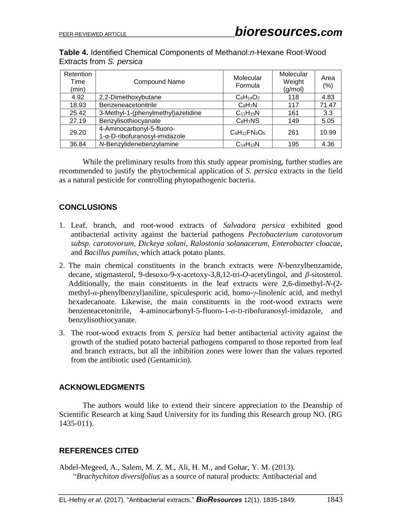
The GC/MS analysis of methanol:n-hexane extracts of the root-wood of S. persica
(Table 4) identified six (6) main components: benzeneacetonitrile (71.47%), 4-
aminocarbonyl-5-fluoro-1-α-D-ribofuranosyl-imidazole (10.99%), benzylisothiocyanate
(5.05%), 2,2-dimethoxybutane (4.83%), N-benzylidenebenzylamine (4.36%), and 3-
methyl-1-(phenylmethyl)azetidine (3.3%). Previously, most identified compounds were
related to alkloidal constituents (i.e., benzeneacetonitrile, and 2,6-dimethyl-N-(2-methyl-
α-phenylbenzyl)aniline) (El-Mostehy et al. 1983; Malik et al. 1987; Bhandari 1990;
Galletti et al. 1993; Darout et al. 2000; Alali and Al-Lafi 2003). In addition,
carbohydrates, steroids, alkaloids, saponins, tannins, triterpenes, glycosides, mucilage,
fats and oils have been reported from leaves and stems extracts of S. oleoides (Arora et
al. 2014).
Phytol and n-hexadecanoic acid were the main components in leaf and stem
extracts of Salvadora oleoides (Samejo et al. 2012). Butanediamide, N,N-
bis(phenylmethyl)-2(S)-hydroxy-butanediamide, N-benzyl-2-phenylacetamide, N-
benzylbenzamide, and benzylurea were isolated from the stems of S. persica (Khalil
2006). Fatty acids esters, such as oleic, linolic, and stearic acid, as well as some
terpenoids, were investigated in the volatile compounds in S. persica crude extract
(Abdelrahman et al. 2003). There were other volatile components, such as benzyl nitrile,
eugenol, thymol, isothymol, eucalyptol, isoterpinolene, and β-caryophyllene (Alali and
PEER-REVIEWED ARTICLE bioresources.com
EL-Hefny et al. (2017). “Antibacterial extracts,” BioResources 12(1), 1835-1849. 1842
Al-Lafi 2003). The aqueous extracts from roots and stems had antimicrobial components
that were anionic, such as sulfate, chloride, thiocynate, and nitrate (Darout et al. 2000).
Root extracts contain salvadourea (Ray et al. 1975) and benzylisothiocynate (Al-Bagieh
1990). Benzyl isothiocyanate was found to be highly active against Gram- Negative
bacteria (Bader et al. 2002; Sofrata et al. 2011).
Table 3. Identified Chemical Components of Methanol:n-Hexane Leaf Extracts of S. persica
Retention Time (min)
Compound Name Molecular Formula
Molecular Weight (g/mol)
Area (%)
3.35
[5,9-Dimethyl-1-(3-phenyl-oxiran-2-yl)-deca-4,8-dienylidene]-(2-phenyl-aziridin-1-yl)-amine
C28H34N2O 414 2.75
3.70 1,2,3-Propanetriol C3H8O3 92 1.91
4.30 9-Hexyl-heptadecane C23H48 324 0.36
4.77 Oxalic acid, isobutyltetradecyl ester C20H38O4 342 0.38
7.19 Benzeneacetonitrile C8H7N 117 0.45
8.44 Dihydro-5-(1-hydroxyethyl)-2(3H)-furanone C6H10O3 130 0.58
9.44 Phenyl-propanedioic acid C9H8O4 180 0.77
13.96 1,4-Diazaspiro[4.4]non-3-en-1-yloxy-3-(dichloromethyl)-2,2-dimethyl-4-oxide C10H15Cl2N2O2 265 4.07
16.33 4,4,6-Trimethyltetrahydro-1,3-oxazine-2-thione C7H13NOS 159 0.61
18.31 9-cis-Octadecenoic acid,(2-phenyl-1,3-dioxolan-4-yl)methyl ester C28H44O4 444 2.11
18.59 4-Fluorobenzoic acid tetradecyl ester C21H33FO2 336 1.08
24.68 Methyl-cis-9-hexadecenoate C17H32O2 268 0.47
24.99 Methyl hexadecanoate C17H34O2 270 11.01
25.22 2-Hydroxy-5,6-epoxy-15-methyl-pregan-20-one C22H34O3 346 0.40
25.48 Hexadecanoic acid C16H32O2 256 7.30
26.43 1,4-Dimethyl-2-octadecylcyclohexane C26H52 364 0.35
27.36 Linoleic acid methyl ester C19H34O2 294 5.77
27.43 Spiculesporic acid C17H28O6 328 13.60
27.61 Phytol C20H40O 296 0.98
27.72 Methyl 16-methylheptadecanoate C19H38O2 298 0.74
27.93 Homo-γ-linolenic acid C20H34O2 306 12.63
28.18 Flavone-4'-OH,5-OH,7-di-O-glucoside C27H30O15 594 0.65
37.30 2,6-Dimethyl-N-(2-methyl-α-phenylbenzyl)aniline C22H23N 301 28.65
PEER-REVIEWED ARTICLE bioresources.com
EL-Hefny et al. (2017). “Antibacterial extracts,” BioResources 12(1), 1835-1849. 1843
Table 4. Identified Chemical Components of Methanol:n-Hexane Root-Wood Extracts from S. persica
Retention Time (min)
Compound Name Molecular Formula
Molecular Weight (g/mol)
Area (%)
4.92 2,2-Dimethoxybutane C6H14O2 118 4.83
18.93 Benzeneacetonitrile C8H7N 117 71.47
25.42 3-Methyl-1-(phenylmethyl)azetidine C11H15N 161 3.3
27.19 Benzylisothiocyanate C8H7NS 149 5.05
29.20 4-Aminocarbonyl-5-fluoro- 1-α-D-ribofuranosyl-imidazole
C9H12FN3O5 261 10.99
36.84 N-Benzylidenebenzylamine C14H13N 195 4.36
While the preliminary results from this study appear promising, further studies are
recommended to justify the phytochemical application of S. persica extracts in the field
as a natural pesticide for controlling phytopathogenic bacteria.
CONCLUSIONS
1. Leaf, branch, and root-wood extracts of Salvadora persica exhibited good
antibacterial activity against the bacterial pathogens Pectobacterium carotovorum
subsp. carotovorum, Dickeya solani, Ralostonia solanacerum, Enterobacter cloacae,
and Bacillus pumilus, which attack potato plants.
2. The main chemical constituents in the branch extracts were N-benzylbenzamide,
decane, stigmasterol, 9-desoxo-9-x-acetoxy-3,8,12-tri-O-acetylingol, and β-sitosterol.
Additionally, the main constituents in the leaf extracts were 2,6-dimethyl-N-(2-
methyl-α-phenylbenzyl)aniline, spiculesporic acid, homo-γ-linolenic acid, and methyl
hexadecanoate. Likewise, the main constituents in the root-wood extracts were
benzeneacetonitrile, 4-aminocarbonyl-5-fluoro-1-α-D-ribofuranosyl-imidazole, and
benzylisothiocyanate.
3. The root-wood extracts from S. persica had better antibacterial activity against the
growth of the studied potato bacterial pathogens compared to those reported from leaf
and branch extracts, but all the inhibition zones were lower than the values reported
from the antibiotic used (Gentamicin).
ACKNOWLEDGMENTS
The authors would like to extend their sincere appreciation to the Deanship of
Scientific Research at king Saud University for its funding this Research group NO. (RG
1435-011).
REFERENCES CITED
Abdel-Megeed, A., Salem, M. Z. M., Ali, H. M., and Gohar, Y. M. (2013).
“Brachychiton diversifolius as a source of natural products: Antibacterial and
PEER-REVIEWED ARTICLE bioresources.com
EL-Hefny et al. (2017). “Antibacterial extracts,” BioResources 12(1), 1835-1849. 1844
antioxidant evaluation of the extracts of wood branches,” J. Pure Appl. Microbiol.
7(3), 1843-1850.
Abdelrahman, H. F., Skaug, N., Whyatt, A. M., and Francis, G. W. (2003). “Volatile
compounds in crude Salvadora persica extracts,” Pharm. Boil. 41(6), 399-404. DOI:
10.1076/phbi.41.6.399.17826
Ahmed, Z., Khan, S. S., Khan, M., Tanveer, A., and Lone, Z. A. (2010). “Synergistic
effect of Salvadora persica extracts, tetracycline and penicillin against
Staphylococcus aureus,” African J. Basic Appl. Sci. 2(1-2), 25-29.
Ahmed, S., Soaad, E., Essawy, E., Mohamed, E. I., and Ewald, S. (2008). “Preliminary
phytochemical and propagation trial with Salvadora persica L,” Agric. For Res. 1,
135-138.
Akhtar, J., Siddique, K., Bi, S., and Mujeeb, M. (2011). “A review on phytochemical and
pharmacological investigations of miswak (Salvadora persica Linn.),” J. Pharm. Bio.
Allied Sci. 3(1), 113-117. DOI: 10.4103/0975-7406.76488
Alali, F., and Al-Lafi, T. (2003). “GC-MS analysis and bioactivity testing of the volatile
oil from the leaves of the toothbrush tree Salvadora persica L.,” Nat. Prod. Res.
17(3), 189-94. DOI: 10.1080/1057563021000040790
Alali, F., Hudaib, M., Aburjai, T., Khairallah, K., and Al-Hadidi, N. (2004). “GC-MS
analysis and antimicrobial activity of the essential oil from the stem of the Jordanian
toothbrush tree Salvadora persica,” Pharm. Biol. 42(8), 577-80. DOI:
10.1080/13880200490901834
Al-Bagieh, N. H., and Almas, K. (1997). “In vitro antimicrobial effects of aqueous and
alcohol extracts of Miswak,” Cairo Dental J. 13, 221-224.
Al-Bagieh, N. H., Idowu, A., and Salako, N. O. (1994). “Effect of aqueous extract of
miswak on the in vitro growth of Candida albicans,” Microbios 80(323), 107-13.
Al-Bagieh, N. H. (1990). “Antiherpes simplex c-virus type 1 activity of
benzyisothicyanate,” Biomed. Lett. 47, 67-70.
Al-Bagieh, N. H. (1992). “Antiherpes simplex virus type 1 activity of
benzylisothiocyanate,” Biomed. Lett. 80, 107-113.
Al-Bagieh, N. H. (1998). “Effect of benzylisothiocyanate on the growth and acid
production of Candida albicans,” Biomed. Lett. 58, 139-145.
Al-Bagieh, N. H., and Weinberg, E. D. (1988). “Benzylisothiocyanate: A possible agent
for controlling dental caries,” Microbios Lett. 39, 143-151.
Al-Bayati, F., and Sulaiman, K. (2008). “In vitro antimicrobial activity of Salvadora
persica L. extracts against some isolated oral pathogens in Iraq,” Turk. J. Biol. 32(1),
57-62.
Ali, H., Konig, G. M., Khalid, S. A., Wright, A. D., and Kaminsky, R. (2002).
“Evaluation of selected Sudanese medicinal plants for their in vitro activity against
hemoflagellates, selected bacteria, HIV-1-RT and tyrosine kinase inhibitory, and for
cytotoxicity,” J. Ethnopharmacol. 83(3), 219-228. DOI: 10.1016/S0378-
8741(02)00245-3
Almas, K., Skaug, N., and Ahmad, I. (2005). “In vitro antimicrobial comparison of
Miswak extract with commercially available non-alcohol mouthrinses,” Int. J. Dent.
Hyg. 3(1), 18-24. DOI: 10.1111/j.1601-5037.2004.00111.x
Almas, K. (2001). “The antimicrobial effects of seven different types of Asian chewing
sticks,” Odontostomatol Trop. 24(96), 17-20.
Almas, K., and Al-Bagieh, N. (1999). “The antimicrobial effects of bark and pulp
extracts of miswak, Salvadora persica,” Biomed. Lett. 60, 71-75.
PEER-REVIEWED ARTICLE bioresources.com
EL-Hefny et al. (2017). “Antibacterial extracts,” BioResources 12(1), 1835-1849. 1845
Al-Mohaya, M. A., Darwazeh, A., and Al- Khudair, W. (2002). “Oral fungal colonization
and oral candidiasis in renal transplant patients: The relationship to Miswak use,”
Oral Surg. Oral Med. Oral Pathol. Oral Radiol. Endod. 93(4), 455-460. DOI:
10.1067/moe.2002.121992
Al-Obaida, M. I., Al-Essa, M. A., Asiri, A. A., and Al-Rahla, A. A. (2010).
“Effectiveness of a 20% miswak extract against a mixture of Candida albicans and
Enterococcus faecalis,” Saudi Med. J. 31(6), 640-643.
Al-Otaibi, M., and Angmar, B. (2004). “Oral hygiene habits and oral health awareness
among urban Saudi Arabians,” Oral Heal. Prev. Dent. 2(4), 389-96.
Arora, M., and Kalia, A. N. (2013). “Isolation and characterization of stigmasterol and β-
sitosterol-D-glycoside from ethanolic extract of the stems of Salvadora persica linn.,”
Int. J. Pharm. Sci. 5(1), 245-249.
Arora, M., Siddiqui, A. A., Paliwal, S., and Sood, P. (2014). “A phyto-pharmacological
overview on Salvadora oleoides Decne,” Indian J. Nat. Prod. Res. 5(3), 209-214.
Ashmawy, N. A., Behiry, S. I., Ali, H. M., and Salem, M. Z. M. (2014). “Evaluation of
Tecoma stans and Callistemon viminalis extracts against potato soft rot bacteria in
vitro,” J. Pure Appl. Microbiol. 8(Special Ed. 2), 667-673.
Bader, A., Flamini, G., Luigi, P., and Morelli, I. (2002). “The composition of the root oil
of Salvadora persica L.,” J. Essen. Oil Res. 14(2), 128-129. DOI:
10.1080/10412905.2002.9699795
Bakkali, F., Averbeck, S., Averbeck, D., and Idaomar, M. (2008). “Biological effects of
essential oils - A review,” Food Chem. Toxicol. 46(2), 446-475. DOI:
10.1016/j.fct.2007.09.106
Balto, H., Ghandourah, B., and Al-Sulaiman, H. (2012). “The efficacy of Salvadora
persica extract in the elimination of the intracanal smear layer: A SEM study,” Saudi
Dent. J. 24 (2), 71-77. DOI:10.1016/j.sdentj.2012.01.002
Bauer, A.W., Kirby, W. M., Sherris, J. C., and Turck, M. (1966). “Antibiotic
susceptibility testing by a standardized single disk method,” Am. J. Clin. Path. 45(4),
493-496.
Behiry, S. I. (2013). Molecular and Pathological Studies on Potato Bacterial Soft Rot
Disease, Ph.D. Dissertation, Alexandria University, Alexandria, Egypt.
Bhandari, M. M. (1990). Flora of the Indian Desert (1st Ed.), Dhrti Printers, New Delhi,
India.
Chaurasia, A., Patil, R., and Nagar, A. (2013). “Miswak in oral cavity - An update,” J.
Oral Boil. Craniofacial Res. 3(2), 98-101. DOI: 10.1016/j.jobcr.2012.09.004
Chawla, H. S. (1983). “A new natural source for topical fluoride,” J. Indian Dent. Assoc.
55(10), 419-422.
Darmani, H., Al-Hiyasat, A. S., Elbetieha, A. M., and Alkofahi, A. (2003). “The effect of
an extract of Salvadora persica (meswak, chewing stick) on fertility of male and
female mice,” Phytomed. 10(1), 63-65.
http://dx.doi.org/10.1078/094471103321648683
Darout, I. A., Christy, A. A., Skaug, N., and Egeberg, K. P. (2000). “Identification and
quantification of some potentially antimicrobial anionic component in miswak
extract,” Indian J. Pharmacol. 32(1), 11-14.
Davies, N. W. (1990). “Gas chromatographic retention indices of monoterpenes and
sesquiterpenes on methyl silicone and Carbowax 20 M phases,” J. Chromatogr. A
503(2015), 1-24.
Edris, A. E., Shalaby, A. S., Fadel, H. M., and Abdel-Wahab, M. A. (2003). “Evaluation
PEER-REVIEWED ARTICLE bioresources.com
EL-Hefny et al. (2017). “Antibacterial extracts,” BioResources 12(1), 1835-1849. 1846
of a chemotype of spearmint (Mentha spicata L.) grown in Siwa Oasis, Egypt,” Eur.
Food Res. Technol. 218(1),74-78. DOI: 10.1007/s00217-003-0802-4
El-Mostehy, M. R., Al-Jassem, A. A., Al-Yassin, I. A., Al-Gindy, A. R., and Shoukry, E.
(1983). “Miswak as an oral health device. Preliminary chemical and clinical
evaluation,” Hamdard. 26(4), 41-50.
Elvin-Lewis, M., Hall, J. B., and Adu-uta, M. (1980). “The dental health of chewing stick
users of southern Ghana, preliminary finding,” J. Prev. Dent. 6, 151-159.
Ezmirly, S. T., Cheng, J. C., and Wilson, S. R. (1978). “Saudi Arabian medicinal plants:
Salvadora persica,” Planta Med. 35(2), 191-192. DOI: 10.1055/s-0028-1097205
FAO, STAT (2013). “FAOSTAT database,” (http://faostat.fao.org), Food and Agriculture
Organization of the United Nations, Rome, Italy.
Fock, I., Collonnier, C., Luisetti, J., Purwito, A., Souvannavong, V., Vedel, F., Servaes,
A., Ambroise, A., Kodja, H., Ducreux, G., and Sihachakr, D. (2001). “Use of
Solanum stenotomum for introduction of resistance to bacterial wilt in somatic
hybrids of potato,” Plant Physiol. Biochem. 39(10), 899-908.
http://dx.doi.org/10.1016/S0981-9428(01)01307-9
Galletti, G., Chiavari, G., and Kahie, Y. (1993). “Pyrolysis/gas chromatography/ion-trap
mass spectrometry of the ‘tooth brush’ tree (Salvadora persica L.),” Rapid Commun.
Mass Spectrom. 7(7), 651-655. DOI: 10.1002/rcm.1290070719
Gardan, L., Gouy, C., Christen, R., and Samson, R. (2003). “Elevation of three
subspecies of Pectobacterium carotovorum to species level: Pectobacterium
atrosepticum sp. nov., Pectobacterium betavasculorum sp. nov. and Pectobacterium
wasabiae sp. nov.,” Int. J. Syst. Evol. Microbiol. 53(Pt 2), 381-391. DOI:
10.1099/ijs.0.02423-0
Habashy, W. H. S., Fawzi, F. G., El-Huseiny, T. M., and Neweigy, N. A. (1993).
“Bacterial wilt of potatoes. II. Sensitivity of the pathogen to antibiotics and
pathogenesis by streptomycin-resistant mutants,” Egyptian Journal of Agricultural
Research 71, 401-412.
Halawany, H. S. (2012). “A review on miswak (Salvadora persica) and its effect on
various aspects of oral health,” Saudi Dent. J. 24(2), 63-69. DOI:
10.1016/j.sdentj.2011.12.004
Hamza, O. J., Bout-van, C. J., Matee, M. I., Moshi, M. J., Mikx, F. H., Selemani, H. O.,
Mbwambo, Z. H., Vander, A. J., and Verweij, P. E. (2006). “Antifungal activity of
some Tanzanian plants used traditionally for the treatment of fungal infections,” J.
Ethnopharmacol. 108(1), 124-132. DOI:10.1016/j.jep.2006.04.026
Kamel, M., Ohtani, K., and Assaf, M. (1992). “Lignan glycosides from stems of
Salvadora persica,” Phytochemistry 31(7), 2469-2471. DOI: 10.1016/0031-
9422(92)83301-E
Kamil, M., Ahmad, F., Jayaraj, A. F., Gunasekhar, C., Thomas, S., Habibullah, M., and
Chan, K. (2000). “Isolation and identification of a flavonol glycoside using high
speed counter current chromatographic technique from the leaves of Salvadora
persica,” Pak. J. Sci. Ind. Res. 43(4), 255-257.
Khalessi, A. M., Pack, A. R. C., Thomson, W. M., and Tompkins, G. R. (2004). “An in
vivo study of the plaque control efficacy of Persica TM: A commercially available
herbal mouthwash containing extracts of Salvadora persica,” Int. Dent. J. 54(5), 279-
283. DOI: 10.1111/j.1875-595X.2004.tb00294.x
Khalil, A. T. (2006). “Benzylamides from Salvadora persica,” Arch. Pharm. Res. 29(11),
952-956. DOI: 10.1007/BF02969277
PEER-REVIEWED ARTICLE bioresources.com
EL-Hefny et al. (2017). “Antibacterial extracts,” BioResources 12(1), 1835-1849. 1847
Laurila, J., Ahola, V., Lehtinen, A., Joutsjoki, T., Hannukkala, A., Rahkonen, A., and
Pirhonen, M. (2008). “Characterization of Dickeya strains isolated from potato and
river water samples in Finland,” Eur. J. Plant. Pathol. 122(2), 213-225. DOI:
10.1007/s10658-008-9274-5
Lopez, M. M., and Biosca, E. G. (2004). “Potato bacterial wilt management: New
prospects for an old problem,” in: Bacterial Wilt Disease and the Ralostonia Species
Complex, C. Allen, P. Prior, and A. C. Hayward (eds.), APS Press, St. Paul,
Minnesota, USA, pp. 205-224
Malik, S., Ahmad, S. S., Haider, S. I., and Muzaffar, A. (1987). “Salvadoricine, a new
alkaloid from the leaves of Salvadora persica,” Tetrahedron Lett. 28(2), 163-164.
DOI: 10.1016/S0040-4039(00)95675-2
Messiha, N. A. S., van Diepeningen, A. D., Farag, N. S., Abdallah, S. A., Janse, J. D.,
and van Bruggen, A. H. C. (2007). “Stenotrophomonas maltophilia: A new potential
biocontrol agent of Ralostonia solanacerum, causal agent of potato brown rot,” Eur.
J. Plant Pathol. 118(3), 211-225. DOI: 10.1007/s10658-007-9136-6
Mickail, K. Y., Bishay, F., Farag, N. S., and Tawfik, A. E. (1974). “Evaluation of
bacterial wilt disease in A.R.E. during the years from 1967–1968 to 1971–1972,”
Agri. Res. Rev. (Cairo) 52, 89-94.
Mohamed, S.A., and Khan, J. A. (2013). “Antioxidant capacity of chewing stick miswak
Salvadora persica,” BMC Complem. Altern. Med. 13(40). DOI: 10.1186/1472-6882-
13-40
Murakoshi, S., and Takahashi, M. (1984). “Trials of some control of tomato wilt caused
by Pseudomonas solanacerum,” Bulletin of the Kanagawa Horticultural Experiment
Station 31, 50-56.
Noumi, E., Snoussi, M., Hajlaoui, H., Valentin, E., and Bakhrouf, A. (2010). “Antifungal
properties of Salvadora persica and Juglans regia L. extracts against oral Candida
strains,” Eur. J. Clin. Microbiol. Infect. Dis. 29(1), 81-88. DOI: 10.1007/s10096-009-
0824-3
Ohtani, K., Kasai, R., Yamasaki, K., Tanaka, O., Kamel, M. S., Assaf, M. H., El-
Shanawani, M. A., and Ali. A. A. (1992). “Lignan glycoside from stems of Salvodora
persica L,” Phytochemistry 31(7), 2469-2471. DOI: 10.1016/0031-9422(92)83301-E
Paliwal, S., Chauhan, R., Siddiqui, A. A., Paliwal, S., and Sharma, J. (2007). “Evaluation
of antifungal activity of Salvadora persica Linn. leaves,” Nat. Prod. Rad. 6(5), 372-
374.
Parrott, N., and Kalibwani, F. (2004). “Organic agriculture in the continents, Africa,” in:
The World of Organic Agriculture Statistics and Emerging Trends, H. Willer and M.
Yussefi (eds.), pp. 55-68.
Pérombelon, M. C. M. (2002). “Potato diseases caused by soft rot erwinias: An overview
of pathogenesis,” Plant Pathol. 51(1), 1-12. DOI: 10.1046/j.0032-
0862.2001.Shorttitle.doc.x
Ray, A. B., Chand, L., and Dutta, S. C. (1975). “Salvadoure, a new urea derivative from
Salvadora persica,” Chem. Ind. 12, 517-518.
Sabet, K. A. (1961). “The occurrence of bacterial wilt of potatoes caused by
Pseudomonas solanacerum (E.F. Smith) in Egypt,” Cairo, General Organisation for
Govt. Print. Offices, 1961.
PEER-REVIEWED ARTICLE bioresources.com
EL-Hefny et al. (2017). “Antibacterial extracts,” BioResources 12(1), 1835-1849. 1848
Salem, M. Z. M. (2013). “Evaluation of the antibacterial and antioxidant activities of
stem bark extracts of Delonix regia and Erythrina humeana grown in Egypt,” J.
Forest Prod. Ind. 2(2), 48-52.
Salem, M. Z. M., Ali, H. M., El-Shanhorey, N. A., and Abdel-Megeed, A. (2013).
“Evaluation of extracts and essential oil from Callistemon viminalis leaves:
Antibacterial and antioxidant activities, total phenolic and flavonoid contents,” Asian
Pac. J. Trop. Med., 6(10):785–791. http://dx.doi.org/10.1016/S1995-7645(13)60139-
X
Salem, M. Z. M., Ali, H. M., and Mohamed, N. H. (2014a). “Evaluation of extracts from
different parts of some tree species against the growth of some human bacterial
pathogens,” J. Pure Appl. Microbio. 8(Spl. Edn. 1), 149-154.
Salem, M. Z. M., Khamis, M. H., El-Shanhorey, N. A., Al-Muwayhi, M. A., Okla, M. K.,
and Ali, H. M. (2014b). “Evaluation of extracts from leaves of Brachychiton
diversifolius R.Br against the growth of some clinical pathogens,” J. Pure Appl.
Microbio. 8(Spl. Edn. 1), 105-109.
Salem, M. Z. M., Abdel-Megeed, A., and Ali, H. M. (2014c). “Stem wood and bark
extracts of Delonix regia (Boj. Ex. Hook): Chemical analysis, antibacterial,
antifungal, and antioxidant properties,” BioResources 9(2), 2382-2395.
Salem, M. Z. M., Elansary, H. O., Elkelish, A. A., Zeidler, A., Ali, H. M., EL-Hefny, M.,
and Yessoufou, K. (2016a). “In vitro bioactivity and antimicrobial activity of Picea
abies and Larix decidua wood and bark extracts,” BioResources 11(4), 9421-9437.
DOI: 10.15376/biores.11.4.9421-9437
Salem, M. Z. M., Zayed, M. Z., Ali, H. M., and Abd El-Kareem, M. S. M. (2016b).
“Chemical composition, antioxidant and antibacterial activities of extracts from
Schinus molle L. wood branch growing in Egypt,” J. Wood Sci. 62(6), 548-561. DOI:
10.1007/s10086-016-1583-2
Saini, S., Yadav, J. P., and Kalia A. N. (2006). “Hypoglycemic activity of Salvadora
persica and S. oleoides in diabetic albino rats,” J. Sci. Pharm. 7 (1), 5-12.
Samejo, M. Q., Memon, S., Bhanger, M. I., and Khan, K. M. (2012). “Chemical
constituents of essential oil of Salvadora oleoides,” J. Pharm. Res. 5(4), 2366-2367.
SAS, (2001). Users Guide: Statistics (Release 8.02), SAS Inst. Inc., Cary, NC, USA,
2001.
Sofrata, A. H., Claesson, R. L., Lingstram, P. K., and Gustafsson, A. K. (2008). “Strong
antibacterial effect of miswak against oral microorganisms associated with
periodontitis and caries,” J. Periodontol. 79(8), 1474-1479. DOI:
10.1902/jop.2008.070506
Sofrata, A., Santangelo, E. M., Azeem, M., Borg-Karlson, A.-K., Gustafsson, A., and
Pütsep, K. (2011). “Benzyl isothiocyanate, a major component from the roots of
Salvadora Persica is highly active against gram-negative bacteria,” PLoS ONE 6(8),
e23045. DOI: 10.1371/journal.pone.0023045
Sylvander, B., and Le Floc’h-Wadel, A., (2000). “Consumer demand and production of
organics in the EU,” AgBioForum 3(2&3), 97-106
Toth, I. K., Bell, K. S., Holeva, M. C., and Birch, P. R. J. (2003). “Soft-rot Erwina: From
genes to genomes,” Mol. Plant Pathol. 4(1), 17-30. DOI: 10.1046/j.1364-
3703.2003.00149.x
PEER-REVIEWED ARTICLE bioresources.com
EL-Hefny et al. (2017). “Antibacterial extracts,” BioResources 12(1), 1835-1849. 1849
Weingartner, D. P., and Shumaker, J. R. (1988). “In row injection of metham sodium and
other soil fumigants for control of nematodes and soil borne potato diseases in
Florida,” 72nd Annual Meeting of The Potato Association of America. American
Potato Journal, Fort Collins, Colorado, USA, 65: 504.
Yabuuchi, E. Y., Kosaku, I., Yano, I., Hotta, H., and Nishiuchi, Y. (1995). “Transfer of
two Burkholderia and an Alcaligenes species to Ralostonia gen. Nov.,” Microbiol.
Immunol. 39(11), 897-904. DOI: 10.1111/j.1348-0421.1995.tb03275.x
Article submitted: October 21, 2016; Peer review completed: January 11, 2017; Revised
version received: January 14, 2017; Accepted: January 17, 2017; Published: January 25,
2017.
DOI: 10.15376/biores.12.1.1835-1849