Embed Size (px)
Citation preview
PEGylated-PLGA microparticles containing VEGF for long term drug delivery 1
2
3
4
5
6
7
8
9
10
11
12
13
14
15
16
17
Teresa Simón-Yarza a, Fabio R. Formiga a, Esther Tamayo a, Beatriz Pelacho b, Felipe
Prosper b, María J. Blanco-Prieto a*
a Pharmacy and Pharmaceutical Technology Department, School of Pharmacy,
University of Navarra, Pamplona, Spain
b Hematology Service and Area of Cell Therapy, Clinic Universidad de Navarra,
Foundation for Applied Medical Research, University of Navarra, Pamplona, Spain
*Address for correspondence: Maria J. Blanco-Prieto, Department of Pharmacy and
Pharmaceutical Technology, School of Pharmacy, University of Navarra, Irunlarrea 1,
E-31080 Pamplona, Spain. Tel.: +34 948 425600 x 6519; fax: +34 948 425649 e-mail:
1
Abstract 18
19
20
21
22
23
24
25
26
27
28
29
30
31
32
33
34
35
36
37
38
39
40
41
42
The potential of poly (lactic-co-glycolic) acid (PLGA) microparticles as carriers
for vascular endothelial growth factor (VEGF) has been demonstrated in a previous
study by our group, where we found improved angiogenesis and heart remodeling in a
rat myocardial infarction model (Formiga et al., 2010). However, the observed
accumulation of macrophages around the injection site suggested that the efficacy of
treatment could be reduced due to particle phagocytosis.
The aim of the present study was to decrease particle phagocytosis and
consequently improve protein delivery using stealth technology. PEGylated
microparticles were prepared by the double emulsion solvent evaporation method using
TROMS (Total Recirculation One Machine System). Before the uptake studies in
monocyte-macrophage cells lines (J774 and Raw 264.7), the characterization of the
microparticles developed was carried out in terms of particle size, encapsulation
efficiency, protein stability, residual poly(vinyl alcohol) (PVA) and in vitro release.
Microparticles of suitable size for intramyocardial injection (5 μm) were obtained by
TROMS by varying the composition of the formulation and TROMS conditions with
high encapsulation efficiency (70-90%) and minimal residual PVA content (0.5%).
Importantly, the bioactivity of the protein was fully preserved. Moreover, PEGylated
microparticles released in phosphate buffer 50% of the entrapped protein within 4 hours,
reaching a plateau within the first day of the in vitro study. Finally, the use of PLGA
microparticles coated with PEG resulted in significantly decreased uptake of the carriers
by macrophages, compared with non PEGylated microparticles, as shown by flow
cytometry and fluorescence microscopy.
On the basis of these results, we concluded that PEGylated microparticles loaded with
VEGF could be used for delivering growth factors in the myocardium.
2
Keywords: PEG, PLGA, macrophage uptake, VEGF, protein delivery. 43
3
1. Introduction 44
45
46
47
48
49
50
51
52
53
54
55
56
57
58
59
60
61
62
63
64
65
66
67
68
The concept of stealth technology came into being during the World War II in
the attempt to escape from radar control. Ever since then, stealth strategy has included
two different approaches: the development of radar absorbing paints, and novel designs
in terms of shape and size. In the field of drug delivery systems (DDS), this concept has
been applied to the ability of these carriers to avoid immunological recognition (Wassef
et al., 1991). As in the military context, the shape (Lin et al., 2011), size (Maldiney et al.,
2011) and material properties (Essa et al., 2011; Zhu et al., 2011) of the delivery system
are crucial.
Recently, some research showing how macrophages have a higher affinity for
specific shapes and sizes has been published (Doshi and Mitragotri, 2010). In this paper
the authors conclude that particles with a size greater than 4 µm suffer less protein
adsorption, which is the stage prior to macrophage phagocytosis. Interestingly, when
comparing these results with the size distribution of bacteria, they found that most of
these have a size between 2 and 3 µm, which favors their opsonization.
In 1978, Van Oss (Van Oss, 1978) described the phagocytosis process as a
surface phenomenon, demonstrating how bacteria that are more hydrophobic than
phagocytes readily become phagocytized, whereas bacteria that are more hydrophilic
than phagocytes resist phagocytosis. At that time, researchers proposed the surface
modification of molecules, by making them more hydrophilic, as a strategy to reduce
phagocytic removal. In the 1970s, pegnology, the art of surface-modifying proteins,
drugs or DDS by attaching molecules of poly(ethylene glycol) (PEG) was proposed by
Abraham Abuchowski and Frank F. Davis (Abuchowski et al., 1977), and this has been
applied effectively in protein therapies, obtaining increased stability (Khondee et al.,
2011), increased resistance to proteolytic inactivation (Turner et al., 2011), decreased
4
69
70
71
72
73
74
75
76
77
78
79
80
81
82
83
84
85
86
87
88
89
90
91
92
93
immunogenicity (Milla et al., 2011), increased circulatory half-lives (Maleki et al.,
2011), and reduced toxicity (Jain, A. and Jain, S.K., 2008), thus improving the delivery
and efficacy of proteins. To date, incorporating PEG seems to hold the most promising
benefits while showing the lowest harmful effects (Owens and Peppas, 2006) and
modified drugs are already on the market, most of which are PEGylated proteins (Pasut
et al., 2008), such as interferon alpha (Fried et al., 2002), L-asparaginase (Abuchowski
et al., 1984), granulocyte colony-stimulating factor (Tanaka et al., 1991) and uricase
(Davis et al., 1981). However, despite the advances in the field of protein therapy,
stealth technology is still emerging within the area of DDS. In fact, just one PEGylated
delivery system has come onto the market (Knop et al., 2010): a PEGylated liposome
containing doxorubicin for the treatment of cancer.
Our group recently published a study in which poly (lactic-co-glycolic) acid
(PLGA) microparticles encapsulating the vascular endothelial growth factor (VEGF)
were intramyocardially implanted in a ischemia-reperfusion animal model (Formiga et
al., 2010). Benefits of the therapy were observed in terms of enhanced angiogenesis and
notable reduction of harmful remodeling, but when we studied the continued presence
of the particles at the injection site over time, a macrophage accumulation around the
particles depot was observed, which could limit the efficacy of the treatment. To
overcome this challenge, in the present study our aim was to develop and characterize in
vitro PEG-PLGA microparticles loaded with VEGF for their subsequent use in
cardiovascular disease. The uptake of VEGF-PEGylated microparticles was studied by
flow cytometry and fluorescence microscopy using two different monocyte-macrophage
cell lines. Non PEGylated PLGA microparticles were used for comparison.
5
2. Materials and methods 94
95
96
97
98
99
100
101
102
103
104
105
106
107
108
109
110
111
112
113
114
115
116
117
118
2.1. Materials
Human recombinant VEGF was from R&D Systems (Minneapolis, MN, USA).
PLGA with a lactic:glycolic ratio of 50:50 Resomer® RG 503H (Mw 34KDa),
poly[(D,L-lactide-co-glycolide)- co-PEG] diblock Resomer® RGP d 5055 (5% PEG)
and Resomer® RGP d 50105 (10% PEG) were provided by Boehringer-Ingelheim
(Ingelheim, Germany). PEG 400, sodium azide, Rhodamine B isothiocyanate and
human serum albumin (HSA) were provided by Sigma–Aldrich (Barcelona, Spain).
Dichloromethane and acetone were obtained from Panreac Quimica S.A. (Barcelona,
Spain). Poly(vinyl alcohol) (PVA), 88% hydrolyzed (MW 125000), was from
Polysciences, Inc. (WA, USA). Rabbit polyclonal anti-human VEGF-A (clone A-20, sc-
152) was supplied by Santa Cruz Biotechnology (Santa Cruz, CA, USA). ECL™ anti-
Rabbit IgG horseradish peroxidase-linked whole antibody was from Amersham
Biosciences (Buckinghamshire, UK). Mouse monoclonal anti-rat CD68 was provided
by AbD Serotec (Oxford, UK). All the Western blot reagents were purchased from
BioRad unless specified in the text.
The murine monocyte-macrophage cells lines J774 and Raw 264.7 were
provided by Dr. Latasa (CIMA, University of Navarra). Human umbilical venous
endothelial cells (HUVECs) were extracted from umbilical cords from donors, after
informed consent according to the guidelines of the Committee on the Use of Human
Subjects in Research at the Clinic Universidad de Navarra. CellTiter 96® AQueous One
Solution Cell Proliferation Assay (MTS) was obtained from Promega.
6
2.2. Preparation of PLGA and PEG-PLGA microparticles 119
120
121
122
123
124
125
126
127
128
129
130
131
132
133
134
135
136
137
138
139
140
VEGF-loaded microparticles were prepared by the double emulsion solvent
evaporation method using TROMS (Formiga et al., 2010; Garbayo et al., 2009). Briefly,
the organic solution composed of 4 ml of a mixture of dicloromethane/acetone (3:1)
containing 50 mg of Resomer RG503 was injected into the inner aqueous phase, which
consisted of 50 µg of VEGF in 10 mM phosphate, 50 mM sodium chloride (PBS), 5 mg
of HSA and 5 µl of PEG400. The primary emulsion (W1/O) was recirculated through
the system for 90 sec under a turbulent regime at a flow rate of 38 ml/min. The first
emulsion was injected into 20 ml of the external aqueous phase (W2) composed of 20
ml of a PVA solution resulting in the formation of a double emulsion (W1/O/W2) which
was homogenized by circulation through the system for 45 sec. The resulting double
emulsion was stirred at room temperature for at least 3 hours to allow solvent
evaporation and microparticle formation. Finally, microparticles were washed three
times with ultrapure water and lyophilized (Genesis 12EL, Virtis). For PEGylated
microparticles, 50 mg of a mixture of Resomer® 503H and Resomer® RGP d 5055 or
Resomer® RGP d 50105 (1:1) were dissolved in the organic phase and microparticles
were prepared as described above
The composition of the different phases and TROMS parameters were varied to
achieve an adequate particle size (of around 5 μm) for intramyocardial administration,
(Formiga et al., 2010).
2.3. Microparticle characterization
2.3.1. Particle size, size distribution and zeta potential 141
The mean particle size and size distribution of the microparticles were 142
determined by laser diffractometry using a Mastersizer-S® (Malvern Instruments, 143
7
Malvern, UK). Microparticles were dispersed in distilled water and analyzed under 144
continuous stirring. The results were expressed as mean volume, in micrometers. 145
Samples were measured in triplicate. 146
The zeta potential was measured using Zetaplus® (Brookhaven instruments, NY, 147
US). Samples were diluted with distilled water and each experiment was repeated three 148
times. 149
150
151
2.3.2. Residual PVA
The residual PVA associated with microparticles was determined by a 152
colorimetric method based on the formation of a colored complex between two adjacent 153
hydroxyl groups of PVA and an iodine molecule (Joshi et al., 1979). Briefly, 2 mg of 154
lyophilized microparticles were resuspended in 2 ml of NaOH 0.5 M for 15 min at 155
60 °C. Each sample was neutralized with 900 μl of 1 N HCl and the volume was 156
adjusted to 5 ml with distilled water. Next, 3 ml of a 0.65 M solution of boric acid, 0.5 157
ml of a solution of I2/KI (0.05 M/0.15 M) and 1.5 ml of distilled water were added. 158
After 15 min of incubation, the absorbance of the samples was measured at 690 nm 159
using an Agilent 8453 UV–visible spectrophotometer (Agilent Technologies, Palo Alto, 160
CA, USA). A standard plot of PVA was prepared under identical conditions. 161
Measurements were performed in triplicate. 162
163
164
2.3.3. Drug loading and encapsulation efficiency
The amount of VEGF encapsulated in the microparticles was determined by 165
dissolving 1 mg of microparticles in 50 μl of DMSO. VEGF containing samples were 166
diluted in 350 μl of PBS for western-blot analysis. SDS-PAGE was performed onto 167
8
12% polyacrylamide gels. Following electrophoresis the proteins were transferred onto 168
nitrocellulose membranes which were then blockaded using 5% nonfat dried milk in 169
Tris Buffered Saline (TBS) with 0.05% Tween 20, for 1h at room temperature (RT). 170
Membranes were incubated for 2.5 h at RT with rabbit antihuman VEGF-A antibody 171
(A-20: sc-152, 1:2000 dilution). The bounded antibody was detected with horseradish 172
peroxidase (HRP)-conjugated donkey anti-rabbit IgG antibody (1 h, RT, 1:2000 173
dilution). Chemiluminiscence detection was performed using LumiLight Plus western 174
blotting substrate (Roche Diagnostics, Mannheim, Germany). The VEGF signal was 175
quantified by densitometry using the Quantity One software (Bio-Rad Laboratories Inc., 176
Munich, Germany). Samples containing defined quantities of VEGF were diluted under 177
the same conditions (PBS and DMSO) and used as standard curve. 178
179
180
2.4. In vitro release studies
VEGF loaded microparticles (1 mg, n=3) were resuspended in 0.25 ml PBS pH 181
7.4 with 0.02% (w/v) sodium azide used as a bacteriostatic agent. Incubation took place 182
under orbital shaking in rotating vials (FALC F200, Falc instruments, Treviglio, Italy) 183
at 37 °C. At predefined times, the tubes were centrifuged (20000 g, 10 min) and the 184
supernatant was removed and frozen at -80ºC until it was analyzed by western-blot. The 185
removed solution was replaced with an equal volume of fresh release buffer to maintain 186
sink conditions. Release profiles were expressed in terms of cumulative release and 187
plotted versus time. 188
189
190
191
192
2.5. VEGF bioactivity
The bioactivity of the VEGF released from the microparticles was evaluated in
vitro by determining the proliferative capacity of a human umbilical vein endothelial
9
193
194
195
196
197
198
199
200
201
202
203
204
205
206
207
208
209
210
211
212
213
214
215
216
217
cell (HUVECs) after VEGF treatment. Cells were obtained from human umbilical cord
by 0.1% collagenase II digestion (Jaffe et al., 1973) and expanded in F12K medium
(ATCC 30-2004) supplemented with 30 μg/mL endothelial cell growth supplement
(ECGS, BD Biosciences), 10% fetal bovine serum, 1% sodium heparin and 1%
penicillin/streptomycin.
For the proliferation assay, the cells were plated into 96-well culture plates at a
density of 3× 10³ cells/well. After 12 hours, cells were treated with 10 and 25 ng/ml of
free VEGF or released from the microparticles. Culture medium and release medium
from non-loaded microparticles were used as control. After 72 h incubation time under
normal culture conditions, proliferation in each group was measured using MTS assay.
2.6. Uptake of microparticles by macrophages
The microparticle (PEGylated and non-pelylated microparticles) uptake study
was analyzed in two different monocyte-macrophage cell lines by fluorescence
microscopy and flow cytometry.
2.6.1. Fluorescence microscopy
Fluorescent-labeled microparticles with Rhodamin B isothiocyanate (0.5 mg/ml)
were prepared by adding the marker to the inner aqueous phase. Microparticles were
prepared as described above. The uptake of fluorescence particles was evaluated in the
monocyte-macrophage J774 cell line. Cells were plated into a 6 well culture plates at a
70% confluence in serum free RPMI medium containing 1% penicillin/streptomycin.
Four hours later culture medium (control), Rhodamine B isothiocyanate PLGA or PEG-
PLGA microparticles were added at a final concentration of 0.33 mg/ml. After 3 hours,
culture supernatant containing microparticles was removed and the wells were washed
10
218
219
220
221
222
223
224
225
226
227
228
229
230
231
232
233
234
235
236
237
238
239
240
241
242
three times with PBS. Fluorescence microparticles inside the cells were visualized using
an EVOSfl fluorescence microscope (Euroclone, Milan, Italy). The fluorescent signal
(corresponding to particle uptake) was quantified using the ImageJ software. Ten fields
per well were randomly analyzed (experiments performed in triplicate). The signal
emitted was normalized to the cell number in each field.
2.6.2. Flow cytometry
For flow cytometry studies, RAW 264.7 cells were seeded at a 30 % confluence
in DMEM 10% serum at 37 ºC and allowed to adhere to the 6 well plate for 48 h. Then
the medium was removed and cells were incubated with serum free DMEM for 4 h.
PLGA or PEG-PLGA microparticles previously suspended in DMEM were then added
(0.33 mg/ml), whereas the control group received only DMEM. At different time
intervals (from 30 minutes up to 3 h) the medium was removed, cells were detached,
collected and washed three times with PBS. After centrifugation (1500 g, 5 min), the
cells were suspended and fixed with 2 % formaldehyde solution for their analysis. Cell
complexity or cell granularity was studied by flow cytometry analysis using a BD
FACSCalibur flow cytometer for the acquisition of samples. The side scatter (SSC)
parameter was recorded as reflecting internal properties of cells (e.g. granularity and
refractive index). Data were analyzed using the CellQuest software.
2.7. Statistics
Results are expressed as mean ± SD. Statistical significance was tested on the
basis of Student’s t test at 95 % confidence intervals.
11
3. Results and discussion 243
244
245
246
247
248
249
250
251
252
253
254
255
256
257
258
259
260
261
262
263
264
265
266
267
3.1. Preparation of PLGA and PEG-PLGA microparticles
Among the different methods available for protein encapsulation, TROMS was
selected because it is a semi-industrial technique capable of encapsulating fragile
molecules while maintaining their native properties (Formiga et al., 2010; Garbayo et al.,
2008). Since our final goal is to inject the microparticles in the ischemic heart, the aim
when preparing the polymeric microparticles was to obtain a size between 5 and 8 µm,
which has been shown to be compatible with intramyocardial administration (Formiga
et al., 2010).
During the manufacturing process, size was shown to be affected by polymer
composition. The best results, in terms of feasibility, reproducibility and adequate
particle size distribution for intramyocardial injection were obtained with the polymer
containing 10% of PEG, and so this polymer was selected for the subsequent
experiments.
Using the same formulation and maintaining the TROMS parameters to prepare
PEGylated and non-PEGylated microparticles, the size was increased for the PEG-
PLGA co-polymer. Therefore TROMS parameters, mainly first and second emulsion
circulation times, were studied in order to achieve PEGylated particles with the desired
diameter (Table 1). The circulating times selected were 90 seconds for the first emulsion
and 45 seconds for the second emulsion, obtaining a particle size of approximately 6.6
µm (Table 2). Other factors were also studied, such as TROMS needle inner diameter,
polymer % (w/v) in the organic phase and PVA % (w/v) in the external aqueous phase.
Selected parameters are resumed in Table 2.
12
3.2. Microparticle characterization 268
3.2.1. Particle size and zeta potential 269
270
271
272
273
274
275
276
277
278
279
280
281
282
283
284
285
286
287
288
289
290
291
292
As stated above, particles with an average size close to 6 µm were obtained for
both types of microparticles, which have been demonstrated to be compatible with
intramyocardial injection (Formiga et al., 2010).
As shown in Table 3, surface charge values were negative for PEGylated and
non-PEGylated particles. However, PEGylated microparticles showed a decreased
negative charge (-8.79±0.61 mV vs. -18.10±0.71 mV). This may be attributed to the
presence of the PEG chains in the surface of the particle (Essa et al., 2010). Moreover, it
has been previously described that a higher PEG chain density on the surface of the
particles decreases the mobility of the PEG chains and thus decreases the steric
hindrance properties of the PEG layer (Owens and Peppas, 2006). On the other hand, if
the PEG concentration is too low, opsonins will attach to the surface and the stealth
effect will be decreased. Therefore, in order to achieve an intermediate surface chain
concentration between the “mushroom” and the “brush” conformation (low and high
PEG concentration respectively), a ratio composition of 1:1 (w/w) of polymers Resomer
503H: Resomer RGP d 50105 was finally selected (Table 3).
3.2.2. Residual PVA
PVA contained in the two types of microparticles was less than 2%, being lower
for the PEGylated microparticles (Table 3). This lower adsorption of PVA in the surface
modified particles could be explained as a consequence of the increased degree of
hydrophilicity due to PEG chains, reducing PVA interaction (Essa et al., 2010). In any
case, these concentrations are much lower than those reported in the literature for PLA
microparticles (Gref et al., 2001)
13
3.3. Drug loading and encapsulation efficiency 293
294
295
296
297
298
299
300
301
302
303
304
305
306
307
308
309
310
311
312
313
314
315
316
317
In a previous study, VEGF-PLGA microparticles with high encapsulation
efficiency were obtained (Formiga et al., 2010). In the present paper, this growth factor
was entrapped into PEG-PLGA microparticles obtaining very high encapsulation values,
between 80 and 100 % for both PLGA and PEG-PLGA particles, as determined by
western-blot. Moreover, western-blot allowed us to confirm that no degradation of
VEGF occurred during the encapsulation process, showing a single characteristic band
corresponding to 21 kDa (results not shown).
The ability to quantify proteins by light emitting chemiluminescence detection
has been previously studied, highlighting the hotspots which have been taken into
account in this research (Dickinson and Fowler, 2002). The results of encapsulation
efficiency obtained were also indirectly confirmed in the bioactivity assays (section 3.5).
In the cell proliferation study, when treating cells with the protein released from the
particles, VEGF concentration was calculated considering the encapsulation values. If
cell proliferation is in accordance with the expected VEGF concentration, it is possible
to confirm the encapsulation efficiency values, and in this sense the obtained results
allow us to consider the western-blot technique as a reliable method to measure the
encapsulation efficiency.
3.4. In vitro release studies
The amount of VEGF released from the microparticles was measured by an in
vitro assay, to confirm that the particles really retain the protein for a period of time and
allow a sustained release.
When comparing both types of microparticles, the burst effect was higher for the
PEGylated ones, which released approximately 50 % of VEGF within the first 4 hours,
14
while 30 % of the entrapped peptide was released from the PLGA particles (Figure 1).
Moreover, the plateau was reached after 24 hours for the surface modified particles,
whereas for the PLGA particles it occurred after three days. This different release
behavior is attributable firstly to the fact that burst effect is mainly due to the protein
located in the surface of the particle (Essa et al., 2010; Yoncheva et al., 2009). The
presence of PEG chains increases the surface of the particle and, as a consequence, a
greater amount of protein attaches to it. Secondly, as PEG chains are hydrophilic, when
they are in an aqueous medium, like the release buffer, they are dissolved and this
makes it easier for the buffer to get into the matrix, allowing the protein to be released.
In any case, it has to be taken into account that a slower protein release is expected in
vivo, as previously demonstrated (Blanco-Prieto et al., 2004). The main reason for the
slower in vivo kinetics is the low availability of water in the tissue compared with the
318
319
320
321
322
323
324
325
326
327
328
329
330
331
332
333
334
335
336
337
338
339
340
341
342
in
vitro conditions, in which the PLGA microparticles are incubated in PBS at 37°C and
shaken. Moreover, the tissue environment surrounding the microparticles will slow the
release of VEGF in vivo.
3.5. VEGF bioactivity
VEGF is a growth factor well known for its angiogenic activity (Carmeliet and
Jain, 2011). In this sense, it has been demonstrated to promote proliferation of
endothelial cells.
In order to confirm that VEGF bioactivity was preserved during the
encapsulation/release processes, we tested the ability of VEGF released from the
PEGylated particles to stimulate proliferation of HUVEC. Considering protein load and
in vitro release profile, cells received the same dose of free VEGF and VEGF released
from PEGylated microparticles (10 and 25 ng/ml). Both treatments induced the same
15
343
344
345
346
347
348
349
350
351
352
353
354
355
356
357
358
359
360
361
362
363
364
365
366
367
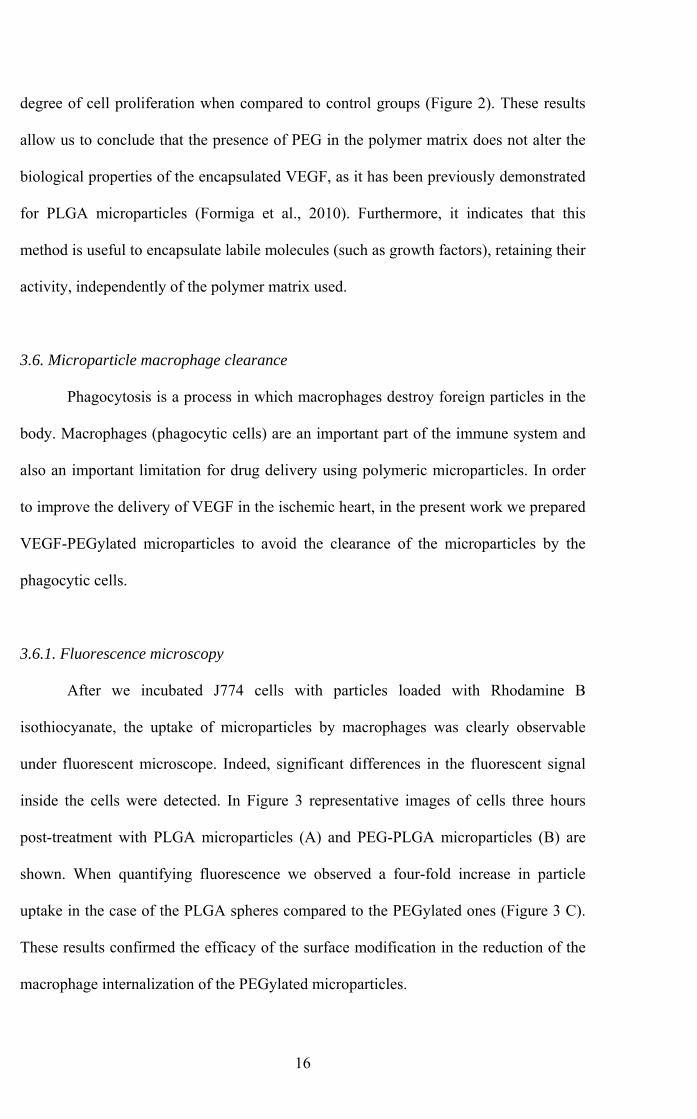
degree of cell proliferation when compared to control groups (Figure 2). These results
allow us to conclude that the presence of PEG in the polymer matrix does not alter the
biological properties of the encapsulated VEGF, as it has been previously demonstrated
for PLGA microparticles (Formiga et al., 2010). Furthermore, it indicates that this
method is useful to encapsulate labile molecules (such as growth factors), retaining their
activity, independently of the polymer matrix used.
3.6. Microparticle macrophage clearance
Phagocytosis is a process in which macrophages destroy foreign particles in the
body. Macrophages (phagocytic cells) are an important part of the immune system and
also an important limitation for drug delivery using polymeric microparticles. In order
to improve the delivery of VEGF in the ischemic heart, in the present work we prepared
VEGF-PEGylated microparticles to avoid the clearance of the microparticles by the
phagocytic cells.
3.6.1. Fluorescence microscopy
After we incubated J774 cells with particles loaded with Rhodamine B
isothiocyanate, the uptake of microparticles by macrophages was clearly observable
under fluorescent microscope. Indeed, significant differences in the fluorescent signal
inside the cells were detected. In Figure 3 representative images of cells three hours
post-treatment with PLGA microparticles (A) and PEG-PLGA microparticles (B) are
shown. When quantifying fluorescence we observed a four-fold increase in particle
uptake in the case of the PLGA spheres compared to the PEGylated ones (Figure 3 C).
These results confirmed the efficacy of the surface modification in the reduction of the
macrophage internalization of the PEGylated microparticles.
16
3.6.2. Flow cytometry 368
369
370
371
372
373
374
375
376
377
378
379
380
381
382
383
384
385
386
387
388
389
390
391
392
Microparticle uptake by macrophages induces changes in cellular granularity
that can be monitored by flow cytometry. Indeed, cells treated with PLGA
microparticles showed high granularity levels over incubation time (up to 3 hours),
indicating that a large number of particles had been internalized during that period.
However, coating the microparticles surface with PEG significantly influenced the
uptake of the microparticles by the macrophages. Cells receiving surface modified
particles maintained cell complexity in the same way as the non treated cells (control).
The differences became significant after incubating the particles for 2 hours in the
culture medium (Figure 4). Results obtained using flow cytometry confirmed the
observation made by fluorescence microscopy, demonstrating that PLGA microparticles
suffer phagocytosis in a more rapid way than PEGylated ones, and consequently
confirming that particles have been successfully PEGylated.
4. Conclusion
In this study we encapsulated VEGF in stealth microparticles, using a co-
polymer of PEG and PLGA, with a percentage of PEG adequate to reduce macrophage
phagocytosis. PEGylated microparticles with high encapsulation efficiency and suitable
size to be implanted in the myocardium were developed. Importantly, the bioactivity of
the loaded therapeutic protein was fully preserved. Microparticles whose surface was
modified by the incorporation of PEG in the formulation illustrated a significantly
decreased uptake by phagocityc cells.
In summary, PEGylation could be a useful approach to obtain growth factor-
loaded microparticles for myocardial administration, minimizing their local clearance
and enhancing the efficacy of the protein therapy in cardiovascular disease.
17

Consequently, the next step will be to test the developed microparticles in vivo, in a rat
model of myocardial infarction.
393
394
18
Acknowledgments 395
396
397
398
399
400
401
402
403
404
405
406
407
408
409
410
411
412
413
414
415
416
417
418
This work was supported by MICCIN PLE2009-0116, PSE SINBAD (PSS 0100000-
2008-1), Caja de Ahorros de Navarra (Programa Tu Eliges: Tu Decides) and the “UTE
project CIMA”. We thank Dr. Estella-Hermoso de Mendoza for the critical reading of
the manuscript.
References
Abuchowski, A., van Es, T., Palczuk, N.C., Davis, F.F., 1977. Alteration of
immunological properties of bovine serum albumin by covalent attachment of
polyethylene glycol. J. Biol. Chem. 252, 3578-3581.
Abuchowski, A., Kazo, G.M., Verhoest, C.R., Jr., Van Es, T., Kafkewitz, D., Nucci,
M.L., Viau, A.T., Davis, F.F., 1984. Cancer therapy with chemically modified enzymes.
I. Antitumor properties of polyethylene glycol-asparaginase conjugates. Cancer
Biochem. Biophys. 7, 175-186.
Blanco-Prieto, M.J., Campanero, M.A., Besseghir, K., Heimgartner, F., Gander, B.
2004. Importance of single or blended polymer types for controlled in vitro release and
plasma levels of a somatostatin analogue entrapped in PLA/PLGA microspheres. J.
Control. Release 96, 437-448);
Carmeliet, P., Jain, R.K., 2011. Molecular mechanisms and clinical applications of
angiogenesis. Nature 473, 298-307.
19
419
420
421
422
423
424
425
426
427
428
429
430
431
432
433
434
435
436
437
438
439
440
441
442
Davis, S., Park, Y.K., Abuchowski, A., Davis, F.F., 1981. Hypouricaemic effect of
polyethyleneglycol modified urate oxidase. Lancet 2, 281-283.
Dickinson, J., Fowler, S., 2002. Quantification of Proteins on Western Blots Using ECL,
in: Walker, J. (Ed.), The Proteins Protocols Handbook, Second ed. Humana Press Inc.,
Totowa, NJ, pp. 429-437.
Doshi, N., Mitragotri, S., 2010. Macrophages recognize size and shape of their targets.
PLoS One 5, e10051.
Essa, S., Rabanel, J.M., Hildgen, P., 2010. Effect of polyethylene glycol (PEG) chain
organization on the physicochemical properties of poly(D, L-lactide) (PLA) based
nanoparticles. Eur. J. Pharm. Biopharm. 75, 96-106.
Essa, S., Rabanel, J.M., Hildgen, P., 2011. Characterization of rhodamine loaded PEG-
g-PLA nanoparticles (NPs): effect of poly(ethylene glycol) grafting density. Int. J.
Pharm. 411, 178-187.
Formiga, F.R., Pelacho, B., Garbayo, E., Abizanda, G., Gavira, J.J., Simon-Yarza, T.,
Mazo, M., Tamayo, E., Jauquicoa, C., Ortiz-de-Solorzano, C., Prosper, F., Blanco-
Prieto, M.J., 2010. Sustained release of VEGF through PLGA microparticles improves
vasculogenesis and tissue remodeling in an acute myocardial ischemia-reperfusion
model. J. Control. Release 147, 30-37.
20
443
444
445
446
447
448
449
450
451
452
453
454
455
456
457
458
459
460
461
462
463
464
465
466
467
Formiga, F.R., Tamayo, E., Simon-Yarza, T., Pelacho, B., Prosper, F., Blanco-
Prieto,M.J., 2011.Angiogenic therapy for cardiac repair based on protein delivery
systems. Heart Fail. Rev. (In press).
Fried, M.W., Shiffman, M.L., Reddy, K.R., Smith, C., Marinos, G., Goncales, F.L., Jr.,
Haussinger, D., Diago, M., Carosi, G., Dhumeaux, D., Craxi, A., Lin, A., Hoffman, J.,
Yu, J., 2002. Peginterferon alfa-2a plus ribavirin for chronic hepatitis C virus infection.
N. Engl. J. Med. 347, 975-982.
Garbayo, E., Ansorena, E., Lanciego, J.L., Aymerich, M.S., Blanco-Prieto, M.J., 2008.
Sustained reléase of bioactive glycosylated glial cell-line derived neurotrophic factor
from biodegradable polymeric microspheres. Eur. J. Pharm. Biopharm. 69, 844-851.
Garbayo, E., Montero-Menei, C.N., Ansorena, E., Lanciego, J.L., Aymerich, M.S.,
Blanco-Prieto, M.J., 2009. Effective GDNF brain delivery using microspheres-a
promising strategy for Parkinson's disease. J. Control. Release 135, 119-126.
R. Gref, P. Quellec, A. Sanchez, P. Calvo, E. Dellacherie, M.J. Alonso, Development
and characterization of CyA-loaded poly(lactic acid)-poly(ethylene glycol)PEG micro-
and nanoparticles, Comparison with conventional PLA particulate carriers, Eur. J.
Pharm. Biopharm. 51 (2001) 111-118.
Jaffe, E.A., Nachman, R.L., Becker, C.G., Minick, C.R., 1973. Culture of human
endothelial cells derived from umbilical veins. Identification by morphologic and
immunologic criteria. J. Clin. Invest. 52, 2745-2756.
21
468
469
470
471
472
473
474
475
476
477
478
479
480
481
482
483
484
485
486
487
488
489
490
491
492
Jain, A., Jain, S.K., 2008. PEGylation: an approach for drug delivery. A review. Crit
Rev. Ther. Drug Carrier Syst. 25, 403-447.
Joshi, D.P., Lan-Chun-Fung, Y.L., Pritchard, J.G., 1979. Determination of poly(vinyl
alcohol) via its complex with boric acid and iodine. Analytica Chimica Acta 104, 153-
160.
Khondee, S., Olsen, C.M., Zeng, Y., Middaugh, C.R., Berkland, C., 2011. Noncovalent
PEGylation by Polyanion Complexation as a Means To Stabilize Keratinocyte Growth
Factor-2 (KGF-2). Biomacromolecules 12, 3880-3894.
Knop, K., Hoogenboom, R., Fischer, D., Schubert, U.S., 2010. Poly(ethylene glycol) in
drug delivery: pros and cons as well as potential alternatives. Angew. Chem. Int. Ed.
Engl. 49, 6288-6308.
Lin, S.Y., Hsu, W.H., Lo, J.M., Tsai, H.C., Hsiue, G.H., 2011. Novel geometry type of
nanocarriers mitigated the phagocytosis for drug delivery. J. Control. Release 154, 84-
92.
Maldiney, T., Richard, C., Seguin, J., Wattier, N., Bessodes, M., Scherman, D., 2011.
Effect of core diameter, surface coating, and PEG chain length on the biodistribution of
persistent luminescence nanoparticles in mice. ACS Nano 5, 854-862.
Maleki, A., Madadkar-Sobhani, A., Roohvand, F., Najafabadi, A.R., Shafiee, A.,
Khanahmad, H., Cohan, R.A., Namvar, N., Tajerzadeh, H., 2011. Design, modeling,
and expression of erythropoietin cysteine analogs in Pichia pastoris: Improvement of
22
493
494
495
496
497
498
499
500
501
502
503
504
505
506
507
508
509
510
511
512
513
514
515
516
mean residence times and in vivo activities through cysteine-specific PEGylation. Eur. J.
Pharm. Biopharm. (In press).
Milla, P., Dosio, F., Cattel, L., 2011. PEGylation of Proteins and Liposomes: A
Powerful and Flexible Strategy to Improve the Drug Delivery. Curr. Drug Metab. (In
press).
Owens, D.E., Peppas, N.A., 2006. Opsonization, biodistribution, and pharmacokinetics
of polymeric nanoparticles. Int. J. Pharm. 307, 93-102.
Pasut, G., Sergi, M., Veronese, F.M., 2008. Anti-cancer PEG-enzymes: 30 years old,
but still a current approach. Adv. Drug Deliv. Rev. 60, 69-78.
Tanaka, H., Satake-Ishikawa, R., Ishikawa, M., Matsuki, S., Asano, K., 1991.
Pharmacokinetics of recombinant human granulocyte colony-stimulating factor
conjugated to polyethylene glycol in rats. Cancer Res. 51, 3710-3714.
Turner, K.M., Pasut, G., Veronese, F.M., Boyce, A., Walsh, G., 2011. Stabilization of a
supplemental digestive enzyme by post-translational engineering using chemically-
activated polyethylene glycol. Biotechnol. Lett. 33, 617-621.
Van Oss, C.J., 1978. Phagocytosis as a surface phenomenon. Ann. Rev. Microbiol. 32,
19-39.
23
517
518
519
520
521
522
523
524
525
526
527
528
529
Wassef, N.M., Matyas, G.R., Alving, C.R., 1991. Complement-dependent phagocytosis
of liposomes by macrophages: suppressive effects of "stealth" lipids. Biochem. Biophys.
Res. Commun. 176, 866-874.
Yoncheva, K., Lambov, N., Miloshev, S., 2009. Modification of biodegradable
poly(malate) and poly(lactic-co-glycolic acid) microparticles with low molecular
polyethylene glycol. Drug Dev. Ind. Pharm. 35, 449-454.
Zhu, Z., Xie, C., Liu, Q., Zhen, X., Zheng, X., Wu, W., Li, R., Ding, Y., Jiang, X., Liu,
B., 2011. The effect of hydrophilic chain length and iRGD on drug delivery from
poly(epsilon-caprolactone)-poly(N-vinylpyrrolidone) nanoparticles. Biomaterials 32,
9525-9535.
24
Figure Legends 530
531
532
533
534
535
536
537
538
539
540
541
542
543
544
545
546
547
548
549
550
551
Figure 1. In vitro release profiles. VEGF released from PLGA and PEG-PLGA MPs is
represented as a % of the total VEGF load in the particles.
Figure 2. VEGF bioactivity: HUVECs treated with VEGF and VEGF released from the
microparticles (MPS-VEGF) at the same concentration (10 and 25 ng/ml) proliferate in
the same ratio when compared to control groups. *P<0.05 and ***P<0.001.
Figure 3. Macrophage uptake studied by fluorescent microscopy. Cells observed under
fluorescent microscope after incubating them with PLGA (A) and PEG-PLGA (B)
microparticles containing Rhodamine isocyanate. More Rhodamine is visualized inside
the cells treated with the PLGA microparticles. This differences are significantly
different when quantified (C), indicating that these have been phagocyted in a larger
number than those with the PEG chains in the surface. ***P<0.001.
Figure 4. Macrophage uptake studied by flow cytometry. The highest observed cell
granularity value (measured as Side Scatter Cell) has been assigned 100%. Cell
complexity increases when cells are treated with PLGA microparticles, whereas cells
treated with culture medium or PEG-PLGA microparticles did not have altered
complexity three hours after the treatment. *P<0.05 and **P<0.01.
25