Embed Size (px)
Citation preview
Pel is a cationic exopolysaccharide that cross-linksextracellular DNA in the Pseudomonas aeruginosabiofilm matrixLaura K. Jenningsa, Kelly M. Storeka,1, Hannah E. Ledvinaa, Charlène Coulonb, Lindsey S. Marmontc,d, Irina Sadovskayab,Patrick R. Secora, Boo Shan Tsenga, Michele Sciane, Alain Fillouxf, Daniel J. Wozniakg,h,i, P. Lynne Howellc,d,and Matthew R. Parseka,2
aDepartment of Microbiology, University of Washington, Seattle, WA 98195; bEquipe Biochimie des Produits Aquatiques, Université du Littoral-Côted’Opale, 62327 Boulogne-sur-mer, France; cProgram in Molecular Structure and Function, The Hospital for Sick Children, Toronto, ON, Canada M5G 0A1;dDepartment of Biochemistry, Faculty of Medicine, University of Toronto, Toronto, ON, Canada M5S 1A8; eSchool of Pharmacy, University of Washington,Seattle, WA, 98195; fDivision of Cell and Molecular Biology, Faculty of Natural Science, Center for Molecular Microbiology and Infection, Imperial CollegeLondon, London SW7 2AZ, United Kingdom; gDepartment of Microbial Infection and Immunity, The Ohio State University, Columbus, OH 43210;hDepartment of Microbiology, The Ohio State University, Columbus, OH 43210; and iCenter for Microbial Interface Biology, The Ohio State University,Columbus, OH 43210
Edited by Margaret J. McFall-Ngai, University of Hawaii at Manoa, Honolulu, HI, and approved July 17, 2015 (received for review March 16, 2015)
Biofilm formation is a complex, ordered process. In the opportunisticpathogen Pseudomonas aeruginosa, Psl and Pel exopolysaccharidesand extracellular DNA (eDNA) serve as structural components of thebiofilm matrix. Despite intensive study, Pel’s chemical structure andspatial localization within mature biofilms remain unknown. Usingspecialized carbohydrate chemical analyses, we unexpectedly foundthat Pel is a positively charged exopolysaccharide composed of par-tially acetylated 1→4 glycosidic linkages of N-acetylgalactosamineand N-acetylglucosamine. Guided by the knowledge of Pel’s sugarcomposition, we developed a tool for the direct visualization of Pelin biofilms by combining Pel-specificWisteria floribunda lectin stain-ingwith confocal microscopy. The results indicate that Pel cross-linkseDNA in the biofilm stalk via ionic interactions. Our data demon-strate that the cationic charge of Pel is distinct from that of otherknown P. aeruginosa exopolysaccharides and is instrumental in itsability to interact with other key biofilm matrix components.
biofilms | exopolysaccharide | extracellular DNA | Pel | Psl
Biofilm infections are inherently difficult to eradicate, owingto increased resistance to antimicrobials and host defenses
(1–3). Biofilms are microbial communities embedded in an extra-cellular matrix (4), composed of exopolysaccharides, extracellularDNA (eDNA), and proteins (5–7). Pseudomonas aeruginosa is anopportunistic pathogen that causes chronic biofilm infections (1)and has the capacity to synthesize three exopolysaccharidesimplicated in biofilm formation: alginate, Psl, and Pel (8, 9). Pslis a neutral polysaccharide consisting of a pentasaccharide repeatcontaining glucose, mannose, and rhamnose (10), and alginate isa negatively charged polymer of guluronic and mannuronic acid(8). Although previous reports suggest that glucose may be theprimary component of Pel, its structure is unknown.The role of Pel in biofilm formation was first identified in a screen
for mutants deficient in pellicle formation (i.e., biofilms forming atthe air–liquid interface of standing cultures) (11). Pel was later shownto be important for initiating and maintaining cell–cell interactions inbiofilms (12, 13). In some circumstances, Pel also can play a role inadherence of cells to a surface (14). Finally, Pel affords biofilmsprotection against certain aminoglycoside antibiotics (12).The importance of Pel in biofilm formation is strain-dependent.
Nonmucoid strains (i.e., strains that make little alginate), use Peland/or Psl as the primary structural scaffold. In the common labo-ratory strain PAO1, Psl is the primary exopolysaccharide pro-duced in the biofilm matrix, although some Pel is produced aswell (13, 15, 16). Deletion of pel genes in PAO1 does not havea significant impact on biofilm development (16). In contrast,Pel is the primary biofilm matrix exopolysaccharide in another
commonly used laboratory strain, PA14, which is incapable ofPsl production (16). Some strains appear to rely on both exo-polysaccharides. Strains characterized by hyperadherence andhyperaggregation, called rugose small colony variants (RSCVs),are frequently isolated from in vivo and in vitro biofilms (17,18). RSCVs harbor mutations (such as in wspF) that result inconstitutive overexpression of both Pel and Psl (19, 20).Biofilm formation is an ordered and sequential process that
often begins with adherence of cells to a surface, followed byformation of large aggregates or microcolonies (21, 22). Poly-mers within the biofilm matrix exhibit spatial localization that iscoordinated with the stages of biofilm development. In maturebiofilms, controlled cell death and lysis occur in the interior ofthe microcolony, releasing eDNA that contributes to the stabilityof the biofilm structure (23–27). eDNA and Psl are spatially sep-arated in mature microcolonies, with eDNA localized primarily tothe base of the microcolony (27) and Psl located at the periphery ofthe microcolony (25).Current data relating to the structure of Pel are limited and
conflicting. It has been reported that PA14 pellicles were glucose-rich compared with a pel deletion mutant (11); however, the
Significance
Exopolysaccharides and extracellular DNA are important struc-tural components that contribute to the self-assembly of largeaggregates or microcolonies that are characteristic of biofilms.Pseudomonas aeruginosa is capable of producing multipleexopolysaccharides, including alginate, Psl, and Pel. At present,little is known about Pel’s chemical structure and its role inmicrocolony formation. Our results demonstrate that Pel iscomposed of cationic amino sugars. Using this knowledge, wehave developed a Pel-specific lectin stain to directly visualize Pelin biofilms. We show that the positive charge on Pel facilitatesits binding to extracellular DNA in the biofilm stalk, and thatPel can compensate for lack of Psl in the biofilm periphery.
Author contributions: L.K.J., K.M.S., A.F., D.J.W., P.L.H., and M.R.P. designed research; L.K.J.,K.M.S., H.E.L., C.C., L.S.M., I.S., P.R.S., B.S.T., and M.S. performed research; K.M.S. contributednew reagents/analytic tools; L.K.J., K.M.S., C.C., L.S.M., I.S., B.S.T., A.F., D.J.W., P.L.H., and M.R.P.analyzed data; and L.K.J. and M.R.P. wrote the paper.
The authors declare no conflict of interest.
This article is a PNAS Direct Submission.1Present address: Department of Microbiology and Immunology, Stanford University,Stanford, CA 94305.
2To whom correspondence should be addressed. Email: [email protected].
This article contains supporting information online at www.pnas.org/lookup/suppl/doi:10.1073/pnas.1503058112/-/DCSupplemental.
www.pnas.org/cgi/doi/10.1073/pnas.1503058112 PNAS | September 8, 2015 | vol. 112 | no. 36 | 11353–11358
MICRO
BIOLO
GY
Dow
nloa
ded
by g
uest
on
May
10,
202
0
predominance of glucose was later attributed to cyclic glucans andnot to the pel locus (28). In another study, analysis of extracellularmaterial from PA14 pellicles identified lipopolysaccharide (LPS)-like material, leading to speculation that Pel is a modified form ofLPS (29). We previously reported that the periplasmic PelA proteinhas de-N-acetylase activity in vitro, suggesting that some of the Pelsugars are deacetylated before being exported from the cell (30).In the present study, we used glycosyl sugar and linkage analyses
with optimized glycosidic cleavage conditions to investigate thecomposition of Pel. We combined lectin staining with confocalmicroscopy to confirm structural analyses and to directly visualizePel in biofilms produced by distinct P. aeruginosa strains. Thesesurprising results indicate that Pel is positively charged, and com-posed of 1→4 linked partially acetylated galactosamine and glu-cosamine sugars. We provide evidence that Pel cross-links eDNAin the biofilm stalk and can structurally compensate for the ab-sence of Psl in the biofilm periphery. Thus, P. aeruginosa has achemically diverse and flexible suite of matrix exopolysaccharides.Knowledge of the composition and localization of Pel is essentialto delineate its functional role in biofilms, and ultimately tofacilitate the development of therapeutic strategies aimed ateradicating the biofilm matrix.
ResultsLPS Biosynthesis and Uridine 5′-Diphosphate–Glucose Are Not Re-quired for Pel Production. Polysaccharide biosynthesis requiresnucleotide-sugar precursors that are incorporated into theelongating polysaccharide chain (8, 31). There is precedence fora functional overlap of LPS and exopolysaccharide biosynthesisenzymes (10), and previous work suggests that Pel and LPSbiosynthesis may be linked (29). Therefore, we mutated path-ways involved in LPS biosynthesis and evaluated whether thisimpacted Pel production. These mutations impaired A band andB band O-antigen production and the LPS core. Pel immuno-blots indicated that mutant strains deficient in LPS precursorsreacted with the Pel antisera, whereas the corresponding mu-tants deficient in Pel did not (Fig. S1). Our data show that Pelproduction occurs even in the absence of different steps ofLPS biosynthesis, demonstrating that Pel is not a modified formof LPS.Glycosyltransferases catalyze the formation of glycosidic bonds by
transferring the sugar moiety from a sugar nucleotide to a specificacceptor molecule (32). To gain insight into the activated sugarmoiety that is used to generate Pel, we purified PelF, the solepredicted glycosyltransferase encoded by the pel operon. We usedisothermal titration calorimetry (ITC) to show that PelF specificallybinds uridine 5′-diphosphate (UDP) with micromolar affinity (Fig.S2), suggesting that a UDP-sugar nucleotide is required for Pelproduction.Pel was previously reported as a glucose-rich polysaccharide (11).
GalU is critical for the production of the nucleotide sugar UDP-glucose (Glc) (33). If Pel were a glucose-rich polysaccharide, thendisrupting UDP-Glc generation should prevent Pel biosynthesis(10). Immunoblots of extracts from a galU mutant revealed robustPel production relative to a Δpel ΔgalU double mutant (Fig. S1).The results demonstrate that Pel production occurs even in theabsence of UDP-Glc, thus suggesting that glucose is not a primarysugar component of Pel.
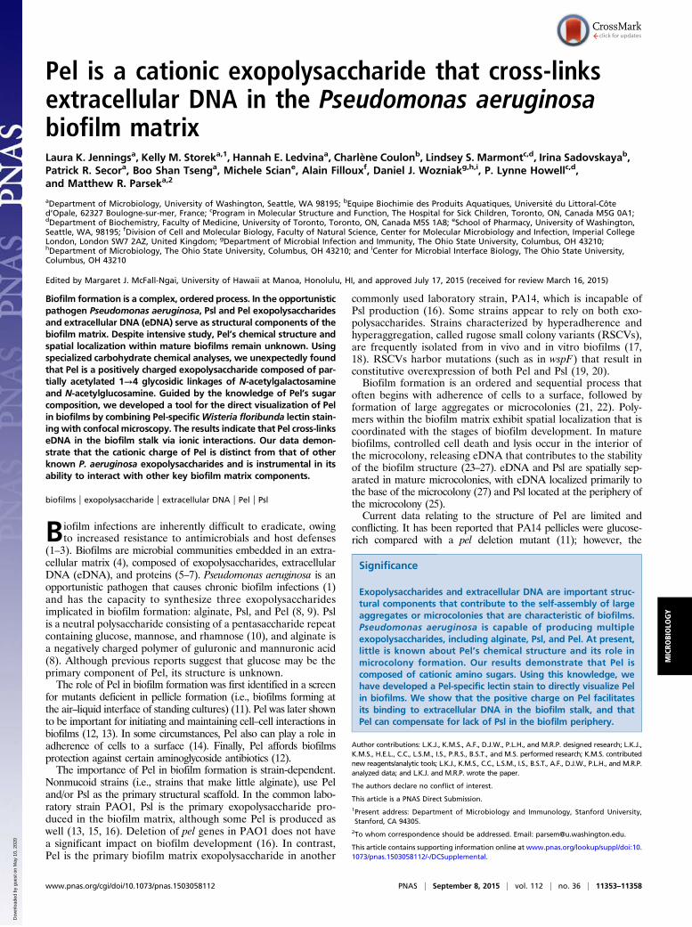
Pel Is Cationic and Has a Cell-Associated and Secreted Form. Webegan the characterization of Pel by investigating the molecularweight and potential charge of the polysaccharide using a strain withthe pel operon under control of the arabinose-inducible PBADpromoter (PAO1 ΔwspF Δpsl PBADpel, herein designated PBADpel).This strain maximizes Pel production. Size-exclusion chromatogra-phy indicated that Pel has two forms: cell-associated and secreted.Cell-associated Pel, which was prepared by EDTA extraction ofPBADpel cell pellets, eluted in the void volume, indicating a size
>80 kDa. Secreted Pel, collected from PBADpel culture super-natants, eluted near the total column volume. We calculated thesize of secreted Pel as 0.5 kDa by comparison with dextranstandards (Fig. 1A and Fig. S3). In this study, we characterizedthe chemical composition of secreted Pel, because this form ismore stable than cell-associated Pel.To determine whether Pel carries a net charge, we performed
ion-exchange chromatography on filtered supernatant of PBADpeland PAO1 ΔwspF Δpsl Δpel (herein designated Δpel) cultures.Culture supernatant was applied to a cation-exchange columnpreequilibrated with a pH 5.5 buffer. Examination of the cation-exchange fractions using Pel antiserum indicated that Pel bound thecolumn and eluted at 1.25 M NaCl (Fig. S4). No binding of Pel wasobserved to an anion-exchange column equilibrated with a pH 7.7buffer. These results suggest that Pel is positively charged at pH 5.5.The isoelectric point of Pel was determined by applying super-
natant from a PBADpel culture to a cation-exchange column underacidic conditions that facilitated binding (pH 5.5), and then grad-ually increasing the pH until Pel eluted (Fig. 1B). Pel was first de-tected in the effluent at pH 7.0. This suggests that the isoelectricpoint of Pel is between 6.7 and 6.9, and thus that Pel carries a netpositive charge at pH values below this range. The isoelectric pointfor Pel is consistent with that of a polysaccharide containing aminosugars; for example, the amino group of chitosan (poly-β-1,4-N-acetylglucosamine <50% acetylated) has a pKa of 6.5.
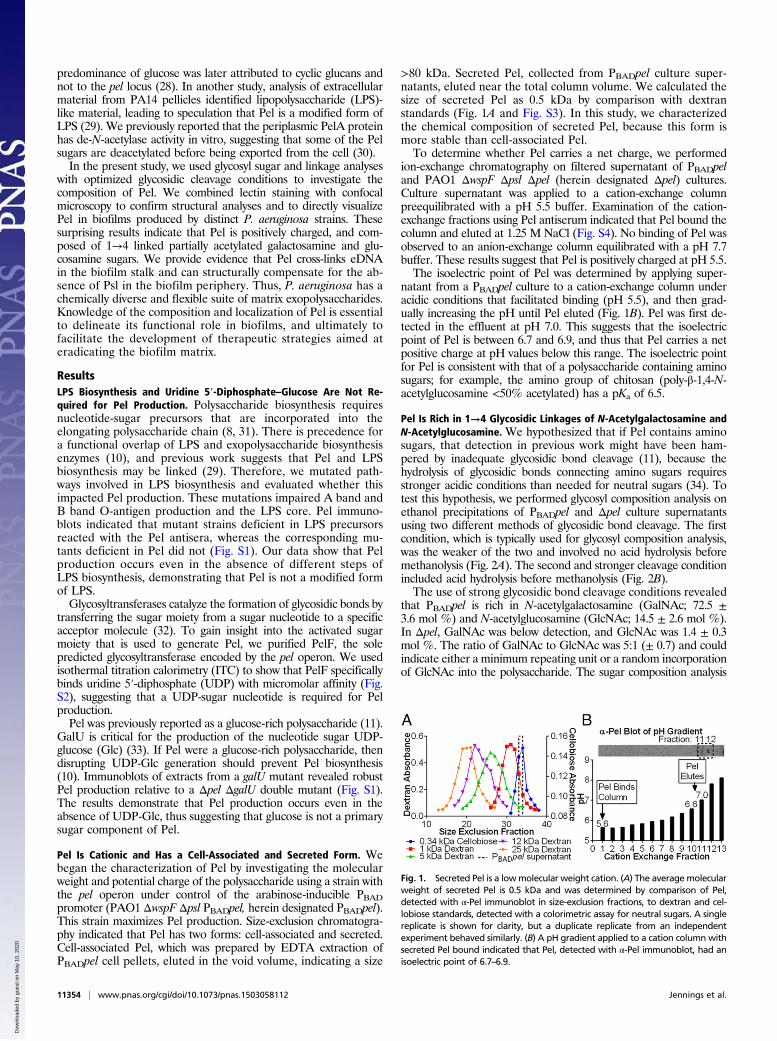
Pel Is Rich in 1→4 Glycosidic Linkages of N-Acetylgalactosamine andN-Acetylglucosamine. We hypothesized that if Pel contains aminosugars, that detection in previous work might have been ham-pered by inadequate glycosidic bond cleavage (11), because thehydrolysis of glycosidic bonds connecting amino sugars requiresstronger acidic conditions than needed for neutral sugars (34). Totest this hypothesis, we performed glycosyl composition analysis onethanol precipitations of PBADpel and Δpel culture supernatantsusing two different methods of glycosidic bond cleavage. The firstcondition, which is typically used for glycosyl composition analysis,was the weaker of the two and involved no acid hydrolysis beforemethanolysis (Fig. 2A). The second and stronger cleavage conditionincluded acid hydrolysis before methanolysis (Fig. 2B).The use of strong glycosidic bond cleavage conditions revealed
that PBADpel is rich in N-acetylgalactosamine (GalNAc; 72.5 ±3.6 mol %) and N-acetylglucosamine (GlcNAc; 14.5 ± 2.6 mol %).In Δpel, GalNAc was below detection, and GlcNAc was 1.4 ± 0.3mol %. The ratio of GalNAc to GlcNAc was 5:1 (± 0.7) and couldindicate either a minimum repeating unit or a random incorporationof GlcNAc into the polysaccharide. The sugar composition analysis
Fig. 1. Secreted Pel is a lowmolecular weight cation. (A) The averagemolecularweight of secreted Pel is 0.5 kDa and was determined by comparison of Pel,detected with α-Pel immunoblot in size-exclusion fractions, to dextran and cel-lobiose standards, detected with a colorimetric assay for neutral sugars. A singlereplicate is shown for clarity, but a duplicate replicate from an independentexperiment behaved similarly. (B) A pH gradient applied to a cation column withsecreted Pel bound indicated that Pel, detected with α-Pel immunoblot, had anisoelectric point of 6.7–6.9.
11354 | www.pnas.org/cgi/doi/10.1073/pnas.1503058112 Jennings et al.
Dow
nloa
ded
by g
uest
on
May
10,
202
0
involved the chemical re-N-acetylation of amino sugars and thus doesnot indicate the degree of acetylation of such sugars; however, insecreted Pel, the amino sugars cannot be 100% acetylated, becauseotherwise the polysaccharide would not carry a charge. The presenceof partially acetylated amino sugars is consistent with the observedde-N-acetylase in vitro enzyme activity of PelA (30).Glycosyl linkage analysis of ethanol precipitations of PBADpel
supernatant revealed that among the residues detected from Gal-NAc and GlcNAc linkages, 4-linked GalNAc and GlcNAc were themost abundant (Fig. 2C). These results suggest that Pel is a linearexopolysaccharide containing 1→4 glycosidic linkages of partiallyacetylated GalNAc and GlcNAc. Consistent with glucosamine as asecondary component of Pel, monoclonal antibodies raised againstpoly-β-1,6-N-acetylglucosamine (PNAG) and chitosan (poly-β-1,4-N-acetylglucosamine) bound samples from a PBADpel strain (Fig. S5A and B). Chitosanase, which catalyzes the specific cleavage of β-1,4acetylated and nonacetylated glucosamine linkages, showed someactivity on pellicle biofilms (Fig. S5C).Given that GalNAc was the most abundant sugar detected, we
screened lectins that recognized GalNAc sugars to identify a Pel-specific lectin. Fluorescein-labeledWisteria floribunda lectin (WFL),which has specificity to GalNAc moieties, bound clusters of plank-tonic PBADpel cells, but not from Δpel cells (Fig. S5D). This findingprovides evidence that in addition to secreted Pel, GalNAc isalso a component of cell-associated Pel.
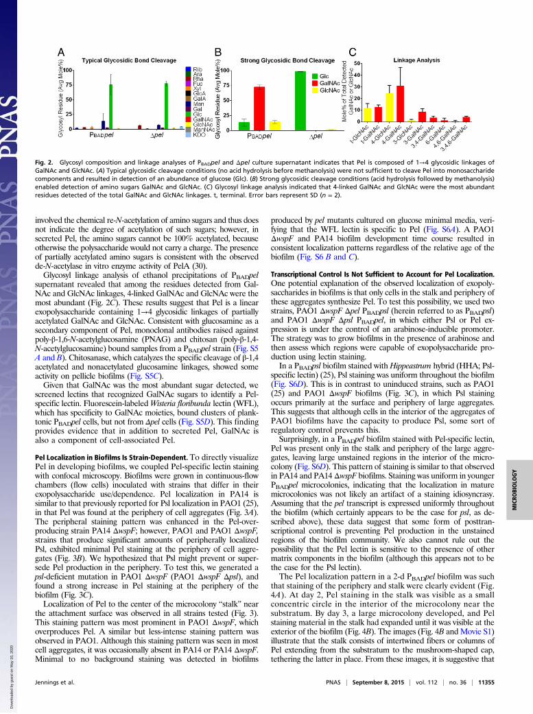
Pel Localization in Biofilms Is Strain-Dependent. To directly visualizePel in developing biofilms, we coupled Pel-specific lectin stainingwith confocal microscopy. Biofilms were grown in continuous-flowchambers (flow cells) inoculated with strains that differ in theirexopolysaccharide use/dependence. Pel localization in PA14 issimilar to that previously reported for Psl localization in PAO1 (25),in that Pel was found at the periphery of cell aggregates (Fig. 3A).The peripheral staining pattern was enhanced in the Pel-over-producing strain PA14 ΔwspF; however, PAO1 and PAO1 ΔwspF,strains that produce significant amounts of peripherally localizedPsl, exhibited minimal Pel staining at the periphery of cell aggre-gates (Fig. 3B). We hypothesized that Psl might prevent or super-sede Pel production in the periphery. To test this, we generated apsl-deficient mutation in PAO1 ΔwspF (PAO1 ΔwspF Δpsl), andfound a strong increase in Pel staining at the periphery of thebiofilm (Fig. 3C).Localization of Pel to the center of the microcolony “stalk” near
the attachment surface was observed in all strains tested (Fig. 3).This staining pattern was most prominent in PAO1 ΔwspF, whichoverproduces Pel. A similar but less-intense staining pattern wasobserved in PAO1. Although this staining pattern was seen in mostcell aggregates, it was occasionally absent in PA14 or PA14 ΔwspF.Minimal to no background staining was detected in biofilms
produced by pel mutants cultured on glucose minimal media, veri-fying that the WFL lectin is specific to Pel (Fig. S6A). A PAO1ΔwspF and PA14 biofilm development time course resulted inconsistent localization patterns regardless of the relative age of thebiofilm (Fig. S6 B and C).
Transcriptional Control Is Not Sufficient to Account for Pel Localization.One potential explanation of the observed localization of exopoly-saccharides in biofilms is that only cells in the stalk and periphery ofthese aggregates synthesize Pel. To test this possibility, we used twostrains, PAO1 ΔwspF Δpel PBADpsl (herein referred to as PBADpsl)and PAO1 ΔwspF Δpsl PBADpel, in which either Psl or Pel ex-pression is under the control of an arabinose-inducible promoter.The strategy was to grow biofilms in the presence of arabinose andthen assess which regions were capable of exopolysaccharide pro-duction using lectin staining.In a PBADpsl biofilm stained with Hippeastrum hybrid (HHA; Psl-
specific lectin) (25), Psl staining was uniform throughout the biofilm(Fig. S6D). This is in contrast to uninduced strains, such as PAO1(25) and PAO1 ΔwspF biofilms (Fig. 3C), in which Psl stainingoccurs primarily at the surface and periphery of large aggregates.This suggests that although cells in the interior of the aggregates ofPAO1 biofilms have the capacity to produce Psl, some sort ofregulatory control prevents this.Surprisingly, in a PBADpel biofilm stained with Pel-specific lectin,
Pel was present only in the stalk and periphery of the large aggre-gates, leaving large unstained regions in the interior of the micro-colony (Fig. S6D). This pattern of staining is similar to that observedin PA14 and PA14ΔwspF biofilms. Staining was uniform in youngerPBADpel microcolonies, indicating that the localization in maturemicrocolonies was not likely an artifact of a staining idiosyncrasy.Assuming that the pel transcript is expressed uniformly throughoutthe biofilm (which certainly appears to be the case for psl, as de-scribed above), these data suggest that some form of posttran-scriptional control is preventing Pel production in the unstainedregions of the biofilm community. We also cannot rule out thepossibility that the Pel lectin is sensitive to the presence of othermatrix components in the biofilm (although this appears not to bethe case for the Psl lectin).The Pel localization pattern in a 2-d PBADpel biofilm was such
that staining of the periphery and stalk were clearly evident (Fig.4A). At day 2, Pel staining in the stalk was visible as a smallconcentric circle in the interior of the microcolony near thesubstratum. By day 3, a large microcolony developed, and Pelstaining material in the stalk had expanded until it was visible at theexterior of the biofilm (Fig. 4B). The images (Fig. 4B andMovie S1)illustrate that the stalk consists of intertwined fibers or columns ofPel extending from the substratum to the mushroom-shaped cap,tethering the latter in place. From these images, it is suggestive that
Fig. 2. Glycosyl composition and linkage analyses of PBADpel and Δpel culture supernatant indicates that Pel is composed of 1→4 glycosidic linkages ofGalNAc and GlcNAc. (A) Typical glycosidic cleavage conditions (no acid hydrolysis before methanolysis) were not sufficient to cleave Pel into monosaccharidecomponents and resulted in detection of an abundance of glucose (Glc). (B) Strong glycosidic cleavage conditions (acid hydrolysis followed by methanolysis)enabled detection of amino sugars GalNAc and GlcNAc. (C) Glycosyl linkage analysis indicated that 4-linked GalNAc and GlcNAc were the most abundantresidues detected of the total GalNAc and GlcNAc linkages. t, terminal. Error bars represent SD (n = 2).
Jennings et al. PNAS | September 8, 2015 | vol. 112 | no. 36 | 11355
MICRO
BIOLO
GY
Dow
nloa
ded
by g
uest
on
May
10,
202
0
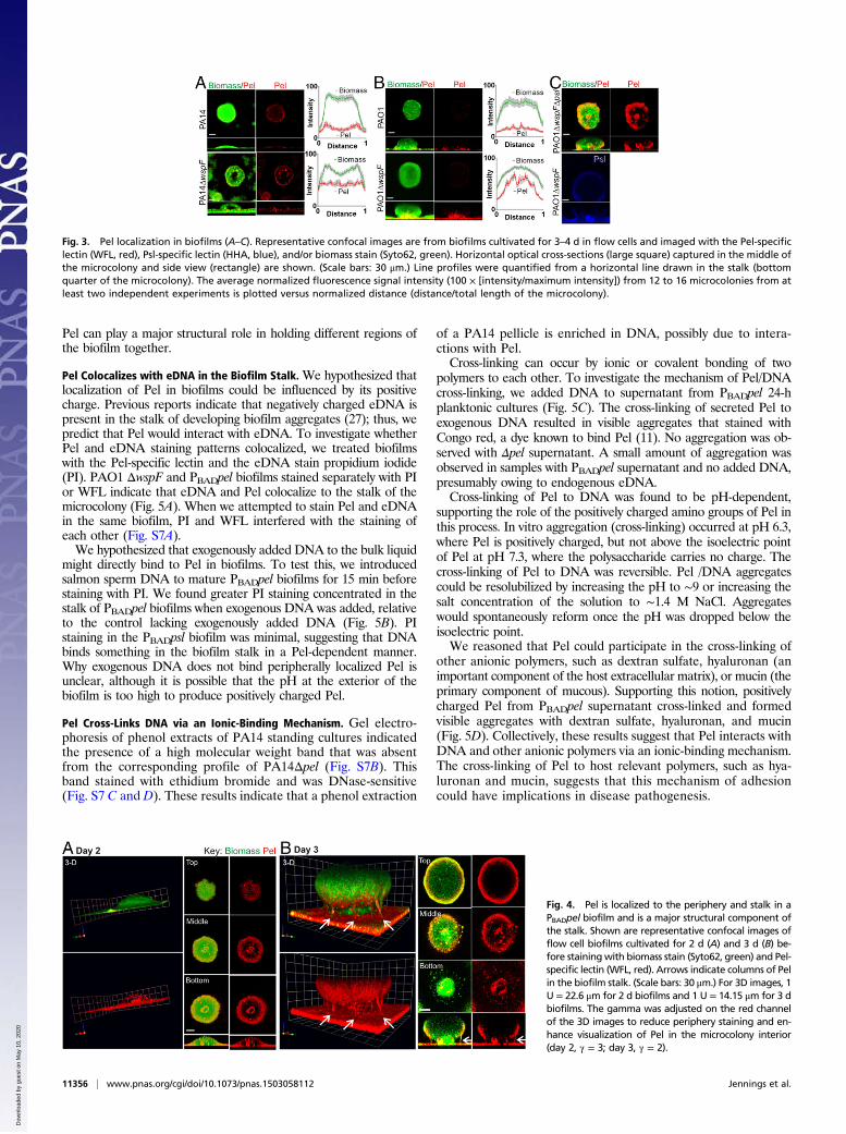
Pel can play a major structural role in holding different regions ofthe biofilm together.
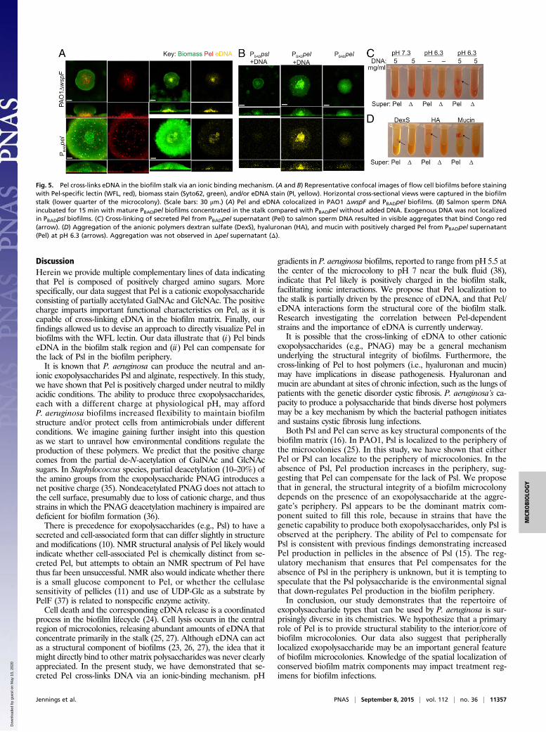
Pel Colocalizes with eDNA in the Biofilm Stalk. We hypothesized thatlocalization of Pel in biofilms could be influenced by its positivecharge. Previous reports indicate that negatively charged eDNA ispresent in the stalk of developing biofilm aggregates (27); thus, wepredict that Pel would interact with eDNA. To investigate whetherPel and eDNA staining patterns colocalized, we treated biofilmswith the Pel-specific lectin and the eDNA stain propidium iodide(PI). PAO1 ΔwspF and PBADpel biofilms stained separately with PIor WFL indicate that eDNA and Pel colocalize to the stalk of themicrocolony (Fig. 5A). When we attempted to stain Pel and eDNAin the same biofilm, PI and WFL interfered with the staining ofeach other (Fig. S7A).We hypothesized that exogenously added DNA to the bulk liquid
might directly bind to Pel in biofilms. To test this, we introducedsalmon sperm DNA to mature PBADpel biofilms for 15 min beforestaining with PI. We found greater PI staining concentrated in thestalk of PBADpel biofilms when exogenous DNA was added, relativeto the control lacking exogenously added DNA (Fig. 5B). PIstaining in the PBADpsl biofilm was minimal, suggesting that DNAbinds something in the biofilm stalk in a Pel-dependent manner.Why exogenous DNA does not bind peripherally localized Pel isunclear, although it is possible that the pH at the exterior of thebiofilm is too high to produce positively charged Pel.
Pel Cross-Links DNA via an Ionic-Binding Mechanism. Gel electro-phoresis of phenol extracts of PA14 standing cultures indicatedthe presence of a high molecular weight band that was absentfrom the corresponding profile of PA14Δpel (Fig. S7B). Thisband stained with ethidium bromide and was DNase-sensitive(Fig. S7 C and D). These results indicate that a phenol extraction
of a PA14 pellicle is enriched in DNA, possibly due to intera-ctions with Pel.Cross-linking can occur by ionic or covalent bonding of two
polymers to each other. To investigate the mechanism of Pel/DNAcross-linking, we added DNA to supernatant from PBADpel 24-hplanktonic cultures (Fig. 5C). The cross-linking of secreted Pel toexogenous DNA resulted in visible aggregates that stained withCongo red, a dye known to bind Pel (11). No aggregation was ob-served with Δpel supernatant. A small amount of aggregation wasobserved in samples with PBADpel supernatant and no added DNA,presumably owing to endogenous eDNA.Cross-linking of Pel to DNA was found to be pH-dependent,
supporting the role of the positively charged amino groups of Pel inthis process. In vitro aggregation (cross-linking) occurred at pH 6.3,where Pel is positively charged, but not above the isoelectric pointof Pel at pH 7.3, where the polysaccharide carries no charge. Thecross-linking of Pel to DNA was reversible. Pel /DNA aggregatescould be resolubilized by increasing the pH to ∼9 or increasing thesalt concentration of the solution to ∼1.4 M NaCl. Aggregateswould spontaneously reform once the pH was dropped below theisoelectric point.We reasoned that Pel could participate in the cross-linking of
other anionic polymers, such as dextran sulfate, hyaluronan (animportant component of the host extracellular matrix), or mucin (theprimary component of mucous). Supporting this notion, positivelycharged Pel from PBADpel supernatant cross-linked and formedvisible aggregates with dextran sulfate, hyaluronan, and mucin(Fig. 5D). Collectively, these results suggest that Pel interacts withDNA and other anionic polymers via an ionic-binding mechanism.The cross-linking of Pel to host relevant polymers, such as hya-luronan and mucin, suggests that this mechanism of adhesioncould have implications in disease pathogenesis.
Fig. 3. Pel localization in biofilms (A–C). Representative confocal images are from biofilms cultivated for 3–4 d in flow cells and imaged with the Pel-specificlectin (WFL, red), Psl-specific lectin (HHA, blue), and/or biomass stain (Syto62, green). Horizontal optical cross-sections (large square) captured in the middle ofthe microcolony and side view (rectangle) are shown. (Scale bars: 30 μm.) Line profiles were quantified from a horizontal line drawn in the stalk (bottomquarter of the microcolony). The average normalized fluorescence signal intensity (100 × [intensity/maximum intensity]) from 12 to 16 microcolonies from atleast two independent experiments is plotted versus normalized distance (distance/total length of the microcolony).
Fig. 4. Pel is localized to the periphery and stalk in aPBADpel biofilm and is a major structural component ofthe stalk. Shown are representative confocal images offlow cell biofilms cultivated for 2 d (A) and 3 d (B) be-fore stainingwith biomass stain (Syto62, green) and Pel-specific lectin (WFL, red). Arrows indicate columns of Pelin the biofilm stalk. (Scale bars: 30 μm.) For 3D images, 1U = 22.6 μm for 2 d biofilms and 1 U = 14.15 μm for 3 dbiofilms. The gamma was adjusted on the red channelof the 3D images to reduce periphery staining and en-hance visualization of Pel in the microcolony interior(day 2, γ = 3; day 3, γ = 2).
11356 | www.pnas.org/cgi/doi/10.1073/pnas.1503058112 Jennings et al.
Dow
nloa
ded
by g
uest
on
May
10,
202
0
DiscussionHerein we provide multiple complementary lines of data indicatingthat Pel is composed of positively charged amino sugars. Morespecifically, our data suggest that Pel is a cationic exopolysaccharideconsisting of partially acetylated GalNAc and GlcNAc. The positivecharge imparts important functional characteristics on Pel, as it iscapable of cross-linking eDNA in the biofilm matrix. Finally, ourfindings allowed us to devise an approach to directly visualize Pel inbiofilms with the WFL lectin. Our data illustrate that (i) Pel bindseDNA in the biofilm stalk region and (ii) Pel can compensate forthe lack of Psl in the biofilm periphery.It is known that P. aeruginosa can produce the neutral and an-
ionic exopolysaccharides Psl and alginate, respectively. In this study,we have shown that Pel is positively charged under neutral to mildlyacidic conditions. The ability to produce three exopolysaccharides,each with a different charge at physiological pH, may affordP. aeruginosa biofilms increased flexibility to maintain biofilmstructure and/or protect cells from antimicrobials under differentconditions. We imagine gaining further insight into this questionas we start to unravel how environmental conditions regulate theproduction of these polymers. We predict that the positive chargecomes from the partial de-N-acetylation of GalNAc and GlcNAcsugars. In Staphylococcus species, partial deacetylation (10–20%) ofthe amino groups from the exopolysaccharide PNAG introduces anet positive charge (35). Nondeacetylated PNAG does not attach tothe cell surface, presumably due to loss of cationic charge, and thusstrains in which the PNAG deacetylation machinery is impaired aredeficient for biofilm formation (36).There is precedence for exopolysaccharides (e.g., Psl) to have a
secreted and cell-associated form that can differ slightly in structureand modifications (10). NMR structural analysis of Pel likely wouldindicate whether cell-associated Pel is chemically distinct from se-creted Pel, but attempts to obtain an NMR spectrum of Pel havethus far been unsuccessful. NMR also would indicate whether thereis a small glucose component to Pel, or whether the cellulasesensitivity of pellicles (11) and use of UDP-Glc as a substrate byPelF (37) is related to nonspecific enzyme activity.Cell death and the corresponding eDNA release is a coordinated
process in the biofilm lifecycle (24). Cell lysis occurs in the centralregion of microcolonies, releasing abundant amounts of eDNA thatconcentrate primarily in the stalk (25, 27). Although eDNA can actas a structural component of biofilms (23, 26, 27), the idea that itmight directly bind to other matrix polysaccharides was never clearlyappreciated. In the present study, we have demonstrated that se-creted Pel cross-links DNA via an ionic-binding mechanism. pH
gradients in P. aeruginosa biofilms, reported to range from pH 5.5 atthe center of the microcolony to pH 7 near the bulk fluid (38),indicate that Pel likely is positively charged in the biofilm stalk,facilitating ionic interactions. We propose that Pel localization tothe stalk is partially driven by the presence of eDNA, and that Pel/eDNA interactions form the structural core of the biofilm stalk.Research investigating the correlation between Pel-dependentstrains and the importance of eDNA is currently underway.It is possible that the cross-linking of eDNA to other cationic
exopolysaccharides (e.g., PNAG) may be a general mechanismunderlying the structural integrity of biofilms. Furthermore, thecross-linking of Pel to host polymers (i.e., hyaluronan and mucin)may have implications in disease pathogenesis. Hyaluronan andmucin are abundant at sites of chronic infection, such as the lungs ofpatients with the genetic disorder cystic fibrosis. P. aeruginosa’s ca-pacity to produce a polysaccharide that binds diverse host polymersmay be a key mechanism by which the bacterial pathogen initiatesand sustains cystic fibrosis lung infections.Both Psl and Pel can serve as key structural components of the
biofilm matrix (16). In PAO1, Psl is localized to the periphery ofthe microcolonies (25). In this study, we have shown that eitherPel or Psl can localize to the periphery of microcolonies. In theabsence of Psl, Pel production increases in the periphery, sug-gesting that Pel can compensate for the lack of Psl. We proposethat in general, the structural integrity of a biofilm microcolonydepends on the presence of an exopolysaccharide at the aggre-gate’s periphery. Psl appears to be the dominant matrix com-ponent suited to fill this role, because in strains that have thegenetic capability to produce both exopolysaccharides, only Psl isobserved at the periphery. The ability of Pel to compensate forPsl is consistent with previous findings demonstrating increasedPel production in pellicles in the absence of Psl (15). The reg-ulatory mechanism that ensures that Pel compensates for theabsence of Psl in the periphery is unknown, but it is tempting tospeculate that the Psl polysaccharide is the environmental signalthat down-regulates Pel production in the biofilm periphery.In conclusion, our study demonstrates that the repertoire of
exopolysaccharide types that can be used by P. aeruginosa is sur-prisingly diverse in its chemistries. We hypothesize that a primaryrole of Pel is to provide structural stability to the interior/core ofbiofilm microcolonies. Our data also suggest that peripherallylocalized exopolysaccharide may be an important general featureof biofilm microcolonies. Knowledge of the spatial localization ofconserved biofilm matrix components may impact treatment reg-imens for biofilm infections.
Fig. 5. Pel cross-links eDNA in the biofilm stalk via an ionic binding mechanism. (A and B) Representative confocal images of flow cell biofilms before stainingwith Pel-specific lectin (WFL, red), biomass stain (Syto62, green), and/or eDNA stain (PI, yellow). Horizontal cross-sectional views were captured in the biofilmstalk (lower quarter of the microcolony). (Scale bars: 30 μm.) (A) Pel and eDNA colocalized in PAO1 ΔwspF and PBADpel biofilms. (B) Salmon sperm DNAincubated for 15 min with mature PBADpel biofilms concentrated in the stalk compared with PBADpel without added DNA. Exogenous DNA was not localizedin PBADpsl biofilms. (C) Cross-linking of secreted Pel from PBADpel supernatant (Pel) to salmon sperm DNA resulted in visible aggregates that bind Congo red(arrow). (D) Aggregation of the anionic polymers dextran sulfate (DexS), hyaluronan (HA), and mucin with positively charged Pel from PBADpel supernatant(Pel) at pH 6.3 (arrows). Aggregation was not observed in Δpel supernatant (Δ).
Jennings et al. PNAS | September 8, 2015 | vol. 112 | no. 36 | 11357
MICRO
BIOLO
GY
Dow
nloa
ded
by g
uest
on
May
10,
202
0
Materials and MethodsMore detailed information is provided in SI Materials and Methods.
Culturing and Purification of Pel Polysaccharide. Bacterial strains are listed inTable S1. Planktonic cultures were maintained on Jensen’s defined me-dium (pH 7.3). Biofilms were grown in continuous-flow cell chambers onglucose minimal media (pH 7). Cell-associated Pel was extracted fromPBADpel and Δpel cell pellets with EDTA. Secreted Pel was collected fromculture supernatants and in some cases purified further using cation-exchangeor ethanol precipitation.
Glycosyl Composition and Linkage Analyses. Carbohydrate analyses wereconducted at the University of Georgia’s Complex Carbohydrate ResearchCenter (CCRC) on ethanol precipitated supernatant from PBADpel and Δpel.Samples for composition analysis were subjected to (i ) typical glycosidiccleavage (no acid hydrolysis before methanolysis) or (ii ) strong glycosidiccleavage (hydrolysis with TFA and hydrochloric acid, followed by meth-anolysis). GC/MS was used to analyze per-O-trimethylsilyl methyl glyco-sides for composition analysis and partially methylated alditol acetates forlinkage analysis.
Lectin Staining and Confocal Microscopy. Flow cell biofilms were stained andthen imaged on a confocal microscope. To determine whether exogenous
DNA bound Pel in biofilms, salmon sperm DNA was added to mature biofilmsand incubated statically for 15 min, and then rinsed, stained with Syto62 andPI, rinsed again, and finally imaged on the confocal microscope.
In Vitro Pel Cross-Linking Experiments. PBADpel and Δpel supernatant weremixed with anionic polymers including DNA, dextran sulfate, hyaluronan,and mucin until dissolved. The pH of the polymer solution was adjusted to7.3 or 6.3. Congo red was added, and aggregates resulting from the cross-linking of the Pel and anionic polymers were visualized.
ACKNOWLEDGMENTS. We thank T. Romeo and S. Schillberg for antibodies,and M. Vedadi and G. Senisterra (Structural Genomics Consortium, Toronto)for ITC assistance. This work was supported by National Institutes of HealthGrants 2R01AI077628 (to M.R.P.) and R01AI097511 (to D.J.W.), and CanadianInstitutes of Health Research Operating Grant 13337 (to P.L.H.). L.K.J. isthe recipient of an American Heart Association Postdoctoral Fellowship(14POST20130017). P.R.S. and B.S.T. are recipients of Cystic Fibrosis Founda-tion Postdoctoral Fellowships. L.S.M. is supported by graduate scholarshipsfrom the Natural Sciences and Engineering Research Council of Canada, theOntario Graduate Scholarship Program, and the Hospital for Sick ChildrenFoundation Student Scholarship Program. P.L.H is the recipient of a CanadaResearch Chair.
1. Costerton JW, Stewart PS, Greenberg EP (1999) Bacterial biofilms: A common cause ofpersistent infections. Science 284(5418):1318–1322.
2. Parsek MR, Singh PK (2003) Bacterial biofilms: An emerging link to disease patho-genesis. Annu Rev Microbiol 57:677–701.
3. Hall-Stoodley L, Costerton JW, Stoodley P (2004) Bacterial biofilms: From the naturalenvironment to infectious diseases. Nat Rev Microbiol 2(2):95–108.
4. Costerton JW, Lewandowski Z, Caldwell DE, Korber DR, Lappin-Scott HM (1995) Mi-crobial biofilms. Annu Rev Microbiol 49(1):711–745.
5. Flemming H-C, Wingender J (2010) The biofilm matrix. Nat Rev Microbiol 8(9):623–633.6. Sutherland IW (2001) The biofilm matrix: An immobilized but dynamic microbial
environment. Trends Microbiol 9(5):222–227.7. Pamp SJ, Gjermansen M, Tolker-Nielsen T (2007) The biofilm matrix: A sticky frame-
work. The Biofilm Mode of Life: Mechanisms and Adaptations, eds Kjelleberg S,Givskov M (Horizon Bioscience, Norfolk, UK), pp 37–69.
8. Franklin MJ, Nivens DE, Weadge JT, Howell PL (2011) Biosynthesis of the Pseudo-monas aeruginosa extracellular polysaccharides, alginate, Pel, and Psl. Front Microbiol2(167):167.
9. Mann EE, Wozniak DJ (2012) Pseudomonas biofilm matrix composition and nichebiology. FEMS Microbiol Rev 36(4):893–916.
10. Byrd MS, et al. (2009) Genetic and biochemical analyses of the Pseudomonas aeru-ginosa Psl exopolysaccharide reveal overlapping roles for polysaccharide synthesisenzymes in Psl and LPS production. Mol Microbiol 73(4):622–638.
11. Friedman L, Kolter R (2004) Genes involved in matrix formation in Pseudomonasaeruginosa PA14 biofilms. Mol Microbiol 51(3):675–690.
12. Colvin KM, et al. (2011) The Pel polysaccharide can serve a structural and protectiverole in the biofilm matrix of Pseudomonas aeruginosa. PLoS Pathog 7(1):e1001264.
13. Yang L, et al. (2011) Distinct roles of extracellular polymeric substances in Pseudo-monas aeruginosa biofilm development. Environ Microbiol 13(7):1705–1717.
14. Vasseur P, Vallet-Gely I, Soscia C, Genin S, Filloux A (2005) The pel genes of thePseudomonas aeruginosa PAK strain are involved at early and late stages of biofilmformation. Microbiology 151(Pt 3):985–997.
15. Ghafoor A, Hay ID, Rehm BHA (2011) Role of exopolysaccharides in Pseudomonasaeruginosa biofilm formation and architecture. Appl Environ Microbiol 77(15):5238–5246.
16. Colvin KM, et al. (2012) The Pel and Psl polysaccharides provide Pseudomonas aeru-ginosa structural redundancy within the biofilm matrix. Environ Microbiol 14(8):1913–1928.
17. Häussler S, et al. (2003) Highly adherent small-colony variants of Pseudomonas aer-uginosa in cystic fibrosis lung infection. J Med Microbiol 52(Pt 4):295–301.
18. Kirisits MJ, Prost L, Starkey M, Parsek MR (2005) Characterization of colony mor-phology variants isolated from Pseudomonas aeruginosa biofilms. Appl EnvironMicrobiol 71(8):4809–4821.
19. Hickman JW, Tifrea DF, Harwood CS (2005) A chemosensory system that regulatesbiofilm formation through modulation of cyclic diguanylate levels. Proc Natl Acad SciUSA 102(40):14422–14427.
20. Starkey M, et al. (2009) Pseudomonas aeruginosa rugose small-colony variants haveadaptations that likely promote persistence in the cystic fibrosis lung. J Bacteriol191(11):3492–3503.
21. Stoodley P, Sauer K, Davies DG, Costerton JW (2002) Biofilms as complex differenti-ated communities. Annu Rev Microbiol 56(1):187–209.
22. O’Toole G, Kaplan HB, Kolter R (2000) Biofilm formation as microbial development.Annu Rev Microbiol 54(1):49–79.
23. Rice KC, et al. (2007) The cidA murein hydrolase regulator contributes to DNA releaseand biofilm development in Staphylococcus aureus. Proc Natl Acad Sci USA 104(19):8113–8118.
24. Webb JS, et al. (2003) Cell death in Pseudomonas aeruginosa biofilm development.J Bacteriol 185(15):4585–4592.
25. Ma L, et al. (2009) Assembly and development of the Pseudomonas aeruginosa bio-film matrix. PLoS Pathog 5(3):e1000354.
26. Whitchurch CB, Tolker-Nielsen T, Ragas PC, Mattick JS (2002) Extracellular DNA re-quired for bacterial biofilm formation. Science 295(5559):1487.
27. Allesen-Holm M, et al. (2006) A characterization of DNA release in Pseudomonasaeruginosa cultures and biofilms. Mol Microbiol 59(4):1114–1128.
28. Sadovskaya I, et al. (2010) High-level antibiotic resistance in Pseudomonas aeruginosa
biofilm: the ndvB gene is involved in the production of highly glycerol-phosphory-lated β-(1→3)-glucans, which bind aminoglycosides. Glycobiology 20(7):895–904.
29. Coulon C, Vinogradov E, Filloux A, Sadovskaya I (2010) Chemical analysis of cellularand extracellular carbohydrates of a biofilm-forming strain Pseudomonas aeruginosaPA14. PLoS One 5(12):e14220.
30. Colvin KM, et al. (2013) PelA deacetylase activity is required for Pel polysaccharidesynthesis in Pseudomonas aeruginosa. J Bacteriol 195(10):2329–2339.
31. Rocchetta HL, Burrows LL, Lam JS (1999) Genetics of O-antigen biosynthesis in Pseu-domonas aeruginosa. Microbiol Mol Biol Rev 63(3):523–553.
32. Rocchetta HL, Burrows LL, Pacan JC, Lam JS (1998) Three rhamnosyltransferases re-
sponsible for assembly of the A-band D-rhamnan polysaccharide in Pseudomonasaeruginosa: A fourth transferase, WbpL, is required for the initiation of both A-bandand B-band lipopolysaccharide synthesis. Mol Microbiol 28(6):1103–1119.
33. Dean CR, Goldberg JB (2002) Pseudomonas aeruginosa galU is required for a com-plete lipopolysaccharide core and repairs a secondary mutation in a PA103 (serogroupO11) wbpM mutant. FEMS Microbiol Lett 210(2):277–283.
34. Merkle RK, Poppe I (1994) Carbohydrate composition analysis of glycoconjugates bygas-liquid chromatography/mass spectrometry. Methods Enzymol 230:1–15.
35. Cerca N, et al. (2007) Molecular basis for preferential protective efficacy of antibodiesdirected to the poorly acetylated form of staphylococcal poly-N-acetyl-β-(1-6)-glu-cosamine. Infect Immun 75(7):3406–3413.
36. Vuong C, et al. (2004) A crucial role for exopolysaccharide modification in bacterialbiofilm formation, immune evasion, and virulence. J Biol Chem 279(52):54881–54886.
37. Ghafoor A, Jordens Z, Rehm BHA (2013) Role of PelF in Pel polysaccharide biosynthesisin Pseudomonas aeruginosa. Appl Environ Microbiol 79(9):2968–2978.
38. Hunter RC, Beveridge TJ (2005) Application of a pH-sensitive fluoroprobe (C-SNARF-4)for pH microenvironment analysis in Pseudomonas aeruginosa biofilms. Appl EnvironMicrobiol 71(5):2501–2510.
39. Hoang TT, Karkhoff-Schweizer RR, Kutchma AJ, Schweizer HP (1998) A broad-host-range Flp-FRT recombination system for site-specific excision of chromosomally-locatedDNA sequences: Application for isolation of unmarked Pseudomonas aeruginosa mu-
tants. Gene 212(1):77–86.40. Penterman J, Singh PK, Walker GC (2014) Biological cost of pyocin production during
the SOS response in Pseudomonas aeruginosa. J Bacteriol 196(18):3351–3359.
11358 | www.pnas.org/cgi/doi/10.1073/pnas.1503058112 Jennings et al.
Dow
nloa
ded
by g
uest
on
May
10,
202
0