Embed Size (px)
Citation preview

FINAL DRAFT
Pelvic Girdle Pain in the Antepartum Population
Physical Therapy Clinical Practice Guidelines
Linked to the International Classification of
Functioning, Disability, and Health from the
Section on Women’s Health and the Orthopaedic
Section of the American Physical Therapy
Association
Authors:
Susan C. Clinton PT DScPT OCS WCS COMT FAAOMPT
Alaina Newell PT DPT WCS CLT-LANA
Patricia A. Downey PT PhD DPT
Kimberly Ferreira PT MSPT PhD
RECOMMENDATIONS
INTRODUCTION
METHODS
CLINICAL GUIDELINES: Impairment/Function-Based Diagnosis
CLINICAL GUIDELINES: Examinations
CLINICAL GUIDELINES: Intervention
RECOMMENDATIONS
AUTHOR/REVIEWER AFFILIATIONS AND CONTACTS
REFERENCES

FINAL DRAFT
RECOMMENDATIONS
Risk Factors: A
Clinicians should utilize the following risk factors: prior history of pregnancy, orthopedic
dysfunctions, increased BMI, smoking as well as work dissatisfaction and a lack of belief of improvement
in the prognosis of PGP. (Recommendation based on strong evidence)
Postural Changes: B
Clinicians should not consider postural changes as indicative of the development and/or intensity
of PGP in the antepartum population. (Recommendation based on moderate evidence)
Clinical Course: A/B
Clinicians should (consider) treat patients with early onset, multiple pain locations, a high number
of positive pelvic pain provocation tests, work dissatisfaction and lack of belief of improvement, as these
are strong/moderate factors in determining the potential for persisting PGP in late pregnancy and post-
partum. (Recommendations based on strong/moderate evidence)
Diagnosis/Classification: B
Clinicians may consider the utilization of the classification system for the diagnosis of the type of
pelvic girdle pain in antepartum population. (Recommendation is based on moderate evidence)
Differential Diagnosis: A
PGP, in this population, should be differentiated from signs and symptoms of serious disease and
psychological factors when the symptoms are not associated with the described clinical course of PGP,
impairments are failing to normalize and the symptoms are worsening with increased disability. This
should include the presence of transient osteoporosis and diastasis rectus abdominus as possible co-
morbidities in this population as well as the presence of pelvic floor muscle, hip and lumbar spine
dysfunction. (Recommendations are based upon strong evidence)
Imaging Studies: F
In the absence of good evidence, expert opinion and foundation science may be used to guide
examination with the use of imaging studies.
Examination-Outcome Measures: A
Clinicians should administer self-reported outcome questionnaires such as Disability Rating
Index, Oswestry Disability Index, Pelvic Girdle Questionnaire, Fear-Avoidance Belief Questionnaire, and
Pain Catastrophizing Scale. These are scales are practical for the determination of baseline disability,
function and pain belief as well as change throughout the clinical course. These should be utilized in
combination with clinical examination for clinical decision. (Recommendations are based on strong
evidence.)

FINAL DRAFT
Examination-Activity Limitation and Participation Restriction Measures: E
While strong evidence exists to support a high risk of falls, no measures have been validated to
objectively assess the dynamic balance and fall risk in antepartum population. (Recommendation is based
on theoretical/foundational evidence)
Intervention- Support Belts: D
Clinicians should consider the application of a support belt in the antepartum population with
PGP. The four studies reviewed investigated different patient populations, had varied intervention groups
and controls, different durations of intervention application and different timing of follow-up. Further
research is needed to clarify initial application, duration and specific antepartum PGP patient
classification for support belt intervention. (Recommendation is based on conflicting evidence.)
Intervention-Exercise: D
Clinicians should consider the use of exercise in the antepartum population with PGP. ACOG
and the Canadian CPG have recommended exercise for health benefits because of the low risk and
minimal adverse effects for the antepartum population. The two systematic reviews as well as the recent
RCTs were non-specific in the application of exercise to heterogeneous groups of Pregnancy Low Back
Pain (PLBP) and PGP. The populations varied in early and late pregnancy and demonstrated a variety of
exercise interventions. No study based the exercise intervention on the classification of PGP proposed by
Albert, et al4 and Cook, et al.
23 (Recommendation is based on conflicting evidence.)
Intervention- Manual Therapy: C
Clinicians may or may not utilize manual therapy techniques including high velocity low
amplitude manipulations for the treatment of PBLP and PGP. This evidence is emerging and treatment
could be considered, as there is little to no reported evidence of adverse effects in the healthy antepartum
population. (Recommendations are based upon weak evidence.)
Introduction
AIM OF THE GUIDELINES
The Section on Women’s Health (SOWH) and the Orthopaedic Section of the American Physical Therapy
Association (APTA) has an ongoing effort to create evidence-based practice guidelines for women’s
health and orthopedic physical therapy management of patients with musculoskeletal impairments
described in the World Health Organization’s International Classification of Functioning, Disability, and
Health (ICF).104
The purposes of these clinical guidelines are to:

FINAL DRAFT
Describe evidence-based physical therapy practice including diagnosis, prognosis, intervention,
and assessment of outcome for musculoskeletal disorders commonly managed by women’s health
and/or orthopedic physical therapists
Classify and define common musculoskeletal conditions using the World Health Organization’s
terminology related to impairments of body function and body structure, activity limitations, and
participation restrictions
Identify interventions supported by current best evidence to address impairments of body function
and structure, activity limitations, and participation restrictions associated with common
musculoskeletal conditions
Identify appropriate outcome measures to assess changes resulting from physical therapy
interventions
Provide a description to policy makers, using internationally accepted terminology, of the practice
of women’s health and/or orthopaedic physical therapists
Provide information for payers and claims reviewers regarding the practice of women’s health
and/or orthopaedic physical therapy for common musculoskeletal conditions
Create a reference publication for women’s health and/or orthopedic physical therapy clinicians,
academic instructors, clinical instructors, students, interns, residents, and fellows regarding the
best current practice regarding women’s health and/or orthopaedic physical therapy
STATEMENT OF INTENT
This guideline is not intended to be construed or to serve as a standard of clinical care. Standards of care
are determined on the basis of all clinical data available for an individual patient and are subject to change
as scientific knowledge and technology advance and patterns of care evolve. These parameters of practice
should be considered guidelines only. Adherence to them will not ensure a successful outcome in every
patient, nor should they be construed as including all proper methods of care or excluding other
acceptable methods of care aimed at the same results. The ultimate judgment regarding a particular
clinical procedure or treatment plan must be made in light of the clinical data presented by the patient, the
diagnostic and treatment options available, and the patient’s values, expectations, and preferences.
However, we suggest that the rationale for significant departures from accepted guidelines be documented
in the patient’s medical records at the time the relevant clinical decision is made.
Methods
SUMMARY OF LITERATURE SEARCH
Content experts within the Section of Women’s Health in partnership with Orthopaedic Section of APTA,
developed a clinical practice guideline for physical therapists in the examination and intervention of
pelvic girdle pain in the antepartum population. Utilizing the ICF terminology, the authors identified
impairments of body function, and structure, activity limitation and participation restrictions that could
(1) categorize patients into mutually exclusive impairment patterns upon which to base intervention
strategies and (2) serve as measures of change in function over the course of an episode of care.
Secondly, the authors described the supporting evidence for the identified impairment pattern
classification as well as interventions for patients with activity limitations and impairments of body
function and structure consistent with the identified impairment pattern classification. It was also
acknowledged by the Section of Women’s Health and Orthopaedic Section of the APTA that a systematic

FINAL DRAFT
search and review solely of the evidence related to diagnostic categories based on International Statistical
Classification of Diseases and Related Health Problems (ICD)140 terminology would not be sufficient for
these ICF-based clinical practice guidelines, as most of the evidence associated with changes in levels of
impairment or function in homogeneous populations is not readily searchable using the current
terminology. For this reason, the authors also searched the scientific literature related to prevalence, risk
factors, examination, classification, outcome measures, and intervention strategies implemented by
physical therapists for pelvic girdle pain in the antepartum population. Thus, the authors of this clinical
practice guideline systematically searched MEDLINE, CINAHL, and the Cochrane Database of
Systematic Reviews (through 2011) for any relevant articles related to prevalence, risk factors,
examination, classification, outcome measures, and intervention strategies for pelvic girdle pain in the
antepartum population. Additionally, when relevant articles were identified their reference lists were
hand-searched in an attempt to identify other articles that might have contributed to the outcome of this
clinical practice guideline. This guideline will be issued in 2015 based upon publications in the scientific
literature prior to July 2012. This guideline will be considered for review in 2020, or sooner, if new
evidence becomes available. Any updates to the guideline in the interim period will be noted on the
Section on Women’s Health (www.womenshealthapta.org) and the Orthopaedic Section
(www.orthopt.org) of the APTA.
CRITICAL APPRAISAL PROCESS AND RELIABILITY
Each literary article was reviewed by two reviewers and required greater than 95% agreement among
reviewers via Key Questions from the Evidence Based Physical Therapy36
for determination of article
quality for the appropriate of level of evidence established by the Centers for Evidence-Based Medicine.
If greater than 95% agreement was not achieved a third reviewer was utilized for quality determination.
Articles were considered “high-quality” if they fulfilled greater than 75% of key questions for the specific
aim of the articles. Articles of less than 75% where considered “lesser-quality” for determination of level
of evidence.
LEVELS OF EVIDENCE
The levels of evidence established by the Center for Evidence-Based Medicine, Oxford, United Kingdom
was utilized to grade individual clinical research articles for diagnostic, prospective and therapeutic
studies.60, 77
I Evidence obtained from high-quality randomized control trials, prospective cohort studies,
diagnostic studies, prognostic studies, or meta-analysis and systematic review (of level I studies)
II Evidence obtained from lesser-quality randomized control trials, retrospective cohort studies,
diagnostic studies or systematic reviews of level II or better) (i.e. Weaker diagnostic criteria and
reference standards, improper randomization, no blinding, <80% follow up)
III Case controlled studies or systematic reviews (of level III studies)
IV Case series, poor cohort studies, or poor reference standards
V Expert opinion without explicit critical appraisal, or based on physiology, bench research or “first
principles”

FINAL DRAFT
GRADES OF EVIDENCE
The overall strength of the evidence supporting recommendations made in this guideline will be graded
according to guidelines described by Guyatt et al46
as modified by Law and MacDermid54
and adopted by
the coordinator and reviewers of this project.46, 54
In this modified system, the typical A, B, C, and D
grades of evidence were modified to include the role of consensus expert opinion and basic science
research to demonstrate biological or biomechanical plausibility.
A Strong evidence A preponderance of level I and/or II studies support the
recommendation. This must include at least 1 level I study
B Moderate evidence A single high-quality RCT or a preponderance of level II studies
support the recommendation
C Weak evidence A single level II study or a preponderance of level III & IV
studies including statements of consensus by content experts
support the recommendation
D Conflicting evidence Higher-quality studies conducted on this topic disagree with
respect to their conclusions. The recommendation is based on
these conflicting studies.
E Theoretical/foundational evidence A preponderance of evidence from animal or cadaver studies,
from conceptual models/principles, or from basic
sciences/bench research support this conclusion.
F Expert opinion Best practice based on the clinical experience of the guidelines
development team
REVIEW PROCESS
The authors in conjunction with the Section of Women’s Health APTA selected reviewers from the
following areas to serve as reviewers of the first draft of this clinical practice guideline:
American College of Obstetrics and Gynecologist guidelines
Coding
Manipulative therapy
Obstetric physical therapy
Orthopedic physical therapy rehabilitation
Outcomes research
Pain science
Pelvic Girdle Pain Rehabilitation
Physical therapy academic education
Women’s health physical therapy education
Comments from these reviewers were utilized by the authors to edit this clinical practice guideline prior
to submission to the Journal of Women’s Health Physical Therapy and the Journal of Orthopaedic and
Sports Physical Therapy. In addition, several physical therapists practicing in antepartum and pelvic
girdle pain rehabilitation physical therapy practices were sent initial drafts of this clinical practice
guideline for assessment.
REVIEWERS:
Joseph J. Godges, DPT, MA - Orthopedic Section, CPG director (Review of outline/format/permission of
Orthopedic Section use of format)

FINAL DRAFT
Anita Bemis-Doughty (coding) (Review of ICF language) - APTA
Nancy Donovan PhD PT – Journal of Women’s Health Editor (Review of guideline intent and content
outline – for Journal on Women’s Health)
Pat Downey PhD PT DPT – Chatham University – Department of Physical Therapy – Program Chair,
Pittsburgh, PA
Kimberly Ferreria PT MSPT PhD(c) – Andrews University Department of Physical Therapy – Entry level
Chair
Valerie L Bobb PT, DPT, WCS, ATC - Baylor Institute for Rehabilitation OutPatient Services, Dallas,
TX
Jill Schiff Boissonnault PT, PhD, WCS - Associate Professor, The George Washington University
Doctorate in Physical Therapy Program, School of Medicine and Health Sciences, Washington DC
Teresa Costello MISCP, BSc (Hons) Physiotherapy, Pg Cert Continence, Dip Acupuncture, Chartered
Physiotherapist, Clinical Specialist in Women's Health and Continence - Teresa Costello Chartered
Physiotherapist & HSE, Longford, Ireland
Karen Litos PT, DPT, WCS – No Mom Left Behind Physical Therapy, E. Lansing, MI
Gillian Healy Bsc physio hons, MISCP - Enable Ireland, Ireland
Rebecca G. Stephenson PT, DPT, MS, CLT, WCS - Brigham and Women’s Hospital, Boston, MA
David A. Hoyle, PT, DPT, MA, OCS, MTC, CEAS - National Director of Clinical Quality:
WorkStrategies, Select Medical
Zacharia Isaac MD, Board certified in physical medicine and rehabilitation and pain management
Division chief of spine care and pain management, Spaulding rehabilitation hospital
Associate chairman, Brigham and Woman's Hospital department of Physical Medicine and
Rehabilitation. Boston, MA
Lennox Hoyte MD – OB/Gyn, University of South Florida Medical Group. Tampa, FL
Tonya Satteson, BA – Bulter, PA (consumer)
CLASSIFICATION
The primary ICD-10 codes and conditions associated with pelvic girdle pain during pregnancy are: R10.2
Pelvic Pain, M54.5 Low back pain, M53.3 Sacrococcygeal disorders not elsewhere classified, O26.9
Pregnancy-related condition, unspecified, R29.3 Abnormal posture, M48.48 Fatigue (stress)
fracture of vertebra, sacral and sacrococcygeal region, M99.04/.05 Segmental and somatic
dysfunction of sacral region/pelvic region, S33.2 Dislocation of sacroiliac and sacrococcygeal joints,
M46.1 Sacroilitis, not elsewhere specified, M46.98 Unspecified inflammatory spondylopathy, sacral
and sacrococcygeal region, M53.2X8 Spinal instabilities of sacral and sacrococcygeal region, S33.6
Sprain and strain of sacroiliac joint, M99.14/.15 Subluxation complex of the sacral region/pelvic

FINAL DRAFT
region, O26.7 Subluxation of symphysis (pubis) in pregnancy, childbirth and the puerperium,
M24.2 Disorder of ligament, M24.4 Recurrent dislocation and subluxation of joint, G96.8 Disorder
of central nervous system specified as central nervous system sensitivity to pain, and F45.4 Pain
disorders related psychological factors.103
The corresponding ICD-9 codes and conditions associated
used in the United States are: 724.2 Lumbago, 724.6 Disorders of sacrum, 739.4 Nonallopathic lesion
of the sacral region, not elsewhere specified, 846.70 Pregnancy backache, 848.5 Public symphysis
sprain/strain, 847.3 Sacroiliac joint pain, 839.42 Subluxation of the sacroiliac joint, and 349.89
Other specified disorders of the nervous system.
The primary ICF body-function codes associated with the previously stated ICD-10 conditions are: b1520
Appropriateness of motion, b1602 Content of thought b2800 Generalized pain, b2801 Pain in body
part, b28013 Pain in back, b6601 Functions related to pregnancy, b7100 Mobility of a single joint,
b7101 Mobility of several joints, b715 Stability of joint functions, b7201 Mobility of the pelvis,
b7300 Power of isolated muscle and muscle groups, b735 Muscle tone functions b7601 Control of
complex voluntary movements, b770 Gait pattern functions, b7800 Sensation of muscle stiffness,
and b7801 Sensation of muscle spasm.102
The primary ICF body-structure codes associated with pelvic girdle pain during pregnancy include: s1100
Structure of cortical lobes, s1101 Structure of midbrain, s1102 Structure of diencephalon, s1103
Basal ganglia and related structures, s1104 Structure of brainstem, and s1200 Structure of spinal
cord, s620 Structure of pelvic floor, s7401 Joints of the pelvic region, s7402 Muscles of the pelvic
region, s7403 Ligaments of fasciae of the pelvic region, s7409 Structure of pelvic region,
unspecified, and s770 Additional musculoskeletal structure related to movement.102
The primary ICF activity and participation codes associated with the above ICD-10 conditions are: d129
Purposeful sensory experiences, specified and unspecified, d230 Carrying out daily routine, d410
Changing basic body position, d415 Maintaining a body position, d430, Lifting and carrying
objects, d455 Moving around, d460 Moving around in different locations, d475 Driving, d640 Doing
housework, d660 Assisting others, d7203 Interacting according to social rules, d770 Intimate
relationships, and d8451 Maintaining a job.102
International Statistical Classification of Disease and Related Health Problems (ICD-10) Codes

FINAL DRAFT
Acute, Subacute, and Chronic Pelvic Girdle Pain with or without Pregnancy Low Back Pain
R10.2 M54.5 M53.3 O26.9 R29.3 M48.48
Pelvic pain Low back pain Sacrococcygeal disorders, not elsewhere classified Pregnancy-related condition, unspecified Abnormal posture Fatigue (stress) fracture of vertebra, sacral and sacrococcygeal region

FINAL DRAFT
International Classification of Functioning, Disability and Health (ICF) Codes
Acute, Subacute, and Chronic Pelvic Girdle Pain with or without with Pregnancy Low Back Pain
Body Function b2801 b28013 b6601
Pain in body part Pain in back Functions related to pregnancy
Body Structure s7401 s7402 s7409 s770
Joints of the pelvic region Muscles of the pelvic region Structure of pelvic region, unspecified Additional musculoskeletal structures related to movement
Activities and Participation d230 d410 d415 d460 d475 d640 d660 d770
Carrying out daily routine Changing basic body position Maintaining a body position Moving around in different locations Driving Doing housework Assisting others Intimate relationships
Acute, Subacute and Chronic Pelvic Girdle Pain with mobility deficits during pregnancy
M99.04/.05 S33.2 M46.1 M46.98
Segmental and somatic dysfunction of sacral region/pelvic region Dislocation of sacroiliac and sacrococcygeal joint Sacroilitis, not elsewhere specified Unspecified inflammatory spondylopathy, sacral and sacrococcygeal region
Acute, Subacute, and Chronic Pelvic Girdle Pain with movement coordination impairments during pregnancy
M53.2X8 S33.6 M99.14/.15 O26.7 M24.2
Spinal instabilities of sacral and sacrococcygeal region Sprain and strain of sacroiliac joint Subluxation complex of the sacral region/pelvic region Subluxation of symphysis (pubis) in pregnancy, childbirth and the puerperium Disorder of ligament
Chronic- recurrent Pelvic Girdle Pain during pregnancy
M24.4 Recurrent dislocation and subluxation of joint
Chronic Pelvic Girdle Pain with Related Generalized Pain during pregnancy
G96.8 F45.4
Disorder of central nervous system specified as central nervous system sensitivity to pain Pain disorders related psychological factors

FINAL DRAFT
d8451 Maintaining a job
Acute, Subacute and Chronic Pelvic Girdle Pain with mobility deficits during pregnancy
Body Function b2801 b7100 b7101 b715 b7201 b7300 b735 b770 b7800 b7801
Pain in body part Mobility of a single joint Mobility of several joints Stability of joint functions Mobility of pelvis Power of isolated muscles & muscle groups Muscle tone functions Gait pattern functions Sensation of muscle stiffness Sensation of muscle spasm
Body Structure s7401 s7402 s7403
Joints of the pelvic region Muscles of the pelvic region Ligaments of fasciae of the pelvic region
Activities and Participation d410 d415 d430 d455 d460 d640 d8451
Changing basic body position Maintaining a body position Lifting and carrying objects Moving around Moving around in different locations Doing housework Maintaining a job
Acute, Subacute, and Chronic Pelvic Girdle Pain with movement coordination impairments during pregnancy
Body Function b2801 b715 b735 b7601
Pain in body part Stability of joint functions Muscle tone functions Control of complex voluntary movements
Body Structure s7401 s7402 s7403
Joints of the pelvic region Muscles of the pelvic region Ligaments of fasciae of the pelvic region
Activities and Participation d410 d415 d430 d455 d640 d660 d770 d8451
Changing basic body position Maintaining a body position Lifting and carrying objects Moving around Doing housework Assisting others Intimate relationships Maintaining a job
Chronic- recurrent Pelvic Girdle Pain during pregnancy
Body Function b2801 b735 b7800 b7801
Pain in body part Muscle tone functions Sensation of muscle stiffness Sensation of muscle spasm
Body Structure s620 s7401 s7402 s7403 s7409 s770
Structure of pelvic floor Joints of the pelvic region Muscles of the pelvic region Ligaments of fasciae of the pelvic region Structure of pelvic region, unspecified Additional musculoskeletal structures related to movement
Activities and Participation d410 d415
Changing basic body position Maintaining a body position

FINAL DRAFT
d430 d455 d460 d640 d660 d770 d8451
Lifting and carrying objects Moving around Moving around in different locations Doing housework Assisting others Intimate relationships Maintaining a job
Chronic Pelvic Girdle Pain with Related Generalized Pain during pregnancy
Body Function b134 b1520 b1602 b2800
Sleep functions Appropriateness of emotion Content of thought Generalized pain
Body Structure s1100 s1101 s1102 s1103 s1104 s1200
Structure of cortical lobes Structure of midbrain Structure of diencephalon Basal ganglia and related structures Structure of brainstem Structure of spinal cord
Activities and Participation d129 d230 d640 d710 d7203 d8451
Purposeful sensory experiences, specified and unspecified Carrying out daily routine Doing housework Intimate relationships Interacting according to social rules Maintaining a job
CLINICAL GUIDELINES: Impairment/Function-Based Diagnosis
PREVALENCE
I The prevalence of pregnancy low back pain (PLBP) and pelvic girdle pain (PGP) is estimated to
occur in 56-72% of the antepartum population with 20% reporting severe symptoms during 20-30 weeks
of gestation.5,35,62,64
33-50% of pregnant females report PGP before 20 weeks of gestation and the
prevalence may reach 60-70% in late pregnancy.44,72,80
RISK FACTORS
I Risk Factors for the development of PGP in this population include a history of multiparity, joint
hypermobility, periods of amenorrhea, increased BMI, and hip and/or lower extremity dysfunction
including the presence of gluteus medius and pelvic floor muscle dysfunction.12,43,63
There is an
association of the development of PGP with a history of trauma to the pelvis and a history of low back
pain and/or pelvic girdle pain especially in a previous pregnancy.20,49,73,82,94,96,98,99
Finally, an association
also exists with work dissatisfaction and lack of belief in improvement.45,101
I Smoking during the antepartum period as well as cessation of smoking in the first trimester had
an increased odds ratio for the development of PGP compared to non-smokers.13

FINAL DRAFT
A Clinicians should utilize the following risk factors: prior history of pregnancy, orthopedic
dysfunctions, increased BMI, smoking as well as work dissatisfaction and a lack of belief of improvement
in the prognosis of PGP. (Recommendation based on strong evidence)
PATHOANATOMICAL FEATURES
Definition of Pelvic Girdle Pain
I European Guidelines: 99
“Pelvic girdle pain arises in relation to pregnancy, trauma, arthritis and osteoarthritis.
Pain is experienced between the posterior iliac crest and the gluteals fold, particularly in the vicinity of
the sacroiliac joint. The pain may radiate in the posterior thigh and can also occur in conjunction with/or
separately in the symphysis.”
Postural Changes
I Franklin and Conner-Kerr measured antepartum postural changes resulting in a significant
increase in lumbar lordosis, sagittal anterior pelvic tilt and posterior head position from the first to third
trimester. The magnitude of postural changes during pregnancy was not indicative of the intensity of
PLBP and PGP in the antepartum population.37
B Clinicians should not consider postural changes as indicative of the development and/or intensity
of PGP in the antepartum population. (Recommendation based on moderate evidence)
Pathophysiology
Vleeming et al99, 100
developed the hypothesis of hormonal and biomechanical factors as potential
contributors to PGP. Stabilization of the pelvis during load transfer is achieved by the two mechanisms
of “form closure” and “force closure”. “Form closure” is achieved when the wedge shaped sacrum fits
tightly between the ilia. This process is maximized by the “force closure” of the muscles, fascia, and
ligaments to provide the joint stability.99, 100
Changes in the ability to manage load transfers due to joint
laxity may account for the development of PGP in this population. A change in adequate force and/or
form closure of the pelvic girdle was previously postulated to occur by the presence of the hormone
relaxin, however, current studies suggest no correlation between relaxin and PGP.14,76
Post mortem
studies completed in 1924 have provided some minimal evidence that the SIJ in pregnant women
demonstrated increased laxity and greater synovial fluid volume.21
Finally, Mens et al64
reported an
increased motion in the pelvic joints in pregnant females with PGP compared to healthy non-painful
pregnant controls.64
The pubic symphysis undergoes anatomical changes during the antepartum period. Symphysis widening
occurs as early as 8-10 weeks gestation and continues to increase an average width of 7mm (3-20mm) at
full-term. Symptoms of pain are more likely to be present if there is a greater than 10mm horizontal or
5mm vertical separation. However, these findings are not representative of a linear correlation.10
CLINICAL COURSE

FINAL DRAFT
I The development and progression of PGP in the antepartum population has been demonstrated to
include an increase in intensity and disability by the end of the antepartum period and persistence into the
post-partum period. The most common time period for PGP to occur is between 14-30 weeks of
gestation. The development of PGP in the first trimester, increasing number of pain locations within the
pelvis (SIJs, pubic symphysis), and the presence of LBP are indicative of a higher intensity of symptoms
in the last trimester. Other factors that also have a high predictive value include a positive Posterior Pelvic
Pain Provocation Test (PPPT) in the first trimester, an increase in the sum scores of compression,
distraction, Flexion Abduction External Rotation Test (FABER) and provocative palpation, along with an
increase in distress and disability ratings.7,45,79,80,81,101
I Persistent pain into the postpartum period has been estimated at 7% - 25% with 1/5th of these
subjects assumed to have serious problems.,6,7,49,69.74,75,107
Of the serious cases, 8-10% continue to have
pain for 1-2 years.6,74,82
Risk factors for persistent pain include all of the factors listed earlier as well as
some additional reports. Albert et al demonstrated that subjects with a higher number of positive pelvic
pain provocation tests in the last trimester, correlated with subjects more likely to have pelvic pain 2 years
after delivery. This group also found that a slower postpartum recovery was seen in subjects with a
greater number of pelvic pain locations.6 Robinson et al
80 also found that subjects were most likely to
have problems at 12 weeks post-delivery with a higher number of pain sites and a history of LBP (pre-
antepartum).80
Work dissatisfaction and lack of belief in improvement were also highly predictive of
persistent pain.45, 101
Clinical Course: A/B
Clinicians should (consider) treat patients with early onset, multiple pain locations, a high number
of positive pelvic pain provocation tests, work dissatisfaction and lack of belief of improvement, as these
are strong/moderate factors in determining the potential for persisting PGP in late pregnancy and post-
partum. (Recommendations based on strong/moderate evidence)
DIAGNOSIS/CLASSIFICATION
II In 2002, Albert et al5 reported on a prospective, epidemiological cohort study in Denmark
conducted over a 1-year period. During this time, 293 (20.1%) of the total sample size were found to
have pelvic joint pain. The authors, through the use of patient reports and a physical examination, were
able to define four classification groups: pelvic girdle syndrome (PGS) (6%), defined as daily pain in
both SIJs and the PS, symphysiolysis (2.3%), defined as daily pain in the pubic symphysis only, one-
sided sacroiliac syndrome (5.5%), and double-sided sacroiliac syndrome (6.3%). All of these
classifications were confirmed by physical examination. One final category was the miscellaneous
category (1.6%), defined as inconsistent objective findings when compared to the patient report.5 Cook et
al25
in 2007, supported the findings of Albert et al.77
B Clinicians may consider the utilization of the classification system for the diagnosis of the type of
pelvic girdle pain in antepartum population. (Recommendation is based on moderate evidence)
DIFFERENTIAL DIAGNOSIS (Red Flags)

FINAL DRAFT
V PGP in the antepartum population can be associated with signs and symptoms of inflammatory,
infective, traumatic, neoplastic, degenerative or metabolic disorders. The Physical Therapist should
proceed with caution or consider a medical referral for any history of trauma, unexplained weight loss,
history of cancer, steroid use, drug abuse, human immunodeficiency virus or immunosuppressed state,
neurological symptoms/signs, fever, and/or systemically unwell.97
Special considerations for pelvic girdle
pain should include symptoms due to uterine abruption or referred pain due to urinary tract infection to
the lower abdomen/pelvic or sacral region.16
Failure to achieve functional improvement, pain that does
not improve with rest and/or severe, disabling pain would require a medical specialist referral.
II Pelvic floor muscle weakness, a risk factor for PGP45
and is associated with weakness of the
abdominal wall in diastasis rectus abdominus (DRA).88
The incidence of DRA in the antepartum
population in the third trimester is 66% with the occurrence in the post-partum population at 39% after 7
weeks to several years.15,78
I Differential diagnosis of pelvic girdle pain should consider the presence of hip dysfunction
including the possibility of a femoral neck stress fracture due to transient osteoporosis. Studies have
demonstrated average bone mineral density decreases with loss of trabecular bone of 1.8 to 3.4% in the
lumbar spine, 3.2±0.5% at the entire hip, 4.3% in the femoral neck, 4.2±0.7% at the distal forearm, and
6% at the calcaneus across trimesters in the antepartum period.18, 65, 70, 95
II Additional hip dysfunctions can include bursitis/tendonitis, chondral damage/loose bodies, capsular
laxity, femoral acetabular impingement, labral irritations/tears, muscle strains, referred pain from L2,3
radiculopathy, osteonecrosis of the femoral head, Paget’s disease, rheumatoid, psoriatic and septic
arthritis.93
Physical examination measures that may be helpful in the diagnostic process can be confusing
as a positive test can implicate either the hip joint or the pubic symphysis.4,25
Ensure proper test
interpretation is based on the location of the pain.
I The Physical Therapist should rule out the presence lumbar spine dysfunctions such as
spondylolisthesis, discal patterns of symptoms that fail to centralize, and neurological screenings that may
reveal the presence of LMN or UMN signs. Bowel/bladder dysfunction should also be considered in
combination with multiple sensory, motor and diminished reflexes that could indicate cauda equina
syndrome, large lumbar disc or other space occupying lesions around the spinal cord or nerve roots.
I A patient pain distribution diagram is most useful for differentiation between PGP and PLBP.
By definition, Pelvic Girdle Pain is located under the PSIS (posterior superior iliac spine), in the gluteals
area, the posterior thigh, and the groin (specifically located over the pubic symphysis). 99
PLBP appears to
be concentrated in the lumbar region above the sacrum.
A PGP, in this population, should be differentiated from signs and symptoms of serious disease
and psychological factors when the symptoms are not associated with the described clinical course of
PGP, impairments are failing to normalize and the symptoms are worsening with increased disability.
This should include the presence of transient osteoporosis and diastasis rectus abdominus as possible co-
morbidities in this population as well as the presence of pelvic floor muscle, hip and lumbar spine
dysfunction. (Recommendations are based upon strong evidence)
IMAGING STUDIES

FINAL DRAFT
V During pregnancy, imaging studies are kept to a minimum to decrease the exposure of the fetus to
radiation or radiopaque and paramagnetic contrast agents. The preferred methods of imaging,
ultrasonography or magnetic resonance, have no known association of adverse fetal effects. Imagining
may be necessary for interventional and or surgical planning as well as to determine the presence of
serious medical conditions.3
F In the absence of good evidence, expert opinion and foundation science may be used to guide
examination with the use of imaging studies.
CLINICAL GUIDELINES: Examinations
This clinical practice guideline will provide clinicians with a core set of examination tests and measures,
with the best available evidence, that enables a clinician to determine (1) the presence of clinical findings
associated with an impairment/pelvic joint pain classification, and (2) changes in impairments of body
function, activity limitations, and participation restrictions over the course of the patient’s episode of care.
Clinicians are expected to choose the most relevant outcome, activity limitation, and/or impairment
measures to utilize based upon the patient’s presentation, needs, and goals. This is especially true for
measures based on patient’s presentation of catastrophization and/or fear.
OUTCOMES MEASURES
Patient reported outcomes have been well established in the orthopedic population. A variety of domains
should be captured in outcome assessment of pelvic girdle pain including pain, generalized disability,
pelvic girdle activity-specific function, work and physical activity limitations, mental processing beliefs
and perceptions.
I A common generalized disability outcome measure is the Disability Rating Index (DRI). DRI was
developed to assess physical disability in patients with disability resulting in common motor functions
including arthritis, neck, shoulder, and low back pain.84
In the antepartum population, those with pelvic
girdle pain have higher DRI scores than those with low back pain.79
I The Oswestry Disability Index (ODI) is a well-established functional outcome measure in the low
back pain population.27, 34
The ODI, along with the Roland-Morris Disability Questionnaire (RMDQ),
have been validated across the spectrum of low back pain, including the antepartum population.41, 86, 101
However, low back pain and pelvic girdle pain are distinct conditions that warrant separate outcome
measures to capture the specific impairments and functional limitations that patient’s describe.
I The Pelvic Girdle Questionnaire (PGQ) is currently the only outcome measure specifically
developed to evaluate impairments and functional limitations of pelvic girdle pain during pregnancy and
post-partum.90
The PGQ was developed to include questions from the DRI, ODI, RMDQ as well as
functional activity questions that were considered clinically relevant by clinicians and a patient focus

FINAL DRAFT
group. The Pelvic Girdle Questionnaire is simple to concurrently administer with fear and
catastrophization outcomes measures.
II Outcome measures can be used to aid the clinician in the assessment of mental processing
concerning the condition of pelvic girdle pain. It has been demonstrated that patients’ beliefs and
perceptions about their pain have been well demonstrated across the spectrum of orthopedic conditions
and in the antepartum population.101
Once such belief is fear-avoidance, which can be used to determine
the relationship of fear related to pelvic girdle pain and its relationship to the ability to perform physical
activities and work. There are studies that suggest that fear related avoidance behavior can have a
predictive function of the development of chronic low back pain.38, 39, 52, 87
The Fear-Avoidance Belief
Questionnaire (FABQ) is a common tool to measure fear beliefs in patients and is divided into physical
activity (FABQ-PA) and work subscales (FABQ-W). At this time, only the FABQ-PA subscale has been
validated in the antepartum population.41
II Catastrophization of a painful condition. It is the perception that the patient will suffer the worst
possible outcome due to their pain experience. This perception has also been linked to the development
of chronicity of the condition,69,74,101,107
and it has been demonstrated that patients who believe they will
improve demonstrate greater improvement than those who do not.67,89
The Pain Catastrophization Scale
(PCS) has three subscales: rumination, magnification, and helplessness and has been utilized is various
populations, including the antepartum population.11,41
Disability Rating Index (DRI)84,41 ICF Category Measurement of limitation in activities and participation
Description The DRI was developed to assess physical disability in patients with chronic pain in the neck, shoulder and low back. It is a 12-item scale of activities of daily living, demanding physical activity, and work-related or more vigorous activities. A mean score is calculated 0-100 with 100 representing the greatest possible disability.
Measurement Method Self-report
Nature of Variable Continuous
Units of Measurement Individual: 0-100 Visual Analog Scale 0= no disability; 100= severe disability
Measurement Properties Test-retest Reliability: ICC (95%)
0.89 (0.79-0.94)
MCD 17.6
SEM 6.34
Internal Consistency (Chronbach α)
0.85
Validity: PGQ Activity subscale (0.83), PGQ Symptom subscale (0.64),ODI (0.71), SF2 (.63)
Oswestry Disability Index (ODI)27,34,41 ICF Category Measurement of limitation in activities and participation
Description A condition-specific outcome measure designed to assess the level of disability in individuals with spinal disorders. The ODI contains 10 sections that evaluate pain and domains of daily living including, personal care, lifting, walking, sitting, standing, sleeping, sexual activity, social activity and traveling. Scores are

FINAL DRAFT
reported on a 0-100% scale with 100% representing severe disability.
Measurement Method Self-report
Nature of Variable Continuous
Units of Measurement Individual items: 5-point Likert scale 0=no disability; 5=severe disability
Measurement Properties Test-retest Reliability: ICC (95%)
0.94 (0.89-0.97)
MCD 11.1
MCID 10pt; 30% 25
SEM 4.02
Internal Consistency (Chronbach α)
0.83
Validity: PGQ Activity subscale (0.72), PGQ Symptom subscale (0.71),DRI (0.71), SF2 (.66)
Pelvic Girdle Questionnaire (PGQ)41,90 ICF Category Measurement of limitation in activities and participation
Description A condition-specific outcome measure designed to assess aspects of quality of life in the antepartum and post-partum population who experience pelvic girdle pain. The PGQ 25-item questionnaire with two subscales, 20-item activity subscale and 5-item symptom subscale. There is 75 possible points that are adjusted (x4/3) to a 0-100% with 100% representing highest impact on quality of life.
Measurement Method Self-report
Nature of Variable Continuous
Units of Measurement Individual items: 4-point Likert scale 0=no impairment/pain; 3=large extent/considerable pain
Measurement Properties PGQ Activity subscale Symptom subscale
Test-retest Reliability: ICC (95%)
0.93(0.87-0.96) 0.93 (0.86-0.96) 0.91 (0.84-0.95)
MCD 14.8 14.4 19.6
SEM 5.33 5.21 7.17
Internal Consistency (Chronbach α)
0.86
Validity: Activity subscale (0.93), Symptom subscale (0.96),DRI (0.76), ODI (0.72), SF2 (.63)
Fear-Avoidance Beliefs Questionnaire, Physical Activity Subscale (FABQ-PA)41,102
ICF Category Measurement of impairment of body function- fear avoidance thoughts and behaviors
Description The FABQ was designed to assess fear-avoidance beliefs associated with low back pain. It consists of 2 subscales Physical Activity (FABQ-PA) and Work (FABQ-W). In the pelvic girdle pain population the FAQB-PA is the primary subscale utilized. The FABQ-PA is a 5-item questionnaire with a summation score (0-24) calculated from items 2-5. A score of 24 represents the highest level of fear-avoidance belief
Measurement Method Self-report
Nature of Variable Continuous

FINAL DRAFT
Units of Measurement Individual: 7-point Likert scale 0= completely disagree; 6= completely agree
Measurement Properties Test-retest Reliability: ICC (95%)
0.88 (0.77-0.93)
MCD 6.1
SEM 2.2
Internal Consistency (Chronbach α)
0.6
Validity: Low validity with PCS (0.27)
Pain Catastrophizing Scale (PCS)41,91 ICF Category Measurement of impairment of body function- pain catastrophic thoughts and
behaviors
Description The PCS was designed to assess individual’s level of catastrophic thinking in regards to pain experience and to predict the chronicity of their pain experience. It allows the patient to reflect on past painful experiences, and indicate the degree to which they experienced each 13 thoughts or feelings when experiencing pain. A summation of the 13-items provides a total possible score of 0-52 with 52 representing the highest level of catastrophization. The scale also has three subscales: rumination, magnification, and helplessness.
Measurement Method Self-report
Nature of Variable Continuous
Units of Measurement Individual: 5-point Likert scale 0= not at all; 4= all the time
Measurement Properties Test-retest Reliability: ICC (95%)
0.92 (0.84-0.96)
MCD 10.5
SEM 3.78
Internal Consistency (Chronbach α)
0.89
Validity: Low validity with FABQ-PA (0.27)
A Clinicians should administer self-reported outcome questionnaires such as Disability Rating
Index, Oswestry Disability Index, Pelvic Girdle Questionnaire, Fear-Avoidance Belief Questionnaire, and
Pain Catastrophizing Scale. These scales are practical for the determination of baseline disability,
function and pain belief as well as change throughout the clinical course. These should be utilized in
combination with clinical examination for clinical decision. (Recommendations are based on strong
evidence.)
ACTIVITY LIMITATION AND PARTICIATION RESTRICTIONS
During the antepartum period, activity limitations and participations restrictions may be warranted in
order to provide the patient an optimal function during pregnancy. This should include modifications of
work and home environments, lifting restrictions, bedrest, positioning, etc. At the present time there are
no functional capacity evaluations that target the disability of pelvic girdle pain in the antepartum

FINAL DRAFT
population. Further studies to validate current Functional Capacity Evaluation methods or development
of additional evaluations are warranted in the antepartum population.
I The antepartum population is at high risk for falls, comparable to the geriatric population.31
Incidences are reported at 26.8% with 35.3% having fallen 2 or greater times during pregnancy.
Individuals during the 7th month have the highest rate of falls, which coincides with peak of prevalence
of pelvic girdle pain in the last trimester of pregnancy.44,72,80
Significant gait pattern and speed changes
have been documented in pregnant and post-partum patients with pelvic girdle pain in comparison to
healthy pregnant women.106,108
I Advancing pregnancy results in increased anterior-posterior postural sway, increased stance
width, and individuals rely greater on visional input for postural balance.22,59
Static balance challenged by
perturbations is not indicative of dynamic falls in pregnancy. Utilization of dynamic balance tests such as
gait speed, 58,106
Short Physical Performance Battery (SPPB) 42
, and Functional Reach Test30
should be
considered in this population for assessment of activity limitations and participation restrictions.
E While strong evidence exists to support a high risk of falls, no measures have been validated to
objectively assess the dynamic balance and fall risk in antepartum population. (Recommendation is based
on theoretical/foundational evidence)
PHYSICAL IMPAIRMENT-BASED MEASURES
Active Straight Leg Raise (ASLR)25,79
ICF Category Measurement of body structure impairment, inability to stabilize
Description In supine, the patient actively raises the involved leg with knee in extension 6"
(20cm) above the table. Then the clinician stabilizes the pelvic with either an SIJ
belt around the pelvic or manually compresses the pelvis tightly. The patient
then repeats the active leg raise. The exam is performed bilaterally if bilateral
involvement is suspected.
Measurement Method A positive result is if the patient has pain during the first raise and is relieved
during the second raise.
Nature of Variable Dichotomous
Units of Measurement Present/Absent
Measurement Properties Test-retest Reliability:
ICC (95%)
Sensitivity 0.44
Specificity 0.83
Positive LR 2
Negative LR 0.8
Compression Test25
/Separation Test4
ICF Category Measurement of body function impairment, pain with compression
Description The patient assumes a side-lying position with the painful side superior. Resting
symptoms are assessed. The clinician then cups the iliac crest and applies a
downward force for 30 seconds through the ilium.
Measurement Method The reproduction of the patient's symptom is considered a positive result.
Nature of Variable Dichotomous
Units of Measurement Present/Absent
Measurement Properties Test-retest Reliability:
ICC (95%)
0.84

FINAL DRAFT
Sensitivity 0.04-0.59
Specificity 0.5-1
Positive LR -
Negative LR 0.45
Distraction Test25
/Compression Test4
ICF Category Measurement of body function impairment, pain with distraction
Description With the patient in a supine position, the clinician crosses his or her arms to form
an "X" at the forearms. The clinician applies a posterior-lateral force on the
ASIS for 30 seconds. If no pain is present after 30 seconds, the clinician applies
a series of vigorous thrust through the ASIS. (This could potentially be a
differentiating factor between the tests).
Measurement Method A positive result is the presence of pain with the testing maneuver.
Nature of Variable Dichotomous
Units of Measurement Present/Absent
Measurement Properties Test-retest Reliability:
ICC (95%)
0.79
Sensitivity 0.13-0.70
Specificity 0.67-1
Positive LR 1.6
Negative LR 0.3-0.87
Gaenslen Test25
ICF Category Measurement of body function impairment, pain with counternutation torque
Description Near the end of the table, the patient assumes a supine position. Resting
symptoms are assessed. The clinician passively raises the non-involved leg into
90o hip flexion with the knee flexed while the opposite leg is off the end of the
table (as in a modified Thomas test position). A downward force is applied to
the involved, extended leg to produce a counternutation torque.
Measurement Method A positive result is pain with the application of the counternutation torque.
Nature of Variable Dichotomous
Units of Measurement Present/Absent
Measurement Properties Test-retest Reliability:
ICC (95%)
Sensitivity 0.47
Specificity 1
Positive LR -
Negative LR 0.57
Flexion Abduction External Rotation (FABER) Test4
ICF Category Measurement of body structure impairment, hip joint or SIJ pathology present
Description With the patient in supine, the clinician passively flexes, abducts, and externally
rotates the involved leg to place the heel on the opposite knee.
Measurement Method A positive test is pain in either SI joints or pubic symphysis. Hip joint pathology
is indicated when pain is present on the medial side of the femur and knee or in
the inguinal area.
Nature of Variable Dichotomous
Units of Measurement Present/Absent
Measurement Properties Test-retest Reliability:
ICC (95%)
0.54
Sensitivity 0.40-0.70

FINAL DRAFT
Specificity 0.99
Positive LR 40-70
Negative LR 0.30-0.61
Hip Passive Range of Motion (PROM)25
, Passive Hip Abduction, Adduction4
ICF Category Measurement of body structure impairment, pain with passive movement
Description In supine, the clinician passive moves the hip into flexion, abduction, adduction
and internal and external rotation in each cardinal plane.48
Measurement Method A positive test is indicated by an increase of pain from baseline.
Nature of Variable Interval, Continuous
Units of Measurement Degrees
Hip PROM Hip Abduction Hip Adduction
Measurement Properties Test-retest Reliability:
ICC (95%)
Sensitivity 0.55 0.17-0.70 0.30-0.67
Specificity 1 1 1
Positive LR - - -
Negative LR 0.45 0.30-0.83 0.33-0.70
Lunge25
ICF Category Measurement of body function impairment, pain with lunge
Description The patient is asked to step forward and shift the weight over the forward leg.
Then the patient flexes the hip and knee of the forward leg to 90 degrees.
Measurement Method A positive test is indicated by an increase of pain from baseline.
Nature of Variable Dichotomous
Units of Measurement Present/Absent
Measurement Properties Test-retest Reliability:
ICC (95%)
Sensitivity 0.44
Specificity 0.83
Positive LR 2.6
Negative LR 0.68
Menell’s Test4
ICF Category Measurement of body structure impairment, pain with joint loading
Description In supine, the involved leg is positioned into 30o abduction and 10
o flexion of the
hip joint. The clinician first compresses then distracts the leg in the sagittal
plane.
Measurement Method A positive test is pain provocation with the maneuver
Nature of Variable Dichotomous
Units of Measurement Present/Absent
Measurement Properties Test-retest Reliability:
ICC (95%)
Sensitivity 0-0.70
Specificity 1
Positive LR -
Negative LR 0.30-1.0
Palpation of Pubic Symphysis4,25
ICF Category Measurement of body structure impairment, pain with palpation

FINAL DRAFT
Description The patient lays supine, the entire anterior aspect of the pubic symphysis is
gently palpated.
Measurement Method A positive test is indicated if the pain persists greater than 5 seconds after
palpation.
Nature of Variable Dichotomous
Units of Measurement Present/Absent
Measurement Properties Test-retest Reliability:
ICC (95%)
0.89
Sensitivity 0-0.81
Specificity 0.5-0.99
Positive LR 0-81
Negative LR 0.19-1
Palpation of Sacroiliac Joints (SIJ)4
ICF Category Measurement of body structure impairment, pain with palpation
Description The patient is in side-lying with slight flexion at the hips and knees. The area
proximal to both SIJ is palpated.
Measurement Method If the pain persists greater than 5 seconds after palpation, it is considered positive
pain with palpation.
Nature of Variable Dichotomous
Units of Measurement Present/Absent
Measurement Properties Test-retest Reliability:
ICC (95%)
0.34
Sensitivity 0-0.49
Specificity 1
Positive LR -
Negative LR 0.51-1
Posterior Pelvic Pain Provoking Test (PPPT)4,79/
Thigh Thrust25
ICF Category Measurement of body function impairment, pain with compression
Description P4: With the patient in a supine position the clinician stands on examination side.
The clinician places the leg into 90o hip flexion and applies a light manual
pressure along the longitudinal axis of the femur. The pelvis is stabilized by the
examiner's hand on the contralateral ASIS.
Thigh Thrust: With the patient in a supine position, clinician stands on the non-
involved side. The involved hip and knee is flexed to 90o and the clinician places
one hand beneath the sacrum for stability. A downward pressure is applied
through the femur to force a posterior translation of the pelvis.
Measurement Method Pain in the posterior hip or near the SIJ is indicative of a positive result.
Nature of Variable Dichotomous
Units of Measurement Present/Absent
Measurement Properties Test-retest Reliability:
ICC (95%)
Sensitivity 0.17-0.93
Specificity 0.67-0.98
Positive LR 1.6-61.5
Negative LR 0.07-0.85
Trendelenburg Test4
ICF Category Measurement of body structure impairment, inability to stabilize
Description The patient stands with her back to the clinician and actively flexes her hip and

FINAL DRAFT
knee to 90o.
Measurement Method A test is positive if the flexed hip descends and if pain is experienced in the
pelvic joints the test becomes a test for classification
Nature of Variable Dichotomous
Units of Measurement Present/Absent
Measurement Properties Test-retest Reliability:
ICC (95%)
Sensitivity 0.18-0.62
Specificity 0.99
Positive LR 18-62
Negative LR 0.38-0.83
*Likelihood Ratios were calculated with SPSS for data from Albert et al.4
The following tables describe the tests and measures from Albert et al4 and Cook et al
25. Albert et
al4 used the tests listed to categorize the Danish pregnant subjects in the four classifications that included
pelvic girdle pain syndrome (PGS), symphysiolysis (pubic symphysis pain), one-sided SI syndrome and
double-sided SI syndrome. The patients were classified based on the reported location(s) of their
symptoms, and the location of pain with provocation testing in the physical examination. The special
tests of separation, compression and hip abduction/adduction yielded an acceptable level of sensitivity for
the pelvic girdle PGS group, whereas the PPPT, Menell’s and FABER tests yielded a higher level of
sensitivity across the PGS, one-sided, and two-sided SI syndromes. Palpation of the PS and the
Trendelenburg test were reported as the best tests for PS involvement.4
Cook et al25
using the same criteria found the same classification with a difference on emphasis
from the findings of the physical examination with pregnant and non-pregnant subjects. This study
reported the strongest diagnostic accuracy was with the ASLR test, thigh thrust, and the lunge due to
higher sensitivities compared to the other tests and measures. Combining the positive pain provocation
findings from the lunge, manual muscle testing (MMT) of the hip and the hip passive range of motion
(ROM) demonstrated the highest, positive likelihood ratios.25
Measurement Properties Based on Classification Groups: Albert et al4
Sensitivity Specificity
PGS Symphysiolysis
1-Sided SI
Syndrome
2-Side SI
Syndrome
Menell’s test 0.70 0 0.54 0.65 1
Trendelenburg test 0.60 0.62 0.19 0.18 0.99
Passive hip abduction 0.70 0.17 0.25 0.37 1
Passive hip adduction 0.67 0.38 0.30 0.30 1
Separation test 0.4 0.13 0.04 0.14 1
Compression Test 0.7 0.13 0.25 0.38 1
PPPT 0.9 0.17 0.84 0.93 0.98
FABER Test 0.7 0.4 0.42 0.4 0.99
Palpation of the Sacroiliac Joints 0.49 0 0.15 0.11 1
Palpation of Pubic Symphysis 0.81 0.6 0 0 0.99
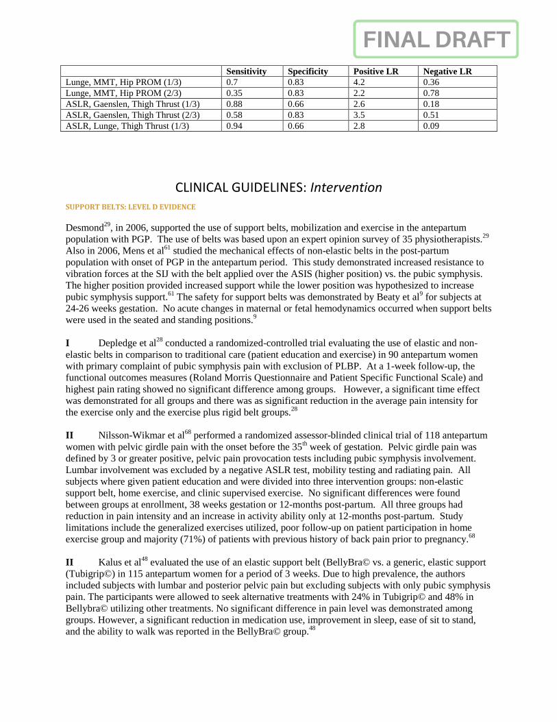
Measurement Properties Based on Test Clusters: Cook et al25
FINAL DRAFT
Sensitivity Specificity Positive LR Negative LR
Lunge, MMT, Hip PROM (1/3) 0.7 0.83 4.2 0.36
Lunge, MMT, Hip PROM (2/3) 0.35 0.83 2.2 0.78
ASLR, Gaenslen, Thigh Thrust (1/3) 0.88 0.66 2.6 0.18
ASLR, Gaenslen, Thigh Thrust (2/3) 0.58 0.83 3.5 0.51
ASLR, Lunge, Thigh Thrust (1/3) 0.94 0.66 2.8 0.09
CLINICAL GUIDELINES: Intervention SUPPORT BELTS: LEVEL D EVIDENCE
Desmond29
, in 2006, supported the use of support belts, mobilization and exercise in the antepartum
population with PGP. The use of belts was based upon an expert opinion survey of 35 physiotherapists.29
Also in 2006, Mens et al61
studied the mechanical effects of non-elastic belts in the post-partum
population with onset of PGP in the antepartum period. This study demonstrated increased resistance to
vibration forces at the SIJ with the belt applied over the ASIS (higher position) vs. the pubic symphysis.
The higher position provided increased support while the lower position was hypothesized to increase
pubic symphysis support.61
The safety for support belts was demonstrated by Beaty et al9 for subjects at
24-26 weeks gestation. No acute changes in maternal or fetal hemodynamics occurred when support belts
were used in the seated and standing positions.9
I Depledge et al28
conducted a randomized-controlled trial evaluating the use of elastic and non-
elastic belts in comparison to traditional care (patient education and exercise) in 90 antepartum women
with primary complaint of pubic symphysis pain with exclusion of PLBP. At a 1-week follow-up, the
functional outcomes measures (Roland Morris Questionnaire and Patient Specific Functional Scale) and
highest pain rating showed no significant difference among groups. However, a significant time effect
was demonstrated for all groups and there was as significant reduction in the average pain intensity for
the exercise only and the exercise plus rigid belt groups.28
II Nilsson-Wikmar et al68
performed a randomized assessor-blinded clinical trial of 118 antepartum
women with pelvic girdle pain with the onset before the 35th week of gestation. Pelvic girdle pain was
defined by 3 or greater positive, pelvic pain provocation tests including pubic symphysis involvement.
Lumbar involvement was excluded by a negative ASLR test, mobility testing and radiating pain. All
subjects where given patient education and were divided into three intervention groups: non-elastic
support belt, home exercise, and clinic supervised exercise. No significant differences were found
between groups at enrollment, 38 weeks gestation or 12-months post-partum. All three groups had
reduction in pain intensity and an increase in activity ability only at 12-months post-partum. Study
limitations include the generalized exercises utilized, poor follow-up on patient participation in home
exercise group and majority (71%) of patients with previous history of back pain prior to pregnancy.68
II Kalus et al48
evaluated the use of an elastic support belt (BellyBra© vs. a generic, elastic support
(Tubigrip©) in 115 antepartum women for a period of 3 weeks. Due to high prevalence, the authors
included subjects with lumbar and posterior pelvic pain but excluding subjects with only pubic symphysis
pain. The participants were allowed to seek alternative treatments with 24% in Tubigrip© and 48% in
Bellybra© utilizing other treatments. No significant difference in pain level was demonstrated among
groups. However, a significant reduction in medication use, improvement in sleep, ease of sit to stand,
and the ability to walk was reported in the BellyBra© group.48

FINAL DRAFT
II Carr et al23
employed a pilot study of the Loving Comfort Back Support© in 40 antepartum
females with pelvic girdle and lumbar pain. Thirty consecutive subjects were enrolled into the
intervention group, with 10 wait-list control subjects. Subjects who wore the support during waking
hours for 2 weeks demonstrated a significant reduction in the number of days/week, hours/day and overall
change in pain compared to controls.23
D Clinicians should consider the application of a support belt in the antepartum population with
PGP. The four studies reviewed investigated different patient populations, had varied intervention groups
and controls, different durations of intervention application and different timing of follow-up. Further
research is needed to clarify initial application, duration and specific antepartum PGP patient
classification for support belt intervention. (Recommendation is based on conflicting evidence.)
EXERCISE: LEVEL D EVIDENCE
The American College of Obstetrics and Gynecology (ACOG) and the Canadian Clinical Practice Guidelines has
issued guidelines for the contraindications, warning signs and recommendations for exercise in the antepartum
population.1, 2,8,26
These are summarized in the table below:
Absolute Contraindications to Exercise Relative Contraindications to Exercise
Hemodynamically significant heart disease
Restrictive lung disease
Incompetent cervix/cerclage
Multiple gestation at risk for premature labor
Persistent second- or third-trimester bleeding
Placenta previa after 26 weeks of gestation
Premature labor during the current pregnancy
Ruptured membranes
Preeclampsia/pregnancy-induced hypertension
Severe anemia
Unevaluated maternal cardiac arrhythmia
Chronic bronchitis
Poorly controlled Type 1 diabetes
Extreme morbid obesity
Extreme underweight (BMI <12)
History of extremely sedentary lifestyle
Intrauterine growth restriction in current pregnancy
Poorly controlled hypertension, seizure disorder or
hyperthyroidism
Orthopedic limitations
Heavy smoker
Warning Signs to Stop Exercise & Consult MD Contraindications During Exercise
Vaginal bleeding
Dizziness or feeling faint
Increased Shortness of Breath
Chest pain
Headache
Muscle weakness
Calf pain or swelling
Uterine contractions
Decreased fetal movement
Fluid leaking from the vagina
Supine Position (relative obstruction of venous
return and therefore decreases cardiac output)
Prolonged Static Standing (decrease in cardiac
output)
Increased basal metabolic rate (heat production)
above non-pregnant levels (increased maternal core
temperature above 1.5c (first 45-60 days gestation)
Avoid activities with high fall risk, abdominal
trauma, potential contact sport and scuba
(decompression sickness)
Recommendations During Exercise Exercise Safety
Heart rate monitoring (difficult during
pregnancy – due to blunted heart rate)
Borg Rate of Perceived Exertion Scale17
Hydration (to keep blood volume up) critical
for heat balance
Energy cost (considered for balancing intensity
Exercise does not cause minimal to no changes on
uterine activity during the final 8 weeks of
pregnancy
Fetal Implications (no evidence): no effect
transplacental transport of oxygen, carbon dioxide,
and nutrients, birth weight, premature labor

FINAL DRAFT
and duration of activity)
Exercise Prescription: include elements to
improve cardiovascular and musculoskeletal
function (American College of Sorts Medicine:
same as non-pregnant in frequency – at least 30
minutes/day)
All without contraindications should be
encouraged to aerobic and strength-training
exercise with reasonable goals.
Water exercise (redistribution of extravascular
fluid into vascular space)
No increased risk of adverse pregnancy or fetal
outcomes.
I Boissonnault et al17
performed a systematic review of exercise intervention on PLBP and PGP in
the antepartum population. Of the 11 studies reviewed, 3 were determined good quality (7-8/10), 6
moderate quality (4-6/10), and 2 poor quality (0-3/10) by the PEDro scale. The heterogeneity of
methodology, patient inclusion criteria, specific exercise protocols, intervention parameters and varied
outcomes measures did not allow for a meta-analysis to be performed.17
Of the 3 good-quality studies, only Elden et al33
conducted a study of the management of PGP in
antepartum women, at the time of enrollment. Subjects were randomized into 3 groups: standard care
(advice, patient education and support belt), exercise group (including standard care) and acupuncture
(including standard care). Exercises included stabilization of the back and pelvis and stretching of hip
external rotators and extensors. The acupuncture group experienced less pain than the exercise group and
they both experienced less than the standard care group.33
The other good-quality studies, Morkved et al40
and Garshasbi and Faghih Zadeh66
, studied
healthy nulliparous women and focused on exercise intervention in order to prevent ‘low back pain’
without distinguishing between lumbar and pelvic girdle pain. Both studies reported less pain the
exercise group compared to controls.40, 66
The authors reported, based on the good-quality studies, support for the intervention of exercise,
either alone or combined with advice, patient education and support belts for the prevention or treatment
of PLBP and PGP.
II In contrast, Lillos and Young56
,performed a systematic review to examine the specific exercise
interventions of core stabilization and lower extremity strengthen in PLBP and PGP.56
Of the 7 studies
reviewed, 5 were included in the Boissonnault et al16
review with 2 of the articles considered good
quality.16,32,40
One article related exercise to generalized, pregnancy-related discomfort and the final
article compared an education program including exercise to a control group.47,87
Based on the included
literature, the authors found no conclusive evidence to support exercise as a standard treatment for PLBP
and PGP.55
I Eggen et al32
investigated the reduction of severity and prevalence of PLBP and PGP via RCT of
supervised group exercise vs. a control group. Healthy subjects (n=257) were enrolled before the 20th-
week of gestation with 18% reporting PGP and 29% reporting PLBP at baseline. Half the subjects were
provided supervised group exercise intervention including 16-20 weeks of 1 time/week group exercise,
home exercise program and ergonomic advice, while the others were followed through routine obstetric

FINAL DRAFT
care. Exercises included aerobic activity; localized back and pelvic exercise, and global strengthening.
Interventions were not differentiated for subjects base on the presence or type of pain. No effect on
severity or prevalence was demonstrated by the exercise intervention in PLBP or PGP.32
I Kluge et al53
investigated the benefit of exercise on pain intensity and functional ability in a RCT
of antepartum women with PLBP, PGP or combination, based on a pain diagram. The intervention group
(n=26) underwent a 10-week progressive exercise program including group training, a home exercise
program and education using a posture and ergonomics brochure. The control group (n=24) only
received the posture and ergonomics brochure. Exercises included stretching, relaxation, breathing and
isometric pelvic stabilization with progressive exercise to include co-activation with gluteals, hip
abductors and quadriceps. While the authors reported low compliance with the exercise intervention, the
exercise group demonstrated a significant reduction in pain intensity as well as a significant difference
between groups for pain and functional ability following the intervention. The control group remained
relatively unchanged regarding pain and functional ability during the intervention period.53
D Clinicians should consider the use of exercise in the antepartum population with PGP. ACOG
and the Canadian CPG have recommended exercise for health benefits because of the low risk and
minimal adverse effects for the antepartum population. The two systematic reviews as well as the recent
RCTs were non-specific in the application of exercise to heterogeneous groups of PLBP and PGP. The
populations varied in early and late pregnancy and demonstrated a variety of exercise interventions. No
study based the exercise intervention on the classification of PGP proposed by Albert et al4, and Cook et
al.25
(Recommendation is based on conflicting evidence.)
MANUAL THERAPY: LEVEL C EVIDENCE
Introduction: Manual therapy in physical therapy can consist of joint manipulation (defined as high
velocity low amplitude force delivered to a joint), and joint mobilization (low velocity passive movement
techniques with the joint’s normal range of motion.) Manual therapy can also include soft tissue
mobilization/manipulation, myofascial release, muscle energy and muscle assisted range of motion.
In the general population, severe adverse effects of joint manipulation to the spine are rare especially
related to the lumbar spine.24, 82, 92
In 2002, Whitman delivered an expert opinion that, based upon support
by numerous articles in the general population, the use of manipulation for acute musculoskeletal
disorders in the antepartum population should be considered to restore normal movement in the lumbar
spine and/or pelvis. There is little to no evidence that spinal manipulation and/or mobilization is harmful
to the antepartum female or the fetus. Normal movement in all directions is advocated despite
hypermobility or laxity in one or more directions.103
III In 2009, Khorsan et al51
published a systematic review on Manipulative Therapy for Pregnancy
and Related Conditions. The review was conducted to evaluate the evidence on treatment effects of
spinal manipulation therapy and/or joint mobilization for back pain, pelvic girdle pain and other related
symptoms during pregnancy. Thirteen articles were included in the review with three studies formally
reporting no adverse effects and two studies reporting contraindications while the rest of the studies did
not include any report of adverse effects. Within the review, low evidence case series and reviews
investigated the relationship of PLBP/PGP and the use of manipulation or mobilization. The side posture

FINAL DRAFT
manipulation was reported with greater frequency and a rotational manipulation was described in one
article. Of these articles, all of the subjects had relief of symptoms with some studies showing 70-91%
relief. Three case reports noted a reduction of pain by the subjects. The authors concluded that expert
opinion exists within the literature that, the relative safety of spinal manipulation and/or mobilization in
the general population exists. This intervention could be considered in the antepartum population for
those without complications within the pregnancy.51
III In a retrospective case series, Lisi57
reported on spinal manipulation in the treatment of PLBP and
PGP. Spinal manipulation was aimed at the lumbar facets and the SI joints. Other interventions were
described as manual mobilization and manual myofascial release. Seventeen cases were reviewed an
average decrease of 5.9 to 1.5 using the numerical pain rating scale. Sixteen cases reported clinical
important improvement based on pain intensity with 2-4 days following two interventions. No adverse
effects were reported in any of the cases.57
I Licciardone et al55
conducted a randomized placebo controlled trial to observe the effects of
osteopathic manipulation therapy vs. sham ultrasound vs. no treatment on antepartum patients with PLBP
and PGP. 127 subjects between the 28th and 30
th week of gestation were entered into the study and
divided into one of three groups: control, sham ultrasound or osteopathic manipulative therapy. The
groups were stratified based on age and gravida. Both intervention groups received treatments for 7 visits
over 9 weeks. Manipulation therapy included soft tissue mobilization, myofascial release, muscle energy
and range of motion mobilization. The osteopath interventionists determined regions of the body to be
treated from the cervical spine to the sacrum. High velocity low amplitude manipulation was not used as
the authors felt a “theoretical risk” was posed due to increasing ligamentous laxity in the antepartum
population. No significant difference were found between groups for level of pain at the end of the
treatment period. The manipulative therapy group demonstrated significantly less deterioration in back
specific function. The authors concluded that the manipulative therapy techniques may not have had a
significant impact on pain, but did lessen or slow down the deterioration of back specific function.55
C Clinicians may or may not utilize manual therapy techniques including high velocity low
amplitude manipulations for the treatment of PLBP and PGP. This evidence is emerging and treatment
could be considered, as there is little to no reported evidence of adverse effects in the healthy antepartum
population. (Recommendations are based upon weak evidence.)
RECOMMENDATIONS
Risk Factors: A
Clinicians should utilize the following risk factors: prior history of pregnancy, orthopedic
dysfunctions, increased BMI, smoking as well as work dissatisfaction and a lack of belief of improvement
in the prognosis of PGP. (Recommendation based on strong evidence)
Postural Changes: B
Clinicians should not consider postural changes as indicative of the development and/or intensity
of PGP in the antepartum population. (Recommendation based on moderate evidence)

FINAL DRAFT
Clinical Course: A/B
Clinicians should (consider) treat patients with early onset, multiple pain locations, a high number
of positive pelvic pain provocation tests, work dissatisfaction and lack of belief of improvement, as these
are strong/moderate factors in determining the potential for persisting PGP in late pregnancy and post-
partum. (Recommendations based on strong/moderate evidence)
Diagnosis/Classification: B
Clinicians may consider the utilization of the classification system for the diagnosis of the type of
pelvic girdle pain in antepartum population. (Recommendation is based on moderate evidence)
Differential Diagnosis: A
PGP, in this population, should be differentiated from signs and symptoms of serious disease and
psychological factors when the symptoms are not associated with the described clinical course of PGP,
impairments are failing to normalize and the symptoms are worsening with increased disability. This
should include the presence of transient osteoporosis and diastasis rectus abdominus as possible co-
morbidities in this population as well as the presence of pelvic floor muscle, hip and lumbar spine
dysfunction. (Recommendations are based upon strong evidence)
Imaging Studies: F
In the absence of good evidence, expert opinion and foundation science may be used to guide
examination with the use of imaging studies.
Examination-Outcome Measures: A
Clinicians should administer self-reported outcome questionnaires such as Disability Rating
Index, Oswestry Disability Index, Pelvic Girdle Questionnaire, Fear-Avoidance Belief Questionnaire, and
Pain Catastrophizing Scale. These are scales are practical for the determination of baseline disability,
function and pain belief as well as change throughout the clinical course. These should be utilized in
combination with clinical examination for clinical decision. (Recommendations are based on strong
evidence.)
Examination-Activity Limitation and Participation Restriction Measures: E
While strong evidence exists to support a high risk of falls, no measures have been validated to
objectively assess the dynamic balance and fall risk in antepartum population. (Recommendation is based
on theoretical/foundational evidence)
Intervention- Support Belts: D
Clinicians should consider the application of a support belt in the antepartum population with
PGP. The four studies reviewed investigated different patient populations, had varied intervention groups
and controls, different durations of intervention application and different timing of follow-up. Further
research is needed to clarify initial application, duration and specific antepartum PGP patient
classification for support belt intervention. (Recommendation is based on conflicting evidence.)

FINAL DRAFT
Intervention-Exercise: D
Clinicians should consider the use of exercise in the antepartum population with PGP. ACOG
and the Canadian CPG have recommended exercise for health benefits because of the low risk and
minimal adverse effects for the antepartum population. The two systematic reviews as well as the recent
RCTs were non-specific in the application of exercise to heterogeneous groups of Pregnancy Low Back
Pain (PLBP) and PGP. The populations varied in early and late pregnancy and demonstrated a variety of
exercise interventions. No study based the exercise intervention on the classification of PGP proposed by
Albert, et al4 and Cook, et al.
23 (Recommendation is based on conflicting evidence.)
Intervention- Manual Therapy: C
Clinicians may or may not utilize manual therapy techniques including high velocity low
amplitude manipulations for the treatment of PBLP and PGP. This evidence is emerging and treatment
could be considered, as there is little to no reported evidence of adverse effects in the healthy antepartum
population. (Recommendations are based upon weak evidence.)
AUTHOR/REVIEWER AFFILIATIONS AND CONTACTS
Susan C. Clinton PT, DScPT, COMT, OCS, WCS
Embody Physiotherapy & Wellness, LLC
University of Pittsburgh – Department of Physical Therapy
Chatham University – Department of Physical Therapy
Alaina Newell PT, DPT, WCS, CLT-LANA
Oncology Rehab
Patricia A. Downey, PT, PhD, DPT
Chatham University – Department of Physical Therapy - Chair
Kimberly Ferreira, PT, MSPT, PhD(c)
Andrews University – Department of Physical Therapy – Entry Level Chair
REFERENCES
1 ACOG. Exercise during pregnancy (FAQ 0119). In ACOG, ed www.acog.org Aug 2011.
2 ACOG. Exercise during pregnancy and the postpartum period. Obstet Gynecol. 2002; 99(1):171-3.

FINAL DRAFT
3 ACOG. Guidelines for diagnostic imaging during pregnancy. In: ACOG, ed www.acog.org 2004,
2009.
4 Albert HB, Godskesen M, Westergaard JG. Evaluation of clinical tests used in classification
procedures in pregnancy-related pelvic joint pain. European Spine Journal. 2000;9:161-166.
5 Albert HB, Godskesen M, Westergaard JG. Incidence of four syndromes of pregnancy-related
pelvic joint pain. Spine. 2002;27:2831-2834.
6 Albert HB, Godskesen M, Westergaard JG. Prognosis in four syndromes of pregnancy-related
pelvic pain. Acta Obstet Gynecol Scand 2001; 80:505-510.
7 Albert HB, Godskesen M, Korsholm L, Westergaard JG. Risk factors in developing pregnancy-
related pelvic girdle pain. Acta Obstet Gynecol Scand. 2006;85(5):539-544.
8 Artal R, O'Toole M. Guidelines of the American College of Obstetricians and Gynecologists for
Exercise During Pregnancy and the Postpartum Period. British journal of sports medicine, 2003;37(1):6-
12.
9 Beaty CM, Bhaktaram VJ, Rayburn WF, Parker MJ, Christensen HD, Chandrasekaran K. Low
Backache During Pregnancy. Acute Hemodynamic Effects of a Lumbar Support. J Reprod Med.
1999;44(12):1007-11.
10 Becker I, Woodley SJ, Stringer MD. The adult human pubic symphysis: a systematic review. J of
Anatomy. 2010;217(5):475-487.
11 Bergbom S, Boersma K, Overmeer T, Linton SJ. Relationship among pain catastrophizing,
depressed mood, and outcomes across physical therapy treatments. Phys Ther. 2011;91(5):754-764.
12 Bewyer KJ, Bewyer DC, Messenger D, et al. Pilot data: association between gluteus medius
weakness and low back pain during pregnancy. Iowa Orthop J 2009; 29:97-99.
13 Biering K, Aagaard Nohr E, Olsen J, Hjollund NH, Nybo Andersen AM, Juhl M. Smoking and
pregnancy-related pelvic pain. BJOG: An Inter J of Obstet & Gynco. 2010;117(8):1019-1026
14 Bjorklund K, et al. Symphyseal distention in relation to serum relaxin levels and pelvic
pain in pregnancy. Acta obstetet gyneco Scan 2000; 79(4): 269-275.
15 Boissonnault JS, Blaschak MJ. Incidence of diastasis recti abdominus during the childbearing
year. Phys Ther 1988; 68(7): 1082-1086.
16 Boissonnault JS, Klestinski JU, Pearcy K. The Role of Exercise in the Management of Pelvic
Girdle and Low Back in Pregnancy: A Systematic Review of the Literature. J Women’s Health.
2012;36(2):69-77.
17 Boissonnault, WG. Primary care for the physical therapist: examination and triage. Elsevier
Health Sciences, 2010.

FINAL DRAFT
18 Boissonnault WG, Boissonnault JS. Transient osteoporosis of the hip associated with pregnancy.
J WH Phys Ther 2005; 29(3): 33-39.
19 Borg, G. and Linderholm, H. Perceived exertion and pulse rate during graded exercise in various
age groups. Acta Medcia Scand. 1067;181: 194-206.
20 Breen TW, Bernard JR, Philppa AG, et al. Factors associated with back pain after childbirth.
Anesthesiology 1994; 81: 29-34.
21 Brooke R. The sacro-iliac joint. J Anat 1924; 58: 299.
22 Butler EE, Colón I, Druzin ML, Rose J. Postural equilibrium during pregnancy: Decreased
stability with an increased reliance on visual cues. Amer J of Obstet and Gynec. 2006;195(4):1104-1108.
23 Carr CA. Use of a Maternity Support Binder for Relief of Pregnancy‐Related Back Pain. Journal
of Obstetric, Gynecologic, & Neonatal Nursing, 2003;32(4):495-502.
24 Cassidy JD, Boyle E, Cote , et al. Risk of Vertebrobasilar stroke and Chiropractic Care: Results
of a Population-Based Case-Control and Case-Crossover Study. Spine. 2008;33(supp 4):S176-S183.
25 Cook C, Massa L, Harm-Ernandes I, et al. Inter-rater reliability and diagnostic accuracy of pelvic
girdle pain classification. J Manipulative Phys Ther. 2007;30(4):252-258.
26 Davies GA, Wolfe LA, Mottola MF, MacKinnon C. Joint SOGC/CSEP Clinical Practice
Guideline: Exercise in Pregnancy and the Postpartum Period. Canadian Journal of Applied Physiology,
2003;28(3):329-341.
27 Delitto A, George SZ, Van Dillen L, Whitman JM, Sowa G, Shekelle P, Denniger TR, Godges
JJ. Low Back Pain: Clinical Practice Guidelines Linked to the International Classification of
Functioning, Disability, and Health from the Orthopaedic Section of the American Physical Therapy
Association. J Orthop Sports Phys Ther. 2012;42(4):A1-A57. doi:10.2519/jospt.2012.0301
28 Depledge J, McNair PJ, Keal-Smith C, and Williams M. Management of Symphysis Pubis
Dysfunction During Pregnancy Using Exercise and Pelvic Support Belts. Phys Ther. 2005;85:1290-
1300.
29 Desmond R. How Women’s Health Physiotherapists Treat Symphysis Pubis Dysfunction. J
Assoc Chartered Physio Women’s Health. 2006;99:63-71.
30 Dunken BW, Weiner DK, Chanlder J, et al. Functional Reach, a clinical measure of balance. J of
Geriontial. 1990; 45(6): M192-197.
31 Dunning K, LeMasters G, Bhattacharya A. A Major Public Health Issue: The High Incidence of
Falls During Pregnancy. Maternal and child health journal, 2010;14(5):720-725.
32 Eggen MH, Stuge B, Mowinckel P, Jensen KS, Hagen KB. Can supervised group exercises
including ergonomic advice reduce the prevalence and severity of low back pain and pelvic girdle pain in
pregnancy? A randomized controlled trial. Phys Ther. 2012;92(6):781-790.

FINAL DRAFT
33 Elden H, Ladfors L, Olsen MF, Ostgaard HC, Hagberg H. Effects of acupuncture and stabilizing
exercises as adjunct to standard treatment in pregnant women with pelvic girdle pain: randomized single
blind controlled trial. BMJ. 2005;330(7494):761.
34 Fairbank JC, Pynsent PB. The Oswestry Disability Index. Spine. 2000;25:2940–2952
35 Fast A, Sharpo D, Ducommun EJ, et al. Low back pain in pregnancy. Spine 1987; 12: 368-71.
36 Fetters L and Tilson J. Evidence Based Physical Therapy. F.A. Davis Company, Philadelphia:
2012; Appendix 162-171.
37 Franklin ME, Conner-Kerr T. An analysis of posture and back pain in the first and third trimesters
of pregnancy. JOSPT 1998; 28:133.
38 Fritz JM, George SZ, Delitto A. The Role of Fear-Avoidance Beliefs in Acute Low Back Pain:
Relationship with Current and Future Disability and Work Status. Pain. 2001;94:7-15.
39 Fritz, JM, Irrgang JJ. A Comparison of a modified Oswestry Low Back Pain Disability
Questionnaire and the Quebec Back Pain Disability Scale. Phys Ther. 2001;81:776-788.
40 Garshasbi A, Faghih Zadeh S. The effect of exercise on the intensity of low back pain in pregnant
women. Int J Gynaecol Obstet. 2005;88(3):271-275.
41 Grotle M, Garratt AM, Krogstad Jenssen H, Stuge B. Reliability and construct validity of self-
report questionnaires for patients with pelvic girdle pain. Phys Ther. 2012;92(1):111-123.
42 Guralnik JM, Simonsick EM, Ferrucci L,et al. A short physical performance battery assessing
lower extremity function: association with self-reported disability and prediction of mortality and nursing
home admission. J of Gerontology 1994;49(2): M85-94.
43 Gutke A, Ostgaard HC, Oberg B. Association between muscle function and low back pain in
relation to pregnancy. J Rehabil Med. 2008;40(4):304-311.
44 Gutke A, Ostgaard HC, Oberg B. Pelvic Girdle pain and lumbar pain in pregnancy: a cohort study
of the consequences in terms of health and functioning. Spine 2006; 31:E149-55.
45 Gutke A, Ostgaard HC, Oberg B. Predicting persistent pregnancy-related low back pain. Spine
2008; 33:E386-93.
46 Guyatt, GH. Sacket DL, Sinclair JC, Hayward R, Cook DJ, and Cook RJ. Users’ Guides to the
Medical Literature: IX. A Method for Grading Health Care Recommendations. JAMA. 1995; 274: 1800-
1804.
47 Haugland KS, Rasmussen S, Daltveit AK. Group intervention for women with pelvic girdle pain
in pregnancy. A randomized controlled trial. Acta Obstet Gynecol Scand. 2006; 85(11):1320-1326.
48 Kalus SM, Kornman LH, Quinlivan JA. Managing back pain in pregnancy using a support
garment: a randomized trial. BJOG. 2008; 115(1):68-75.

FINAL DRAFT
49 Kanakaris NK, Roberts CS, Giannoudis PV. Pregnancy-related pelvic girdle pain: an update.
BMC Medicine. 2011;9(1):15.
50 Kendall FP, McCreary EK, Provance PG, et al. Muscles: Testing and Function, with Posture and
Pain. 5th Ed. 2005.
51 Khorsan R, Hawk C, Lisi AJ, Kizhakkeveettil A. Manipulative Therapy for Pregnancy and
Related Conditions. A Systematic Review. Obstet Gynecol Survey. 2009;64(6):416-427.
52 Klenerman L, Slade PD, Stanley IM, et al. The Prediction of Chronicity in Patients with an
Acute Attack of Low Back Pain in a General Practice Setting. Spine. 1995;20:478-484.
53 Kluge J, Hall D, Louw Q, Theron G, Grove D. Specific exercises to treat pregnancy-related low
back pain in a South African population. Int J Gynaecol Obstet. 2011;113(3):187-191.
54 Law MC, MacDermid J. Evidence Based Rehabilitation: Guide to Practice. 2nd
Ed. Thorofare,
NJ: Slack incorporated; 2008
55 Licciardone JC, Buchanan S, Hensel KL, King HH, Fulda KG, Stoll ST. Osteopathic
manipulative treatment of back pain and related symptoms during pregnancy: a randomized controlled
trial. Am J Obstet Gynecol. 2010;202(1):43 e41-48.
56 Lillos S, Young J. The Effects of Core and Lower Extremity Strengthening on Pregnancy-
Related Low Back and Pelvic Girdle Pain: A Systematic Review. J Women’s Health. 2012;36(3):116-
124.
57 Lisi AJ. Chiropractic spinal manipulation for low back pain of pregnancy: a retrospective case
series. J Midwifery Women’s Health. 2006;51(1):e7-10.
58 Maki BE. Gait changes in older adults: predictors of falls or indicators of fear. J Am Geriatr Soc
1997;45(3): 313-320.
59 McCrory JL, Chambers AJ, Daftary A, Redfern MS. Dynamic postural stability during advancing
pregnancy. J Biomech. 2010;43(12):2434-2439.
60 McPoil TG, Martin RL, Cornwell MW, Wukich DK, Irrgang JJ, Godges JJ. Heel Pain- Plantar
Fasciitis: Clinical Practice Guidelines Linked to the International Classification Function, Disability, and
Health from the Orthopedic Section of the American Physical Therapy Association. J Orthop Sports Phys
Ther. 2008: 38(4); A1-A18.
61 Mens JM, Damen L, Snijders CJ, Stam HJ. The mechanical effect of a pelvic belt in patients with
pregnancy-related pelvic pain. Clin Biomech. 2006;21(2):122-127.
62 Mens JM, Huis in 't Veld YH, Pool-Goudzwaard A. Severity of signs and symptoms in
lumbopelvic pain during pregnancy. Man Ther. 2012;17(2):175-179.
63 Mens JMA, Pool-Goudzwaard A, Stem HJ. Mobility of the pelvic joint in pregnancy-related
lumbopelvic pain: a systematic review. Obstet Gynecol Survey 2009; 64: 200-208.

FINAL DRAFT
64 Mogren IM. Previous physical activity decreases the risk of low back pain and pelvic pain during
pregnancy. Scand J Public Health 2005; 33: 300-306.
65 Møller UK, Streym S, Mosekilde L, Rejnmark L. Changes in bone mineral density and body
composition during pregnancy and postpartum. A controlled cohort study. Osteoporosis International.
2011;23(4):1213-1223.
66 Morkved S, Salvesen KA, Schei B, Lydersen S, Bo K. Does group training during pregnancy
prevent lumbopelvic pain? A randomized clinical trial. Acta Obstet Gynecol Scand. 2007;86(3):276-282.
67 Myers SS, Phillips RS, Davis RB, et al. Patient Expectations as Predictors of Outcome In Patients
with Acute Low Back Pain. J of Gen Internal Med. 2007;23(2):148-153.
68 Nilsson-Wikmar L, Holm K, Öijerstedt R, Harms-Ringdahl K. (2005). Effect of Three Different
Physical Therapy Treatments on Pain and Activity in Pregnant Women with Pelvic Girdle Pain: a
Randomized Clinical Trial with 3, 6, and 12 Months Follow-up Postpartum. Spine, 2005;30(8):850-856.
69 Noren L, Ostgaard S, Johansson G, Ostgaard HC. Lumbar back and posterior pelvic pain during
pregnancy: a 3-year follow-up. Eur Spine J. 2002;11(3):267-271.
70 Oliveri B, Parisi MS, Zeni S, Mautalen C. Mineral and bone mass changes during pregnancy and
lactation. Nutrition. 2004;20(2):235-240.
71 Olsson C and Nilsson-Wikmar L. Health-related quality of life and physical ability among
pregnant women with and without back pain in late pregnancy. Acta Obstet Gynecol Scand 2004; 83:
351-357.
72 Ostgaard HC. Back pain in pregnancy. Acta Orthop Scand 1992; 63(suppl 247): 61.
73 Ostgaard HC, Anderson GBJ. Previous back pain and risk of developing back pain in a future
pregnancy. Spine 1991; 16:432-436.
74 Ostgaard HC, Zentherstrong G, Roos-Hansson E. Back pain in relation to pregnancy: a six-year
follow up. Spine 1997; 22(24): 2945-2950.
75 Ostgaard HC, Roos-Hansson E, Zetherstrong G. Regression of back and posterior pelvic pain
after pregnancy. Spine 1996; 21(23): 2777-2780.
76 Petersen LK, Hvidman L and Uldbjerg N. Normal serum relaxin in women with disabling
pelvic pain during pregnancy. Gynec & obstetric invest 1994; 38(1): 21-23.
77 Phillips B, Ball C, Sackett D, et al. Centre for Evidence-based Medicine Levels of Evidence
Table. Available at: http://www.cebm.net/index.aspx?o=1025 Accessed 7/5/2011, 2009.
78 Ranney B. Diastasis recti and umbilical hernia causes, recognition and repair. S Dakota J Med
1990; 43(10): 5-8.

FINAL DRAFT
79 Robinson HS, Mengshoel AM, Bjelland EK, Vollestad NK. Pelvic girdle pain, clinical tests and
disability in late pregnancy. Man Ther. 2010;15(3):280-285.
80 Robinson HS, Mengshoel AM, Veierod MB, Vollestad N. Pelvic girdle pain: potential risk
factors in pregnancy in relation to disability and pain intensity three months postpartum. Man Ther.
2010;15(6):522-528.
81 Rost CCM, Jacqueline J, Kaiser A, et al. Pelvic pain during pregnancy: a descriptive study of
signs and symptoms of 870 patients in primary care. Spine 2004;29: 2567-72.
82 Rost C, Jacqueline J, Kaiser A, et al. Prognosis of women with pelvic pain during pregnancy: a
long-term follow-up study. Acta Obstet Gynecol Scand 2006; 85: 771-77.
83 Rubinstein SM, Leboeuf_Yde C, Knol DL, et al. Predictors of Adverse Events Following
Chiropractic Care for Patients with Neck Pain. J Manipulative Physio Ther. 2008;31:94-103.
84 Salen BA, Nygren EVS, Nordemar R. The disability rating index: An instrument for the
assessment of disability in clinical settings. J of Clinical Epidem. 1994; 47(12): 1423-1435.
85 Sieben JM, Vlaeyen JW, Tuerlinck S, Portegijs PJ. Pain-Related Fear in Acute Low Back Pain:
The First Two Weeks of a New Episode. Eur J Pain. 2002;6:229-237.
86 Sipko T, Grygier D, Barczyk K, Eliasz G. The Occurrence of Strain Symptoms in the
Lumbosacral Region and Pelvis During Pregnancy and After Childbirth. J of Manip and Phy Ther.
2010;33(5):370-377.
87 Smith SA, Michel Y. A Pilot Study on the Effects of Aquatic Exercises on Discomforts of
Pregnancy. J Obstet Gynecol Neonatal Nurs. 2006;35(3):315-323.
88 Spitznagle TM, Leong FC, van Dillen LR. Prevalence of diastasis recti abdominis in a
urogynogecological population. Inter Urogyencol J 1997; 18(3):321-328.
89 Stomp-van den Berg SGM, Hendriksen IJM, Bruinvels DJ, Twisk JWR, van Mechelen W, van
Poppel MNM. Predictors for postpartum pelvic girdle pain in working women: The Mom@Work cohort
study. Pain. 2012;153(12):2370-2379.
90 Stuge B, Garratt A, Krogstad Jenssen H, Grotle M. The pelvic girdle questionnaire: a condition-
specific instrument for assessing activity limitations and symptoms in people with pelvic girdle pain. Phys
Ther. 2011;91(7):1096-1108.
91 Sullivan MJL, Bishop SR, Pivik J. The Pain catastrophizing scale: development and validation.
Psychol Assess. 1995;7: 524–532.
92 Thiel HW, Bolton JE, Docherty S, et al. Safety of Chiropractic Manipulation of the Cervical
Spine: a Prospective National Survey. Spine. 2007;3223:75-2378; discussion 9
93 Tibor LM, Sekiya JK. Differential diagnosis of pain around the hip joint. Arthroscopy.
2008;24(12):1407-1421.

FINAL DRAFT
94 To WW, Wong MW. Factors associated with back pain symptoms in pregnancy and the
persistence of pain 2 years after pregnancy. Acta Obstet Gynecol Scand 2003; 82:1086-91.
95 To WW, Wong MW. Persistence of back pain symptoms after pregnancy and bone mineral
density changes as measured by quantitative ultrasound--a two year longitudinal follow up study. BMC
Musculoskelet Disord. 2011;12:55.
96 Turgut F, Turgut M, Setinsahin M. A prospective study of persistent back pain after pregnancy.
Euro J Obstet Gynecol Reprod Bio 1998; 80: 45-48.
97 van Tulder M, Becker A, Bekerring T, et al. European guidelines on the management of acute
nonspecific low back pain in primary care [European Commission Research Directorate General Web
site]. 2004. Available at: http://www.backpaineurope.org/web/files/WG1_Guidelines.pdf (accessed
August 13, 2013).
98 Vermani E, Mittal R, Weeks A. Pelvic Girdle Pain and Low Back Pain in Pregnancy: A Review.
Pain Practice. 2010;10(1):60-71.
99 Vleeming A, Albert HB, Ostgaard HC, Sturesson B, Stuge B. European guidelines for the
diagnosis and treatment of pelvic girdle pain. Eur Spine J. 2008;17(6):794-819.
100 Vleeming A, Stoechart R, Volkers ACW, et al. Relationship between form and function in the
sacroiliac joint: part 1: clinical anatomical aspects. Spine 1990; 15:130-32.
101 Vøllestad NK, Stuge B. Prognostic factors for recovery from postpartum pelvic girdle pain. Eur
Spine J. 2009;18(5):718-726.
102 Waddell G, Newton M, Henderson I, Somerville D, Main CJ. A fear-avoidance beliefs
questionnaire (FABQ) and the role of fear-avoidance beliefs in chronic low back pain and disability. Pain
1993;52(2): 157-168.
103 Whitman JM. Pregnancy, Low Back Pain and Manual Physical Therapy Interventions (Guest
Editorial). J Orthop Sports Phys Ther. 2002;32(7):314-317.
104 World Health Organization. International classification of functioning disability and health (ICF).
Geneva, Switzerland: World Health Organization; 2001
105 World Health Organization. ICD-10: International Statistical Classification of Diseases and
Related Health Problems: Tenth Revision. Geneva, Switzerland: World Health Organization; 2005.
106 Wu WH, Meijer OG, Bruijn SM, et al. Gait in Pregnancy-related Pelvic girdle Pain: amplitudes,
timing, and coordination of horizontal trunk rotations. Eur Spine J. 2008;17(9):1160-1169.
107 Wu WH, Meijer OG, Uegaki K, et al. Pregnancy-related pelvic girdle pain (PPP), I: Terminology,
clinical presentation, and prevalence. Eur Spine J. 2004;13(7):575-589.

FINAL DRAFT
108 Wu W, Meijer OG, Jutte PC, Uegaki K, Lamoth CJ, Sander de Wolf G, et al. Gait in patients with
pregnancy-related pain in the pelvis: an emphasis on the coordination of transverse pelvic and thoracic
rotations. Clinical biomechanics. 2002;17: 678-686.