Embed Size (px)
Citation preview


Pelvic tumor in childhood Classification, imaging approach and
radiological findings
M. Mearadji
International Foundation for
Pediatric Imaging Aid
Rotterdam, The Netherlands
Solid pelvic masses in childhood composed from a group of rare and heterogeneous tumors originating from different pelvic organs or structure.
Principally the pelvic mass should be categorized in three groups:
I. Rhabdomyosarcoma
II. Germ cell tumors
III. Other rare and incidental tumors localized in the pelvic region, also to be found anywhere in the body with different origin and histology

• Following earlier report the pelvic tumors are
strongly related to the compartmental location.
• Anterior midline mostly is the location for
rhabdomyosarcoma (RMS).
• Middle midline is the location for a germ cell
tumor as well as a RMS.
• Posterior midline frequently is the origin of a
neurogenic or a germ cell tumor.
• Lateral pelvic region other soft tissue and bone
tumors.
Patient material
The patient material includes 90 cases of
pelvic and testicular tumors in male and
female. The anatomic location, the value
and limitation of performed different
imaging procedures were retrospectively
analyzed.

I. Rhabdomyosarcoma (RMS) • Tumor will arise from primitive cell in any organ.
• Represents 5% - 10% of malignant solid tumor in childhood
• Ranking 4th in frequency after:
– CNS
– Neuroblastoma
– Wilms tumor
• Bimodial presentation, primary peak 2-5 years of age, secondary 12-16 years of age.
• Site classification following intergroup RMS study:
– Head and Neck (35 %)
– Genitortinary system (26 %)
– Extremities (19 %)
– Other bodyparts (20 %)
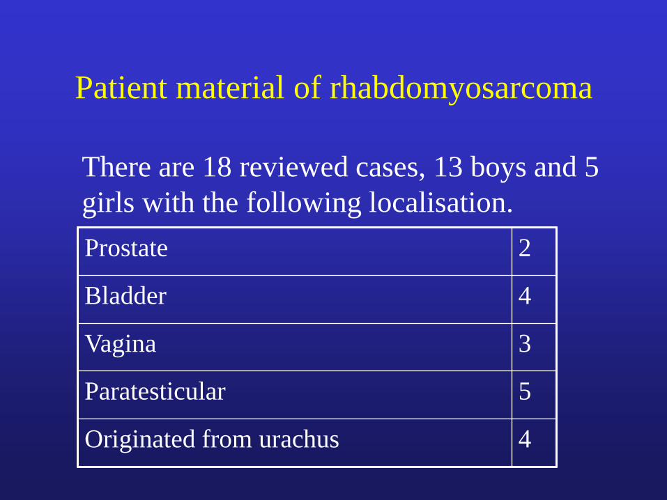
Patient material of rhabdomyosarcoma
There are 18 reviewed cases, 13 boys and 5
girls with the following localisation.
Prostate 2
Bladder 4
Vagina 3
Paratesticular 5
Originated from urachus 4

Imaging approaches of RMS
• Conventional as excretory urogram and micturation cystourethrogram as well as angiography in 4 cases (from more than 20 years ago).
• Later ultrasound routinly used as first diagnostic procedure.
• Use of CT or MRI or both in all remaining cases.

RMS of the prostate
RMS of the prostate grows rapidly and extend outside the
capsule invading the bladder and posterior urethra. This
tumor mostly is histologically botroid of type.
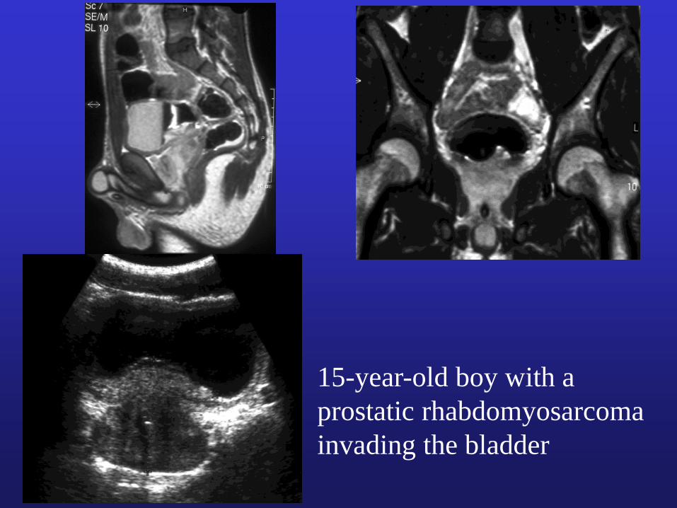
15-year-old boy with a
prostatic rhabdomyosarcoma
invading the bladder
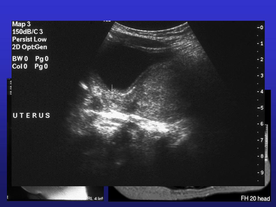
RMS of the vagina
Most RMS of the vagina arise in the anterior wall of the
vagina adjacent to the cervix.

Paratesticular RMS
The RMS of the testis are histological embryonic of aspect
with intermediate prognosis originated of spermatic cord.
The patients are less than 5 years of age.
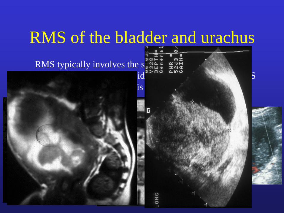
RMS of the bladder and urachus
RMS typically involves the submucousal region of the
trigon, histological is botroid with a good prognosis. RMS
in the dome of the bladder is mostly urachal of origin.
II. Germ cell tumors in children
Germ cell tumors in children are rare entities
contributing more than 3% of all pediatric cancer,
frequently they encountered in the gonads, but
they also are located incidentally in other regions
such as in pineal gland, retroperitoneum as well as
the sacral area.
Histological classification of the germ cell tumors
is complex, principally they can be categorized as
shown in following table.

Germ cell
Unipotential
Germinoma
Seminoma
Dysgerminoma
Gonadoblastoma
Multipotential
Teratoma Embryonal
carcinoma
Benigne
And
Mature
Benigne
And
Immature
Sacrococcygeal
Teratoma
Yolk Sac
tumor Mixed
Chorio
carcinoma

Patient material germ cell tumor (60)
Girls 48 Boys 12
Mature and immature
ovarian teratoma
15 Mature teratoma of the
testis
3
Sacrococcygeaal
teratoma
33
Sacrococcygeal
teratoma
7
Yolk Sac tumor
(testicular)
1
Non seminoma 1
Ovarian teratoma
• Cystic teratoma is by far the most common tumor,
accounting for more than 90 % benign ovarian neoplasms.
On the basis histological examination teratomas are
classified as mature (90 %), immature (containing
embryonic neural elements) and malignant.
• The sonographic appearance of teratomas is variable
depending on relative amount of sebum, serous fluid,
calcium, hair and fat.
• The tissue characteristic, location and extension of
teratomas could be easily demonstrated by MRI and CT.
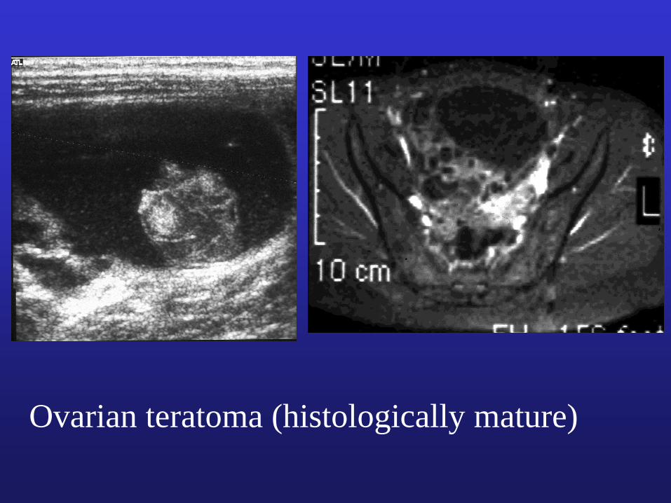
Ovarian teratoma (histologically mature)

Ovarian teratoma
(histologically
immature with
dissemination of glia
tissue)

Primary testicular germ cell tumor
• Teratoma is the principal benign germ cell tumor of the testis.
• Affected boys are usually younger than 4 years of age (85 %).
• Teratomas on later life tend to be more aggressive and malignant.
• Yolk Sac carcinoma, teratocarcinoma and choriocarcinomas are all the malignant type of the testicular germ cell tumor.
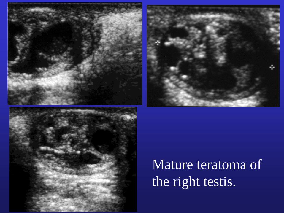
Mature teratoma of
the right testis.
Sacrococcygeal teratoma
• Sacrococcygeal teratoma represents about 40 % of
the germ cell tumors.
• Females are more affected than males (4:1).
• The frequency of malignancy depends on age:
7 % (girls) and 10 % (boys) in infants younger than 2
months of age.
47 % (girls) and 66 % (boys) on later life.
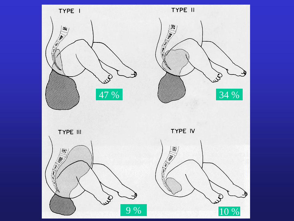
47 % 34 %
9 % 10 %
Sacrococcygeal teratoma
• Imaging appearance of the tumor is variable
depending on the relative amounts of soft tissue
and cystic components.
• Sonography can provide useful information
regarding the internal characteristics of the
sacrococcygeal teratomas.
• MRI is the first modality of choice to determine
the total extent of the mass.


Newborn baby with a
large sacrococcygeal
teratoma type I

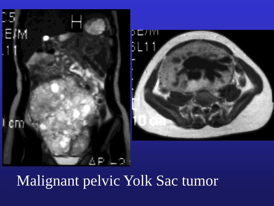
Malignant pelvic Yolk Sac tumor

III. Miscellaneous
• Pelvic masses categorized in this group have no relation to RMS or germ cell tumors, neither histologically nor pathogenetically.
• Some of them however originate from the genital system (4 cases).
• They could be osteogenic or related to pelvic soft tissue (5 cases).
• Neurogenic and lymphatic neoplasms are a subgroup under this category of pelvic masses (3 cases).
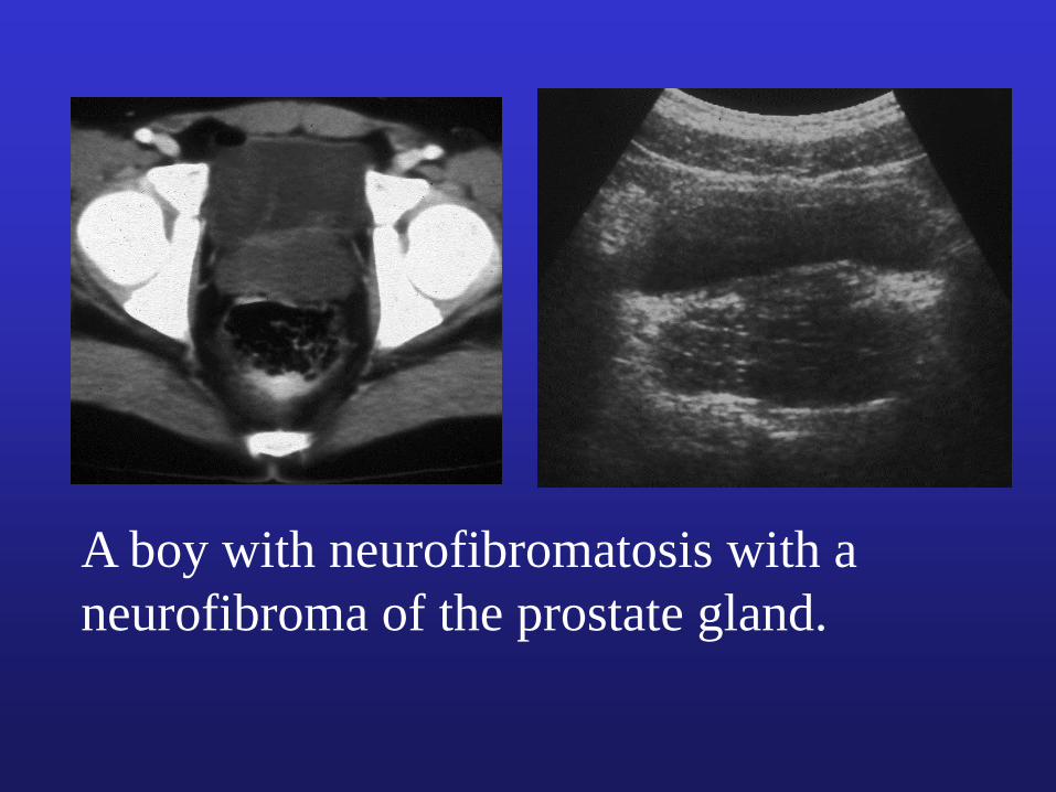
A boy with neurofibromatosis with a
neurofibroma of the prostate gland.

Pelvic Ewing sarcoma
right-sided.
Note the huge soft tissue
Pelvic osteosarcoma
right-sided.
Note the massive
destruction of the ileum

Malignant
fibrohistiocytoma

Primary malignant pelvic lymphoma

Conclusions
• Dividing the pelvic space into three midline compartments
(anterior, middle and posterior) as well as lateral and
pelvic floor is useful to define the tumor type.
• Rhabdomyosarcoma of the lower urogenital system is
more frequent in male patients (ratio 2:1) and originates
from epididymis, prostate bladder and urachus.
• Sacrococcygeal teratoma is the most common germ cell
tumor in girls with an increasing malignancy of ± 10 % in
neonates and up to 66 % in later life.

Conclusions
• Sonography should be considered a sufficient modality in
recognition of scrotal mass. Additional contribution of
other modalities is not relevant.
• Sonography is a sufficient imaging modality in neonatal
sacrococcygeal teratoma type I and cystic abdominal
teratoma on later life if little or no soft tissue is detected.
• Sonography is only an initial procedure for evaluation of
pelvic malignancy, MRI more than CT is required for
recognition of tumoral extension and extrapelvic
metastasis.
• CT and MRI are imaging modalities of choice in diagnosis
of all masses located around close to the urogenital system
ostegenic, or non-osteogenic.