Embed Size (px)
Citation preview

PEPTIC ULCER DISEASE
Hannah Vawda FY1
Objectives
Definition of peptic ulcer Comparison of duodenal & gastric ulcers Aetiology Clinical presentation Management Emergency scenario
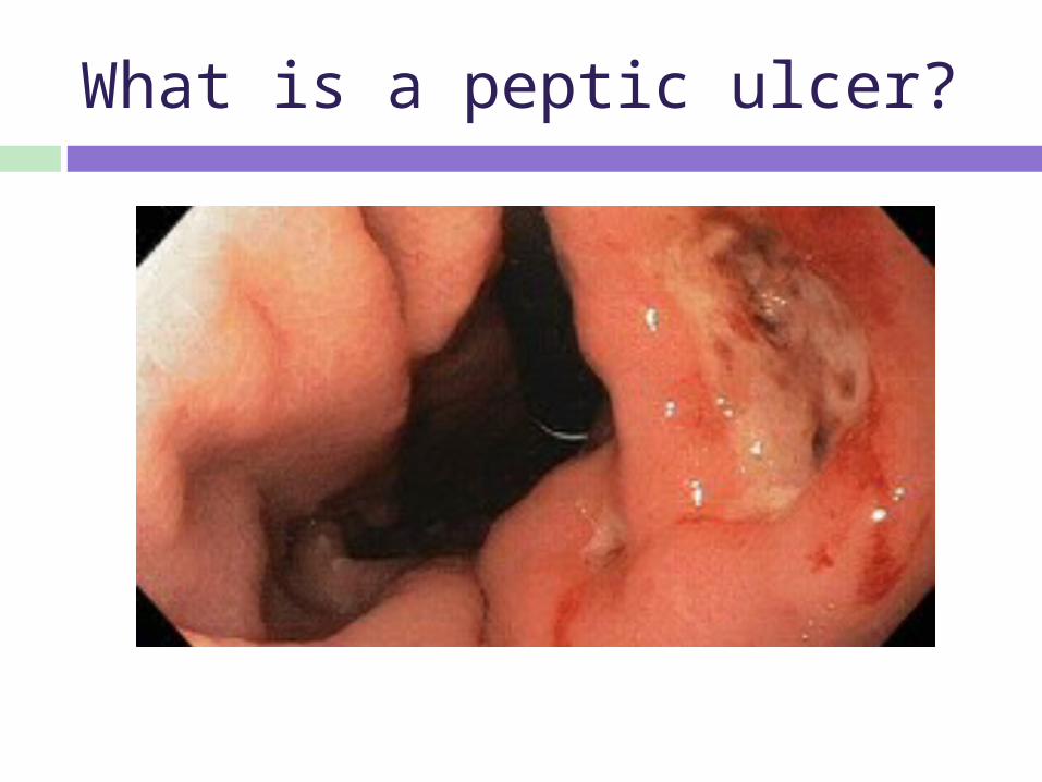
What is a peptic ulcer?
Peptic ulcer
A break in superficial epithelial cells penetrating down to muscularis mucosa

Differences between duodenal & gastric ulcers?
DUODENAL GASTRIC
INCIDENCE
ANATOMY
DURATION (acute/chronic)
MALIGNANCY
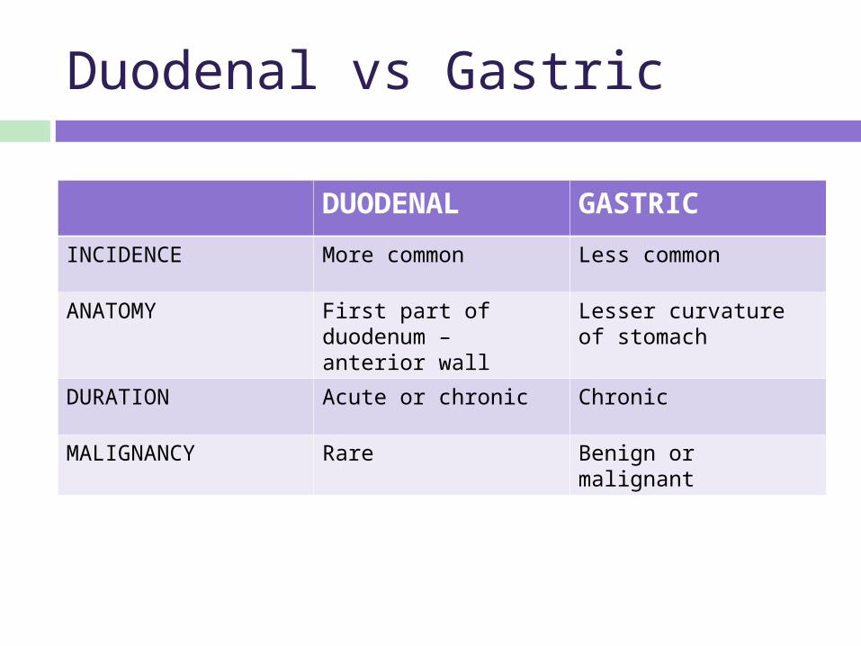
Duodenal vs Gastric
DUODENAL GASTRIC
INCIDENCE More common Less common
ANATOMY First part of duodenum – anterior wall
Lesser curvature of stomach
DURATION Acute or chronic Chronic
MALIGNANCY Rare Benign or malignant

Taking a history I
What risk factors would you ask about in the history?
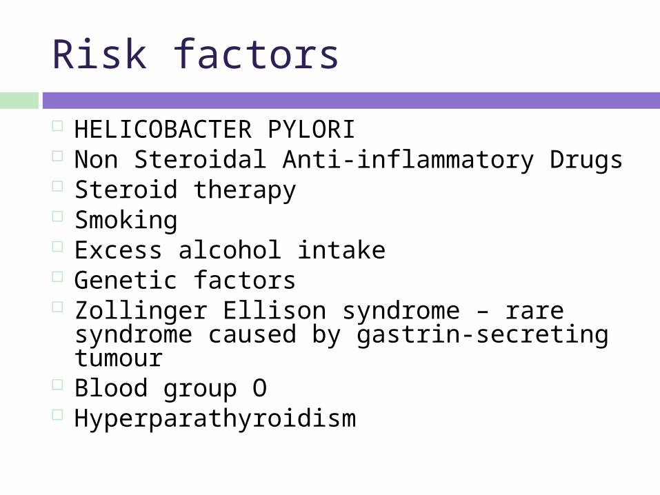
Risk factors
HELICOBACTER PYLORI Non Steroidal Anti-inflammatory Drugs Steroid therapy Smoking Excess alcohol intake Genetic factors Zollinger Ellison syndrome – rare syndrome
caused by gastrin-secreting tumour Blood group O Hyperparathyroidism
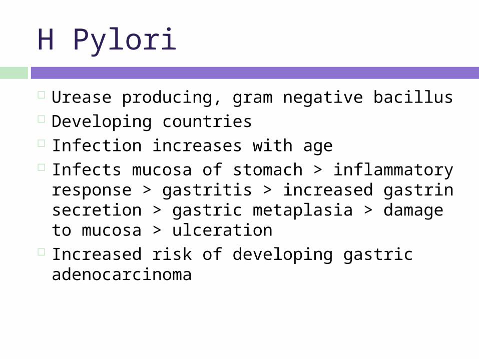
H Pylori
Urease producing, gram negative bacillus Developing countries Infection increases with age Infects mucosa of stomach > inflammatory
response > gastritis > increased gastrin secretion > gastric metaplasia > damage to mucosa > ulceration
Increased risk of developing gastric adenocarcinoma

Taking a history II
Take a focused history
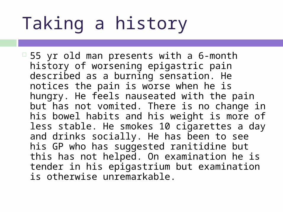
Taking a history
55 yr old man presents with a 6-month history of worsening epigastric pain described as a burning sensation. He notices the pain is worse when he is hungry. He feels nauseated with the pain but has not vomited. There is no change in his bowel habits and his weight is more of less stable. He smokes 10 cigarettes a day and drinks socially. He has been to see his GP who has suggested ranitidine but this has not helped. On examination he is tender in his epigastrium but examination is otherwise unremarkable.
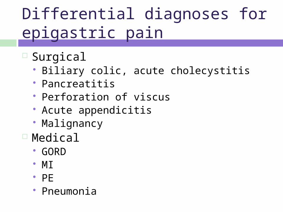
Differential diagnoses for epigastric pain Surgical
Biliary colic, acute cholecystitis Pancreatitis Perforation of viscus Acute appendicitis Malignancy
Medical GORD MI PE Pneumonia
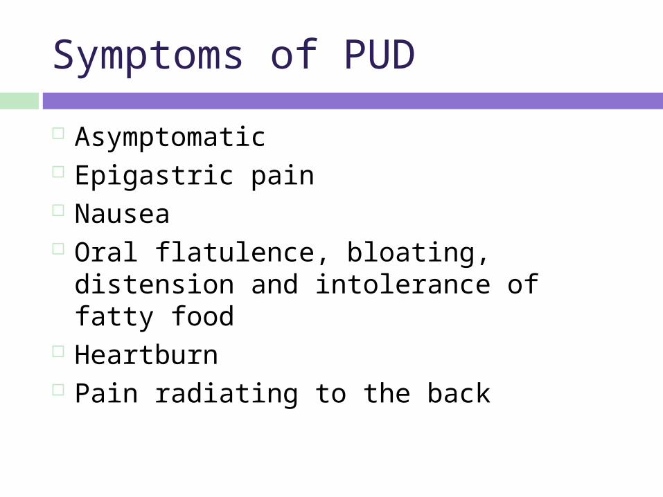
Symptoms of PUD
Asymptomatic Epigastric pain Nausea Oral flatulence, bloating, distension and
intolerance of fatty food Heartburn Pain radiating to the back
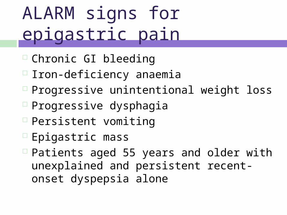
ALARM signs for epigastric pain Chronic GI bleeding Iron-deficiency anaemia Progressive unintentional weight loss Progressive dysphagia Persistent vomiting Epigastric mass Patients aged 55 years and older with
unexplained and persistent recent- onset dyspepsia alone
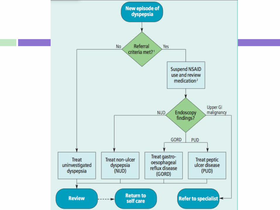
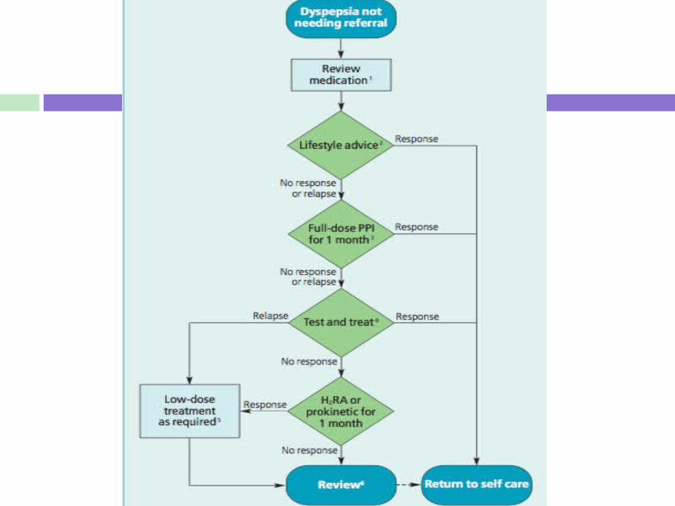
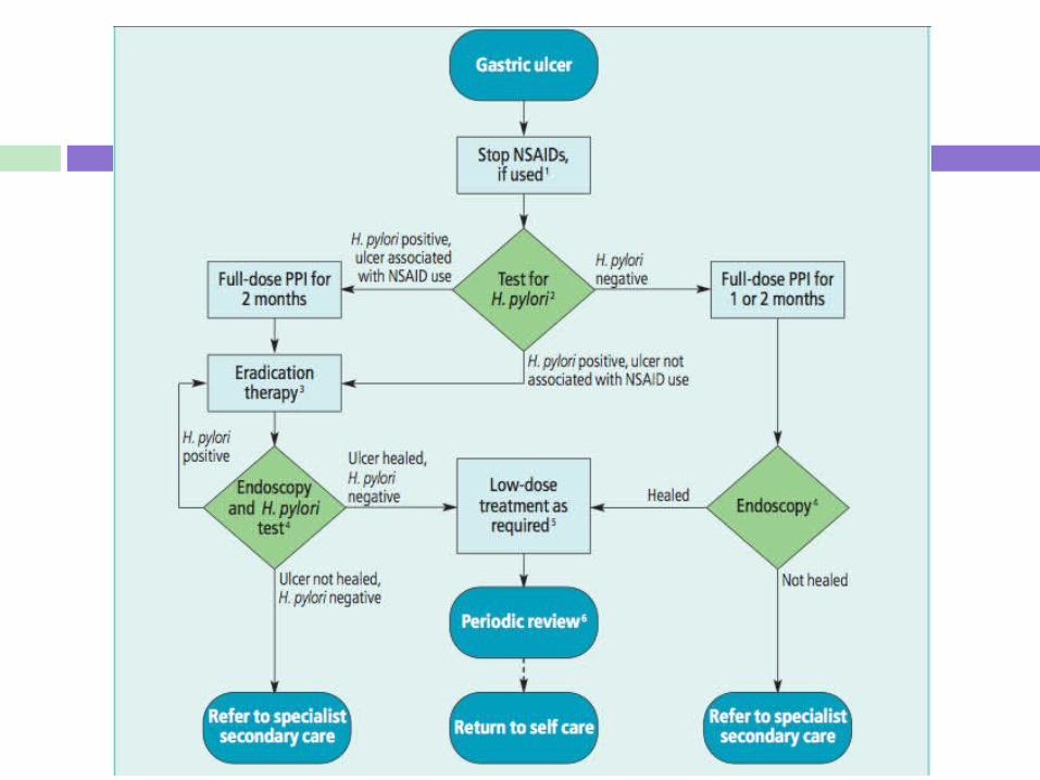
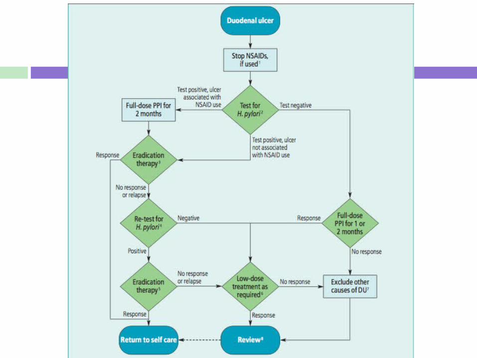
NICE guidance for dyspepsia
Management of dyspepsia
Investigations
H pylori testing
H pylori testing
C urea breath tests Stool antigen tests Serology Endoscopy with biopsy
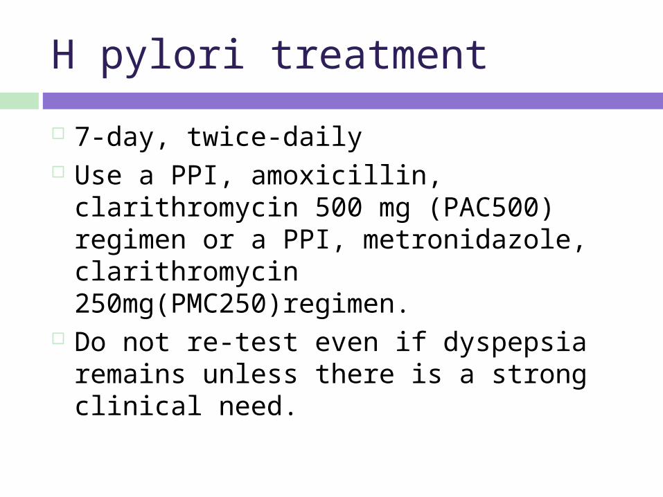
H pylori treatment
7-day, twice-daily Use a PPI, amoxicillin, clarithromycin 500
mg (PAC500) regimen or a PPI, metronidazole, clarithromycin 250mg(PMC250)regimen.
Do not re-test even if dyspepsia remains unless there is a strong clinical need.

Emergency scenario
A 50 year old man is brought into A+E via ambulance. He is vomiting bright red blood and complaining of abdominal pain. You get a quick history from his wife who explains he suffers with heartburn and is on lansoprazole. He was out with his work mates last night and drank quite heavily.
Initial Management I
ABCDE approach Call for help
Initial management II
Airway is clear Breathing – RR 30 breaths/min, Sats 91% OA Circulation – HR 130 beats/min, BP 80/40
mmHg Protect airway & keep NBM High flow oxygen Gain access – 2 large bore cannulae Bloods- FBC, U&Es, LFTs, glucose, clotting, cross
match 6 units Catheterise to monitor urine output
Initial management III
If shocked prompt volume replacement Either colloid or crystalloid solutions Red cell transfusion should be
considered after loss of 30% of the circulating volume
Correct any clotting abnormalities Urgent endoscopy after resuscitation
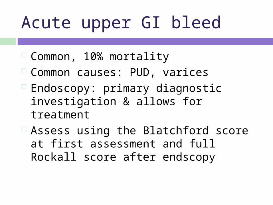
Acute upper GI bleed
Common, 10% mortality Common causes: PUD, varices Endoscopy: primary diagnostic
investigation & allows for treatment Assess using the Blatchford score at first
assessment and full Rockall score after endscopy
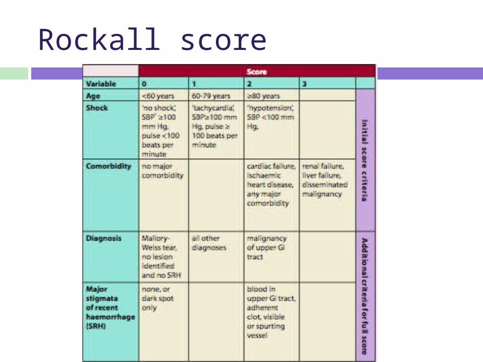
Rockall score
Summary
A peptic ulcer is a break in superficial epithelial cells penetrating down to muscularis mucosa
Duodenal > gastric ulcers Can be asymptomatic H pylori is a predominant risk factor H pylori diagnosed by c urea breath test,
stool antigen or if validated serology, treated with PAC500 or PMC250 regime
Complications of PUD can lead to acute emergency of upper GI bleed