Embed Size (px)
Citation preview
y'
''i " '_I
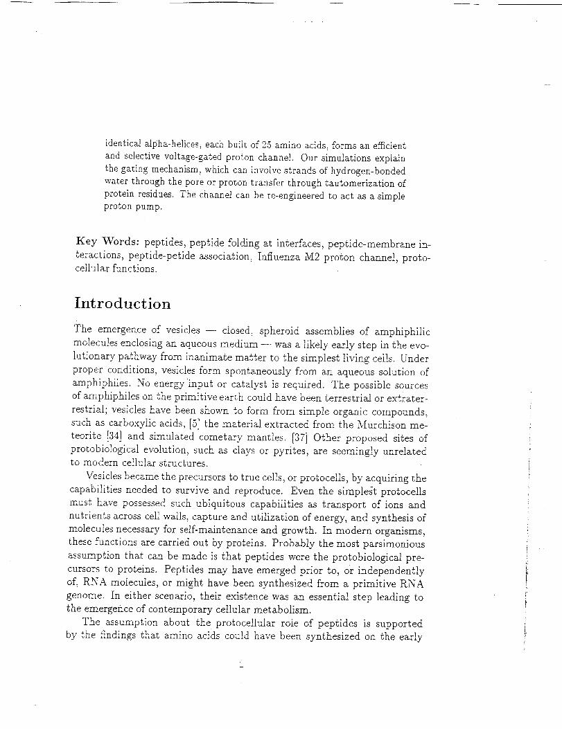
Peptides at Membrane Surfaces and their Rolein Prebiotic Evolution
Andrew Pohorille 1'2, Michael A. Wilson 1'2 and Christophe Chipot s
1Biomolecular and Cellular Modeling Program
NASA Ames Research Center, Moffett Field, CA 94035
2Department of Pharmaceutical Chemistry,
University of California, San Francisco, CA 94143
3Laboratoire de Chimie Th6orique
Unit6 Mixte de Recherche CNRS 7565
Universit( Henri Poinear6 - Nancy I, B.P. 239
54506 Vandoeuvre-16s-Nancy, France
July 12, 2002
Abstract
Protocells had to transport ions and organic matter across mem-
branes separating the interiorofthe cellfrom the environment, capture
and utilizeenergy and transduce environmental signals. In a seriesof
detailed,molecular-levelcomputer simulations we show how these pep-
tides in contact with membranes can acquire ordered structures and
functions. We have investigatedthe stabilityof_ simple c_-helicalpep-
tide containing Leucine (L) and Serine (S) of the form (LSLLLSL)s in
a model membrane system. The parallelin-plane state isthe most sta-
ble Configuration. The transmembrane state ismetastable, and _bout
15 kcal tool-I is required to insert the peptide into the membrane.
We investigated dimers of both (LSLLLSL)3 and glycophorin A, and
show how the free energy of helix association can, at least partially,
offset the free energy of insertion. We have also investigated the trans-
membrane pore of the influenza M2 protein. This aggregate of four
identicalalpha-helices,each builtof25 amino acids,forms an efficient
and selectivevoltage-gatedproton channel. Our simulationsexplain
thegatingmechanism, which can involvestrandsofhydrogen-bonded
water throughthe pore or protontransferthrough tautomerizationof
proteinresidues.The channel can be re-engineeredto act as a simple
protonpump.
Key Words: peptides, peptide folding at interfaces, peptide-membrane in-
teractions, peptide-petide association_ Influenza M2 proton channel, proto-cellular functions.
Introduction
The emergence of vesicles -- closed spheroid assemblies of amphiphilic
molecules enclosing an aqueous medium -- was a likely early step in the evo-
lutionary pathway from inanimate matter to the simplest living cells. Under
proper conditions, vesicles form spontaneously from an aqueous solution of
amphiphiles. No energy input or catalyst is required. The possible sources
of amphiphiles on the primitive earth could have been terrestrial or extrater-
restrial; vesicles have been shown to form from simple organic compounds,
such as carboxylic acids, [.bJ the material extracted from the Murchison me-
teorite [34] and simulated cometary mantles. [37] Other proposed sites of
protobiological evolution, such as clays or pyrites, are seemingly unrelatedto modern cellular structures.
Vesicles became the precursors to true cells, or protocells, by acquiring the
capabilities needed to survive and reproduce. Even the simplest protocells
must have possessed such ubiquitous capabilities as transport of ions and
nutrients across cell walls, capture and utilization of energy, and synthesis of
molecules necessary for self-maintenance and growth. In modern organisms,
these functions are carried out by proteins. Probably the most parsimonious
assumption that can be made is that peptides were the protobiological pre-
cursors to proteins. Peptides may have emerged prior to, or independently
of, RNA molecules, or might have been synthesized from a primitive RNA
genome. In either scenario, their existence was an essential step leading to
the emergence of contemporary cellular metabolism.
The assumption about the protocellular role of peptides is supported
by the findings that amino acids could have been synthesized on the early
earth [21, 66] or deli_;ered from extraterrestrial sources. [30] In particular,
a model for the synthesis and polymerization of amino acids from simple
precursors was recently proposed. [96, 97] Its essential features are that all
reactions occur in the same environmental conditions (one-pot chemistry),
the underlying chemistry resembles modern metabolism, and a mechanism
for autocatalytic feedback is provided. In another model, membrane-assisted
polycondensation of amino acids and peptides was demonstrated. [61] Small
peptides have also been shown to form auto-catalytic, self-correcting net-
works. [56,100] Under appropriate conditions, these networks can select for
peptides built of amino acids of the same chira.lity. [81] Furthermore, small,
functional proteins have been selected from random-sequence libraries with
frequencies that appear to be similar to those observed for equivalent RNA
libraries. [90]
On the basis of their location in the cell, proteins can be divided into
two broad types -- water soluble proteins, which reside in the cytoplasm,
and membrane proteins. This division is useful because these two types of
proteins have different structural properties, functions and evolutionary his-
tories. Integral membrane proteins perform such essential cellular functions
as transport of ions, nutrients and waste products across cell walls, transduc-
tion of environmental signals, regulation of cell fusion, recognition of other
cells, energy capture and its conversion into high-energy compounds. In fact,
30-40% of genes in modern organisms codes for membrane proteins. [18, 94]
In many instances, no alternative means of performing membrane-related
functions are known, or even have been postulated.
Although contemporary membrane proteins can be quite complex, their
transmembrane fragments are usually remarkably simple. The most common
structural motif for these fragments is a bundle of o_-helices, [7, 66] but occa-
sionally is a ,£-barrel. [89] This is in contrast to water-soluble proteins, which
exhibit a much wider range of structural motifs. Moreover, some membrane
proteins retain their functionality even if a large fraction of the protein is
removed, [69] whereas water-soluble proteins almost never do so. Another
protobiologically relevant property is that some simple, o_-helical synthetic
or natural membrane peptides aggregate spontaneously and form functional
complexes exhibiting sequence-dependent specificity.[2,7,8] Finally,short
membrane peptides can acquire well-definedsecondary structures,such as a.-
helices,_-strands or/3-turns,[7,35,50,77] whereas water-solublepeptides of
similarlength are typicallydisordered.These properties of membrane pep-
tidessuggest that theirassociations,especiallyin c_-helicalconformations, are
excellent candidates for mediators of transmembrane functions in protocells.
The conceptual framework to study membrane proteins is provided by
the canonical "two-stage" model of their folding. [38,77, 78] According to this
model, helices are formed and inserted into the bilayer in the first stage. This
isfollowed by specificinteractionsofthese helicesthat resultinhigher order,
functional tertiary structures. The latter step proceeds without significant
modifications of the already existing elements of the secondary structure.
Thus, in line with this model, functional membrane peptides have to fulfill
several conditions. First, they must fold into an ordered structure. Second,
they must be inserted into the membrane. Finally, they must be able to
self-assembleinto functional aggregates (e.g. channels) that exhibit speci-
ficitytowards well-definedprocesses or substrates. For example, channels
that transport both positive and negative ions, regardless of size, would have
been of very limited utility to a protocell. This simple view at the emergence
of membrane proteins raises a host of questions. Which amino acid sequences
are capable offoldingand how do membranes mediate thisprocess? What de-
termines the stability of the inserted, transmembrane orientation of a peptide
relative to the orientation parallel to the water-membrane interface? How
strong are helix-helixinteractionsin the membrane? How does a sequence
modulate the interhelicalrecognitionthat drivesthe associationof helices?
What mechanisms are responsiblefor the specificityof self-assembledtrans-
membrane peptides?
In this paper, we describe the results of computer simulations aimed at
providing at least partial answers to these questions and, by doing so, help
to understand better the role of peptides in the origin and early evolution
of membrane-related cellular functions. After a brief description of the the-
oretical approach we will discuss folding of small peptides, their insertion
into the membrane, interhelical recognition and selectivity of self-assembled
channels. The paper closes with a summary presented in the context of the
origin and early evolution of membrane proteins.
Molecular Dynamics Computer Simulations
The best approach to the computational study of biological systems at a
molecular level has been the molecular dynamics method. In this section, we
outline the basic aspects of this method that are common to the simulations
described in this paper. Several books [4, 46] provide exhaustive descriptions
of molecular dynamics, as applied to chemical and biologicalsystems.
In molecular dynamics, Newton's equations of motion are solved numeri-
callyfor allof the atoms in the system under study using an iterativeproce-
dure. From the positions,velocitiesand forces acting on the atoms at time
t,new positionsand velocitiesat-time t + 8t can be calculated ifthe forces
do not change appreciably during the interval81, called "the time step".
Typically,thistime step isequal to 1 femtosecond (i0-Is s). By repeating
thisprocedure many times we obtain a time-history of the system, called
a trajectory. State-of-the-art molecular dynamics trajectories for biologica]
systems extend currently for t nanosecond to 1 microsecond (10 .9 s - 10 .6
s), which means that they require 106 - t09 time steps.
To solve Newton's equations of motion, the forces acting on the atoms in
the system must be known. These forces depend, in turn, on the interactions
between the atoms and are computed from a potential energy function, which
describes the stretching of chemical bonds, the bending of valence angles, the
rotation of dihedral angles and the electrostatic and van der Waals interac-
tions between atoms. Much work has been invested in the construction of
potential energy functions that successfully reproduce properties of water-
soluble proteins, [20, 64] pure membranes [12, 41,88] and their mixtures with
small solutes and peptides. [6,31,86] In our simulations, we took advantage of
tkis work. For water, the TIP4P potential model was used, [49] which repro-
duces m any of the thermodynamic properties of liquid water. The potentials
that were used to describe the interactions between phospholipids have al-
ready been successfully applied in simulations of bilayer systems. [27, 39, 92]
The proteins were described using the AMBER all-atom force field. [20]
The simulated systems consisted of a model peptide either embedded in
a bilayer solvated by water or located at a model water-membrane interface.
All components of the system were represented at the atomic level. The
system was placed in a box -- the primary simulation cell. The number of
atoms in the system is typically in the 104-10 s range. The corresponding
cross-sectionM length of the water-membrane interface varied between 4 and
6 rim. To obtain results for a macroscopic system from such simulations, the
content of the primary cell was periodically replicated in space, forming an
infinite lattice of identical simulation cells. This is a standard approach to
removing edge effects in molecular simulations.
In many instances, considerable savings of computer time without a sig-
nificant sacrifice of accuracy can be achieved by smoothly truncating inter-
actions between two atoms in the system at a specified distance, typically
equal to 8-10.4. However, phospholipid head groups and ionizable side chains
of amino acids carry large charges, which interact non-negligibly over long
distances, extending beyond the primary simulation cell. In these instances,
truncation of interatomic interactions may be insufficiently accurate. The
most common method for calculating long-ranged effects is called Particle-
Mesh Ewald (PME) [40]. In PME, the long-range, electrostatic interactions
are evaluated through the solution of a differential equation on a grid, using
the Fast Fourier Transform (FFT) method. We have employed this approach
in our simulations of transmembrane channels in phospholipids. [85]
Many properties of the system may be computed directly from a molec-
ular dynamics simulation trajectory. Thermodynamic properties, such as
the temperature, pressure, or membrane surface tension, cain be expressed
as simple averages over the series of configurations that form the trajectory.
Structural quantities, such as conformationM parameters characterizing the
protein or its individual residues may also be computed in the same fashion.
All these quantities calculated from computer simulations can be directly
compared with the same quantities measured experimentally.
Often, a goal of computer simulations is to determine how the free en-
ergy of the system changes in the course of a chemical or biochemical pro-
cess. These changes are directly related to the relative stabilities of different
states of the system. Since free energies cannot be expressed as statistical
averages of mechanical properties, special techniques are require for their
evaluation. [13, 46] Two of these techniques were used in the simulations
discussed in the subsequent sections.
In one approach, a series of simulations is performed, in which the sys-
tem is constrained to several, overlapping ranges, or "windows", along an
appropriately chosen, physical degree of freedom, often called "the reaction
coordinate", _. For example, to compute the free energy change accompa-
nying the insertion of a peptide into the membrane, the distance between
the center of mass of the peptide and the center of the lipid bilayer could be
defined as such a coordinate, For each window, the probability, _(_), of find-
ing the system at different values of the chosen coordinate is obtained. This
probability defines the change of the free energy along [, AA(4), through therelation:
= - BTlog (I)
where kB is the Boltzmann constant and T is the temperature of the system.
AA(_) over all windows is obtained from the requirement that it must be a
continuous function of the chosen coordinate.
Another method for estimating free energy changes associated with the
point mutations of a given amino acid in a peptide, or with evolution of the
system along a "reaction coordinate", is based on the free energy pertur-
bation method [104]. For point mutations, this method is implemented via
an "alchemical transformation", in which the residues of interest in the wild
type are perturbed into those of the mutant. In practice, residues are altered
by modifying separately their point charges, van der Waals parameters and
internal coordinates -- i.e. shrinking or growing chemical bonds -- as a
function of a coupling parameter. [51] In the simulations described in this
paper, creation or annihilation of non-bonded and internal parameters were
carried out using the single topology approach, thus eliminating the need
for defining distinct topologies for both the initial and the final states of the
mutation. [72]
Folding of Simple Peptides at Water-Mem-
brane Interfaces
Membrane peptides capable of performing cellular functions must fold into
well-defined secondary structures. In fact, there are no known examples
of integral membrane proteins that have largely disordered transmembrane
segments. According to the "two-state" model, local folding is the first
step towards self-assembly of membrane proteins into functional structures.
In cells, this process proceeds at the water-membrane interface, but in
laboratory experiments it can be observed also at Water-micelle, water-
oil or water-air interfaces. Numerous experimental studies demonstrated
that short peptidescan._f0! d in_t_o,highly stable structures at aqueous i_nt__er-
faces. [9, 16, 28, 29, 35, 43, 48, 99] A crucial, common characteristic of these
interfaces is that a nonpolar phase is adjacent to water.
The ability of small peptides to organize at aqueous interfaces was ex-
amined by performing a series of large-scale, molecular dynamics computer
simulations of several peptides composed of two amino acids, nonpolar L-
leucine (L) and polar L-glutamine (Q) peptides differed in size and sequence
of the amino acids. The simplest models studied were dipeptides LL, LQ, QL
and QQ at the water-hexane interface [23,24]. Although these peptides were
too short to form secondary structures, they represented very good models
for examining conformational preferences of the peptide backbone as a func-
tion of the sequence. It was found that the preferred conformations of the
backbone corresponded to optimal interactions of the side chains with the
media of similar polarity. Dipeptides containing one polar (Q) and one non-
polar (L) residue adopted orientations, in which Q was immersed in water
and L was buried in hexane. Dipeptides made of two polar or two nonpolar
amino acids adopted orientations which maximized the exposure of their side
chains to hexane and water, respectively.
The studies of L/Q peptides were extended to two heptamerG LQQLLQL
and LQLQLQL, designed to maximize the interfacial stability of an cz-helix
and a /?-strand, respectively, by exposing their polar side chains to water
and their nonpolar side chains to a nonpolar phase. [24] Such structures are
called amphipathic. Both amphipathic structures were highly stable at the
interface. A similar result was obtained for the LQQLLQQLLQL undecamer,
which is amphipathic in the a-helical conformation [24]. When this peptide
was initially assigned the/?-strand conformation, which is not amphipathic,
it rapidly underwent several conformational transitions to adopt a nearby
amphipathic (but not helical) structure. [22] During the course of the simu-
lation a few intramolecular hydrogen bonds along the backbone were formed
that are characteristic of a helical structure. All the intermediate structures
were amphipathic. However, complete refolding into the expected a-helix
was not observed in a 160 ns trajectory.
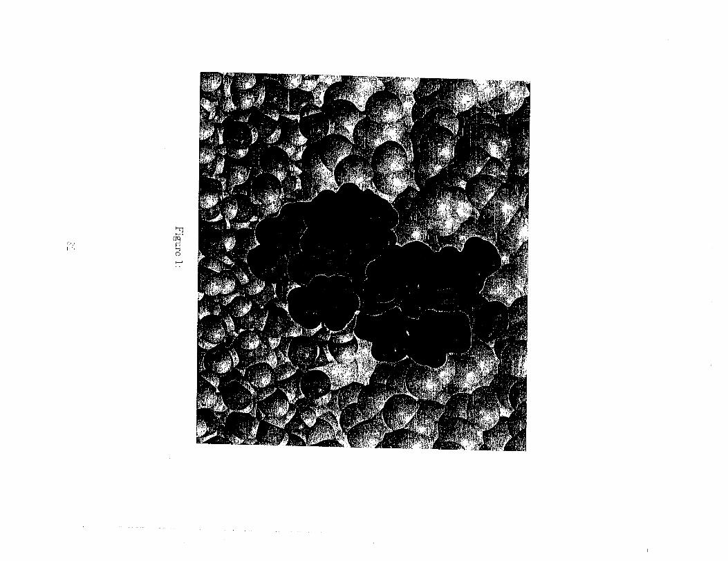
Finally, the transition of an undecamer, composed entirely of L-leucine
residues,from a disorderedstructurein aqueous solution to an a-helixat the
interfacebetween water and hexane was investigated.[25]Complete folding
ofa peptide was accomplished forthe firsttime in computer simulationsthat
explicitlyincluded the solutionenvironment. The poly-L-leucinepeptide was
initiallyplaced in water as a random Coil.It rapidly translocatedto the in-
terface;in this environment, the nonpolar L side chains could be partially
removed from the aqueous medium. Once at the interface,the peptide folded
intoa helixin 36 ns. The finalstructureisshown in Fig. i. During thispro-
cess,some polar groups in the backbone became dehydrated_ facilitatingfor-
mation of intramolecularhydrogen bonds along the backbone. The resulting
structure of the peptide became more hydrophobic and partitionedfurther
intothe hexane phase. This,in turn,created a favorableenvironment for the
emergence of additional structure-formingintramolecular hydrogen bonds.
The most favorableorientationof the folded peptide was parallelto the in-
terface.The freeenergy of perpendicular orientationswith the N-terminus
buried in hexane was lessfavorable by only 4 kcal tool-i. In contrast,the
perpendicular orientation with C-terminus inside the hexane phase was less
favorable than the parallel orientation by 12 kcal mo1-1 because polar groups
in the backbone at this end of the peptide are not involved in intramolecular
hydrogen bonding but, instead, prefer to be hydrated: This result indicates
that there is a preferred direction of peptide insertion into the membrane.
The simulations reveal several basic principles that govern the sequence-
dependent organization of peptides at interfaces and thereby determine their
ability to perform protocellular functions.
Short peptides tend to accumulate at interfaces and acquire ordered struc-
.... tures, provided..that--they_ha.v-e-a-proper--seq-uen-ce-of-polar and--nonpolar
residues. The specific identity of the amino acids appears to be less im-
portant for this process, a desirable protobiological property. The driving
force that enables or enhances secondary structure formation for proteins
interacting with or incorporated into membranes is the hydrophobic effect,
which is manifested at aqueous interfaces as a tendency for polar and nonpo-
iar groups of the solute to segregate into the aqueous and nonpolar phases,
respectively. The emerging amphipathic structures are strongly favored.
Among these structures, c_-helices are especially stable because they are
further stabilized by intramolecular hydrogen bonding interactions. In bulk
water, these interactions do not contribute to the stability of helices be-
cause of competing interactions between hydrogen bonding centers and watermolecules.
If peptides consist of nonpolar residues only, they become inserted into
the nonpolar phase. As demonstrated by the example of the C-leucine unde-
career, nonpolar peptides tend to fold into an a-helix as they partition into
the nonpolar medium. Once in the nonpolar environment, the peptides can
readily change their orientation with respect to the interface from parallel
to perpendicular, for example in response to local electric fields. [91] The
ability of nonpolar peptides to respond to changes in external conditions
may have provided a simple mechanism for transmission of signals from the
environment to the interior of a protocel].
Free Energetics of Insertion of Peptides into
Membranes
According to the two-statemodel, interfacialfoldingof transmembrane pro-
teinsis followed by their insertion into the bilayer. In the previous section, we
have already noted one example of thisphenomenon in the discussionof the
poly-g-leucineundecamer. However, thisexample isnot typicalbecause the
undecamer isnot long enough to span the membrane. A transmembrane a-
helicalpeptide must contain appro_mately 20 residuesto extend for the full
width of a membrane that is 3 nm thick. We studied one such peptide built
of only two amino acids - L-leucine and L-serine (S). The peptide contains 21
residues in the sequence (LSLLLSL)a. This sequence has been chosen such
that in the c_-helical form the peptide is amphipatic, i.e. all serine residues,
which are polar, lie along the same face of the c_-helix. Despite its simplicity,
(LSLLLSL)3 exhibits several interesting properties. It was shown experimen-
tally that the peptide formed transmembrane, tetrameric ion channels in the
presence of an electric field. [55] Depending on the direction of the field, the
channels could transport either positive or negative ions. When the elec-
tric field was removed, the channels persisted on time scales of milliseconds
before the individual peptides reverted to their resting state parallel to the
water-membrane interfacel indicating that the transmembrane channels do
not correspond to the global free energy minimum but, instead, are weaklymetastable.
Experimental results, however, provide no information about stability of
individual helices.in the transmembrane orientation. This will depend on
the balance o.f hydrophobic forces, which tend to drive the nonpolar leucine
residues into the membrane interior, the hydrophilic forces, which are fa-
vorable when the serine residues are located in the aqueous solution, and
the interactions of unsaturated hydrogen bonding sites at both ends of the
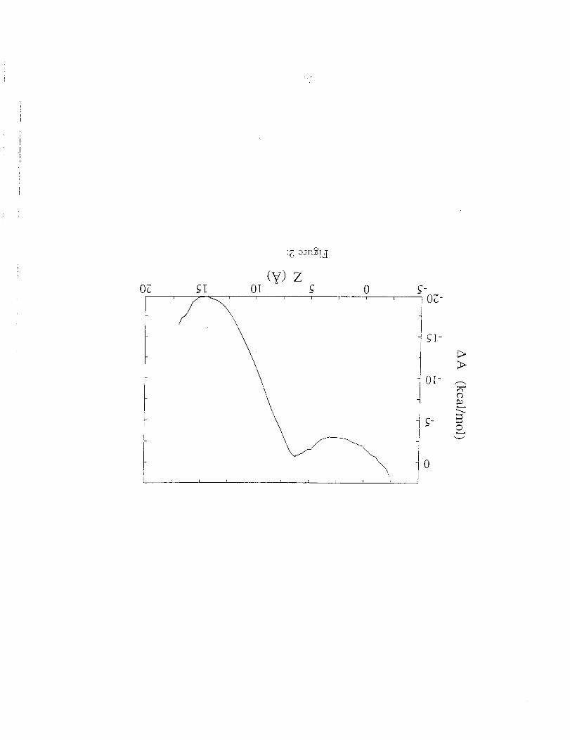
s-helix with the water. To clarify this issue we calculated the free energy
of inserting the peptide into a model membrane system using a variant of
the free energy window method_ described briefly in the section on molecular
dynamics. The resulting free energy curve is shown in Fig. 2 as a function of
the location of the center-of-mass of the pept!de relative to the center of the
membrane (z = 0). The water-membrane interface is located at z = -13.
The orientation of the peptide is dependent on the location of its center-of-
mass. At z = 0 the peptide is approximately perpendicular to the plane of
4
L_'
the membrane, with an end in each of the aqueous phases. In contrast, at
z = 13, the peptide lies paral]el to the interface such that the serine residues
point towards the water. The two main features of the curve in Fig. 2 are
that the in-plane state is approximately 20 kcal tool -1 more stable then the
transmembrane state and that the latterstate corresponds to a broad and
shallow free energy minimum. This means that the transmembrane state of
the peptide is, at best, only weakly metastable and a single peptide in this
state will quickly convert to the in-plane state in the absence of an electric
field.
These features of the free energy surface were borne out by independent
simulations with the peptide initially located in either an in-plane or trans-
membrane orientation. The in-plane state was stable over the course of a
10 ns simulation. The peptide backbone remained entirely a-helical and the
serine residues always pointed towards the water. The transmembrane pep-
tide adopted a variety of mixed a-helical and 310-helical arrangements. This
is due to a mismatch between the length of the peptide and the width of the
membrane. The peptide in the a-helical conformation is slightly longer than
the width of the membrane. Converting part of the backbone to 310 lengthens
the helix and allows for the formation of additional, energetically favorable,
serine-water hydrogen bonds. In simulations of a system with a somewhat
thicker hydrophobic membrane core i the peptide remained a-helical.
A total of three trajectories were started with the peptide in the trans-
membrane state. On the basis of the free energy calculations it was expected
that this state would not be stable and, over time, the peptide would move to
the water-membrane interface. In two of the simulations, the peptide sponta-
neously converted from the transrnembrane to the in-plane state after ? and
9 ns, respectively. In the third simulation, however, the peptide remained
transmembrane after 18 ns. This is due to the asymmetry in the ends of the
peptide. As found for the undecamer of poly-Leucine, the C-terminus inter-
acts with the water much more strongly than the N-terminus. The fiat free
energy curve in the region near z : 0 indicatesthat the peptide can read-
ilydiffuse towards either of the two water-membrane interfaces. In the two
simulations, in which the peptide converted to the in-plane state, it initially
diffused in the direction that required dehydration of the N-terminus. Since
this end interacts with water relatively weakly the conversion appears to pro-
ceed quickly With little or no free energy barrier. In contrast, in the third
simulations, the peptide initially diffused in the opposite direction, which
would have required dehydration of the C-terminus. Considering highly fa-
vorable interactionof thisterminus with water such dehydration process is
unlikely.Instead,itisexpected that in a sufficientlylong molecular dynam-
icstrajectorythe peptide would diffuseback to the centerof the bilayerand,
eventually, converted to the in-plane state by dehydrating its N-terminus.
Helix Association in Membranes
Single transmembrane helices are rarely capable of performing biological
functions. Instead, they form functional units after self-assembling into
higherorder structures.However, not allhelicesself-assemble.Consequently,
itisnecessaryto understand sequence-specificinterhelicalrecognitionbefore
we can predictthe kinds ofstructuresthat Could have formed in protocellular
membranes.
The simplest models for peptide association are helical dimers. Although
they cannot form channels, some are biologically active. Moreover, it is
assumed that dimer formation is the first step in aggregation into higher order
assemblies. For example, it has been suggested that tetrameric channels are
formed as "dimers of dimers". [102]
Conveniently, simple experimental models have recently been developed
for helix associations in membranes. [47, 59,103] The best-studied system,
both structurally and thermodynamically, is the 24-residue transmembrane
region of glycophorin A (GpA). CpA forms non-covalent dimers through the
reversible association of its membrane-spanning domain, [17,58] which adopts
an c_-helical conformation. [63] On the basis of extensive random mutagenesis
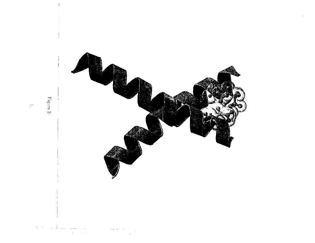
investigations, a model of the GpA transmembrane dimer has been proposed:
in which helix-helix association results from the specific interaction of seven
residues located on one face of each c_-helix. [1, 57, 59, 93] This model has
been recently confirmed by NMR spectroscopy of 40-residue peptides that
contained the transmembrane segment, solvated in detergent micelles.[63]
This study also showed that the c_-helicesformed a right-handed, coil-coiled
structure.This model isshown in Fig. 3
The dissociationfree energy of the CpA dimer in a detergent pen-
taoxyethylene (C8E5) has been estimated to be about 9.0 kcal mol -I. [44]
It has been also demonstrated that single-residuemutations can markedly
influencethe freeenergy of associationof the helices.Mutants in which ei-
therone of two leucineresidueswere substitutedwith alanine or glycine was
substitutedwith isoleucinewere found to be lessstable than the wild-type
dimer by i-3 kcal/mol. [42,44] In the model of the GpA dimer, all these
residues are involved in interhelicalinteractions.
To gain insight into the various contributions that govern the specific in-
teractions of transmembrane s-helices, we simulated both the dissociation of
helices and the effect of point mutations on the free energy of dissociation. [26]
Any mutation that destabilizes the dimer must decrease the dissociation free
energy.
In the first step, we simulated the wild-type dimer of GpA in a lamella
of dodecane, placed between two lamellae of water. The width of the dode-
cane layer was approximately the same as that of the hydrophobic core of a
palmitoyloleylphosphatidylcholine (POPC membrane. The starting structure
was based on the NMR model of the dimer. [63] The comparison between the
calculated, time-averaged structure of the dimer after 600 ps of molecular
dynamics trajectory and the nuclear Overhauser effect data of MacKenzie et
a/. [63] provided an assessment of the accuracy of our model and the poten-
tial energy functions utilized. The distance root mean square deviation was
less than 1.5 ,_and the monomers remained a-helical.
Next, we separated the helices in a series of molecular dynamics simu-
lations using the distance separating the centers of mass of the two helices
as the reaction coordinate, _. The free energy of dissociation was estimated
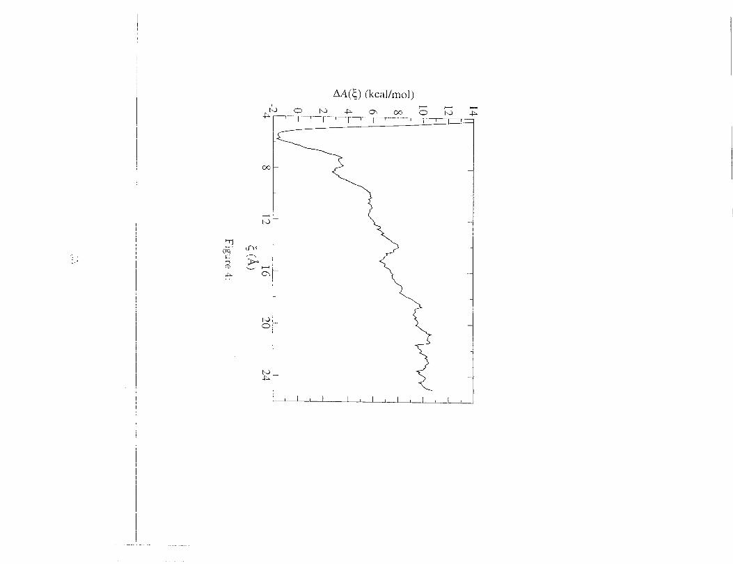
using flee energy perturbation theory. [104] The Complete pathway joining
the dimer at _ __ 6.5 ._, to the dissociated state at _ _ 20 _, was divided
into a series of intermediate states corresponding to different values of the
reaction coordinate. The free energy of dissociation is given as the sum of
free energy differences between consecutive states.
As can be seen in Fig. 4, the estimated free energy is approximately equM
to 11.4±0.3 kcM mo1-1. By comparison, the experimentally determined free
energy of dissociation in the detergent pentaoxyethylene (C8E5) is equal
to 9.0+0.1 kcal tool -1. [44] Since the molecular environments of GpA in
the computational and experimental studies are different -- i.e. dodecane
versus C8E5 -- the free energies are not expected to be identical, but should
be similar. Given that the volume accessibie to the a-helix dimer is much
smaller in C8E5 than in dodecane, which corresponds to a smaller entropic
contribution, we expect the association would be favored in the micellar
environment. This hypothesis is supported by the observation that the Dee
energy of dissociation is higher dodecane than C8E5.
Based on similar considerations, it is expected that the influence of the
surroundings on the computed point mutations would, in principle, be lira-
_3
ited due to compensation of entropic effects in the free energy differences.
However, this may not be necessarily the case. The I76L point mutation was
carried out by decoupling the annihilation of the electrostatic and the van der
Waals and internal parameter contributions. Not too surprisingly, tl_e free
energy difference for the electrostatic term averaged to 0.0 kcal mo1-1. The
contribution due to the modification of the van der Waals parameters and
the participatingchemical bonds and valence angles,and, therefore,the total
freeenergy difference,isequal to 0.4 kcaltool-1,which somewhat underesti-
mates the experimental value of 1.7kcM tool-I.[44]Itshould be emphasized,
however, that, as the L-isoleucineistransformed into L-alanine,the disrup-
tiveeffectof the mutation witnessed in C8E5 should be lesspronounced in
dodecane, because of the greater volume accessibleto the a-helicesin that
environment.
The free energy change upon single-point mutation is fairly small. In con-
trast, the free energy of dissociation is large and positive. This might suggest
that the diner is strongly favored for both the wild-type and the mutants.
This would indeed be the case ifwe only considered the equilibrium between
the associated state and separated, trg_sr_erabrar_ehelices.However, this
comparison isnot appropriatebecause, as we have already pointed out, the
transmembrane orientationof helicesisnot stable.The freeenergy of inser-
tionofa helicalpeptide intothe membrane ispositive(unfavorable)and may
be substantial.[I0,ii] Thus, the equilibrium that needs to be considered is
between the transmembrane dimer and the individual helicesat the water-
membrane interface.This equilibrium is governed by the bMancebetween
the unfavorable freeenergy ofinsertionintothe membrane and favorablefree
energy of interhelicalassociation:This balance could be subtle,and mod-
est changes in eitherterm could shiftthe equilibrium, possibly disruptingdimerization.
So far,we have only discussed the possibilitythat peptide association
followsinsertioninto the membrane. Alternatively,peptides might assem-
ble at interfacesand only then become inserted into a lipidbilayer. This
might seem plausibleespeciallyfor amphipathic peptides because theirag-
gregates could be stabilizedby interhelicalhydrogen bonds between polar
residues. Then, nonpolar residues would be exposed to the environment,
which should promote insertion into the nonpolar membrane. To test this
mechanism of aggregation, we have carried out simulation studies on diners
of the (LSLLLSL)3 peptide, which has already been discussedin the previous
section.When the dimer was placed parallelto the plane of the membrane at
~r-
the water-membrane interface, it dissociated in less than 2 ns. The interfacial
water molecules successfully competed for the serine hydrogen bonding sites,
which led to the loss of serine-serine hydrogen bonds. Additionally, interac-
tions between electrical dipoles associated with the a-helices are highly unfa-
vorable in a parallel arrangement. These results indicate that self-assembly
of peptides at interfaces is unlikely. In contrast, a transmembrane dimer was
found to be stable over the course of a 15 ns simulation. Near the ends of the
dimer, serine-serine hydrogen bonds were lost in favor of water-serine hydro-
gen bonds, allowing water molecules to penetrate the membrane around the
peptide. This, in turn, might increase rates of non-specific permeation of ions
and polar solutes across membranes. [33, 70, 71,76, 98] However, the serine-
serine hydrogen bonds in the middle of the dimer remained intact, keeping
the helices in the dimer together. These results confirm that association of
two peptides appears to increase the stability of the transmembrane state
relative to isolated monomers.
Simulation of a Model Transmembrane Proton
Transport System
Aggregates of membrane proteins are of special interest if they can perform
important cellular functions. One such function is transport of protons across
membranes, which is an essential process for both bioenergetics of modern
ceils and the origins of cellular life. All living systems convert environmen-
tal energy into chemical energy by using transmembrane proton gradients to
drive the synthesis of adenosine triphosphate (ATP) from adenosine diphos-
phate (ADP). ATP, in turn, is used as a source of energy to drive many
cellular reactions. The ubiquity of this process in biology suggests that even
the earliest cellular systems relied on proton gradients to harvest the energy
needed for their survival and growth from the environment. In contempo-
rary cells, proton transfer is assisted by large, complex proteins embedded
in membranes. Could ti_e same process have been accomplished with the
aid of similar, but much simpler peptides that could have existed in the
protobiologicaI milieu?
To answer this question it is desirable to have a protein model which
is small, has a well known structural motif, yet which operates with the
efficiency and control of more complex proteins. This led us to study the
__)
Influenza-A M2 protein, which forms small, voltage-gated proton channels.
[4,5,60,67,73,74,82,9.5,101] The M2 protein contains 97 amino acids, including
a single transmembrane domain 19 residues long. Not all residues, however,
are essential for transport. Active channels have been reconstituted from a
synthetic peptide containing a subset of only 25 amino acids, including the
transmembrane region, with no loss in specificity or efficiency. [36]
In lipid bilayers, four identical protein fragments, each folded into an o_-
helix, aggregate to form small channels spanning the membrane. Protons are
conducted through a narrow pore in the middle of the channel. Compared
with a well-studied, proton-permeating peptide, gramicidin A, the rate of
proton transport across the truncated M2 channel is over 1000-fold faster.
Remarkably, in contrast to gramicidin A, the M2 channel is virtually imper-
meable to alkali ions, such as Na + and K +. This combination of efficiency
and specificity makes M2 an excellent, simple model to study the formation
of proton gradients across membranes.
The channel is large enough to contain water molecules and is normally
filled with water. In analogy to the mechanism of proton transfer in some
other channels, [3, 84] it has been postulated that protons are translocated
along the network of properly aligned water molecules filling the pore. This
mechanism, however, must involve an additional, important step because the
channel contains four L-histidine (H) amino acid residues, one from each of
the helices, which are sufficiently large to occlude the pore and interrupt
the water network. The L-histidine residues have been implicated in gating
protons. Due to their size, they ensure channel selectivity by blocking small
ions, such as Na + and K +, from permeating the membrane but provide a
mechanism for proton transport. The role of the L-histidines in gating is
supported by findings that point mutations, in which the L-histidines are
substituted by other residues greatly impede the ability of M2 to transport
protons.
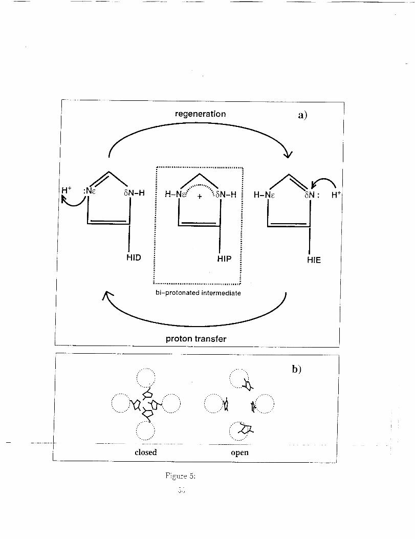
Two mechanisms of gating have been proposed, which rely on the ability
of each L-histidine to Become positively charged by accepting an additional
proton. In one mechanism, all four L-histidines acquire a proton and, due to
repulsion between their positive Charges, move away from one another, thus
opening the channel. [83] The alternative mechanism involves the ability of
protons to move between different atoms in a molecule (tautomerization). In
this mechanism, a proton is captured on one side of the gate while a second
proton is released from the opposite side, and the molecule returns to the
initial state through tautomerization. [73] These two mechanisms are shown
schematically in Fig. 5.
Atomic-level molecular dynamics simulations were designed to test these
two mechanisms. [75, 86] The model system used in the study contained
a bilayer membrane made of phospholipid, dimyristoylphosphatidylcholine
(DMPC), which is a good model of the biological membranes forming cellu-
lar boundaries. Both sides of the bi]ayerwere surrounded by water which
simulated the environment inside and outside the cell. Embedded in the
membrane was a channel made of 25 amino acids fragments of the Influenza-
A M2 protein,and enough sodium counterionsto maintain system neutrality.
Several protonation statesof L-histidineresidues were considered. They rep-
resented differentintermediate statesof the channel predicted by the two
proposed mechanisms of proton transport. The simulations revealed that
allintermediate statesof the system involved in the tautomerization mech-
anism were structurally stable and the arrangement of water molecules in
the channel was conducive to the proton transport. In contrast, in the
four-protonated state,postulated to exist in the gate-opening mechanism_
the electrostaticrepulsion between the L-histidineresidues appeared to be
so large that the channel lost its structural integrity and one helix moved
away from the remaining three. These resultsindicate that translocation
along a network of water molecules in the channel and tautomerization of
the L-histidineresidues isa likelymechanism of proton transport whereas a
mechanism involvingprotonation of allfour L-histidinesisunlikely.A possi-
bilityof gate opening after protonation of two rather than four n-histidines
has not been excluded.
These results not only explain how a simple protein system can achieve
highly efficient and selective passive proton transport 0.e. transport along
the concentration gradient) across cell walls, but also how the system can be
genetically re-engineered to become a simple directional, reversible proton
pump. First, M2 must be coupled with a chromophore capable of releasing a
proton in response to light. Several very simple chromophores, such as poly-
cyclic aromatic hydrocarbons, are already known. In fact, some of them have
been shown to dissolvein membranes and generate transient,light-induced
proton gradients. [32] To maintain the proton gradient, it must ensured that
release of the pumped proton is followed by reprotonation of the chromophore
with a proton from the opposite side of the membrane. This will involve ma-
nipulating the sequence of amino acids along the pore. Cysteine scanning
mutagenesis has already shown that the replacement of the pore-lining, but
not other residues can modify the propertiesof the channel. [87]Other de-
signs are possible, based on coupling electron and proton transfer using iron
or quinones. These have been recently shown to be of possible protobiological
relevance. [14] If such an experimental effort were to be successful, it would
demonstrate that protein-based proton pumps could have emerged early in
protobiological evolution. Furthermore, such a pump could be used to prol
vide energy to laboratory-built models of protocells and cell-like structures
built for biotechnological applications. [53]
Summary
Many proteins that perform essentialcellularfunctions are embedded in
membranes that encapsulate cellsor cellularcomponents. These proteins
or proteincomplexes are among the largestmacromolecular structuresfound
in ceilsand their mode of actionis often complicated and subtle. This ap-
pears to create a seriousdifficultyfrom the originof lifepoint of view. If
the functionsperformed by membrane proteins are essentialto the existence
of even the simplest cells,how could they have been performed, even less
efficientlyor selectively,by much simpler peptides?
In this paper we have argued that the emergence of integral membrane
proteins may have been quite feasible. In fact, this may be much easier to
envision than the emergence of water-soluble proteins. We have supported
our arguments with results of our molecular dynamics computer simulations
and a considerable body of evidence from other experimental and theoretical
studies. The prerequisite for the formation of functional membrane pro-
reins was the existence of peptides containing 20-25 amino acid, which were
sufficiently long to span a membrane. This length requirement is rather mod-
est, considering that functional water-soluble proteins need to be markedly
larger. For example, the shortest, protobiologically relevant proteins contain
45 amino acids [90] and the simplest self-reproducing protein system consists
of proteins built of 33 amino acids: [56]
Many peptides are attracted to water-membrane or water-oil interfaces.
Once at the interface, most nonpolar peptides spontaneously fold into c_-
helices. Peptides that contain both polar and nonpolar amino acids tend to
adopt amphipathic structures, in which amino acid side chains are immersed
in media of similar polarity. Whenever the sequence permits, peptides fold
into amphipathic helices at interfaces. The formation of ordered, helical
structures is primarily governed by the sequence of polar and nonpolar amino
-f
acids. Considering that specific identities of side chains is less important, the
existence of helical peptides in interfaciat, protocellular environments should
not have been rare.
Helical peptides located parallel to the interface could insert into the
membrane and adopt a transmembrane conformation. However, insertion of
a single helix usually involves a positive free energy change, even for fully
nonpolar peptides. The main reason why insertion is unfavorable is that
polar groups in the peptide backbone and some side chains, which remain
at least partially hydrated in water, become completely desolvated. The loss
of solvation free energy is smaller for helices than for disordered structures
because polar groups in the backbone are involved in intramolecular hydrogen
bonding.
The unfavorable free energy of insertion can be regained by spontaneous
association of peptides in the membrane into homomeric or heteromeric mul-
timers. The first step in this process is the formation of dimers, although the
most common structures involve aggregates of 4-7 helices. The helices could
readily arrange themselves such that they form pores capable of transporting
ions and small molecules across membranes. The stability of transmembrane
aggregates of simple proteins is often marginal and, therefore, it can be regu-
lated by environmental conditions, such as external electric fields or the spe-
cific nature of phospholipid headgroups, [15,19, 54, 91] or by small changes in
the sequence of amino acids. [42,44] This ability to respond to environmental
signals might have led to the earliest, although quite imprecise, regulation of
transmembrane functions.
Clearly, a key step in the earliest evolution of integral membrane proteins
was the emergence of selectivity for specific substrates. The selectivity of
early channels was determined to some extent by all residues lining their
lumen, which interact with substrates via electrostatic and van der Vv'aals in-
teractions. [79] However, many contemporary simple channels employ filters
or gates as the primary way to achieve selectivity. [68,73,80] From the evolu-
tionary standpoint it is a very convenient solution because it requires placing
just one or only a very few properly chosen residues in certain positions along
the channel rather than imposing conditions on the whole sequence.
Many additional steps were required before simple aggregates of trans-
membrane peptides reached the structural and functional complexity, diver-
sity and refinement of contemporary integral membrane proteins. The helices
were connected by extra-membrane, hydrophilic linkers to stabilize them in-
side the membrane. The resulting, large proteins aggregated to even larger,
higher-order structures. In many instances this step involved gene dupli-
cation. Protein sequences became optimized for highly specificfunctions.
Perhaps most importantly,membrane proteinsacquired large,water-sohble
domains, which play a regulatory role or help to supply energy for active
transport. This more advanced evolution of membrane proteins has been a
subject of extensivestudies.[77]In the process,some intriguingconnections
between ion channels and enzymes have been uncovered. [62]The evolution-
ary history of membrane proteins isspecialinterestbecause it opened the
doors for the emergence of multicelhlar organisms endowed with nervous
systems.
Acknowledgments
This work was supported by the grants from the NASA Exobiology Program
and from the NASA Astrobiology Institute.
References
[1] Adams, P. D., D. M. Engelman, and A. T. Brfinger: 1996, 'Improved
prediction for the structure of a dimeric transmembrane domain of gly-
cophorin A obtained through global searching'. Proteins: Structure,
Function and Genetics 26, 257-261.
[2] ._kerfeldt, K., J. Lear, Z. Wasserman, L. Chung, and W. DeGrado: 1993,
'Synthetic peptides as models for ion channel proteins'. Ace. Chem. Res.
26, 191-197.
[3jAkeson, M. and D. Deamer: 1990, 'Proton conductance by the Grami-
cidin water wire- model for proton conductance in the FIF0 ATPases'.
Biochim. Biophys. Acta 60, 101-109.
[4] Allen, M. and D. Tildesley: 1987, Computer Simulation of Liquids. Ox-
ford: Oxford University Press.
[5] Apel, C., D. Deamer, and M. Mautner: 2002, 'Self-assembled vesicles of
monocarboxylic acids and alcohols: conditions for stability and for the
encapsulation of biopolymers'. Biochim. Biophjjs. Actai -Biomernbranes
1559, 1-9.
[6] Bassolino-Klimas, D., H. Alper, and T. Stouch: 1995, 'Mechanism of
solute diffusion through lipid bilayer membranes by molecular dynamics
simulation'. Y. Am. Chem. Soc. 117, 4118-4129.
[7"] Bayley, H.: 1999, 'Designed membrane channels and pores'. Curt. Opin.
Biotech. 10, 94-103.
[s] Bechinger, B.: 2000, 'Understanding peptide interactions with the lipid
bilayer: a guide to membrane protein engineering'. Curt. Opin. Chem.
Biol. 4, 639-644.
[gj Bechinger, B., M. Zasloff, and S. Opella: 1993, 'Structure and orien-
tation of the antibiotic peptide magainin in membranes bu solid-state
nuclear-magnetic-resonance spectroscopy'. Prof. Sci. 2, 2077-2084.
[10] Ben-Shaul, A., N. Ben-Tal, and B. Honig: 1996, 'Statistical thermody-
namic analysis of peptide and protein insertion into lipid membranes'.
Biophys. J. 71, 130-137.
[llJ Ben-Tal, N., A. Ben-Shaul, A. Nicholls, and B. Honig: 1996, 'Free-
energy determinants of a-helix insertion into lipid bilayers'. Biophys. Y.
70, 1803-t812.
[12] Berger, O., O. Edholm, and F. Jghnig: 1997, 'Molecular dynamics sim-
ulations of a fluid bilayer of dipalmitoylphosphatidytcholine at full hy-
dration, constant pressure; and constant temperature'. Biophys. J. 72,2002-2013.
[13] Berne, B. and J. Straub: 1997, 'Novel methods of sampling phase space
in the simulation of biological systems'. Curt. Opin. Strt_ct. Biol. 7",
181-189.
[14] Bernstein, M., J. Dworkin, S. Sandford, and L. Allamandola: 2001,
'Ultraviolet Irradiation of Naphthalene in H20 Ice: Implications for
Meteorites and Biogenesis'. Meteoritics and Planetary Science 36, 351-358.
[15] Biggin, P. C. and M. S. Sansom: 1996, 'Simulation of voltage-dependent
interactions of a-helical peptides with lipid bilayers'. Biophys. Chem.
6O, 99-110.
[16] Btondelle,S., J. Ostreh, R. Houghten,and E. Perez-Paya:1995,'In-duced conformational statesof amphipathic peptides in aqueous/lipidenvironments'.Bioph;is. Y. 68,351-359.
[17] Bormann, B. J., W. J. Knowles, and V. T. Marchesi: 1989, 'Synthetic
peptides mimic the assembly of transmembrane glycoproteins'. J. Biol.
Chem. 264, 4033-403"/.
[18] Boyd, D., C. Schierle, and J. Beckwith: 1998, 'How many membrane
proteins are there'. Protein Sci. 7, 201-205.
[19] Cafiso, D.: 1994, 'Alamethicin: A peptide model for voltage gating and
protein-membrane interactions'. Ann. Rev. Bioph_;s. Biomol. Struct. 23,141-165.
[2o] Case, D., D. Pearlman, J. Caldwell, T. Cheatham, III, W. Ross, C.
Simmerling, T. Darden, K. Merz, R. Stanton, A. Cheng, J. Vincent, M.
Crowley, V. Tsui, R. Radmer, Y. Duan, J. Pitera, I. Massova, C. Siebel,
U. Singh, P. Wiener, and P. Kollman: t999, 'AMBER 6'. University ofCalifornia, San Francisco.
[21] Chang, S.: 1993, 'Prebiotic synthesis in planetary environments'. In:
J. M. Greenberg (ed.): The Chemistry/ of Life's Origir_s. Amsterdam:
Kluwer, pp. 259-299.
[22] Chipot, C., B. Maigreit, and A. Pohorille: 1999, _Early events in the fold-
ing of an amphipathic peptide at the water-hexane interface. A multi-
nanosecond molecular dynamics study'. Proteins S_rzLct. Fw_c. Gerze_.
36, 383-399.
[23] Chipot, C. and A. Pohorille: 1997a, 'Conformational Equilibria of Ter-
minally Blocked Single Amino Acids at the Water-Hexane Interface. A
Molecular Dynamics Study'. Y. Phys. Chem. B 102, 281-290.
[24] Chipot, C. and A. Pohorille: 1997b, _Structure and Dynamics of Small
Peptides at Aqueous Interfaces. A Multi-Nanosecond Molecular Dynam-
ics Study.'. Y. Mol. Struct. (THEOCHEM) 398,529-535.
[25] Chipot, C. and A. Pohorille: 1998, 'Folding and translocation of {he un-
decamer of poly-L-leucine across the water-hexane interface. A molecular
dynamics study'. Y. Am. Chem. Soc. 120, 11912-11924.

[26] Chipot, C. and A. Pohorille: 2002. Unpublishedresults.
[27] Chiu, S.-W., M. Clark, V. Balaji, S. Subramaniam, H. L. Scott, andE. Jakobsson:1995,'Incorporation of surfacetensioninto moleculardy-namicssimulation of an interface:A fluid phaselipid bilayermembrane'.Biophys. J. 69, 1230-1245.
[28] Chung, L., L. J.D., and W. Degrado: 1992, 'Fluoresence studies of the
secondary structure and orientation of a model ion channel peptide in
phospholipid vesicles'. Biochemistry 31, 6608-6616.
[29] Cornut, I., B. Desbat, J. Turlet, and J. Dufourcq: 1996, 'In situ study
by polarization modulated Fourier transform infrared spectroscopy of
the structure and orientation of lipids and amphipathic peptides at the
air-water interface'. Biophys. J. 70,305-312.
[30] Cronin, J. and S. Chang: 1993, 'Organic matter inmeteorites: Molecular
and isotopic analyses of the Murcheson meteorite'. In: J. M. Greenberg
(ed.): The Chemistry of Life's Origins. Amsterdam: Kluwer, pp. 200-258.
[31] Damodaran, K., K. Merz, Jr., and B. Gaber: 1995, 'Interaction of small
peptides with lipid bilayers'. Biophys. J. 69, 1299-1308.
[32] Deamer, D.: 1992, 'Polycyclic aromatic hydrocarbons: primitive pig-
ment systems in the prebiotic environment'. Advances in Space Research
12, 183-189.
[33] Deamer, D. and J. Nichols: 1989, :Proton flux mechanisms in model and
biological membranes', or. Membrane Biol. 10T, 91-103.
[34] Deamer, D. W. and R. M. Pashley: 1989, 'Amphiphilic compomenets of
the Murchison Carbonaceous Chondrite: Surface Properties and Mem-
brane Formation'. Origins Life EvoI. Biosphere 19, 21-38.
[35] DeGrado, W. and J. Lear: 1985, 'Induction of peptide conformation
at apolar/water interfaces. 1. A study with model peptides of defined
hydrophobic periodicity'. J. Am. Chem. Soc. 107, 7684-7689.
[36] Duff, K. C. and R. H. Ashley: 1992, 'The Transmembrane Domain of
Influenza A M: Protein forms Amantidine Sensitive Proton Channels in
Planar Lipid Bilayers'. Virology 190, 485-489.
[37] Dworkin, J., D. Deamer,S. Sandford,and L. Atlamandola: 2001,'Self-assemblyingamphiphilic molecules: Synthesisin simulated interstel-lar/precometary ices'. Proc. Natl. Aead. Sci. U.S.A. 98,815-819.
[38]Engelman, D. and T. Steitz:1981, 'The spontaneous insertionof pro-
teins into and across membranes: The helical hairpin hypothesis'. Ceil23, 411-422.
I39] Essmann, U., L. Perera, and M. Berkowitz: 1995a, 'The origin of the
hydration interaction of lipid bilayers from MD simulation of dipalmi-
toylphosphatidylcholine membranes in gel and liquid crystalline phases.'.
Langmuir II, 4519-4531.
[40] Essmann, U., L. Perera, M. Berkowitz, T. Darden, H. Lee, and L. Ped-
ersen: 1995b, 'A smooth particle mesh Ewald method', or. Chem. Phys.103(19), 8577-8593.
[41] Feller, S. E.: 2000, 'Molecular dynamics simulations of lipid bilayers'.
Curt. Opin. Colloid Interface Sci. 5, 217.-223.
[42]Fisher, L. E., D. M. Engelman, and J. N. Sturgis: 1999, 'Detergents
modulate dimerization, but not helicity,of the glycophorin A trans-
membrane domain'. Y. Mol. Biol. 293, 639-651.
[43] Flach, C., J. Brauner, J. Taylor, R. Baldwin, and R. Mendelsohn: 1994,
'External reflection FTIR of peptide monolayer films in situ at the
air/water interface: Experimental design, spectra-structure correlations
and effects of hydrogen-deuterium exchange'. Biophys. J. 67, 402-410.
[44] Fleming, K. C., A. L. Ackerman, and D. M. Engetman: 1997, 'The
effect of point mutations on the free energy of transmembrane c_-helix
dimerization', or. Mol. Biol. 272,266-275.
[4,5] Forrest, L. and M. Sansom: 2000, 'Membrane simulations: bigger and
better?'. Curr. Opin. Struct. Bio. 10, 17.4-181.
[46] Frenkel, D. and B. Stair: 1986, Understandin 9 Molecular Simulations.
San Diego: Academic Press.
[47j Gratowski, H., J. D. Lear, and W. F. DeGrado: 2001, 'Polar side chains
drive the association of model transmembrane peptides'. Proc. Natl.
Acad. Sci. USA 98, 880-888.
[48] Ishiguro, R., N. Kimura, and S. Takahashi: 1993, 'Orientation of fusion-
active synthetic peptides in phospholipid bilayers: Determination by
Fourier-transform infrared-spectroscopy'. Bioehem. 32, 9792-9797.
[49J Jorgensen, W., J. Chandrasekhar, J. Madura, R. Impey, and M. Klein:
1983, _Comparison of simple potential functions for simulating liquid
water'. Y. Chem. Ph!/s. 79, 926-935.
[50] Keire, D. and F. T.G.: 1996, 'The conformation of substance P in lipid
environments'. Biophvs. J. 7.0, 1716-1727.
[5i]
[52]
[53]
[54]
Kollman, P.: 1993, Tree Energy Calculations -- Applications to Chem-
ical and Biochemical Phenomena'. Chem. Rev. 93, 2395.
Kraulis, P.: 1991, '_TV_OLSCRIPT: A program to produce both detailed
and schematic plots of protein structures'. Y. App. Cr_/s. 24, 946-950.
Lanyi, J. K. and A. Pohorille: 2001, _Proton pumps: mechanism of
action and applications'. Tre_ds Biotecho[. 19, 140-144.
Lear, J., J. Schneider, P. Kienker, and W. DeGrado: 1997, 'Electro-
static effects on ion selectivity and rectification in designed ion channel
peptides'. Y. Am. Chem. Soc. 119, 3212-3217.
[55] Lear, J., Z. Wasserman, and W. DeGrado: 1988, _Synthetic amphiphilic
peptide models for protein ion channels'. Science 240, 1177-1181.
[56] Lee, D. H., K. Severin, Y. Yokobayashi, and M. Ghadiri: 1997, 'Emer-
gence of symbiosis in peptide self-replication through a hypercyclic net-
work'../VatzLre 390(6660), 591-594.
[57] Lemmon, M. A. and D. M. Engelman: 1987, 'Specificity and promiscuity
in membrane helix interactions'. Quarterly Reviews of Bioph_/sics. 27.,157-218.
[58] Lemmon, M. A., J. Flanagan, J. Hunt, B. Adair, B. J. Bormann, C.
Dempsey, and D. M. Engelman: 1992a, 'Glycophorin A dimerization
is driven by specific interactions between transmembrane c_-helices'. Y.
Biol. Chem. 267., 7683-7689.
_5
[59] Lemmon, M. A., J. Flanagan, H. R. Treutlein, J. Zhang, and D. M. En-
gelman: 19995, 'Sequence specificty in the dimerization of transmem-
brane c_-helices'. Biochemistry 31, 12719-12725.
[60] Lin, T. I. and C. Schroeder: 2001, 'Definitive assignment of proton
selectivity and attoampere unitary current to the M2 ion channel protein
of influenza A virus'. Y. Virol. 75, 3647-3656.
[61j Luisi, P. L., P. Walde, M. Blocher, and D. J. Liu: 2000, 'Research on
the origin of life: Membrane-assisted polycondensations of amino acids
and peptides'. Chimia 54, 52-53.
[62] L.Y., J. and J. Y.N.: 1992, 'Tracing the roots of ion channels'. Cell 69,715-718.
[63J MacKenzie, K. R., J. H. Prestegard, and D. M. Engelman: 1997, _A
transmembrane helix dimer: Structure and implications'. Science 276,131-133.
[64] MacKerel1, Jr., A., B. Brooks, C. Brooks, III, L. Nilsson, B. Roux, and
M. Won, Y.and Karplus: 1998, 'CHARMM: The Energy Function and
Its Parameterization with an Overview of the Program'. In: et al..
Schleyer, P.v.R. (ed.): The Encyclopedia of Computational Chemistry,
Vol. 1, New York: John Wiley and Sons, pp. 271-277.
[65] Miller, S.: 1953, 'A production of amino acids under under possible
primitive earth conditions'. Science 117, 528-529.
[66] Montal, M.: 1995, 'Molecular mimicry in channel-protein structure'.
Curt. @in. Strwct. Biol. 5,501-506.
[67J Mould, J., J. E. Drury, S. M. Frings, U. B. Kaupp, A. Pekosz, R. A.
Lamb, and L. H. Pinto: 2000, !Permeation and activation of the M2 ion
channel of influenza A virus'. Y. Biol. Chem. 275, 31038-31050.
[68J Murata, K., K. Mitsuoka, T. Hirai, T. Walz, P. Agre, J. Heymannh,
A. Engel, and Y. Fujiyoshi: 2001, 'Structural determinants of water
permeation through aquaporin-l'. Nature 40Y, 599-605.
[69] Oblatt-Montal, M., L. Buhler, T. Iwamoto, J. Tomich, and M. Mon-
tal: 1993, 'Synthetic peptides and four-he!ix bundle proteins as model
[7o]
[71]
[72]
[73]
[74]
[75]
[76j
I77]
[78]
systems forthe pore-forming structureof channel proteins.I.Transmem-
brane segment M2 of the nicotiniccholinergicreceptor channel isa key
pore-lining structure'. J..Biol. Chem. 268, 14601-14607.
Paula, S., A. Volkov, and D. Deamer: 1998, :Permeation of Halide An-
ions through Phospholipid Bilayers Occurs by the Solubility-Diffusion
Mechanism'. Biophys. J. 74, 319-327.
Paula, S., A. Volkov, A. Van Hoeck, T. Haines, and D. W. Deamer:
1996, 'Permeation of protons, potassium ions, and small polar molecules
through phospholipid bilayers as a function of membrane thickness'.
Biophys. Y. 70, 339-348.
Pearlman, D.: 1994, 'A Comparison of Alternative Approaches to Free
Energy Calculations'. Y. Phys. Chem. 98, 1487-1493.
Pinto, L., G. Dieckmann, C. Gandhi, C. Papworth, J. Braman, M.
Shaughnessy, J. Lear, R. Lamb, and W. DeCrado: t997, 'A Function-
ally Defined Model for the M2 Proton Channel of Influenza A Virus
Suggests a Mechanism for its Ion Selectivity'. Proc. Natl. Acad. Sci.
USA 94, 11301-11306.
Pinto, L., L. Holsinger, and R. Lamb: 1992, 'Influenza Virus M2 Protein
had Ion Channel Activity.'. Cell 69,517-528.
Pohorille, A., M. Wilson, C. Chipot, N. M.H., and K. Schweighofer:
1999, 'Interactions of small molecules and peptides with membranes'.
In: J. Lesczynski (ed.): Computational Molecular Biolo9y, Theoretical
and Computational Chemistry. Amsterdam: Elsevier, pp. 485-526.
Pohorille, A. and M. A. Wilson: 2001, 'Unassisted and assisted ion
transport across membranes: Insights from computer simulations'. Cell.
Mol. Biol. Left. 6, 369-374.
Popot, J. L. and D. M. Engelman: 2000, 'Helical membrane protein
folding, stability, and evolution'. Armu. Rev. Biochem. 69, 881-922.
Popot, J. L., D. M. Engelman, O. Gurel, and G. Zaccai: t990, 'Tertiary
structure of bacteriorhodopsin. Positions and orientations of helices A
and B in the structural map determined by neutron diffraction'. J. Mo[.
Biol. 210,829-847.
2'7
[791Roux, B., S. Berneche,and W. Im: 2000, 'Ion channels,permeation,and electrostatics:Insight into the function of KcsA'. Biochemistry 44,13295-13306.
I80] Roux, B. and R. MacKinnon: 1999, 'The cavity and pore helices the
KcsA K + channel: Electrosta_:ic stabilization of monovalent cations'.
Science 424, 100-102.
[81j Saghatelian, A., Y. Yokobayashi, K. Soltani, and M. R. Ghadiri: 2001,
'A chiroselective peptide replicator'. Nature 409, 797-801.
[82] Sakaguchi, T., L. quiang, L. Pinto, and R. Lamb: 1997, 'The Active
Oligomeric State of the Minimalistic Influenza Virus M2 Ion Channel is
a Tetramer'. Proc. Natl. Acad. Sci. USA 94, 5000-5005.
[83J Sansom, M., I. Kerr, and H. Son: 1997, 'The Influenza A Virus M2 Ion
Channel: A Molecular ModeIing and Simulation Study.'. Virolofy 23a,163-173.
[84] Schumaker, M. F., 1%.Pomes, and B. Roux: 2001, 'Framework model for
single proton conduction through gramicidin'. Biophys. J. 80, 12-30.
[85] Schweighofer, K. and A. Pohorille: 2000, 'Computer simulation of ion
channel gating: The M2 channel of influenza A virus in a lipid bilayer'.Biophys. J. 78, 150-163.
[86] Shen, L., D. Bassolino-Klima]s, and T. Stouch: 1997, 'Transmembrane
helix structure, dynamics, and interactions: Multi-nanosecond, molec-
ular dynamics simulations'. Biophys. J. 73, 3-20.
[87] Shuck, K., R. A. Lamb, and L. It. Pinto: 2000, 'Analysis of the pore
structure of the influenza A virus Mi ion channel by the substituted-
cysteine accessibility method'. J. Yirol. 74, 7755-7761.
[88] Smondyrev, A. M. and M. L. Berkowitz: 1999, 'Molecular dynamics
simulation of DPPC bilayer in DMSO'. Biophys. J. 7'6, 2472-2478.
[89] Song, L., M. Hobaugh, C. Shustak, S. Cheley, and H. Bayley: 1996,
_Structure of staphylococcal a-hemolysin, a heptameric transmembrane
pore'. Science 27"4, 1859-1865.
[90] Szostak,J. W., P. L. Luisi, and D. P. Barrel: 2001, 'Synthesizing life'.
Nature 409, 387-390.
[91] Tieleman, D., H. Berendsen, and M. Sansom: 2001, 'Voltage-dependent
insertion of alamethicin at phospholipid/water and octane/water inter-
faces'. Biophys. J. 80, 33t-346.
[92J Tieleman, D., S. Marrink, and K. Berendsen: 1997, 'A computer per-
spective of membranes: Molecular dynamics studies of lipid bilayer sys-
tems'. Biochim. Biophys. Acta 1331, 235-270.
[93] Treutlein, H. R., M. A. Lemmon, D. M. Engelman, and A. T. Briinger:
1992, 'The glycophorin A transmembrane domain dimer: Sequence-
specific propensity for a right-handed supercoil of helices'. Biochemistry
31, 12726-12732.
[94] Waltin, E. and G. von Heijne: 1998, 'Genome-wide analysis of integral
membrane proteins from eubacterial, archaean, and eukaryotic organ-
isms'. Protein Sci. 7, 1029-1038.
[9,5]Wang, C., I<. Takeuchi, L. Pinto, and R. Lamb: 1993, 'Ion channel ac-
tivity of Influenza A virus M: protein -- Characterization of the aman-
tadine block'. Y. Virology 67, 5585-5594.
[96] Weber, A. L.: 1998, 'Prebiotic amino acid thioester synthesis: Thiol-
dependent amino acid synthesis from formose substrates (formaldehyde
and glycolaldehyde) and ammonia'. Ori 9. Life. Evol. Biosph. 28, 259-270.
[97] Weber, A. L.: 2001, 'The sugar model: Catalysis by amines and amino
acid products'. Ori 9. L/fe. Evol. Biosph. 31, 71-86.
[98] Wilson, M. and A. Pohorille: 1996, 'Mechanism of Unassisted Ion Trans-
port across Membrane Bilayers'. Y. Am. Chem. Soc. 118, 6580-6587.
[99] Wu, Y., K. He, S. Ludtke, and H. Huang: 1995, 'X-ray diffraction
study of lipid bilayer membranes interacting with amphiphilic helical
peptides: Diphytanoyl phosphatidylcholine with alamethicin at low con-
centrations'. Biophys. Y. 68, 2361-2369.
[100] Yao, S., I. Ghosh, R. Zutshi, and J. Chmielewski: 1998, 'Selective am-
plification by auto- and cross-catalysis in a replicating peptide system'.Nature 396, 447-450.
[101] Zhong, Q., T. Hisslein, P. Moore, D. Newns, P. Pattnaik, and M. Klein:
1998a, 'The M2 channel of influenza A virus: A molecular dynamics
study'. FEBS Letters 434, 265-271.
[102] Zhong, Q., Q. Jiang, P. Moore, D. Newns, and M. Klein: 1998b, 'Molec-
ular Dynamics Simulation of a Synthetic Ion Channel'. Bioph_./s. J. 74,3-10.
[103] Zhou, F. X., H. J. Merianos, A. T. Bffinger, and D. M. Engelman: 2001,
'Polar residues drive association of polyleucine transmembrane helices'.
Proc. Natl. Acad. Sci. USA 98, 2250-2255.
[104j Zwanzig, R.: 1954, 'High temperature equation of state by a perturba-
tion method. I Nonpolar gasses'. Y. Chem. Phys. 22, 1420-1426.
r
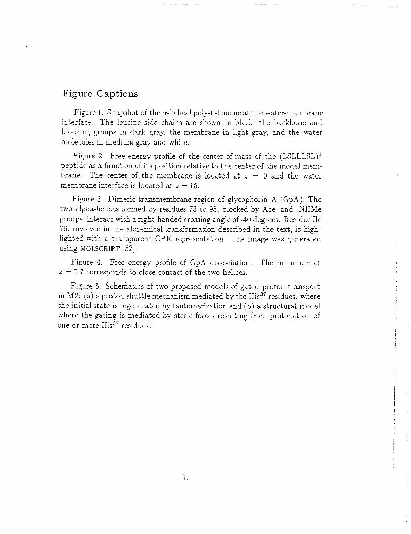
Figure Captions
Figure 1. Snapshot of the c_-helical poly-L-leucine at the water-membrane
interface. The leueine side chains are shown in black, the backbone and
blocking groups in dark gray, the membrane in light gray, and the water
molecules in medium gray and white.
Figure 2. Free energy profile of the center-of-mass of the (LSLLLSL) s
peptide as a function of its position relative to the center of the model mem-
brane. The center of the membrane is located at z = 0 and the water
membrane interface is located at z = 15.
Figure 3. Dimeric transmembrane region of glycophorin A (GpA). The
two alpha-helices formed by residues 73 to 95, blocked by Ace- and -NHMe
groups, interact with a right-handed crossing angle of-40 degrees. Residue Ile
76, involved in the alchemical transformation described in the text, is high-
lighted with a transparent CPK representation. The image was generated
using MOLSORIPT [52]
Figure 4. Free energy profile of GpA dissociation. The minimum at
z = 5.7 corresponds to close contact of the two helices.
Figure 5. Schematics of two proposed models of gated proton transport
in M2: (a) a proton shuttle mechanism mediated by the His ar residues, where
the initial state is regenerated by tautomerization and (b) a structural model
where the gating is mediated by steric forces resulting from protonation ofone or more His ar residues.
Q 7-Jl.
[..j
_oc_q
_D
.°
0_
] I i f
(y) zg
i i i i l
T 1 I
0I-
0
>>
©
_Jq
<" ,
AA(_) (kcal/mol)
I,.)
, I I 1 I [ I--_--____l___x__
, _jr-1, _(lq
_o
H+ :N_
regeneration a)
i
HID HIE
proton transfer
, !
"-. o.'
closed
.°_.
_',,... ° o_
• * o.. •. o-....,
",..o .. ,. .... .,'
'.-..°..•
open
Figure 5:
Sz
b)
]