Embed Size (px)
Citation preview
Perifosine Inhibits Multiple Signaling Pathways in Glial
Progenitors and Cooperates With Temozolomide
to Arrest Cell Proliferation in Gliomas In vivo
Hiroyuki Momota,1Edward Nerio,
1and Eric C. Holland
1,2
Departments of 1Cancer Biology and Genetics and 2Surgery (Neurosurgery) and Neurology,Memorial Sloan-Kettering Cancer Center, New York, New York
Abstract
Perifosine is an oral Akt inhibitor which exerts a markedcytotoxic effect on human tumor cell lines, and is currentlybeing tested in several phase II trials for treatment of majorhuman cancers. However, the efficacy of perifosine in humangliomas has not been established. As Akt is activated in f70%of human glioblastomas, we investigated the impact ofperifosine on glia in culture and on a mouse glioma modelin vivo . Here we show that perifosine strongly reduces phos-phorylation levels of Akt and extracellular signal-regulatedkinase (Erk) 1/2, induces cell cycle arrest in G1 and G2, andcauses dose-dependent growth inhibition of mouse glialprogenitors in which Akt and/or Ras-Erk 1/2 pathways areactivated. Furthermore, because temozolomide is a commonoral alkylating agent used in the treatment of gliomas, weinvestigated the effect of perifosine in combination withtemozolomide. We observed an enhanced effect when bothwere used in culture. With these results, we combinedperifosine and temozolomide as treatment of platelet-derivedgrowth factor B–driven gliomas in mice. Animal studiesshowed that perifosine and temozolomide combination ther-apy was more effective than temozolomide treatment alone(P < 0.01). These results indicate that perifosine is an effectivedrug in gliomas in which Akt and Ras-Erk 1/2 pathways arefrequently activated, and may be a new candidate for gliomatreatment in the clinic. (Cancer Res 2005; 65(16): 7429-35)
Introduction
Gliomas are the most common primary brain tumors (1). Themost aggressive subtypes, glioblastoma multiforme, makes up>50% of newly diagnosed gliomas. Despite aggressive therapy withsurgery, radiation, and chemotherapy, prognosis for patients withglioblastoma multiforme is poor with a median survival of f1 year(2). Many newer forms of treatment, including immunotherapy andgene therapy, have been tested in glioblastoma multiforme, butthese have not been efficacious. Presently, the best chemothera-peutic drugs for glioblastoma multiforme are alkylating agents suchas carmustine and temozolomide (2, 3).The alkylphospholipids compose a novel class of antitumor
agents with structural similarity to platelet-activating factors.Unlike most chemotherapeutic drugs which target the nuclear
DNA, alkylphospholipid interacts with the cell membrane andblocks signal transduction pathways (4). Perifosine is the first oralalkylphospholipid that exerts a marked cytotoxic effect in anumber of human tumor cell lines and has shown few side effectsin clinical phase I trials (5, 6).Recent studies have suggested the molecular mechanism of
perifosine action and the capacity to synergize with radiation orother anticancer drugs. Perifosine interferes with recruitment ofAkt to the plasma membrane and inhibits Akt phosphorylation andactivation (4). This drug also causes inhibition of extracellularsignal-regulated kinase (Erk) 1/2 and activation of c-Jun NH2-terminal kinase (JNK) and p21, resulting in cell cycle arrest in G1
and G2 (7–9). However, the details of the mechanisms of actionremain unclear, and this agent has not been studied fully in gliomacells. The Ras-Erk 1/2 signal transduction cascade is activated innearly all cases of glioblastoma multiforme (10, 11) and Akt isactivated in f70% (12–15). Moreover, evidence from ourlaboratory suggests that forced combined Ras and Akt activationin glial progenitors is sufficient to induce glioblastoma multiformein mice (14). Therefore, we hypothesized that the blockade of Aktand Ras-Erk 1/2 pathways by perifosine would have anti–glioblastoma multiforme effects.To develop a new paradigm for glioma therapy, we used mouse
glial progenitors in vitro , in which Akt and/or Ras-Erk 1/2 pathwaysare activated, and mouse glioma model in vivo . In this study, weused our established mouse models of gliomas and showed thedose-dependent growth inhibitory effects of perifosine in vitro .Perifosine blocked both Akt and Ras-Erk 1/2 signaling pathwaysand induced cell cycle arrest in G1 and G2. We show that perifosineenhances the effect of temozolomide, and does so better thanmTOR inhibitor alone. Finally, we show that perifosine andtemozolomide have additive effects on cell cycle of platelet-derivedgrowth factor (PDGF)-driven gliomas in vivo .
Materials and Methods
DNA constructs and infection of primary brain cell cultures. Thereplication-competent ALV splice acceptor (RCAS)/tv-a system used in this
work have been described previously (14). RCAS-LacZ plasmid was a gift
from Yi Li (Baylor College of Medicine, Houston, TX). RCAS-K-Ras, which
carries the gene encoding the G12D point mutant-activated K-Ras, waskindly provided by Galen Fisher (New York, NY). RCAS-Akt/HA, which
carries the activated form of Akt designated Akt-Myr D11-60 and has a HA
tag sequence added to the 3V-end of the cDNA, was a gift from Peter Vogt
(Scripps Research Institute, La Jolla, CA). RCAS-PDGF-B carries the entirecoding sequence of PDGF-B chain. Transfection of RCAS constructs into
chicken DF-1 fibroblasts and infection of primary glial progenitors from
Ntv-a transgenic mice by the various RCAS virions were done as describedpreviously (16). Ntv-a mice that express the TVA receptor from the nestin
promoter have been published (17). The mice are a mixed genetic
background of C57BL6, 129, Balb/C, and FVB/N.
Note: Supplementary data for this article are available at Cancer Research Online(http://cancerres.aacrjournals.org/).
Requests for reprints: Eric C. Holland, Department of Surgery (Neurosurgery) andNeurology, Memorial Sloan-Kettering Cancer Center, 1275 York Avenue, New York, NY10021. Phone: 212-639-3017; Fax: 646-422-2062; E-mail: [email protected].
I2005 American Association for Cancer Research.doi:10.1158/0008-5472.CAN-05-1042
www.aacrjournals.org 7429 Cancer Res 2005; 65: (16). August 15, 2005
Research Article
Research. on June 28, 2018. © 2005 American Association for Cancercancerres.aacrjournals.org Downloaded from

Cell culture. Glial progenitors transformed with RCAS-LacZ (Ntv-a/LacZ), RCAS-K-Ras (Ntv-a/Ras), RCAS-Akt/HA (Ntv-a/Akt), RCAS-K-Ras +
Akt/HA (Ntv-a/Ras + Akt), and RCAS-PDGF-B (Ntv-a/PDGF) were cultured
in DMEM supplemented with 10% FCS, 100 units/mL penicillin, and 100 Agstreptomycin. Human glioma cell lines, T98G and U373MG, were obtainedfrom American Type Culture Collection (Manassas, VA) and were cultured
in the same medium. All cells were cultured in a humidified atmosphere at
37jC and 5% CO2.
Drugs. Perifosine was provided by Keryx Biopharmaceuticals (New York,NY). Temozolomide was from Schering-Plough (Kenilworth, NJ). Rapamycin
was from Wyeth (Philadelphia, PA). Perifosine stock solutions were
prepared by dissolving perifosine in PBS for cell culture, and in 0.9% NaCl
solution for mouse treatment both at 25 mmol/L concentration.Temozolomide stock solutions were prepared by dissolving temozolomide
in DMSO at 100 mmol/L concentration for cell culture, and at 40 mg/mL
concentration for mouse treatment. Temozolomide solution for mice wasdiluted in saline (5 mg/mL) and administered. Rapamycin stock solutions
were prepared by dissolving in ethanol at 100 Amol/L concentration.
Western blot analysis. Whole-cell protein extracts were prepared by
cold lysis of cell pellets. M-Per (Pierce, Rockford, IL) lysis buffer was
supplemented with 30 mmol/L sodium fluoride, 1 mmol/L sodium
vanadate, 0.5 mmol/L phenylmethylsulfonyl fluoride, 100 mmol/L NaCl, 1
mmol/L EDTA, and protease inhibitor cocktail tablets (Roche, Indian-
apolis, IN). Protein concentrations were determined by bicinchoninic acid
assay (BCA) method (Bio-Rad, Hercules, CA). Samples (100 Ag) were
separated by 11% SDS-PAGE gel, and transferred onto polyvinylidene
difluoride membrane (Millipore, Bedford, MA). Membranes were blocked
with 5% nonfat milk in PBS-0.1% Tween 20. Primary and secondary
antibodies were diluted in the same solution. Signal was visualized using
enhanced chemiluminescence (Amersham Biosciences, Piscataway, NJ).
Primary antibodies against Akt (#9272), phospho-Akt (P-Akt; Ser473; #9271),
S6 kinase (S6K; #9202), P-S6K (Thr389; #9205), 4E-BP1 (#9452), P-4E-BP1
(Ser65; #9451), Erk 1/2 (#9102), P-Erk 1/2 (Ser217/221; #9101), P-JNK (Thr183/
Tyr185; #9251), P-p38 (Thr180/Tyr182; #9211), and P-eIF4E (Ser209; #9741; Cell
Signaling, Beverly, MA) were used at 1:1,000 dilution, and anti-Actin (I-19;
Santa Cruz Biotechnology, Santa Cruz, CA) was used at 1:500 dilution.
Secondary peroxidase-conjugated anti-rabbit antibody (Amersham Bio-
sciences) and anti-mouse antibody (Roche) were used at 1:1,000 dilution.
Platelet-derived growth factor-BB ELISA. Whole-cell protein was
extracted using the same lysis buffer as above, and 100 Ag of protein were
used for ELISA. PDGF-BB ELISA was done using a Quantikine kit (R&DSystems, Minneapolis, MN) according to the manufacturer’s instructions.
The absorbance at 590 nm was recorded and the absorbance at 540 nm was
subtracted from the readings at 590 nm using the 96-well plate reader
(Thermo Electron Corporation, Vantaa, Finland).Cell growth assay. Four thousand cells ( for 10% serum) or 5,000 cells
( for 1% serum) per well were plated in 96-well culture plates with 100 AL of
culture medium. After applying drugs, cells were cultured for 2 days. Viable
cells were determined using 3-(4,5-dimethylthiazol-2-yl)-2,5-diphenyltetra-zolium bromide (MTT) assay (Cell Proliferation Kit I; Roche). The
absorbance at 590 nm was recorded using the 96-well plate reader (Thermo
Electron Corporation).Cdk kinase assay. After treatment with perifosine, the cells were washed
twice with cold PBS and lysed in radioimmunoprecipitation assay buffer [50
mmol/L Tris-HCl (pH 7.4), 250 mmol/L NaCl, 5 mmol/L EDTA, 0.5% NP40,
protease inhibitor cocktail tablets (Roche), 10 Ag/mL soybean trypsininhibitor, and 2 mmol/L phenylmethylsulfonyl fluoride]. The cell lysates
were sonicated twice for 30 seconds, incubated on ice for 30 minutes, and
supernatants were recovered by centrifuging at 14,000 rpm at 4jC for 10
minutes. Protein concentrations were determined by BCA method (Bio-Rad). Proteins (200 Ag) were immunoprecipitated using specific antibodies
against Cdk1 and Cdc2 (Santa Cruz Biotechnology), and collected with
protein A-Sepharose slurry (Repligen, Waltham, MA) at 4jC with agitation.The immunoprecipitate was washed thrice with the lysis buffer. The Cdk
kinase reactions in the immunoprecipitate were done in a kinase reaction
buffer [20 mmol/L Tris-HCl (pH 7.4), 7.5 mmol/L MgCl2, 1 mmol/L
dithrothrectol] containing 30 Amol/L ATP, [g-32P]ATP (3,000 Ci/mmol;
Amersham Biosciences), and 40 Ag histone H1 (Roche) at 37jC for 30minutes. Phosphorylated histone H1 was resolved on 12% SDS-PAGE gels
followed by autoradiography.
Cell cycle analysis. Cells treated with perifosine were trypsinized and
washed in PBS at each time point. Aliquots of 1 � 106 cells were fixed with70% methanol at 4jC for at least 12 hours before centrifugation. The cell
pellets were treated with a solution containing 50 Ag/mL propidium iodide
(Sigma, St. Louis, MO) and 2 mg/mL RNase A (Roche) for 30 minutes at
room temperature. Stained cells were analyzed using a FACScan flowcytometer (Becton Dickinson, San Jose, CA) and evaluated with CellQuest
and FlowJo software.
Giemsa staining of cultured cells. The cells were on four-well glass
slides (Nagle Nunc International, Naperville, IL) and treated with perifosine.After 24 hours of incubation, medium was aspirated from the chamber. The
cells were washed with PBS (pH 7.4), and fixed in methanol at room
temperature for 10 minutes. Then, 75 AL of Giemsa stain (LabChem, Inc.,Pittsburgh, PA) was diluted in 1 mL of water, and applied for 5 minutes. The
stain was discarded and the cells were washed with water and visualized
under a light microscope. The percentage of multinuclear cells was
semiquantitatively analyzed by counting cells in three random fields, usinga light microscope at 200� magnification.
Generation of tumor-bearing mice. DF1 cells transfected with RCAS-
PDGF and double-transgenic Ef-luc Ntv-a mice have been described
previously (18). Neonatal Ef-luc Ntv-a mice received intracranial injectionsof f104 DF1 cells producing the RCAS-PDGF retrovirus. Mice were then
routinely screened with bioluminescence imaging, and image-positive mice
were followed over time, treated, and sacrificed.Drug treatment of tumor-bearing mice. Image-positive Ef-luc Ntv-a
mice were treated daily with i.p. administration of buffer alone as a control, or
i.p. administration of 100 mg/kg temozolomide, or oral administration of 30
mg/kg perifosine, or a combination with perifosine and temozolomide for 3to 5 days. The mean doses of the treatments were: Control, 5 (all five);
temozolomide, 3.75 (three to five); perifosine, 3.75 (three to four); and
perifosine + temozolomide, 3 (all three). Control buffer solution consisted of
5% DMSO and 1% Tween 80 in distilled water.Brain sectioning, H&E staining, and immunohistochemistry. These
procedures were done as described previously (16). Primary antibody against
Ki-67 (VP-K451; Vector, Burlingame, CA) was used at 1:500 dilution overnightat 4jC. Secondary biotinylated anti-rabbit antibody (Vector) was used at
1:250 dilution for 1 hour at room temperature. Antibodies were diluted in 5%
goat serum PBS-Tween 0.1%. Peroxidase signal was developed using the
avidin-biotin complex kit (Vector). Ki-67-positive cells were counted in one ofthe most strongly staining fields per section (1,500-2,500 cells/field).
Statistical analysis. Comparison between two groups was made using
Student’s t test. Data represent the mean F SD. P values of < 0.05 were
considered statistically significant.
Results
We initially investigated the effects of perifosine on human gliomacell lines. We tested the effects of this drug at a variety ofconcentrations on two commonly used lines, T98G and U373MG.These cell lines are both mutant for PTEN and therefore haveconstitutively active Akt signaling. We treated cell cultures for 48hours with a range of concentrations and found a substantialdifference in cell viability between the two cell lines (P < 0.001;Fig. 1A). We interpreted the difference to genetic and signalingvariation seen in primary gliomas and cell lines derived from them.Because this compound is effective by its action on the signalingpathways, we proceeded with the investigation of perifosine activityusing glial cell cultures with defined signaling alterations. Wegenerated glial progenitor cell cultureswith constitutively active Ras,Akt, and PDGF autocrine loop signaling using the RCAS/tv-a system(14). Briefly, central nervous system progenitor cells were derivedfrommice expressing tv-a (the receptor for the avail retroviral vectorRCAS) as a transgene from the nestin promoter (Ntv-a cells). RCAS
Cancer Research
Cancer Res 2005; 65: (16). August 15, 2005 7430 www.aacrjournals.org
Research. on June 28, 2018. © 2005 American Association for Cancercancerres.aacrjournals.org Downloaded from

vector infection, therefore, transfers gene to nestin expressingprogenitor cells. These cells were infected by RCAS vectors encodingLacZ as a control, activated K-Ras, Akt, or PDGF-B, and have beendescribed previously (16). To verify the expression levels ofoncoproteins in RCAS infected glial progenitors, we did Westernblot and ELISA analyses. K-Ras is overexpressed in Ntv-a/Ras andNtv-a/Ras + Akt–infected lines (Fig. 1B). P-Akt levels are increased inNtv-a/Ras + Akt and Ntv-a/Akt cells, whereas total Akt levels areequal in all cell lines. Virally transduced and constitutively active Aktcontains a hemaglutinin epitope tag and was found only in Ntv-a/Ras + Akt and Ntv-a/Akt cells. K-Ras expression and P-Akt levels
were minimal in Ntv-a/LacZ cells. Although K-Ras and Akt are thedownstream components of the PDGFR signaling cascade, increasein K-Ras and P-Akt levels in Ntv-a/PDGF cells were marginal as hasbeen described previously (19). ELISA analysis showed substantialincrease in PDGF-BB levels in Ntv-a/PDGF cells (Fig. 1C ; results ofone representative experiment are shown). The level of PDGF-BB inlysates of Ntv-a/PDGF cells was >15-fold higher than in the other celllines analyzed.Perifosine inhibition of cell growth is highly dependent on
serum concentration.We did cell proliferation assays to determineperifosine concentration required to inhibit growth of these cellswith various signaling abnormalities. Perifosine elicited cell growthinhibition in a dose-dependentmanner in all cells tested. The IC50 forgrowth of cell lines was determined by MTT assay. When the cellswere cultured for 48 hours in 10% FCS-supplementedmedia, the IC50
for cells with constitutively active PDGF, Ras, or Akt signaling wassimilar and found to be f45 Amol/L (Fig. 2A). Ntv-a/LacZ controlcells weremore resistant to perifosine than the other cell lines at thisserum concentration (P = 0.0006 at 50 Amol/L). The effect ofperifosine was enhanced markedly in all cell lines when the FCSconcentration in the medium was reduced to 1% (Fig. 2B).Interestingly, although the control cells were the most resistant toperifosine at high serum concentrations, the PDGF autocrine-drivencells were the most resistant to perifosine at low serum concen-trations (P = 0.0045 at 5 Amol/L, P < 0.0001 at 10 Amol/L), implyingthat PDGF signaling could provide protection from serumdeprivation in the presence of perifosine. In the following experi-ments in this study, we used 10% FCS to assess the molecularmechanisms of action of perifosine.Perifosine inhibits Akt and Ras signaling pathways in glial
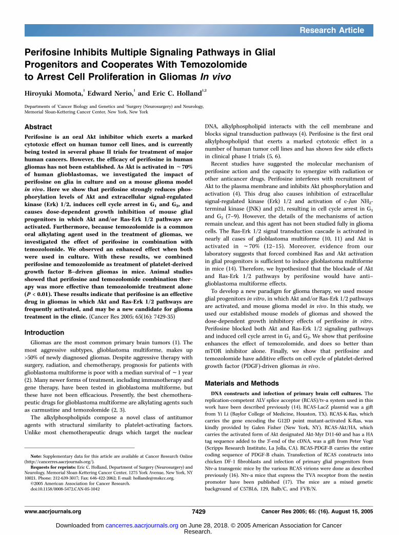
cells. We used Western blot analysis to determine the effect ofperifosine on signaling pathways in our modified glial progenitors.We specifically investigated the PDGF-driven cells as Akt is wild-typeand they have relatively low Akt activity, the RCAS-Akt infected cellsas these have a myristoylated and constitutively active Akt that ishighly phosphorylated, and the RCAS-LacZ infected cells as acontrol. Each cell type showed a unique response to perifosine overtime. The Ntv-a/LacZ cells showed minimal effect of perifosine onAkt or Erk 1/2 pathways but did show some effect on 4E-BP1phosphorylation at later time points (Fig. 3A). The Ntv-a/PDGF cellsshowed a strong block in Akt and S6K phosphorylation by 3 hourswith a decrease in 4E-BP1 and Erk 1/2 phosphorylation by 6 hours(Fig. 3B). The cells expressing the constitutively active Akt showedblockade of Akt phosphorylation by 3 hours but a delayed effect ofS6K phosphorylation. The effect on Erk 1/2 and 4E-BP1 was similarto the Ntv-a/PDGF cells (Fig. 3C). Phospho-JNK and phospho-p38levels did not change substantially or were below detection levels,and phospho-eIF4E levels were also did not change significantly atthis drug concentration (data not shown). As Akt is activated but notmutated in human gliomas, we chose to investigate the effects ofperifosine on glia using the PDGF-driven cells.Perifosine inhibits Cdk2 and Cdc2 kinase activities and
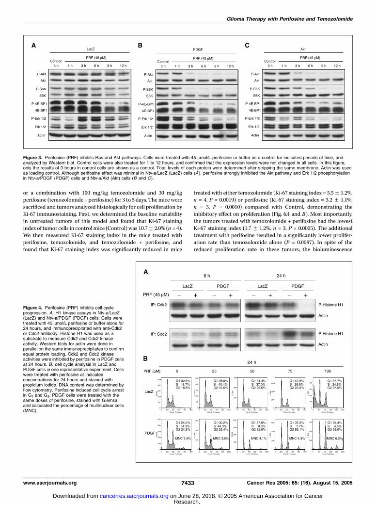
induces G1 and G2 arrest. The reduction of cell numbers seen inFig. 2 implied that perifosine might be affecting cell proliferation. Toinvestigate the impact of perifosine on cell cycle, we did Cdk2 andCdc2 kinase assays. Cdk2 kinase activity was strongly inhibited inNtv-a/PDGF cells after treatment with 45 Amol/L perifosine for 24hours, whereas the effect on Cdc2 was milder (Fig. 4A). By contrast,the effects of perifosine on the Cdk2 in either cell type were notobserved after 6 hours of treatment. As illustrated in Fig. 3B , at 6hours, there is a strong effect on Akt, S6, 4E-BP1, and Erk 1/2 in the
Figure 1. A, growth-inhibitory effect of perifosine (PRF) on human glioma celllines. Cells were incubated in the medium with 10% FCS for 48 hours withindicated concentrations of perifosine. Cell viability was determined by MTTassay. *, P < 0.001, compared with U373MG. Points, mean; bars, F SD. B,expression of viral proteins in mouse glial progenitors. Western blot for K-Rasand Akt. Viral Akt protein is hemaglutinin epitope–tagged. Ntv-a/LacZ (LacZ)cells were used as a control cell line in this study. Actin was used as loadingcontrol. C, PDGF-BB ELISA in one representative experiment. Whole celllysates were analyzed in this assay. Ntv-a/PDGF (PDGF); Ntv-a/Ras (Ras);Ntv-a/Ras + Akt (Ras + Akt); Ntv-a/Akt (Akt).
Glioma Therapy with Perifosine and Temozolomide
www.aacrjournals.org 7431 Cancer Res 2005; 65: (16). August 15, 2005
Research. on June 28, 2018. © 2005 American Association for Cancercancerres.aacrjournals.org Downloaded from

Ntv-a/PDGF cells. The data implies that the effects of perifosine onCdk2 are likely to be secondary to cell cycle arrest induced byalterations in the signaling pathways rather than a direct effect ofperifosine on Cdk2. We also examined the relative levels ofendogenous cyclin A, p27, and p21 by Western blotting in Ntv-a/PDGF cells, but no significant changes were observed at 10% FCSconcentration up to 12 hours (Supplementary Fig. S1).The above data implies that perifosine affects proliferation in
these glial cells, we therefore did flow cytometry cell cycle analysis ofNtv-a/LacZ and Ntv-a/PDGF cells treated with various doses ofperifosine for 24 hours. At doses up to 75 Amol/L, there was littleeffect on the Ntv-a/LacZ cells (P = 0.0020; for further statisticalanalysis of all points, see Supplementary Fig. S2), but at 100 Amol/L,these cells developed a significant percentage of sub-G1 cellsconsistent with cell death. By contrast, in the Ntv-a/PDGF culture,perifosine caused a dose-dependent loss of cells in S phase (Fig. 4B).At lower perifosine concentrations (25-50 Amol/L), cells arrestedpredominantly in G1 (P = 0.0068 at 25 Amol/L). At higher doses(75-100 Amol/L), Ntv-a/PDGF cells arrested predominantly in G2
(P = 0.0028 and P = 0.0006 at 75 and 100 Amol/L, respectively). The G2
arrest seen at high perifosine doses was also observed in the otherprogenitors as well (Supplementary Fig. S3). Cells with 4 N or greaterDNA content might be either G2 arrested or multinucleated.Therefore, we stained the treated cells with Giemsa and calculated
the percentage of multinuclear cells. Only f6% of all treated cellswere found to be multinuclear; thus, the majority of the 4 N cellswere arrested in G2.Relative effects of mTOR inhibition and perifosine-mediated
Akt inhibition. One effect of Akt blockade is a reduction of mTORactivity, however, perifosine has several other effects than simplymTOR inhibition such as affecting non-mTOR pathways down-stream of Akt as well as mitogen-activated protein kinase. SeveralmTOR inhibitors are available (rapamycin) or in clinical trials. Wereasoned that given the central role of Akt activity in gliomasblockade of the Akt pathway would ultimately be part of acombination of drugs used to treat these tumors. Therefore, wecompared the effects of rapamycin and perifosine as single agentsand in combination with the cytotoxic chemotherapy currentlyused for glioma temozolomide on our cell cultures. First, wedetermined doses of rapamycin and perifosine that equallyinhibited phosphorylation of S6K. Western blot analysis of cellstreated with various concentrations of the drug doses showed that0.1 nmol/L of rapamycin and 45 Amol/L of perifosine achievedequal blockade of S6K phosphorylation (Fig. 5A). We noted that atthese doses, rapamycin showed a slight increase in P-Akt levelsconsistent with feedback effects, as has been reported previously(20, 21), and did not see inhibition of Erk 1/2 or 4E-BP1phosphorylation. This limited effect of rapamycin contrasts withthe effect of perifosine on P-Akt, P-S6K, P-4E-BP1, and P-Erk 1/2 asnoted earlier in this manuscript.We then investigated the effects of these drugs and combinations
on our cells in culture. We found that as single agents, perifosine hada greater effect on Ntv-a/PDGF cells (Supplementary Fig. S4; P <0.0001) but had minimal effects on the Ntv-a/LacZ cells (P = 0.1034).Finally, whereas rapamycin had aminimal effect on cells treatedwithtemozolomide (P = 0.0252), perifosine substantially enhanced theinhibitory effects of temozolomide in Ntv-a/PDGF cells (P < 0.0001).Therefore, perifosine results in blockade of several components ofsignal transduction and seems to cooperate with temozolomidebetter than inhibition of mTOR alone with rapamycin.We then did flow analysis of Ntv-a/PDGF cells with 300 Amol/L
temozolomide and 45 Amol/L perifosine alone and in combinationto better define the effects of the combination of these two drugsover time. The analysis indicated that at these doses, thecombination of these two drugs achieved a more complete arrestof the cell cycle than temozolomide alone (Fig. 5B , P < 0.05; forfurther statistical analysis, see Supplementary Fig. S5). Finally, wedetermined the schedule of temozolomide and perifosine treat-ment that had the greatest effect on cells in culture. When we usedtemozolomide and perifosine sequentially, the growth-inhibitoryeffect was more pronounced when temozolomide was used firstfollowed by perifosine than the reverse (Supplementary Fig. S6; P <0.005). We found that simultaneous treatment with the two drugsgave the most potent effect in these cells (P < 0.005, compared withtemozolomide followed by perifosine treatment).Combination therapy of perifosine and temozolomide
reduces tumor proliferation in vivo . The above data indicatesthat perifosine results in blockade of several signaling pathways andachieves a cell cycle arrest in culture that significantly enhances theeffect of temozolomide in culture. Given the enhanced effect ofperifosine over rapamycin in combination with temozolomide, wechose to test temozolomide and perifosine as single agents and incombination against a PDGF-driven glioma model in vivo . Weidentified mice with tumors by bioluminescence imaging and eithertreated themwith 100 mg/kg temozolomide, or 30 mg/kg perifosine,
Figure 2. Perifosine (PRF) inhibits growth in mouse glial progenitors. Cells wereincubated for 48 hours with indicated concentrations of perifosine. Cell viabilitywas determined by MTT assay. A, cells were cultured in the medium with10% FCS. Fifty percent growth inhibition (IC50) dose was determined in thiscondition. B, perifosine effect in 1% FCS condition. *, P < 0.001; **,P < 0.005, compared with the other cell lines. Points, mean; bars, F SD.Ntv-a/LacZ (LacZ); Ntv-a/PDGF (PDGF); Ntv-a/Ras (Ras); Ntv-a/Ras + Akt(Ras + Akt); Ntv-a/Akt (Akt).
Cancer Research
Cancer Res 2005; 65: (16). August 15, 2005 7432 www.aacrjournals.org
Research. on June 28, 2018. © 2005 American Association for Cancercancerres.aacrjournals.org Downloaded from
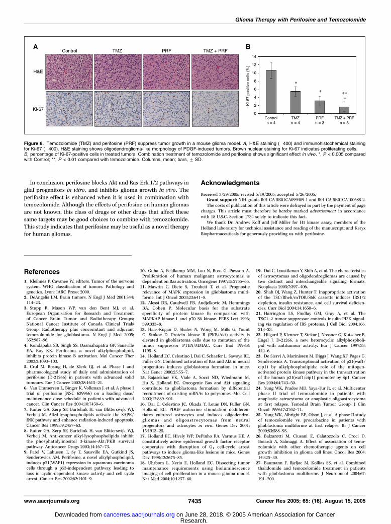
or a combination with 100 mg/kg temozolomide and 30 mg/kgperifosine (temozolomide + perifosine) for 3 to 5 days. Themice weresacrificed and tumors analyzed histologically for cell proliferation byKi-67 immunostaining. First, we determined the baseline variabilityin untreated tumors of this model and found that Ki-67 stainingindex of tumor cells in controlmice (Control) was 10.7F 2.0% (n = 4).We then measured Ki-67 staining index in the mice treated withperifosine, temozolomide, and temozolomide + perifosine, andfound that Ki-67 staining index was significantly reduced in mice
treated with either temozolomide (Ki-67 staining index = 5.5F 1.2%,n = 4, P = 0.0019) or perifosine (Ki-67 staining index = 3.2 F 1.1%,n = 3, P = 0.0010) compared with Control, demonstrating theinhibitory effect on proliferation (Fig. 6A and B). Most importantly,the tumors treated with temozolomide + perifosine had the lowestKi-67 staining index (1.7 F 1.2%, n = 3, P = 0.0005). The additionaltreatment with perifosine resulted in a significantly lower prolifer-ation rate than temozolomide alone (P = 0.0087). In spite of thereduced proliferation rate in these tumors, the bioluminescence
Figure 3. Perifosine (PRF) inhibits Ras and Akt pathways. Cells were treated with 45 Amol/L perifosine or buffer as a control for indicated periods of time, andanalyzed by Western blot. Control cells were also treated for 1 to 12 hours, and confirmed that the expression levels were not changed in all cells. In this figure,only the results of 3 hours in control cells are shown as a control. Total levels of each protein were determined after stripping the same membrane. Actin was usedas loading control. Although perifosine effect was minimal in Ntv-a/LacZ (LacZ) cells (A), perifosine strongly inhibited the Akt pathway and Erk 1/2 phosphorylationin Ntv-a/PDGF (PDGF) cells and Ntv-a/Akt (Akt) cells (B and C ).
Figure 4. Perifosine (PRF) inhibits cell cycleprogression. A, H1 kinase assays in Ntv-a/LacZ(LacZ) and Ntv-a/PDGF (PDGF) cells. Cells weretreated with 45 Amol/L perifosine or buffer alone for24 hours, and immunoprecipitated with anti-Cdk2or Cdc2 antibody. Histone H1 was used as asubstrate to measure Cdk2 and Cdc2 kinaseactivity. Western blots for actin were done inparallel on the same immunoprecipitates to confirmequal protein loading. Cdk2 and Cdc2 kinaseactivities were inhibited by perifosine in PDGF cellsat 24 hours. B, cell cycle analysis in LacZ andPDGF cells in one representative experiment. Cellswere treated with perifosine at indicatedconcentrations for 24 hours and stained withpropidium iodide. DNA content was determined byflow cytometry. Perifosine induced cell cycle arrestin G1 and G2. PDGF cells were treated with thesame doses of perifosine, stained with Giemsa,and calculated the percentage of multinuclear cells(MNC).
Glioma Therapy with Perifosine and Temozolomide
www.aacrjournals.org 7433 Cancer Res 2005; 65: (16). August 15, 2005
Research. on June 28, 2018. © 2005 American Association for Cancercancerres.aacrjournals.org Downloaded from

imaging of the mice after perifosine treatment was quite variable(data not shown). The reason for the variability is not clear but mayreflect the activity of the E2F1 promoter in the G2 phase of the cellcycle and variable G1 and G2 blockade in the tumors.
Discussion
We show growth inhibitory effects of perifosine and itsmolecular mechanism through signal transduction and cell cycleprogression in glial progenitors in vitro . Our work shows thatperifosine blocks Akt and Ras-Erk 1/2 signaling in mouse glialprogenitors. These cell lines are engineered to have activated Rasand Akt signaling pathways, which are the pathways mostcommonly activated in human malignant gliomas. Therefore, thegrowth-inhibitory effects of perifosine are more significant in thesecells than in control cells which are infected with LacZ gene. Wehave also confirmed that perifosine inhibits Cdk2 and Cdc2activities in our cells, and blocks cell cycle progression in G1 and G2
as reported in squamous carcinoma cells (9). These inhibitoryeffects on Cdk2 and Cdc2 were not observed after 6 hours oftreatment, and we suspect that it is a secondary effect on thesignaling pathways. Interestingly, perifosine effect was dependenton serum concentration. Therefore, we think that the inhibitingeffect of perifosine may be compensated for by growth factorscontained in the serum.Different cell and cancer types have characteristic signal
transduction pathways and might be expected to show differingresponses to small molecule inhibitors of specific signalingcomponents. In this light, we have found some characteristics ofglial progenitor cell responses to perifosine that are either unique orat least different from that found in other cell types. For example,the IC50 for perifosine in our mouse glial progenitor cell lines(45 Amol/L) is substantially higher than those reported before suchas glioma (IC50 = 3.1 Amol/L), or other cancer cell lines (0.2-19.9Amol/L) including melanoma, lung, colon, squamous cell, breast,and prostate cancer (4, 9, 22). Second, in glial progenitors expressingconstitutively active Akt, perifosine treatment leads to reduction inP-Akt and a slower but nonetheless substantial effect on P-S6K. Thisfinding differs from results reported with PC3 prostate carcinomacells which have myristoylated form of Akt and are resistant toperifosine action (4). This differential response to perifosine mightbe caused by several factors including different perifosine concen-trations used (45 versus 5 Amol/L), different length of treatment(3 hours versus 30 minutes), and different cell types. Third, the JNKpathway is not significantly up-regulated in glial progenitor cellsunlike what is seen in human leukemia cell lines (7). Finally, p21expression does not increase significantly in glial cells treated withperifosine and phosphorylation of Erk 1/2 is suppressed in our cellsat the IC50, in contrast to the human leukemia cell lines (23).To evaluate the potential of perifosine for combination
chemotherapy, we chose to examine temozolomide, which is acommon drug for glioma therapy. We found that the addition ofperifosine to temozolomide enhanced cell cycle arrest and growthinhibition. Because perifosine is known to enhance radiation-induced apoptosis in human leukemia cell lines (7), and bothradiation and temozolomide cause DNA damage, perifosine mayenhance the effects of temozolomide. We showed the combinationeffect of temozolomide and perifosine in vivo by immunohisto-chemistry where the results of Ki-67 staining in tumor cells supportthe cell culture results. Although the combination of perifosine andtemozolomide only trended toward being more effective thanperifosine monotherapy, it is possible that this finding wouldbecome statistically significant if more animals were used.We previously showed thatmTOR blockade results in inhibition of
proliferation in PDGF-induced mouse gliomas (18). As we show herein the same tumors, blockade of the Akt signaling pathway canenhance temozolomide effect, and Akt blockade is more effectivethan mTOR blockade when combined with temozolomide. Further-more, in sequential treatmentwith temozolomide and perifosine, wefound the growth-inhibitory effect is more pronounced whentemozolomide is administered before perifosine rather than after.These results may help guide schedules of drug combination whentemozolomide is used with other drugs that inhibit Ras and Aktsignaling pathways. Temozolomide has become the first choice ofchemotherapy for gliomas because of its efficacy, convenience, andtolerability (3, 24, 25). Moreover, recent studies suggest thattemozolomide may enhance the effectiveness of other chemother-apeutic agents (26, 27). However, impact of temozolomide on long-term disease control is limited and new drugs are needed.
Figure 5. Relative effect of mTOR and Akt inhibition, and combined effect ofperifosine and temozolomide in vitro. A, Ntv-a/PDGF (PDGF) cells were treatedwith indicated concentrations of rapamycin (RPM) or perifosine (PRF) for 6hours, and analyzed by Western blot. The dose of 0.1 nmol/L rapamycin and45 Amol/L perifosine block S6K phosphorylation equally. Actin was used asloading control. B, cell cycle analysis in PDGF cells in one representativeexperiment. Cells were treated with buffer alone (Control), 45 Amol/L perifosine(PRF), 300 Amol/L temozolomide (TMZ), or 300 Amol/L temozolomide and45 Amol/L perifosine (TMZ + PRF) for indicated periods of time, stained withpropidium iodide, and analyzed by flow cytometry. Perifosine and temozolomidecombination treatment shows marked cell cycle arrest.
Cancer Research
Cancer Res 2005; 65: (16). August 15, 2005 7434 www.aacrjournals.org
Research. on June 28, 2018. © 2005 American Association for Cancercancerres.aacrjournals.org Downloaded from
In conclusion, perifosine blocks Akt and Ras-Erk 1/2 pathways inglial progenitors in vitro , and inhibits glioma growth in vivo . Theperifosine effect is enhanced when it is used in combination withtemozolomide. Although the effects of perifosine on human gliomasare not known, this class of drugs or other drugs that affect thesesame targets may be good choices to combine with temozolomide.This study indicates that perifosine may be useful as a novel therapyfor human gliomas.
Acknowledgments
Received 3/29/2005; revised 5/19/2005; accepted 5/26/2005.Grant support: NIH grants R01 CA 5R01CA099489-1 and R01 CA 5R01CA100688-2.The costs of publication of this article were defrayed in part by the payment of page
charges. This article must therefore be hereby marked advertisement in accordancewith 18 U.S.C. Section 1734 solely to indicate this fact.
We thank Dr. Andrew Koff and Jeff Miller for H1 kinase assay; members of theHolland laboratory for technical assistance and reading of the manuscript; and KeryxBiopharmaceuticals for generously providing us with perifosine.
References
1. Kleihues P, Cavanee W, editors. Tumor of the nervoussystem. WHO classification of tumors. Pathology andgenetics. Lyon: IARC Press; 2000.
2. DeAngelis LM. Brain tumors. N Engl J Med 2001;344:114–23.
3. Stupp R, Mason WP, van den Bent MJ, et al.European Organisation for Research and Treatmentof Cancer Brain Tumor and Radiotherapy Groups;National Cancer Institute of Canada Clinical TrialsGroup. Radiotherapy plus concomitant and adjuvanttemozolomide for glioblastoma. N Engl J Med 2005;352:987–96.
4. Kondapaka SB, Singh SS, Dasmahapatra GP, SausvilleEA, Roy KK. Perifosine, a novel alkylphospholipid,inhibits protein kinase B activation. Mol Cancer Ther2003;2:1093–103.
5. Crul M, Rosing H, de Klerk GJ, et al. Phase I andpharmacological study of daily oral administration ofperifosine (D-21266) in patients with advanced solidtumours. Eur J Cancer 2002;38:1615–21.
6. Van Ummersen L, Binger K, Volkman J, et al. A phase Itrial of perifosine (NSC 639966) on a loading dose/maintenance dose schedule in patients with advancedcancer. Clin Cancer Res 2004;10:7450–6.
7. Ruiter GA, Zerp SF, Bartelink H, van Blitterswijk WJ,Verheij M. Alkyl-lysophospholipids activate the SAPK/JNK pathway and enhance radiation-induced apoptosis.Cancer Res 1999;59:2457–63.
8. Ruiter GA, Zerp SF, Bartelink H, van Blitterswijk WJ,Verheij M. Anti-cancer alkyl-lysophospholipids inhibitthe phosphatidylinositol 3-kinase-Akt/PKB survivalpathway. Anticancer Drugs 2003;14:167–73.
9. Patel V, Lahusen T, Sy T, Sausville EA, Gutkind JS,Senderowicz AM. Perifosine, a novel alkylphospholipid,induces p21(WAF1) expression in squamous carcinomacells through a p53-independent pathway, leading toloss in cyclin-dependent kinase activity and cell cyclearrest. Cancer Res 2002;62:1401–9.
10. Guha A, Feldkamp MM, Lau N, Boss G, Pawson A.Proliferation of human malignant astrocytomas isdependent on Ras activation. Oncogene 1997;15:2755–65.
11. Mawrin C, Diete S, Treuheit T, et al. Prognosticrelevance of MAPK expression in glioblastoma multi-forme. Int J Oncol 2003;23:641–8.
12. Alessi DR, Caudwell FB, Andjelkovic M, HemmingsBA, Cohen P. Molecular basis for the substratespecificity of protein kinase B; comparison withMAPKAP kinase-1 and p70 S6 kinase. FEBS Lett 1996;399:333–8.
13. Haas-Kogan D, Shalev N, Wong M, Mills G, YountG, Stokoe D. Protein kinase B (PKB/Akt) activity iselevated in glioblastoma cells due to mutation of thetumor suppressor PTEN/MMAC. Curr Biol 1998;8:1195–8.
14. Holland EC, Celestino J, Dai C, Schaefer L, Sawaya RE,Fuller GN. Combined activation of Ras and Akt in neuralprogenitors induces glioblastoma formation in mice.Nat Genet 2000;25:55–7.
15. Rajasekhar VK, Viale A, Socci ND, Wiedmann M,Hu X, Holland EC. Oncogenic Ras and Akt signalingcontribute to glioblastoma formation by differentialrecruitment of existing mRNAs to polysomes. Mol Cell2003;12:889–901.
16. Dai C, Celestino JC, Okada Y, Louis DN, Fuller GN,Holland EC. PDGF autocrine stimulation dedifferen-tiates cultured astrocytes and induces oligodendro-gliomas and oligoastrocytomas from neuralprogenitors and astrocytes in vivo . Genes Dev 2001;15:1913–25.
17. Holland EC, Hively WP, DePinho RA, Varmus HE. Aconstitutively active epidermal growth factor receptorcooperates with disruption of G1 cell-cycle arrestpathways to induce glioma-like lesions in mice. GenesDev 1998;12:3675–85.
18. Uhrbom L, Nerio E, Holland EC. Dissecting tumormaintenance requirements using bioluminescenceimaging of cell proliferation in a mouse glioma model.Nat Med 2004;10:1257–60.
19. Dai C, Lyustikman Y, Shih A, et al. The characteristicsof astrocytomas and oligodendrogliomas are caused bytwo distinct and interchangeable signaling formats.Neoplasia 2005;7:397–406.
20. Shah OJ, Wang Z, Hunter T. Inappropriate activationof the TSC/Rheb/mTOR/S6K cassette induces IRS1/2depletion, insulin resistance, and cell survival deficien-cies. Curr Biol 2004;14:1650–6.
21. Harrington LS, Findlay GM, Gray A, et al. TheTSC1–2 tumor suppressor controls insulin-PI3K signal-ing via regulation of IRS proteins. J Cell Biol 2004;166:213–23.
22. Hilgard P, Klenner T, Stekar J, Nossner G, Kutscher B,Engel J. D-21266, a new heterocyclic alkylphospholi-pid with antitumour activity. Eur J Cancer 1997;33:442–6.
23. De Siervi A, Marinissen M, Diggs J, Wang XF, Pages G,Senderowicz A. Transcriptional activation of p21(waf1/cip1) by alkylphospholipids: role of the mitogen-activated protein kinase pathway in the transactivationof the human p21(waf1/cip1) promoter by Sp1. CancerRes 2004;64:743–50.
24. Yung WK, Prados MD, Yaya-Tur R, et al. Multicenterphase II trial of temozolomide in patients withanaplastic astrocytoma or anaplastic oligoastrocytomaat first relapse. Temodal Brain Tumor Group. J ClinOncol 1999;17:2762–71.
25. Yung WK, Albright RE, Olson J, et al. A phase II studyof temozolomide vs. procarbazine in patients withglioblastoma multiforme at first relapse. Br J Cancer2000;83:588–93.
26. Balzarotti M, Ciusani E, Calatozzolo C, Croci D,Boiardi A, Salmaggi A. Effect of association of temo-zolomide with other chemotherapic agents on cellgrowth inhibition in glioma cell lines. Oncol Res 2004;14:325–30.
27. Baumann F, Bjeljac M, Kollias SS, et al. Combinedthalidomide and temozolomide treatment in patientswith glioblastoma multiforme. J Neurooncol 2004;67:191–200.
Figure 6. Temozolomide (TMZ) and perifosine (PRF) suppress tumor growth in a mouse glioma model. A, H&E staining (�400) and immunohistochemical stainingfor Ki-67 (�400). H&E staining shows oligodendroglioma-like morphology of PDGF-induced tumors. Brown nuclear staining for Ki-67 indicates proliferating cells.B, percentage of Ki-67-positive cells in treated tumors. Combination treatment of temozolomide and perifosine shows significant effect in vivo . *, P < 0.005 comparedwith Control; **, P < 0.01 compared with temozolomide. Columns, mean; bars, F SD.
Glioma Therapy with Perifosine and Temozolomide
www.aacrjournals.org 7435 Cancer Res 2005; 65: (16). August 15, 2005
Research. on June 28, 2018. © 2005 American Association for Cancercancerres.aacrjournals.org Downloaded from

2005;65:7429-7435. Cancer Res Hiroyuki Momota, Edward Nerio and Eric C. Holland
In vivoCell Proliferation in Gliomas Progenitors and Cooperates With Temozolomide to Arrest Perifosine Inhibits Multiple Signaling Pathways in Glial
Updated version
http://cancerres.aacrjournals.org/content/65/16/7429
Access the most recent version of this article at:
Material
Supplementary
http://cancerres.aacrjournals.org/content/suppl/2005/08/25/65.16.7429.DC1
Access the most recent supplemental material at:
Cited articles
http://cancerres.aacrjournals.org/content/65/16/7429.full#ref-list-1
This article cites 24 articles, 9 of which you can access for free at:
Citing articles
http://cancerres.aacrjournals.org/content/65/16/7429.full#related-urls
This article has been cited by 23 HighWire-hosted articles. Access the articles at:
E-mail alerts related to this article or journal.Sign up to receive free email-alerts
Subscriptions
Reprints and
To order reprints of this article or to subscribe to the journal, contact the AACR Publications
Permissions
Rightslink site. (CCC)Click on "Request Permissions" which will take you to the Copyright Clearance Center's
.http://cancerres.aacrjournals.org/content/65/16/7429To request permission to re-use all or part of this article, use this link
Research. on June 28, 2018. © 2005 American Association for Cancercancerres.aacrjournals.org Downloaded from