Embed Size (px)
Citation preview
Submitted 5 November 2018Accepted 12 February 2019Published 23 April 2019
Corresponding authorGiuseppina Laganà,[email protected],[email protected]
Academic editorThimios Mitsiadis
Additional Information andDeclarations can be found onpage 8
DOI 10.7717/peerj.6606
Copyright2019 Venza et al.
Distributed underCreative Commons CC-BY 4.0
OPEN ACCESS
Periodontal condition in growing subjectswith Marfan Syndrome: a case-controlstudyNicolò Venza1, Carlotta Danesi1, Diego Contò1, Francesco Fabi2,Gianluca Mampieri1, Federica Sangiuolo2 and Giuseppina Laganà1
1Department of Clinical Sciences and Translational Medicine, University of Roma ‘‘Tor Vergata’’, Roma, Italia2 Laboratory of Medical Genetics, Department of Biomedicine and Prevention, University of Rome‘‘Tor Vergata’’, Rome, Italy
ABSTRACTBackground. Marfan’s syndrome (MFS) is a systemic disorder of connective tissuecaused by mutations in the extracellular matrix protein fibrillin-1. Orofacial char-acteristics may be useful in identification of the syndrome. Severe periodontitis issometimes observed in MFS patients, but no in-depth information has been reportedin Italian groups of growing subjects withMFS. The aim of this study was to analyze theperiodontal condition on a group of growing subjects affected by MFS, in comparisonwith a typically developed control group.Methods. A group of 16 subjects with diagnosed MFS were recruited from theCentre for Rare Diseases for Marfan Syndrome and Related Disorders of Tor VergataUniversity Hospital. The Marfan Group (MG) was compared with a Control Group(CG) composed by 20 nonsyndromic subjects. The periodontal clinical parameters likeMarginal Gingival Thickness (GT), Plaque Index (PI), Bleeding On Probing (BOP) andModified Periodontal Screening and Recording (PSR) were assessed.Results. The mean value of PI in MG was 59%, instead in CG it reached 21%. Analysisshowed a significant difference betweenMG and CG also for the BOP. InMG the meanvalue of BOP attained 36% and in CG it reached 16%. A statistical significant differenceof distribution of PSR index between the two groupswas found for all sextant examined.Discussion. Patients with Marfan syndrome reveal a higher presence of plaque andconsequently a generalized inflammation in the oral cavity when compared with acontrol group.
Subjects DentistryKeywords Marfan Syndrome, Periodontal condition, Growing subjects, Gingival condition
INTRODUCTIONMarfan’s syndrome (MFS) is a systemic disorder of connective tissue caused bymutations inthe extracellularmatrix protein fibrillin-1; its prevalence has been estimated as 1 in 5–10,000individuals (Pepe et al., 2016). MFS was firstly described in a 5-year-old girl by the Frenchpediatrician Antoine Marfan in 1896 (Marfan, 1896). Later, the peculiar features detectedby Marfan were associated with ectopia lentis (i.e., dislocation of the ocular lens) andmitral valve disease. In 1955, McKusick (McKusick, 1955) described aortic dissection andregurgitation in patients with Marfan syndrome and classified it as a heritable disorder of
How to cite this article Venza Nò, Danesi C, Contò D, Fabi F, Mampieri G, Sangiuolo F, Laganà G. 2019. Periodontal condition in grow-ing subjects with Marfan Syndrome: a case-control study. PeerJ 7:e6606 http://doi.org/10.7717/peerj.6606
connective tissue, affecting cardiovascular, ocular, and musculoskeletal systems. Its geneticcause was detected in 1991, whenDietz et al. (1991) reported that a specific mutation resultsin classic Marfan syndrome. Most cases of MFS take origins frommutations in the gene forfibrillin-1 (FBN1), located on chromosome 15q-21.1 (Loeys et al., 2010). According to theliterature, FBN1 is the only major gene involved in Marfan Syndrome (Cook et al., 2015).
Clinical manifestations typically involve the cardiovascular, skeletal, and ocular systems.The diagnosis is based on a combination of major and minor clinical features, described byBerlin classification system in 1986 (Beighton et al., 1988). A revised version ofGhent criteriafor MFS was released in 2010 (Loeys et al., 2010). The major life-threatening manifestationof MFS is aortic aneurysm with high risk of dissection. Individuals with MFS often havemultiple abnormal cardiac valves with prolapse and regurgitation. Consequently, theseindividuals are at greater risk to contract bacterial infective endocarditis than those whodo not suffer from any structural heart disease (Suzuki et al., 2015a).
Orofacial characteristics may be useful in the identification of the syndrome, consistingin: long and narrow face, maxillary/mandibular retrognatia, temporomandibular jointalterations, high arched palate, dental crowding, posterior crossbite and sleep disorders(Bostanci, Korkut & Unlu, 2017; Paoloni et al., 2018; Laganà et al., 2018). It is known thatMarfan syndrome is an autosomal dominant disorder, affecting the elastic system fibers.The responsible gene for Marfan syndrome has been identified as FBN1, which encodesthe major microfibrillar protein, fibrillin-1 that play a crucial role in the compositionof elastic system fibers (Dietz et al., 1991; Maslen et al., 1991). MFS has been shown toincrease the susceptibility to severe periodontal disease in association with periodontalligament dysfunction due to microfibril insufficiency, suggesting that FBN-1 microfibrilformation plays a central role in periodontal ligament formation. Notably, the elasticfibers of the periodontal ligament, known as oxytalan fibers, primarily consist of FBN-1microfibrils and do not contain significant amounts of elastin. Therefore, the periodontalligament is likely more susceptible than other connective tissues to breakdown in theMFS mouse model. Thus, periodontal disease is a useful model to assess the effect ofMFS on inflammatory tissue destruction (Handa et al., 2018). Thus, MFS patients may besusceptible to periodonto-pathogenic bacteria that invade the periodontal area, causingconnective tissue disorders too.
The periodontal ligament (PDL) is a specialized connective tissue composed of collagenand elastic system fibers and situated between the cementum covering the root of teethand the alveolar bone socket (Beertsen, McCulloch & Sodek, 1997).
Furthermore, severe periodontitis is sometimes observed in MFS patients, but noin-depth information has been reported in growing subjects with MFS.
Previous studies revealed a strong relationship between periodontal diseases andpathophysiology of cardiovascular complications in MFS patients (Suzuki et al., 2014;Suzuki et al., 2015a; Suzuki et al., 2015b).
To our knowledge, few studies were conducted on animals (Handa et al., 2018) or adultsubjects (Staufenbiel et al., 2013) but no study has been conducted on growing subjectswith Marfan Syndrome analyzing the periodontal index. Considering the high risk of
Venza et al. (2019), PeerJ, DOI 10.7717/peerj.6606 2/11
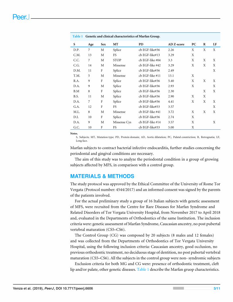
Table 1 Genetic and clinical characteristics of Marfan Group.
S Age Sex MT PD AD Z-score PC R LF
D.P. 7 M Splice cb EGF-like#36 2.26 X X XC.M. 13 M FS cb EGF-like#13 3.29 XC.C. 7 M STOP cb EGF-like #04 3.3 X X XC.G. 14 M Missense cb EGF-like #42 3.29 X X XD.M. 11 F Splice cb EGF-like#36 2.49 XT.M. 5 M Missense cb EGF-like #11 13.1 XR.A. 9 F Splice cb EGF-like#36 5.40 X X XD.A. 9 M Splice cb EGF-like#36 2.93 X XB.M 8 F Splice cb EGF-like#36 2.30 X XB.S. 11 M Splice cb EGF-like#36 2.90 X XD.A. 7 F Splice cb EGF-like#36 4.41 X X XG.A. 12 F FS cb EGF-like#33 3.57 XM.L. 8 M Missense cb EGF-like #41 3.72 X X XD.I. 10 F Splice cb EGF-like#36 2.74 XD.A. 9 M Missense Cys cb EGF-like #14 3.57 X XG.C. 10 F FS cb EGF-like#33 5.00 X
Notes.S, Subjects; MT, Mutation type; PD, Protein domain; AD, Aortic dilatation; PC, Palatal constriction; R, Retrognatia; LF,Long face.
Marfan subjects to contract bacterial infective endocarditis, further studies concerning theperiodontal and gingival conditions are necessary.
The aim of this study was to analyze the periodontal condition in a group of growingsubjects affected by MFS, in comparison with a control group.
MATERIALS & METHODSThe study protocol was approved by the Ethical Committee of the University of Rome TorVergata (Protocol number: 4544/2017) and an informed consent was signed by the parentsof the patients involved.
For the actual preliminary study a group of 16 Italian subjects with genetic assessmentof MFS, were recruited from the Centre for Rare Diseases for Marfan Syndrome andRelated Disorders of Tor Vergata University Hospital, from November 2017 to April 2018and, evaluated in the Departments of Orthodontics of the same Institution. The inclusioncriteria were: genetic assessment ofMarfan Syndrome, Caucasian ancestry, no post pubertalvertebral maturation (CS5–CS6).
The Control Group (CG) was composed by 20 subjects (8 males and 12 females)and was collected from the Departments of Orthodontics of Tor Vergata UniversityHospital, using the following inclusion criteria: Caucasian ancestry, good occlusion, noprevious orthodontic treatment, no deciduous stage of dentition, no post pubertal vertebralmaturation (CS5–CS6). All the subjects in the control group were non- syndromic subjects
Exclusion criteria for both MG and CG were: presence of orthodontic treatment, cleftlip and/or palate, other genetic diseases. Table 1 describe the Marfan gruop characteristics.
Venza et al. (2019), PeerJ, DOI 10.7717/peerj.6606 3/11
The study teeth included both maxillary and mandibular arches. Locally, teeth withrestorations within 1 mm of the gingival margin, crowded, ectopically positioned, or withhistory of surgical procedures, were excluded.
A single examiner performed all clinical measurements using a plane-faced dental mirrorand Michigan O probe with Williams markings coded at 1, 2, 3, 5, 7, 8, 9, and 10 mm.Intra-examiner reliability was performed by repeating the clinical measurements on threerandomly-selected patients one week later.
The periodontal clinical parameters were assessed in the following order:1. Marginal gingival thickness (GT) was assessed by visibility of the out-line of the probe
through the marginal gingiva (visible, thin, not visible, thick) (Kan, Rungcharassaeng& Lozada, 2003);
2. The plaque index (PI) was recorded based on the scale of Silness & Loe (1964). Aperiodontal probe was used, recording the variables in six localizations of each tooth;
3. Bleeding on probing (BOP) were recorded using a manual probe at six points (buccal-mesial, mid-buccal, buccal-distal, lingual-mesial, mid-lingual, lingual-distal) on a rightupper molar, an upper incisor, a left upper molar, a right lower molar, a lower incisorand a left lower molar;
4. Modified Periodontal Screening and Recording (PSR) (Landry & Jean, 2002) indiceswere produced through oral health assessments. To record probing depths, the mouthwas divided into sextants. Gingival crevices around each tooth were examined, andresearchers recorded the deepest probing depth found in each sextant.All statistical analyses were performed with the aid of statistical software (Statistical
Package for Social Sciences, version 16.0, SPSS Inc., Chicago, USA). Values of P < 0.05were considered significant. Descriptive statistics were used to describe both sample groups(SG andCG) in terms of age, sex and gingival thickness. Statistical significance of differencesbetween groups for qualitative variables were assessed using the Chi-squared test; t -testfor unpaired data was applied for assessing the comparison of the quantitative variablesbetween groups.
RESULTSA total of 9 males and 7 females (mean age of 9.4 ± 2.3 years) with confirmed MFS wereevaluated in the Department of Orthodontics of Tor Vergata University. 8 males and 12females with a mean age of 10.0 ± 2.6 years composed the Control Group. About theMarginal gingival thickness, in the MG, 5 subjects showed thick marginal gingiva and 11subjects showed thin marginal gingival thickness. Similar results were found in the CG,in which 6 subjects showed thick marginal gingiva and 15 subjects showed thin marginalgingival thickness.
The mean value of PI registered in the MG was 59% instead in the CG it reached 21%.T -test for unpaired data showed a statistical significant difference (P < 0.05) between thetwo groups.
Analysis showed a significant difference (P < 0.05) between MG and CG also for theBOP evaluated for all the subjects in both groups. In MG the mean value of BOP attained
Venza et al. (2019), PeerJ, DOI 10.7717/peerj.6606 4/11
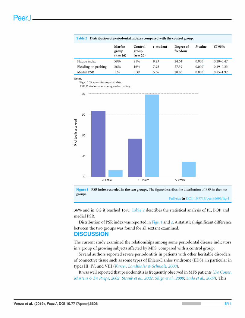
Table 2 Distribution of periodontal indexes compared with the control group.
Marfangroup(n= 16)
Controlgroup(n= 20)
t -student Degree offreedom
P value CI 95%
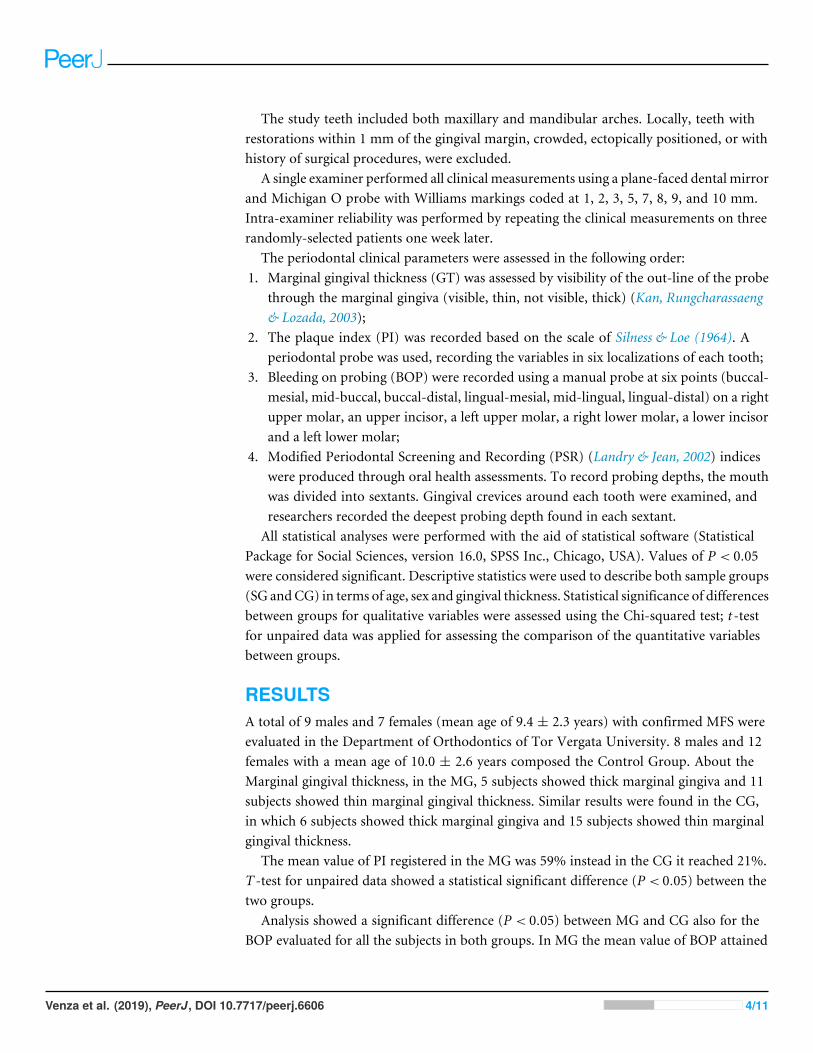
Plaque index 59% 21% 8.23 24.64 0.000* 0.28–0.47Bleeding on probing 36% 16% 7.95 27.39 0.000* 0.19–0.33Medial PSR 1.69 0.39 5.36 20.86 0.000* 0.85–1.92
Notes.*Sig < 0.05, t -test for unpaired data.PSR, Periodontal screening and recording.
Figure 1 PSR index recorded in the two groups. The figure describes the distribution of PSR in the twogroups.
Full-size DOI: 10.7717/peerj.6606/fig-1
36% and in CG it reached 16%. Table 2 describes the statistical analysis of PI, BOP andmedial PSR.
Distribution of PSR index was reported in Figs. 1 and 2. A statistical significant differencebetween the two groups was found for all sextant examined.DISCUSSIONThe current study examined the relationships among some periodontal disease indicatorsin a group of growing subjects affected by MFS, compared with a control group.
Several authors reported severe periodontitis in patients with other heritable disordersof connective tissue such as some types of Ehlers-Danlos syndrome (EDS), in particular intypes III, IV, and VIII (Karrer, Landthaler & Schmalz, 2000).
It was well reported that periodontitis is frequently observed in MFS patients (De Coster,Martens & De Paepe, 2002; Straub et al., 2002; Shiga et al., 2008; Suda et al., 2009). This
Venza et al. (2019), PeerJ, DOI 10.7717/peerj.6606 5/11
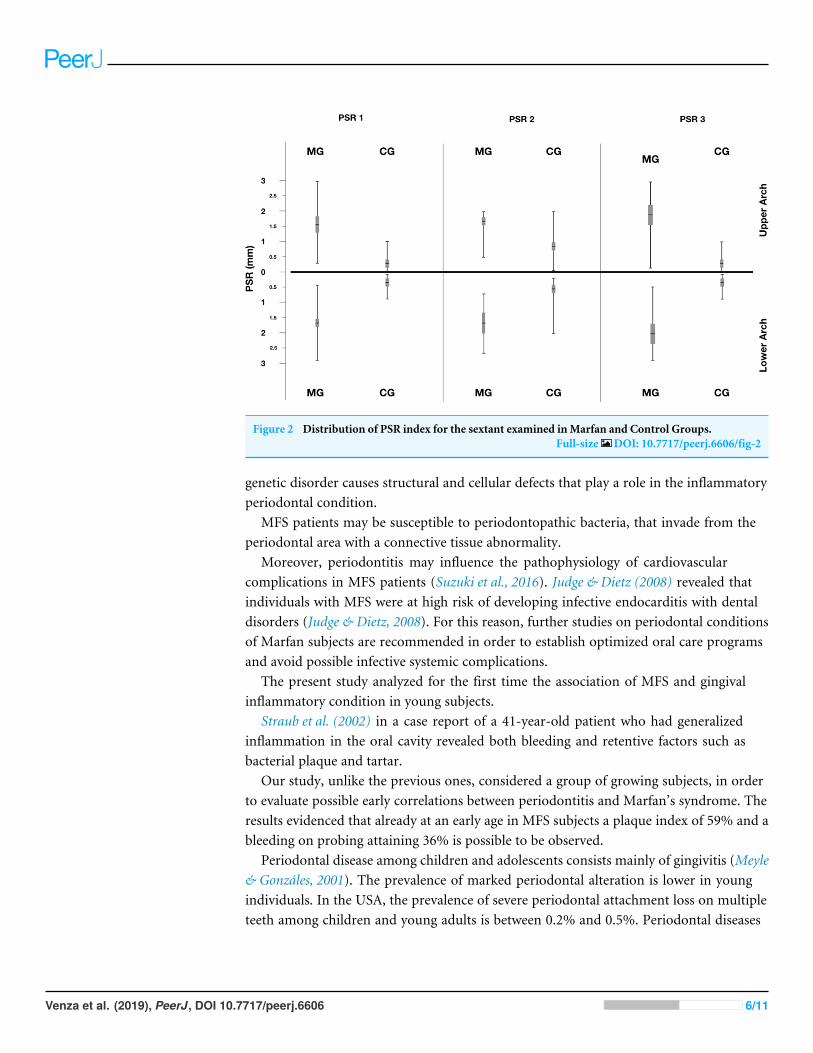
Figure 2 Distribution of PSR index for the sextant examined inMarfan and Control Groups.Full-size DOI: 10.7717/peerj.6606/fig-2
genetic disorder causes structural and cellular defects that play a role in the inflammatoryperiodontal condition.
MFS patients may be susceptible to periodontopathic bacteria, that invade from theperiodontal area with a connective tissue abnormality.
Moreover, periodontitis may influence the pathophysiology of cardiovascularcomplications in MFS patients (Suzuki et al., 2016). Judge & Dietz (2008) revealed thatindividuals with MFS were at high risk of developing infective endocarditis with dentaldisorders (Judge & Dietz, 2008). For this reason, further studies on periodontal conditionsof Marfan subjects are recommended in order to establish optimized oral care programsand avoid possible infective systemic complications.
The present study analyzed for the first time the association of MFS and gingivalinflammatory condition in young subjects.
Straub et al. (2002) in a case report of a 41-year-old patient who had generalizedinflammation in the oral cavity revealed both bleeding and retentive factors such asbacterial plaque and tartar.
Our study, unlike the previous ones, considered a group of growing subjects, in orderto evaluate possible early correlations between periodontitis and Marfan’s syndrome. Theresults evidenced that already at an early age in MFS subjects a plaque index of 59% and ableeding on probing attaining 36% is possible to be observed.
Periodontal disease among children and adolescents consists mainly of gingivitis (Meyle& Gonzáles, 2001). The prevalence of marked periodontal alteration is lower in youngindividuals. In the USA, the prevalence of severe periodontal attachment loss on multipleteeth among children and young adults is between 0.2% and 0.5%. Periodontal diseases
Venza et al. (2019), PeerJ, DOI 10.7717/peerj.6606 6/11
Figure 3 Periodontal condition in a growing subject withMarfan Syndrome. Figure shows gingivalconditions in a subject affected by Marfan Syndrome. Gingival tissue is typically hypertrophic in subjectswith Marfan Syndrome. Photo by Dr. Giuseppina Laganà.
Full-size DOI: 10.7717/peerj.6606/fig-3
in young individuals can develop as a consequence of a local or a systemic factor (Löe &Brown, 1991).
In a study carried out by Suzuki et al. in 2013, that analyzed a group of 40Marfan subjectsand 15 control group’s subjects, MS patients presented periodontitis more frequently thancontrol group subjects at the same age. In addition, patients withMS presented significantlysevere periodontitis, compared to control group subjects (Suzuki et al., 2015b).
The data from our study show that patients with Marfan syndrome have a gingivalinflammatory condition statistically significant higher than patients in GC. Furthermore,the presence of pockets up to 4 mm has also been detected, especially on permanent teeth(Fig. 3).
Considering the increased risk of cardiovascular complications suffering from Marfansyndrome, early diagnosis is essential to the success of treatment andprevention of infection.To combat the severity and progression of periodontal disease among persons with thesesystemic diseases, a preventive strategy should be implemented that includes more frequentthan 6 months professional oral hygiene, chlorhexidine mouth rinse, antibiotic therapywhen indicated, fluoride gel, and/or professionally applied fluoride varnish (Alrayyes &Hart, 2011).
CONCLUSIONSTo the best of our knowledge, this is the first study analyzing the periodontal index ina group of growing subjects affected by MFS. Based on our data, patients with Marfansyndrome reveal a higher presence of plaque and consequently a generalized inflammationin the oral cavity. Moreover, no signs of severe periodontal attachment loss on multipleteeth were on growing subjects with MFS.
Venza et al. (2019), PeerJ, DOI 10.7717/peerj.6606 7/11
ACKNOWLEDGEMENTSThe authors wish to express their gratitude to all the staff of the Centre for Rare Diseasesfor Marfan Syndrome and related disorders.
ADDITIONAL INFORMATION AND DECLARATIONS
FundingThe authors received no funding for this work.
Competing InterestsThe authors declare there are no competing interests.
Author Contributions• Nicolò Venza conceived and designed the experiments, prepared figures and/or tables,approved the final draft.
• Carlotta Danesi performed the experiments, analyzed the data.• Diego Contò performed the experiments.• Francesco Fabi analyzed the data, prepared figures and/or tables.• Gianluca Mampieri contributed reagents/materials/analysis tools, authored or revieweddrafts of the paper, approved the final draft.
• Federica Sangiuolo analyzed all the genetic data of the studied subjects.• Giuseppina Laganà conceived and designed the experiments, contributed reagents/-materials/analysis tools, authored or reviewed drafts of the paper, approved the finaldraft.
Human EthicsThe following information was supplied relating to ethical approvals (i.e., approving bodyand any reference numbers):
The study protocol was approved by the Ethical Committee of the University of RomeTor Vergata (Protocol number: 4544/2017) and an informed consent was signed by theparents of the patients involved.
Data AvailabilityThe following information was supplied regarding data availability:
The raw measurements are available in the Supplemental File. The raw data show all themeasurements taken and the statistical analysis.
Supplemental InformationSupplemental information for this article can be found online at http://dx.doi.org/10.7717/peerj.6606#supplemental-information.
Venza et al. (2019), PeerJ, DOI 10.7717/peerj.6606 8/11
REFERENCESAlrayyes S, Hart TC. 2011. Periodontal disease in children. Disease-a-Month
57(4):184–191 DOI 10.1016/j.disamonth.2011.03.004.BeertsenW,McCulloch CA, Sodek J. 1997. The periodontal ligament: a unique,
multifunctional connective tissue. Periodontology 2000 13:20–40DOI 10.1111/j.1600-0757.1997.tb00094.x.
Beighton P, De Paepe A, Danks D, Finidori G, Gedde-Dahl T, Goodman R, Hall JG,Hollister DW, HortonW,McKusick VA, Opitz JM, Pope FM, Pyeritz RE, RimoinDL, Sillence D, Spranger JW, Thompson E, Tsipouras P, Viljoen D,Winship I,Young James I, Reynolds F. 1988. International nosology of heritable disorders ofconnective tissue, Berlin, 1986. American Journal of Medical Genetics 29(3):581–594DOI 10.1002/ajmg.1320290316.
Bostanci B, Korkut E, Unlu N. 2017. Dental findings in marfan syndrome: acase report. Journal of Istanbul University Faculty of Dentistry 3;51(2):61–67DOI 10.17096/jiufd.78944.
Cook JR, Carta L, Galatioto J, Ramirez F. 2015. Cardiovascular manifestations in Marfansyndrome and related diseases; multiple genes causing similar phenotypes. ClinicalGenetics 87(1):11–20 DOI 10.1111/cge.12436.
De Coster PJ, Martens LC, De Paepe A. 2002. Oral manifestations of patients withMarfan syndrome: a case-control study. Oral Surgery, Oral Medicine, Oral Pathology,Oral Radiology 93(5):564–572 DOI 10.1067/moe.2002.121430.
Dietz HC, Cutting GR, Pyeritz RE, Maslen CL, Sakai LY, Corson GM, Puffenberger EG,Hamosh A, Nanthakumar EJ, Curristin SM, Stetten G, Meyers DA, FrancomanoCA. 1991.Marfan syndrome caused by a recurrent de novo missense mutation in thefibrillin gene. Nature 25;352(6333):337–339.
Ganburged G, Suda N, Saito M, Yamazaki Y, Isokawa K, Moriyama K. 2010. Di-lated capillaries, disorganized collagen fibers and differential gene expression inperiodontal ligaments of hypomorphic fibrillin-1 mice. Cell and Tissue Research341(3):381–395 DOI 10.1007/s00441-010-1021-5.
Handa K, Abe S, Suresh VV, Fujieda Y, IshikawaM, Orimoto A, Kobayashi Y, YamadaS, Yamaba S, Murakami S, Saito M. 2018. Fibrillin-1 insufficiency alters periodontalwound healing failure in a mouse model of Marfan syndrome. Archives of OralBiology 90:53–60 DOI 10.1016/j.archoralbio.2018.02.017.
Judge DP, Dietz HC. 2008. Therapy of Marfan syndrome. Annual Review of Medicine59:43–59 DOI 10.1146/annurev.med.59.103106.103801.
Kan JYK, Rungcharassaeng K, Lozada JL. 2003. Immediate placement and provisional-ization of maxillary anterior single implants. 1-Year prospective study. InternationalJournal of Oral and Maxillofacial Implants 18:31–39.
Karrer S, Landthaler M, Schmalz G. 2000. Ehlers-Danlos syndrome type VIII with severeperiodontitis and apical root resorption after orthodontic treatment. Acta Dermato-Venereologica 80:56–57 DOI 10.1080/000155500750012586.
Venza et al. (2019), PeerJ, DOI 10.7717/peerj.6606 9/11
Laganà G, Venza N, Paoloni V, Bertoldo F, Ruvolo G, Cozza P. 2018. A 3D geomet-ric morphometric analysis of the palatal morphology in Marfan’s syndrome: apreliminary study. Journal of Clinical and Diagnostic Research 12(1):ZC14–ZC17DOI 10.7860/JCDR/2018/31188.11099.
Landry RG, JeanM. 2002. Periodontal screening and recording (PSR) index: precursors,utility and limitations in a clinical setting. International Dental Journal 52(1):35–40DOI 10.1111/j.1875-595X.2002.tb00595.x.
Löe H, Brown LJ. 1991. Early onset periodontitis in the United States of America. Journalof Periodontology 62:608–616 DOI 10.1902/jop.1991.62.10.608.
Loeys BL, Dietz HC, Braverman AC, Callewaert BL, De Backer J, Devereux RB,Hilhorst-Hofstee Y, Jondeau G, Faivre L, Milewicz DM, Pyeritz RE, Sponseller PD,Wordsworth P, De Paepe AM. 2010. The revised Ghent nosology for the Marfansyndrome. Journal of Medical Genetics 47:476–485 DOI 10.1136/jmg.2009.072785.
Marfan AB. 1896. Un cas de déformation congénitale des quatre membres, plusprononcée aux extrémités caractérisée par l’allongement des os avec un certain degréd’amincissement. Bulletins et mémoires de la Société Médicale des Hôpitaux de Paris13:220–226.
Maslen CL, Corson GM,Maddox BK, Glanville RW, Sakai LY. 1991. Partial sequence ofa candidate gene for the Marfan syndrome. Nature 25;352(6333):334–337.
McKusick VA. 1955. The cardiovascular aspects of Marfan’s syndrome: a heritabledisorder of connective tissue. Circulation 11:321–342 DOI 10.1161/01.CIR.11.3.321.
Meyle J, Gonzáles JR. 2001. Influences of systemic diseases on periodontitis in childrenand adolescents. Periodontology 2000 26:92–112 DOI 10.1034/j.1600-0757.2001.2260105.x.
Paoloni V, Cretella Lombardo E, Placidi F, Ruvolo G, Cozza P, Laganà G. 2018.Obstructive sleep apnea in children with Marfan syndrome: relationships betweenthree-dimensional palatal morphology and apnea-hypopnea index. InternationalJournal of Pediatric Otorhinolaryngology 112:6–9 DOI 10.1016/j.ijporl.2018.06.014.
Pepe G, Giusti B, Sticchi E, Abbate R, Gensini GF, Nistri S. 2016.Marfan syn-drome: current perspectives. The Application of Clinical Genetics 9:55–65DOI 10.2147/TACG.S96233.
Shiga M, Saito M, Hattori M, Torii C, Kosaki K, Kiyono T, Suda N. 2008. Characteristicphenotype of immortalized periodontal cells isolated from a Marfan syndrome type Ipatient. Cell and Tissue Research 331(2):461–472 DOI 10.1007/s00441-007-0528-x.
Silness J, Loe H. 1964. Periodontal disease in pregnancy. II. Correlation between oralhygiene and periodontal condtion. Acta Odontologica Scandinavica 22:121–135DOI 10.3109/00016356408993968.
Staufenbiel I, Hauschild C, Kahl-Nieke B, Vahle-Hinz E, Kodolitsch Yvon, BernerM, Bauss O, GeurtsenW, Rahman A. 2013. Periodontal conditions in patientswith Marfan syndrome—a multicenter case control study. BMC Oral Health 13:59DOI 10.1186/1472-6831-13-59.
Straub AM, Grahame R, Scully C, Tonetti MS. 2002. Severe periodontitis inMarfan’s syndrome: a case report. Journal of Periodontology 73(7):823–826DOI 10.1902/jop.2002.73.7.823.
Venza et al. (2019), PeerJ, DOI 10.7717/peerj.6606 10/11

Suda N, Shiga M, Ganburged G, Moriyama K. 2009.Marfan syndrome and its disorderin periodontal tissues. Journal of Experimental Zoology Part B: Molecular andDevelopmental Evolution 15;312B(5):503–509 DOI 10.1002/jez.b.21278.
Suzuki J, Aoyama N, Izumi Y, IsobeM, Komuro I, Hirata Y. 2015a. Effect of periodon-titis on cardiovascular manifestations in Marfan syndrome. Critical common role ofTGF-β. International Heart Journal 56(2):121–124 DOI 10.1536/ihj.14-247.
Suzuki J, Imai Y, Aoki M, Fujita D, Aoyama N, Tada Y, Akazawa H, Izumi Y, IsobeM,Komuro I, Nagai R, Hirata Y. 2015b.High incidence and severity of periodontitisin patients with Marfan syndrome in Japan. Heart and Vessels 30(5):692–695DOI 10.1007/s00380-013-0434-y.
Suzuki J, Imai Y, Aoki M, Fujita D, Aoyama N, Tada Y,Wakayama K, Akazawa H,Izumi Y, IsobeM, Komuro I, Nagai R, Hirata Y. 2014. Periodontitis in cardiovascu-lar disease patients with or without Marfan syndrome—a possible role of Prevotellaintermedia. PLOS ONE 189(4):e95521 DOI 10.1371/journal.pone.0095521.
Suzuki J, Imai Y, Aoki M, Fujita D, Takeda N, Aoyama N,Wakayama K, Ikeda Y,Kumagai H, Akazawa H, Izumi Y, IsobeM, Komuro I, Hirata Y. 2016. Periodontitismay deteriorate sinus of valsalva dilatation in marfan syndrome patients. Interna-tional Heart Journal 27;57(4):456–460 DOI 10.1536/ihj.15-395.
Venza et al. (2019), PeerJ, DOI 10.7717/peerj.6606 11/11