Embed Size (px)
Citation preview
Perioperative involvement of
the cardiologist
Dr Kiran Patel
Consultant Cardiologist, Heart of England Trust & Medical Director, NHS England (West Midlands)


Outline
• Preoperatively: who needs to be seen?• Cardiac patients
• Non-cardiac patients post/for risk stratification
• Perioperative issues:
ESC 2014 guidelines – a good document
By 2035
• 25% more operations in Europe
• 50% more elderly people
• More obesity & DM


Heart Failure?

BNP

Mortality statistics for HF
• 40% die within 1 year of diagnosis
• 10% pa mortality thereafter
• Mortality rates globally predicted to 115-
127% from 1990-2020 – mainly in S Asia
NYHA II NYHA III NYHA IV
CHF 12 26 56
Other 24 15 11
SCD 64 59 33
1 yr mortality 3-25% 10-45% 50-80%
MERIT Ix 1999
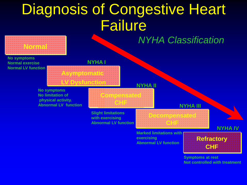
Diagnosis of Congestive Heart Failure
NYHA Classification
Symptoms at rest
Not controlled with treatment
No symptoms
Normal exercise
Normal LV function
Normal
No symptoms
No limitation of
physical activity.
Abnormal LV function
NYHA I
Asymptomatic
LV Dysfunction
Slight limitations
with exercising
Abnormal LV function
NYHA II
Compensated
CHF
Marked limitations with
exercising
Abnormal LV function
NYHA III
Decompensated
CHFNYHA IV
Refractory
CHF
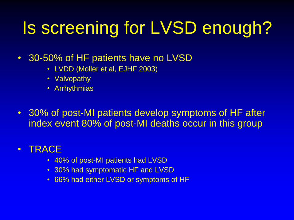
Is screening for LVSD enough?
• 30-50% of HF patients have no LVSD• LVDD (Moller et al, EJHF 2003)
• Valvopathy
• Arrhythmias
• 30% of post-MI patients develop symptoms of HF after index event 80% of post-MI deaths occur in this group
• TRACE• 40% of post-MI patients had LVSD
• 30% had symptomatic HF and LVSD
• 66% had either LVSD or symptoms of HF

LVDD
LV vel
t
Long Fx
Radial Fx
Compensation with
Preserved EF

LVEF and NYHA
• No correlation – why not?• Prevalence of asymptomatic LVSD similar to symptomatic
LVSD (McDonagh et al, Lancet 1997)
• EF dependent upon• Preload
• Afterload
• Chronotropy
• Inotropy
• Rate of fall in EF correlated with prognosis

Aetiology
• Cardiac– Ischaemic (65%)
– HT
– Valvular
– Arrhythmic
– Pericardial
• HOCF– Pregnancy
– Pagets etc.
• Systemic– Vasculitis
– Infection• Chagas, viral
– Genetic• HCM, DCM, DMD
– Metabolic• DM, amyloid, sarcoid,
storage disorders
– Toxic• EtOH, drugs, Fe
overload

Differential diagnosis
• Lung disease
• Obesity
• Mechanical • chest wall or diaphragm
abnormalities
• Fluid retention• Drug induced
• Venous insufficiency
• Renal failure
• Liver failure
• Hypoalbuminaemia
• PE
• Anaemia
• Thyroid disease
• Deconditioning
• Depression/anxiety

Precipitants of decompensation
• ACS: Angina/MI
• Arrhythmia
• Valvopathy deterioration
• Myocarditis
• Tamponade
• Dissection
• Shunts
• HT crisis
• Anaemia
• Alcohol
• Infection
• Iatrogenic – XS fluids, drugs
• Pregnancy
• PE
• Thyroid disease
• Brain injury
• Renal failure
• Asthma
• Drug abuse

HF therapy menu
• Starters• Diuretics
• Rehabilitation
• Main course • ACEI
• blockers
• Spironolactone
• Eplerenone
• ARB
• Digoxin
• Levosimendan
• Dessert• CRT
• ICD
• Coffee (not hungry) • High risk surgery
• After dinner mints (still hungry?)
• Transplantation
• VAD – bridge or destination
• Carriages• Palliative care
• EECP
• Sleep disordered breathing
• Plasmapheresis
• Reincarnation/after-life• Gene therapy
• Stem cells
• Nesiritide
• rEPO or iv Fe
• Myosin activators
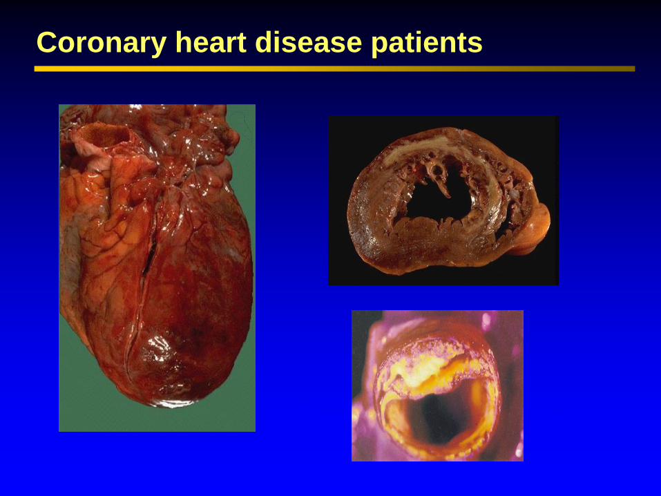
Coronary heart disease patients
Viable
Necrotic

Ischaemic cascade
Ischaemia
Perfusion defect
Diastolic dysfunction
Systolic dysfunction
ECG changes
Angina
MRI
MPI
Echo
MRI
ETT
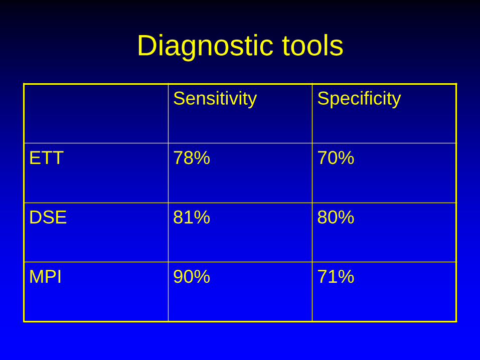
Diagnostic tools
Sensitivity Specificity
ETT 78% 70%
DSE 81% 80%
MPI 90% 71%

Non invasive angiography
• Limited by spatial and temporal resolution
• MRI
• CTCA• High radiation dose
• Limited in pregnancy and renal failure
• Ca scoring• High -ve predictive value: 99.4% 5 yr survival if 0% in non
diabetics (Raggi et al, JACC 2004)

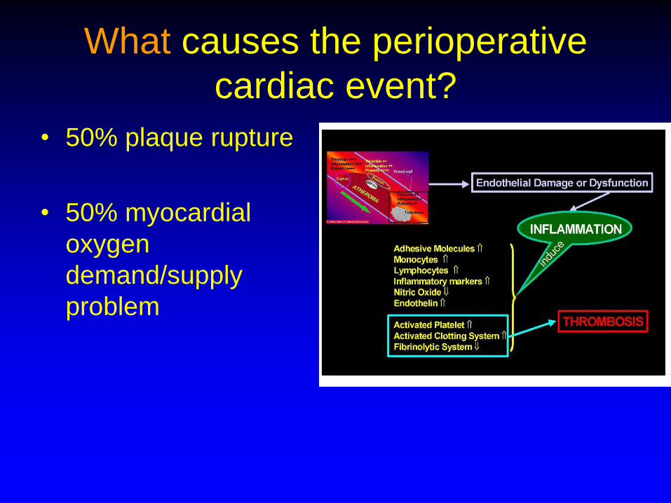
What causes the perioperative
cardiac event?
• 50% plaque rupture
• 50% myocardial
oxygen
demand/supply
problem

Pathophysiology of perioperative
MI
• neurohormmonal activation
• shear stress
• platelet activation
• coronary spasm
• catecholamines
• endogenous tPA
• Supply-Demand imbalance• Post-operative pain
• Fluid shifts
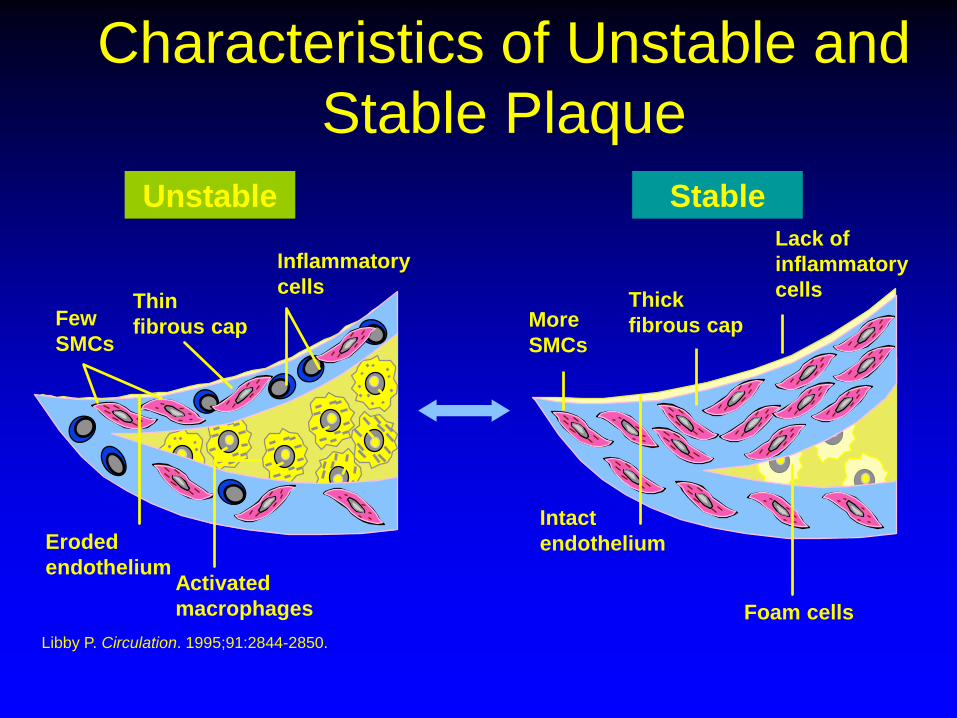
Characteristics of Unstable and
Stable Plaque
Thin
fibrous cap
Inflammatory
cells
Few
SMCs
Eroded
endotheliumActivated
macrophages
Thick
fibrous cap
Lack of
inflammatory
cells
Foam cells
Intact
endothelium
More
SMCs
Libby P. Circulation. 1995;91:2844-2850.
Unstable Stable

Perioperative events: The Q
• Not • Who needs Ix?
» Ix may precipitate a cascade of events of
revascularisation when not indicated to reduce risk
• BUT• Who is at risk and what is that risk?
• What causes perioperative events?
• How can we reduce them?
• Would cardiac referral be indicated in its own
right?

Surgical Risk by Intervention
Glance et al

Fitness for surgery:
Revised Cardiac Risk Index
• Factor• High risk surgery**
• IHD*
• CCF
• Cerebrovascular
disease
• IDDM
• Cr>152
No of
factors
CV
0 0.4%
1 1.1%
2 4.6%
>3 9.7%
Lee et al, Circulation 1999
*asymptomatic prior CABG or PCI excluded**intraperitoneal, intrathoracic, supra-inguinal vascular

Revised Cardiac Risk Index (RCRI)
MACE rate*
No of Variables
(RCRI Class)
Derivation
Cohort % (no)
Validation
Cohort % (no)
0 (Class I)0 0.5 (5/1071) 0.4 (2/488)
1 (Class II) 1.3 (14/1106) 0.9 (5/567)
2 (Class III) 4.0 (18/506) 7.0 (12/258)
3 or more
(Class IV)
9.0 (19/210) 11.0 (12/109)
*AMI, pulmonary oedema, VF, primary cardiac arrest, 3rd heart block

Fitness for surgery: NSQIP
(Gupta) Scoring system (2011)Risk of MI or cardiac arrest (developed as
RCRI suggested to have poor
discriminator ability)
ASA status
• ASA 1 = Normal healthy patient
• ASA 2 = Mild systemic disease
• ASA 3 = Severe systemic disease
• ASA 4 = Severe systemic disease that
is a constant threat to life
• ASA 5 = Moribund patients who are
not expected to survive without the
operation
5 Variables
• ASA status
• Age
• Creatinine (133 or ?)
• Functional
(independent/partially
or totally dependent)
• Procedure
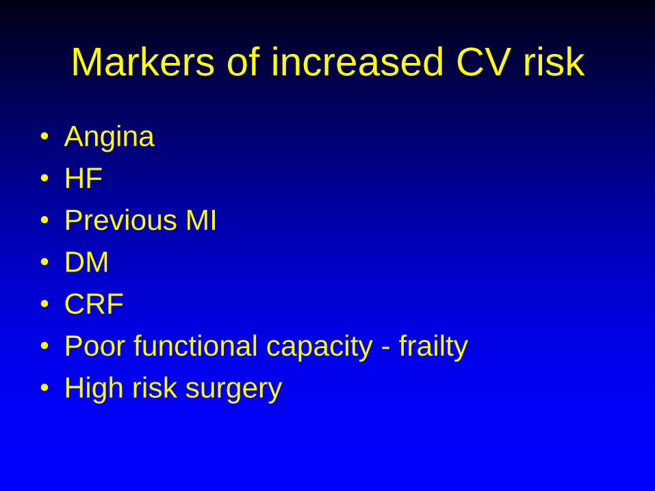
Markers of increased CV risk
• Angina
• HF
• Previous MI
• DM
• CRF
• Poor functional capacity - frailty
• High risk surgery

Clinical Predictors ACC/AHA
Minor Intermediate Major
•Advanced age
•Abnormal ECG
•Rhythm not
sinus
•Poor functional
capacity
•History of CVA
•High BP
•Mild angina
•Prior MI
•CCF (ever)
•DM
•Unstable
angina
•Decomp CCF
•Significant
arrhythmia
•Severe valve
disease
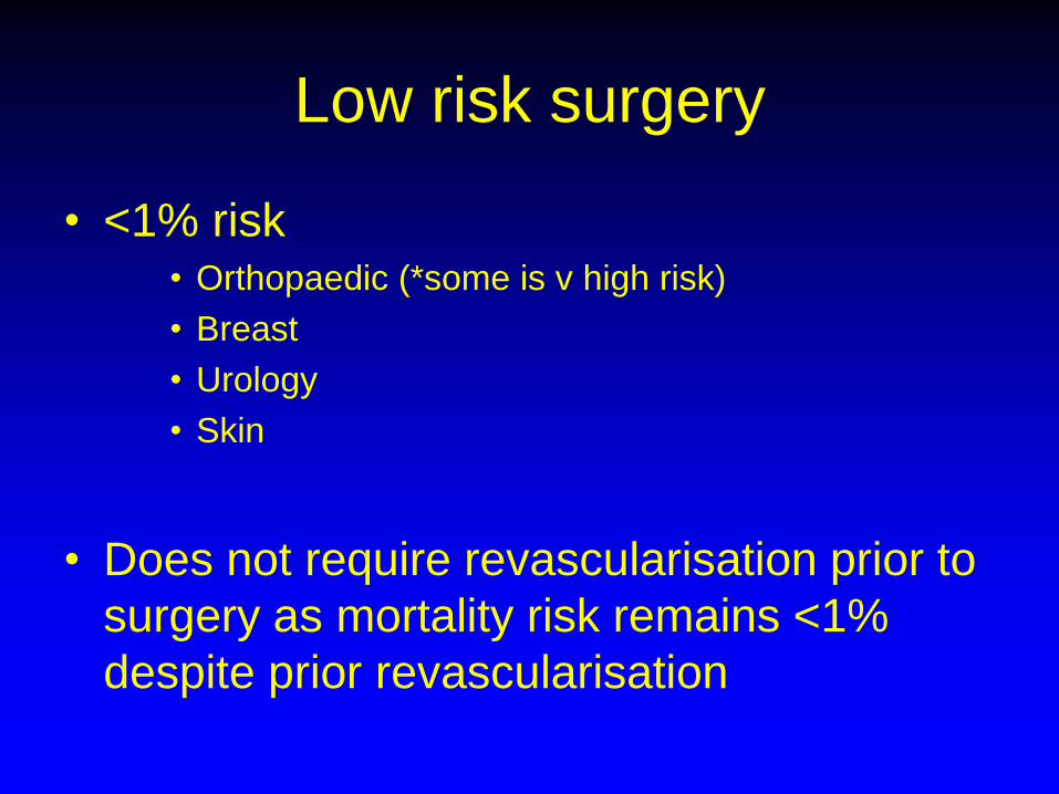
Low risk surgery
• <1% risk• Orthopaedic (*some is v high risk)
• Breast
• Urology
• Skin
• Does not require revascularisation prior to
surgery as mortality risk remains <1%
despite prior revascularisation

Assessing CV risk for non cardiac
surgery
• >50% of patients with fatal MI after non cardiac surgery shown to have unstable coronary plaques
• Preoperative ETT or DSE does not simulate the adrenergic stress of surgery
• Perioperative MI associated with LMS and 3VD

Can Cardiac Drugs reduce
Perioperative CV risk?
• Atenolol• given preoperatively and continued during hospitalisation
reduces mortality compared to placebo
• Benefits sustained for up to 2 years
• Bisoprolol• DECREASE trial in patients undergoing vascular surgery*
• Reduction in death and non fatal MI
• Statins • No large scale trial yet but plaque stabilisation useful in PCI,
CABG etc.
• ACEI, antiplatelet agents etc: no trial evidence

Perioperative Risk Modification

Perioperative blockade
• Improved myocardial oxygenation (-vely
inotropic and chronotropic)
• Antiinflammatory
• 16 fewer non fatal MIs per 1000 patients
treated BUT at expense of 3 strokes and 3
deaths (Bangalore et al, metaanalysis
Lancet Dec 2008)

Betablocker evidencePoldermans et al NEJM 1999
• N= 112 with 1 or more cardiac risk factors
and a positive stress echo
• Vascular surgery
• Bisoprolol 5-10 mg versus placebo
• Cardiovascular death @ 30 days:
• 3.4% bisoprolol v 17% placebo

Poldermans et al NEJM 1999
• RCRI Class I or II and positive stress echo
on betablockers had risk similar to those
with negative stress echo
• But risk posed by significant ischaemia*
not really modified by betablockers
*Class III and IV angina

Perioperative blockade
• Mangano et al• RCT of 200 patients
• No mortality benefit of atenolol 50-100 mg iv given 30 minutes before surgery then orally throughout hospitalisation BUT 50% reduction in ischaemia
• DIPOM• 921 DM patients undergoing non-cardiac surgery
• No benefit of metoprolol
• DECREASE study - discredited• 112 patients undergoing vascular surgery
• 90% reduction in mortality for patients given bisoprolol 30 days preoperatively

Perioperative Beta blockade
• Removing discredited DECREASE studies
demonstrates increased risk of• Mortality
• Stroke
• Hypotension
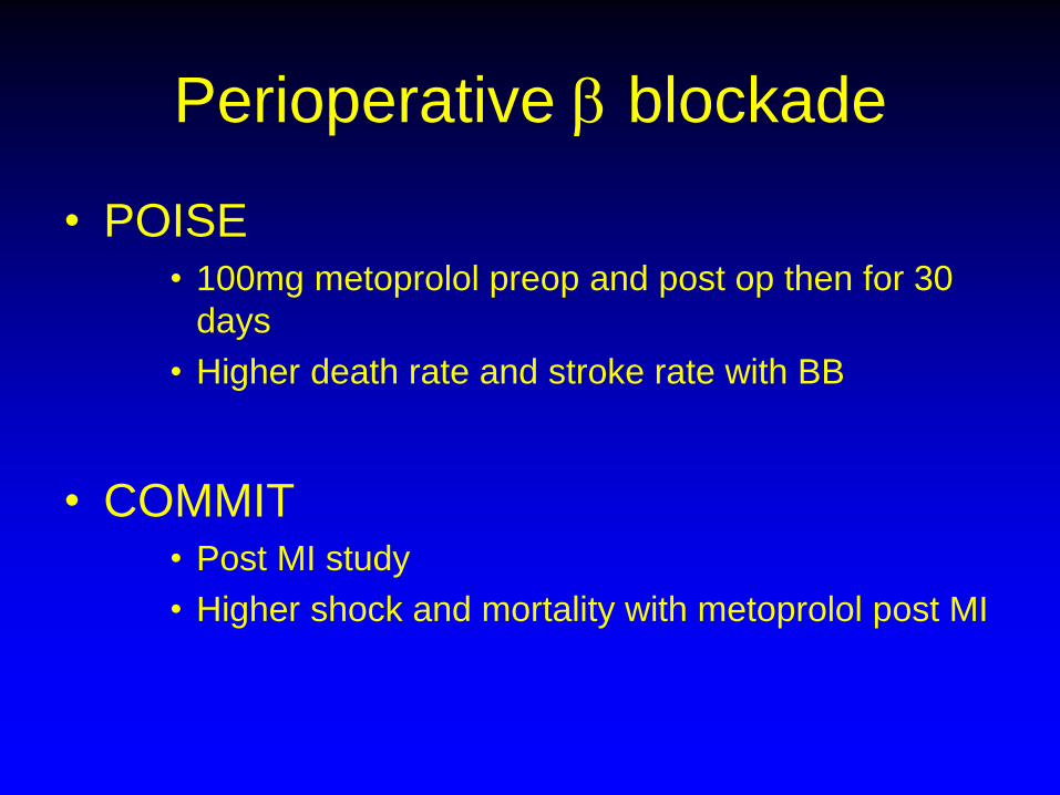
Perioperative blockade
• POISE• 100mg metoprolol preop and post op then for 30
days
• Higher death rate and stroke rate with BB
• COMMIT• Post MI study
• Higher shock and mortality with metoprolol post MI

Perioperative blockade
• 782 000 patients studied
• 122 000 received blockers – timing variable
• Revised cardiac risk index (1 point for each of
the following)• High risk surgery
• IHD
• Cerebrovascular disease
• Renal insufficiency
• DM
Lindenauer et al, NEJM 2005
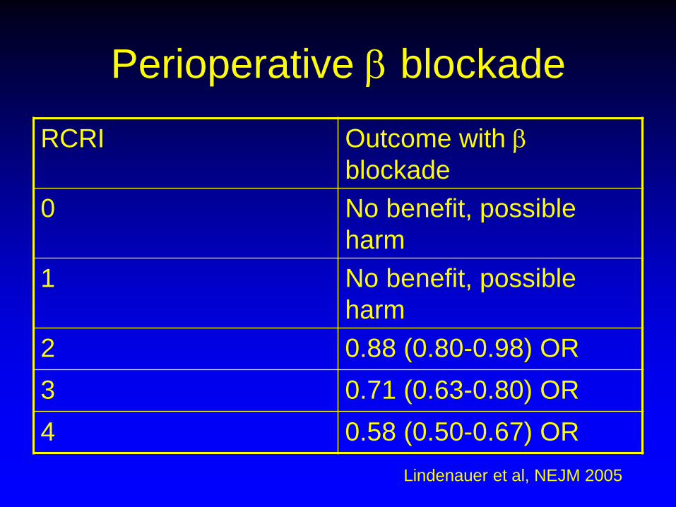
Perioperative blockade
Lindenauer et al, NEJM 2005
RCRI Outcome with
blockade
0 No benefit, possible
harm
1 No benefit, possible
harm
2 0.88 (0.80-0.98) OR
3 0.71 (0.63-0.80) OR
4 0.58 (0.50-0.67) OR

Perioperative blockade
• Meta-analysis of 22 trials• NNT – no clear benefit of metoprolol but benefit seen with
atenolol in elderly
• Devereaux et al, BMJ 2005
• Meta-analysis of 5 trials• NNT = 3-8
• Auerbach and Goldman, JAMA 2002
• Meta-analysis of 11 trials• NNT = 32
• Stevens et al 2004

Perioperative statins
• 3 studies show reduction in M&M in
patients undergoing vascular surgery• O’Neil-Callahan et al, 2005
• Kertai et al 2004
• DECREASE III (2008)
• Pleiotropic effect –plaque stabilisation likely to
explain mechanism of reduced perioperative
MI/ACS
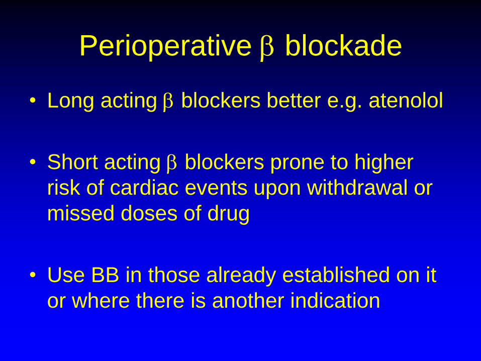
Perioperative blockade
• Long acting blockers better e.g. atenolol
• Short acting blockers prone to higher
risk of cardiac events upon withdrawal or
missed doses of drug
• Use BB in those already established on it
or where there is another indication

Coronary disease?

Revascularise first? CASS Registry
• Patients with previous CABG do have 50-70%
lower perioperative risk for non-cardiac surgery
• However, factoring in the risk of CABG produces
an overall higher risk for the non-cardiac surgery
• CABG therefore must be indicated in its own
right on grounds of symptoms or prognosis

Prophylactic revascularisation
• No benefit of coronary revascularisation in stable CHD patients prior to elective surgery (McFalls et al, NEJM 2004)
• CTCA preoperatively does not improve overall net risk classification (Sheth et al, 2015)
• Most studies exclude patients with – LMS stenosis >50%
– unstable coronary disease
– severe AS
– LVEF<20%
if these can be excluded, no revascularisation required
• No difference in CHD event rates or mortality seen

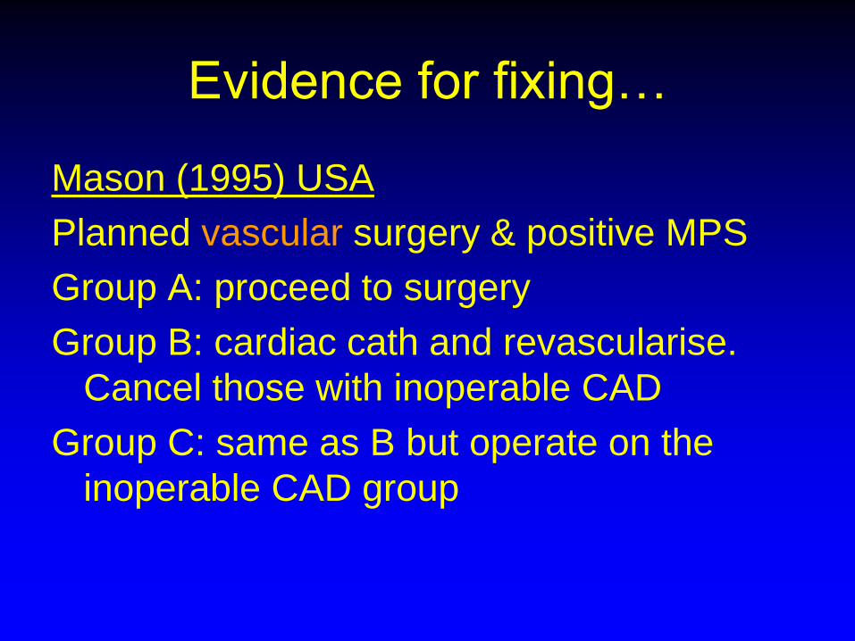
Evidence for fixing…
Mason (1995) USA
Planned vascular surgery & positive MPS
Group A: proceed to surgery
Group B: cardiac cath and revascularise.
Cancel those with inoperable CAD
Group C: same as B but operate on the
inoperable CAD group

Mason - results
End points: Mortality, non-fatal MI, stroke
Overall:
Group A (surgery no angio) did best
BUT
if risk of vascular surgery high – then pre-
op angiography (and treatment) did have
slightly lower mortality

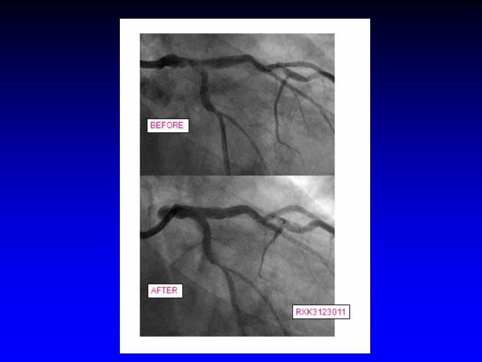
Percutaneous intervention
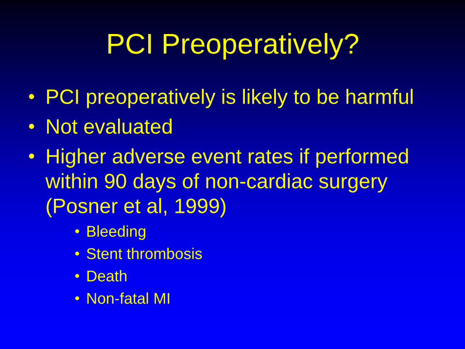
PCI Preoperatively?
• PCI preoperatively is likely to be harmful
• Not evaluated
• Higher adverse event rates if performed
within 90 days of non-cardiac surgery
(Posner et al, 1999)• Bleeding
• Stent thrombosis
• Death
• Non-fatal MI

Any surgery with 40 days PCI
Kaluza (2000)
N = 40; observational; mean 13/7 post PCI
MI 7
Major Bleeds 11
Deaths 8
MI and DEATH = STENT THROMBOSIS
(all occurred within 14/7 of PCI)
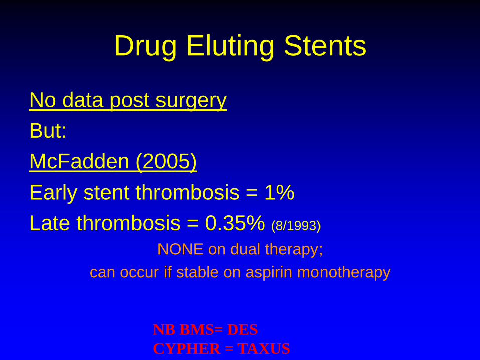
Drug Eluting Stents
No data post surgery
But:
McFadden (2005)
Early stent thrombosis = 1%
Late thrombosis = 0.35% (8/1993)
NONE on dual therapy;
can occur if stable on aspirin monotherapy
NB BMS= DES
CYPHER = TAXUS

Dual antiplatelet Rx and non
cardiac Sx
• Risk of SAT increased • Surgery is prothrombotic
• Stopping clopidogrel 5 days prior in elective Sx
• Minimise risk• Stop clopidogrel for 5 days prior only
• Restart clopidogrel 300mg post op
• Continue aspirin

CAD: To fix or not?: In general
‘Fix at your Peril’
• Negative tests are highly predictive of good
outcome
• Positive tests are a very poor predictor of
poor outcome…….and furthermore, overall
event rates are higher in the ‘fixed’ population
• However, high risk patients may benefit from
strategy of prior coronary revascularisation
indicated in its own right
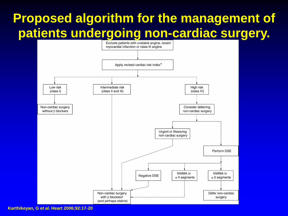
Karthikeyan, G et al. Heart 2006;92:17-20
Proposed algorithm for the management of patients undergoing non-cardiac surgery.

Statins and Plaque Stability
• Thin caped atheroma forms at areas of low shear stress
• Stablising plaques• Increase fibrosis
• Reduce lipid core
• How do statins work?• Shrink lesions – no
• Anti-inflammatory – yes
• Reduce lipid at core of plaques – yes
• Increase plaque fibrosis – yes
• Increase calcification of plaques - yes

Perioperative issues
What causes the perioperative
cardiac event?
• 50% plaque rupture
• 50% myocardial
oxygen
demand/supply
problem


What's at my disposal?
• Tn
• ECG
• Echo
• Cardiologist

Troponin: It is prognostic
In every condition in which it has been assessed, elevated troponin levels correlate with an adverse prognosis e.g. include heart failure, atrial fibrillation, renal failure, pulmonary embolism, sepsis and surgery.
These findings also apply to chronic stable disease e.g. in stable outpatients with risk factors for coronary disease, the level of hsTnT detected in stored blood samples correlated closely with prognosis, with the highest risk group, who had hsTnT of over 14 ng/L, having a four-times higher risk of death over a mean follow-up period of 9.4 years.

Echo: 3 patterns of LV remodelling

LV remodelling post-MI
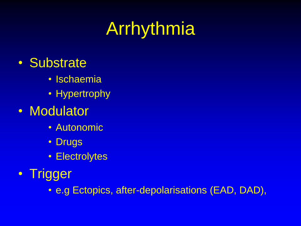
Arrhythmia
• Substrate• Ischaemia
• Hypertrophy
• Modulator• Autonomic
• Drugs
• Electrolytes
• Trigger • e.g Ectopics, after-depolarisations (EAD, DAD),

Delacretaz, E. N Engl J Med 2006;354:1039-1051
Main Mechanisms and Typical Electrocardiographic Recordings of Supraventricular Tachycardia

Adenosine
• 6mg terminates 60% of SVT, 12mg
terminates 90%
• Contraindicated in transplant recipients

Haemodynamic consequences of
new onset/uncontrolled AF
• Reduced cardiac output
– Tachycardia – risk of cardiomyopathy
– Loss of atrial contraction
– Irregular ventricular response
– Impaired diastolic function leading to HF e.g.
HT, LVH

Anticoagulant: Warfarin duration?
• New onset AF: clexane within 24hrs and consider cardioversion (chemical or DCCV)
• Stable: At least 3/52 before and 4/52 after DCCV (electrical restoration of SR is not immediately followed by atrial regular contraction) – consider amiodarone loading
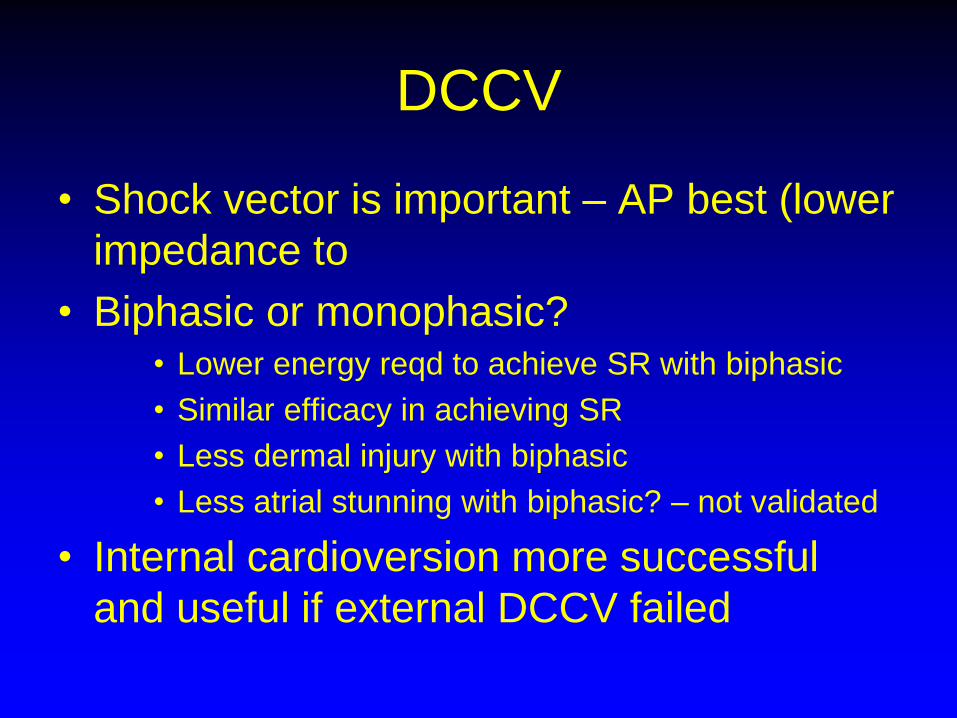
DCCV
• Shock vector is important – AP best (lower
impedance to
• Biphasic or monophasic?• Lower energy reqd to achieve SR with biphasic
• Similar efficacy in achieving SR
• Less dermal injury with biphasic
• Less atrial stunning with biphasic? – not validated
• Internal cardioversion more successful
and useful if external DCCV failed

VT

DEFINITION
• 3 or more ventricular ectopic beats in rapid
succession
• sustained if >30s duration or necessitates
cardioversion or pacing due to severe
hypotension
• accelerated idioventricular rhythm -
<120bpm. Usually seen post-MI.
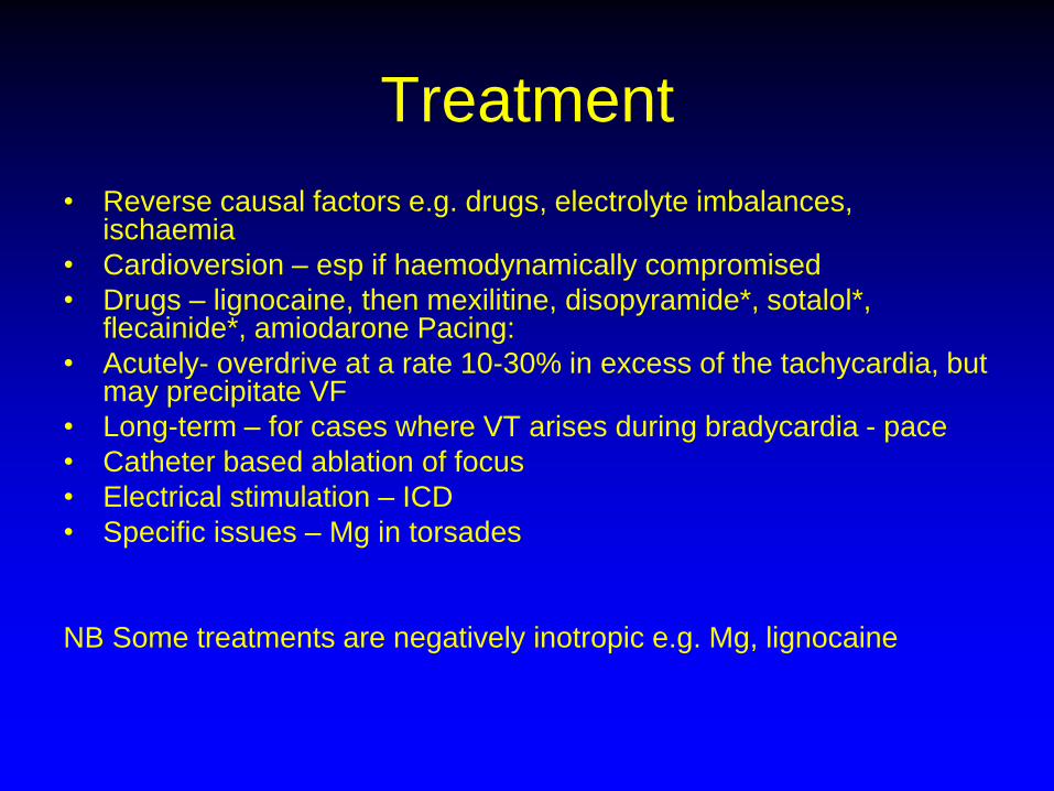
Treatment
• Reverse causal factors e.g. drugs, electrolyte imbalances, ischaemia
• Cardioversion – esp if haemodynamically compromised
• Drugs – lignocaine, then mexilitine, disopyramide*, sotalol*, flecainide*, amiodarone Pacing:
• Acutely- overdrive at a rate 10-30% in excess of the tachycardia, but may precipitate VF
• Long-term – for cases where VT arises during bradycardia - pace
• Catheter based ablation of focus
• Electrical stimulation – ICD
• Specific issues – Mg in torsades
NB Some treatments are negatively inotropic e.g. Mg, lignocaine
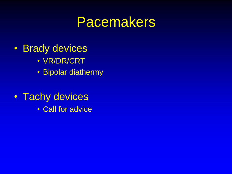
Pacemakers
• Brady devices• VR/DR/CRT
• Bipolar diathermy
• Tachy devices• Call for advice
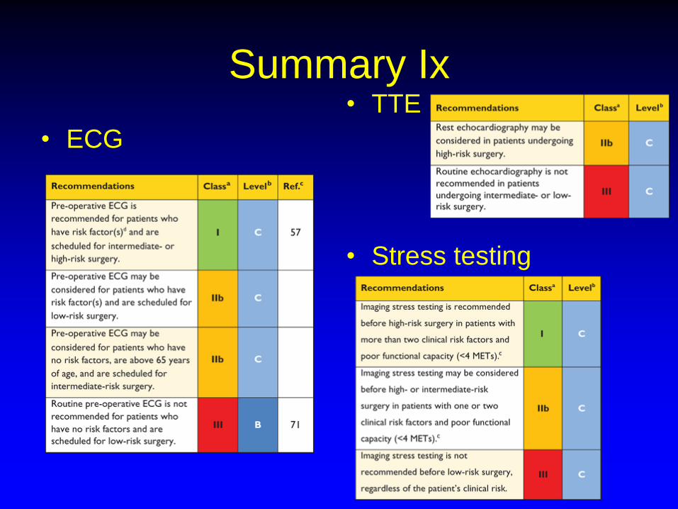
Summary Ix
• ECG
• TTE
• Stress testing

Summary
• Preoperatively: who needs to be seen?• Cardiac patients
» Optimise
» Beware of recent revascularisation - hang fire
• Non-cardiac patients post/for risk stratification» Most OK to proceed without cardiac revascularisation in
particular
» High risk – consider formal risk stratification and risk reduction – BB, statins as indicated
• Perioperative issues: » Tn, ECG, Echo, Cardiologist