Embed Size (px)
Citation preview
Anestesia Pediatrica e Neonatale, Vol. 8, N. 1, Febbraio-Marzo 2010
Perioperative management of a patient with PEHO-Syndrome: Delayed recovery of
neuromuscular blockade after rocuronium
Torsten Birkholz1, Christian Knorr2, Hans-Georg Topf3, Henrik Köhler3, Andrea Irouschek1,
Joachim Schmidt1
1Department of Anesthesiology, 2Department of Pediatric Surgery and 3Department of Pediatrics
University of Erlangen-Nuremberg,
Krankenhausstr. 12
D-91054 Erlangen
Germany
Correspondence to Torsten Birkholz, MD
e-mail: [email protected]
Abstract:
The PEHO syndrome (progressive encephalopathy with brain, facial and peripheral edema,
hypsarrhythmia and optic atrophy) is a very rare autosomal recessive disorder with
progressive encephalopathy. For diagnosis and therapy, many patients may require deep
sedation or even general anesthesia. There are no previous reports on the perioperative
management in PEHO-patients. Distinct obstacles have to be obeyed. Lack of pharyngeal
control might bear an imminent risk of aspiration. Muscular hypotonia and an immobile state
rules out the use of succinylcholine. In the case reported here, a 10-month old boy underwent
laparoscopic assisted percutaneous jejunostomy. The anesthetic management consisted of
total intravenous anesthesia with propofol and remifentanil, preceded by a rapid sequence
induction with 0.6 mg/kg rocuronium. In our patient, recovery from neuromuscular blockade
was grossly delayed. Therefore, with all applications of neuromuscular blocking agents in
patients with PEHO-syndrome, continuous neuromuscular monitoring is indispensable even
on the intensive care unit to determine the end of the neuromuscular block and to exclude
residual curarisation.
Anestesia Pediatrica e Neonatale, Vol. 8, N. 1, Febbraio-Marzo 2010
2
Introduction:
The PEHO syndrome (progressive encephalopathy with edema, hypsarrhythmia and optic
atrophy) is a rare autosomal recessive neurodegenerative disorder with an onset within the
first months of life. This infantile progressive encephalopathy was reported first in 1991 by
Salonen et al. with an estimated incidence of 1:74000 [9, 11]. Only a few patients have been
described in Europe, North America, Australia and Japan. [4, 6, 10]. Diagnosis presupposes
the exclusion of other disorders, and is made by clinical criteria and magnetic resonance
imaging (MRI). Clinical criteria are an infantile onset of profound muscular hypotonia with
concomitant spasms and epilepsia becoming apparent between the second and 52th week of
life, showing an interictal enzephalographic pattern of hypsarrhythmia. Additionally, arrest of
mental development and optic nerve atrophy with the absence or loss of visual fixation with
becomes evident by the age of two years. Typical MRI findings are progressive cerebellar and
brainstem atrophy. Distinct dysmorphic stigmata as epicantic folds, narrow forehead, short
nose, outward turning ear lobes, open mouth and in particular edema of face and limbs with
short tapering fingers are reported [4, 6, 11].
Children with PEHO syndrome may require general anesthesia for diagnostic and therapeutic
procedures such as MRI, placement of central venous line or endoscopic procedures. Data on
sedation, anaesthesia and drugs used during the anesthestic management in patients with
PEHO syndrome are lacking.
Anestesia Pediatrica e Neonatale, Vol. 8, N. 1, Febbraio-Marzo 2010
3
Case report:
We report the perioperative management of a 10 month old boy (8.3 kg, 62 cm) with PEHO
syndrome who was scheduled for laparoscopic assisted percutaneous endoscopic jejunostomy
(PEJ).
In our patient, all diagnostic features of PEHO syndrome were present. Therapy-resistant
epileptic seizures lead to prolonged episodes of apnoea. Along with aspiration pneumonia
caused by gastric regurgitation, the physical status required long time hospitalisation.
For the anesthesiological procedure, premedication with 0.05 mg/kg midazolam intravenously
allowed the transfer of the now sleeping infant from the preoperative holding area to the
prewarmed operation theater. Routine monitoring including ECG, pulse oximetry (SpO2), and
noninvasive blood pressure (Siemens SC 9000 XL, Siemens AG, Erlangen, Germany) as well
as neuromuscular transmission monitoring with quantitative acceleromyography at the
adductor pollicis muscle using TOF-Watch SX equipment (Organon, Dublin, Ireland) were
established. A modified rapid sequence induction technique was applied according to the
guidelines of the german scientific workgroup of pediatric anesthesia [10]: With the upper
part of the body in a 30° upright position and after 3 mins of preoxygenation, a continuous
infusion of 0.5 µg/kg/min remifentanil was started. After additional 2 Minutes, 4 mg/kg
propofol were injected. After loss of consciousness and successful pressure controlled mask
ventilation (FiO2 1.0, peak pressure 12 cm H2O, zero PEEP, 20/min), auto calibration of the
TOF-Watch SX was performed before starting train-of-four-stimulation every 15 sec. The
peak effect of the subsequently applied intravenous 0.6 mg/kg rocuronium was awaited before
conducting endotracheal intubation (Micro Cuff ID 3.5, Kimberly-Clark Health Care,
Roswell, Georgia, USA).
Anaesthesia was maintained as total intravenous anesthesia (TIVA) with remifentanil 0.5 –
0.25 µg/kg/min and propofol 6 mg/kg/h, adapted to surgical stimuli and hemodynamic
response. Pressure-controlled ventilation (40% oxygen in air) was adjusted to normocapnia
(petCO2 34–38 mmHg, Kion, Siemens AG, Erlangen, Germany). Body temperature was
maintained inside a range of 36.0 – 37.0 °C using a warming blanket system (Bair hugger,
Eden Prarie, MN, USA). A venous blood sample showed normal electrolytes, acid-base
laboratory, blood glucose and lactate both after induction of anesthesia and at the end of
surgery.
Surgery was uneventful. However, recovery from the neuromuscular block was only 5% of
the first twitch at the end of surgery (92 min after administration of the intubation dose of
Anestesia Pediatrica e Neonatale, Vol. 8, N. 1, Febbraio-Marzo 2010
4
rocuronium). We decided to transfer the sedated, endotracheally intubated and mechanically
ventilated infant to the pediatric intensive care unit (PICU) to await the offset of the
neuromuscular blockade. Three hours after admission on the PICU, repeated assessments
showed a TOF-ratio larger than 0.9 suggesting that the child had full recovery of the
neuromuscular transmission. It was breathing spontaneously and could be extubated
thereafter. The further course was uneventful.
Anestesia Pediatrica e Neonatale, Vol. 8, N. 1, Febbraio-Marzo 2010
5
Discussion:
The perioperative anesthetic management had to face typical main features of the PEHO
syndrome. Distinct risks and limitations to the anesthesiological procedure had to be obeyed.
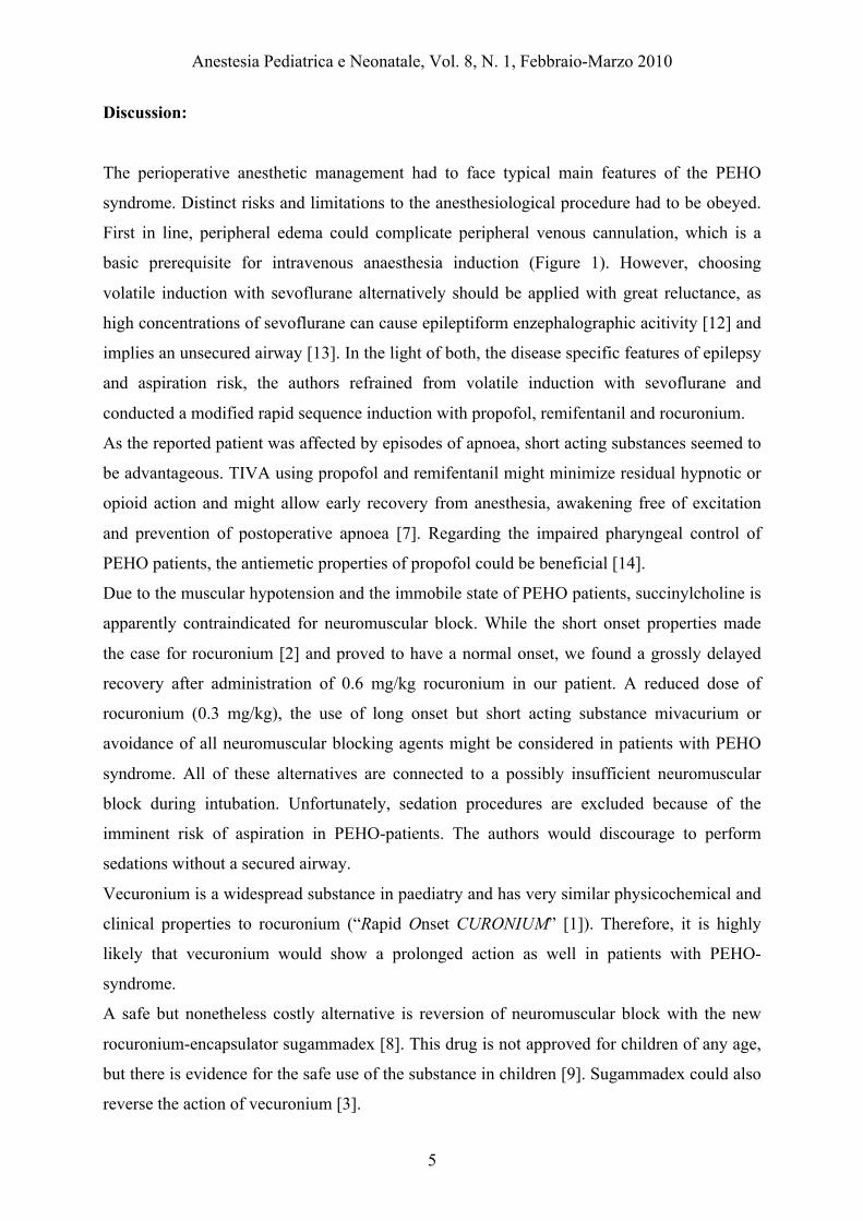
First in line, peripheral edema could complicate peripheral venous cannulation, which is a
basic prerequisite for intravenous anaesthesia induction (Figure 1). However, choosing
volatile induction with sevoflurane alternatively should be applied with great reluctance, as
high concentrations of sevoflurane can cause epileptiform enzephalographic acitivity [12] and
implies an unsecured airway [13]. In the light of both, the disease specific features of epilepsy
and aspiration risk, the authors refrained from volatile induction with sevoflurane and
conducted a modified rapid sequence induction with propofol, remifentanil and rocuronium.
As the reported patient was affected by episodes of apnoea, short acting substances seemed to
be advantageous. TIVA using propofol and remifentanil might minimize residual hypnotic or
opioid action and might allow early recovery from anesthesia, awakening free of excitation
and prevention of postoperative apnoea [7]. Regarding the impaired pharyngeal control of
PEHO patients, the antiemetic properties of propofol could be beneficial [14].
Due to the muscular hypotension and the immobile state of PEHO patients, succinylcholine is
apparently contraindicated for neuromuscular block. While the short onset properties made
the case for rocuronium [2] and proved to have a normal onset, we found a grossly delayed
recovery after administration of 0.6 mg/kg rocuronium in our patient. A reduced dose of
rocuronium (0.3 mg/kg), the use of long onset but short acting substance mivacurium or
avoidance of all neuromuscular blocking agents might be considered in patients with PEHO
syndrome. All of these alternatives are connected to a possibly insufficient neuromuscular
block during intubation. Unfortunately, sedation procedures are excluded because of the
imminent risk of aspiration in PEHO-patients. The authors would discourage to perform
sedations without a secured airway.
Vecuronium is a widespread substance in paediatry and has very similar physicochemical and
clinical properties to rocuronium (“Rapid Onset CURONIUM” [1]). Therefore, it is highly
likely that vecuronium would show a prolonged action as well in patients with PEHO-
syndrome.
A safe but nonetheless costly alternative is reversion of neuromuscular block with the new
rocuronium-encapsulator sugammadex [8]. This drug is not approved for children of any age,
but there is evidence for the safe use of the substance in children [9]. Sugammadex could also
reverse the action of vecuronium [3].
Anestesia Pediatrica e Neonatale, Vol. 8, N. 1, Febbraio-Marzo 2010
6
In our case, full neuromuscular recovery has to be awaited on the intensive care unit. In the
clinical practice of neuromuscular block, clinical signs of recovery (lifting of the head,
adduction of the arm) are insufficient to assess full neuromuscular recovery [5]. Especially
residual curarisation of the pharynx and the larynx could not be ruled out clinically and
require quantitative neuromuscular monitoring. In our case, full neuromuscular recovery was
essential to minimize the risk of aspiration. To assess neuromuscular block,
acceleromyography is a well-established quantitative technique, which should be available in
each unit where neuromuscular blocking drugs are utilized. It is easy to apply, non-invasive
and well evaluated [5]. In such cases, interdisciplinary management as described here might
be beneficial.
For the clinical practice, it has to be concluded that with the use of neuromuscular blocking
agents in PEHO patients, monitoring of neuromuscular transmission is indispensable.
Furthermore, the postoperative availability of intensive care treatment for patients with PEHO
syndrome should be strongly recommended. Further reports on the onset and offset of
nondepolarizing neuromuscular blocking agents in PEHO patient should be emphasized.
Anestesia Pediatrica e Neonatale, Vol. 8, N. 1, Febbraio-Marzo 2010
7
Figure 1: Hand edema in PEHO syndrome
Anestesia Pediatrica e Neonatale, Vol. 8, N. 1, Febbraio-Marzo 2010
8
References:
1. Agoston S, Vandenbrom RH and Wierda JM (1992) Clinical pharmacokinetics of
neuromuscular blocking drugs. Clin Pharmacokinet 22;94-115 2. Di Filippo A and Gonnelli C (2009) Rapid sequence intubation: a review of recent
evidences. Rev Recent Clin Trials 4;175-178 3. Duvaldestin P, Kuizenga K, Saldien V, et al A randomized, dose-response study of
sugammadex given for the reversal of deep rocuronium- or vecuronium-induced neuromuscular blockade under sevoflurane anesthesia. Anesth Analg 110;74-82
4. Field MJ, Grattan-Smith P, Piper SM, et al (2003) PEHO and PEHO-like syndromes:
report of five Australian cases. Am J Med Genet A 122A;6-12 5. Hemmerling TM and Le N (2007) Brief review: Neuromuscular monitoring: an update
for the clinician. Can J Anaesth 54;58-72 6. Klein A, Schmitt B and Boltshauser E (2004) Progressive encephalopathy with edema,
hypsarrhythmia and optic atrophy (PEHO) syndrome in a Swiss child. Eur J Paediatr Neurol 8;317-321
7. Mani V and Morton NS (2009) Overview of total intravenous anesthesia in children.
Paediatr Anaesth 8. Naguib M (2007) Sugammadex: another milestone in clinical neuromuscular
pharmacology. Anesth Analg 104;575-581 9. Salonen R, Somer M, Haltia M, et al (1991) Progressive encephalopathy with edema,
hypsarrhythmia, and optic atrophy (PEHO syndrome). Clin Genet 39;287-293 10. Schmidt J, Strauß J, Becke K, et al (2007) Handlungsempfehlung zur Rapid sequence
induction im Kindesalter. Anaesthesiologie und Intensivmedizin 46;S88-93 11. Somer M (1993) Diagnostic criteria and genetics of the PEHO syndrome. J Med Genet
30;932-936 12. Sonkajarvi E, Alahuhta S, Suominen K, et al (2009) Topographic
electroencephalogram in children during mask induction of anaesthesia with sevoflurane. Acta Anaesthesiol Scand 53;77-84
13. Thorn K, Thorn SE and Wattwil M (2006) The effects on the lower esophageal
sphincter of sevoflurane induction and increased intra-abdominal pressure during laparoscopy. Acta Anaesthesiol Scand 50;978-981
14. Vasileiou I, Xanthos T, Koudouna E, et al (2009) Propofol: A review of its non-
anaesthetic effects. Eur J Pharmacol