Embed Size (px)
Citation preview

ACT A
V ol. 27
PAL A EON T 0 LOG I C A
1 982
POLONICA
No . 1-4
ALTANGEREL PERLE, TERESA MARYANSKA and HALSZKA OSMOLSKA
GOYOCEPHALE LATTIMOREI GE N. ET SP. N., A NEW FLAT-HEADEDPACHYCEPHALOSAUR (ORNITHISCHIA, DINOSAURIA) FROM THE
UPPER CRETACEOUS OF MONGOLIA
PERLE, A. , MARYANSKA, T . a n d OSMOLSKA, H . : G oy ocepha le lattimorei gen. etsp. n ., a new fl at-he aded pachyce phalosau r (Orn lt hlschia , Dinosa u r ia) from theUpper Cretaceous of Mongolia. Acta Palaeont. Polonlca , 27, 1-4, 115-127, Dec ember1982. Issued J anua r y 1983.
A new r epresentative of the p a ch y cephalosaur family Homalocephalidae Dong isdescribed , which comes from th e Upper Cr etaceous deposits of the Gobi- Des ert,Mongolian P eople's R epublic . It Is more primitiv e a n d probably stratigraphicallyolder than H omalocephale cala thoce r cos know n from the Upper Cretaceous NemegtFormation of the Gobi Desert. The diagnosis of H. ca la t llOcercos Is r evised.
Ke y w 0 r d s : dinosaurs, O rnlthlsc hia , P achyeephalosauria , Cretaceous, Mo ngolia .
Altanger el P erle , Labdratory of s t ratigrap hy and Paleont ology, G eo logical Inst tt u t e, A ca demy of Sctences of the Mongolian P eople'S R epublic, Ulan B a t or ,Mongotian People'S R epublic.T eresa M a r y ailska , Muzeum Ziemi, P olsk a A kademia Nauk, at. N a Ska r p ie 20/26,0D-488 W a r szaw a. H a l szka Osmolska, Z aklad P aleobtol ogit , polska A k ade m i a Nau k,at. Z w t r k i i W tgu ry 93, 02-089 Warszaw a , P oland. R eceived : De cembe r 1981.
INTRODUCTION
In 1977, an incomplete skeleton was collected by the senior author atthe locality Boro Khovil 10 km west of Dzamyn Khond (60 km west ofBulgan Somon and south of the Arts Bogd Ull Mountain) in the southernGobi Desert, Mongolia. The precise age of the red sandstone deposits atBoro Khovil is still undetermined. It is probable that they are not olderthan the Djadokhta Formation, judging from the mutual relations of theBoro Khovil beds and the sediments of the Djadokhta Formation exposedin the vicinity. This material supplements our knowledge of the flat-headed pachycephalosaur family Homalocephalidae Dong, 1978. Dongassigned to this family: Yaverlandia Galton, 1971, Wannanosaurus Hou,1977, Micropachycephalosaurus Dong, 1978 in addition to the nomin ativegenus Homalocephale Maryanska et Osm6lska , 1974. The Hornalocephali-
8'

116 A. PERLE, T. MARYANSKA & H . OSMOLSKA
dae are characterized by the flat skull roof and the relatively largesupratemporal fenestrae. The new genus described here displays thesecharacters. The Homalocephalidae presently known are small to mediumsized dinosaurs ranging from about 60 em to 150 em (total length), thatare known so far only from the Eurasian continent. They display anatomical characters that distinguish them from the Pachycephalosauridae,thus we accept Dong's opinion that the family constitutes a natural, separate unit within the suborder P achyc ephalosauria Maryanska et Osm6lska,1974.
Wall and Galton 1979 considered the separate subordinal status for pachyeephalosaurs not justified. We intend to reconsider the problem in a future paper.
In addition to the flat-headed pachycephalosaurs, the dome-headedmembers of the family Pachycephalosauridae Sternberg, 1945 (sensuDong 1978) also occur in the Asian Upper Cretaceous. These include"S tegoceras" bexelli Bohlin, 1953, Tylocephale Maryanska et Osm6lska,1974, Prenocephale Maryanska et Osm6lska, 1974. We considered (Maryanska and Osm6lska 1974) that members of. the family Pachycephalosauridae were characterized by well pronounced heterodonty (presence ofcaniniform premaxillary teeth except for Stegoceras in which these arenot caniniform). It is now known that heterodonty was also present in thehomalocephalid here described Goyocephale lat timorei gen. et. sp. n.
Abbreviations used: GI SPS - Geological Institute, Academy of Sciences of the Mongolian P eople's Republic, Ulan Bator; ZPAL - Institute ofP aleobiology, Polish Acad emy of Science, Warszawa.
The material here described is store d in the Laboratory of Stratigraphyand Paleontology, Geological Institute, Academy of Sciences of 'the Mon,golian People's Republic, Ulan Bator.
Acknowledgements. - The authors thank Dr. Phillip Currie (provincialMuseum of Alberta, Edmonton) and Dr. Michael E. Howgate (UniversityCollege, London) who corrected the English and offered a valuable criticism.
SYSTEMATICS
Suborder Pachycephalosauria Maryanska et Osm6lska, 1974Family Homalocephalidae Dong, 1978 nom.corr. (= Homalocephaleridae
Dong, 1978)Genus Homalocephale Maryanska et Osm6lska, 1974
Homalocephale calathocercos Maryanska et Osm6lska, 1974(pI. 42: 13)
Ho~otype: GI SPS 100/1201 (= GI SPS 100/51 i n Maryanska and Osm61ska 1974 :56).

A NEW PACHYCEPHALOSAUR 117
Revised diagnosis. - Supratemporal fenestra rounded , interfenestral brigde broad , equal about the t rans vers e w idth of supra temporal fenestra. Interfrontal andfrontoparietal sutures di stinct. Infratemporal fenestra broad anteroposteriorly andhigh dorsoventrally. Orbit large and nearly round. Dorsal part of quadrate deflectedposteriorly. Occiput moderately concave, deepened at the centre. Foramen magnumrelatively large, about one seven th of width of the occiput. Occipital condyle large.Ventral maxillary edge bowed laterally along it s posterior portion. Cranial roofroughly ornamented. Sacrum incuding 6 coalesced vertebrae. Preacetabular processof ilium with gently convex dorsal surfa ce ; medial iliac flange long anteroposteriorly:postacetabular process curved downwards in lateral view.
Stratigraphic and geographic range. - Nernegt F ormation, Upper Cretaceou s,Nernegt, Nernegt Ba sin, Gobi Desert, Mongolia.
Genus Goyocephale nov.
Type species: Goyocephale lattimorei sp . n.; genus monotypic.Derivation of the name: Goyo (mong.) - decorated, elegant ; cephale (gr.) - head.Diagnosis, geographic and stra tigraphic r ange as for the type species.
Goyocephale lattimorei sp. n.(pls, 41, 42: 1-12, 43-45)
Holotype: GI SPS 100/1501, di sarticulated skeleton in cluding complete skull roof,damaged occiput (fr agments of parietals, squamosals and exoccipitals), basicranialregion (ba sioccipital with condyle and fragmentary basisphenoid), left postorbitalbar, fragments of quadrates and of [ugals, premaxillae and maxillae with dentition,mandibles with dentit ion , a tlas intercentrum, 2 spinal processes of dor sals, sac r umwith fragments of neural arches, a nearly complete series of 32 caudal centra (7 po sterior ones with fragmentary arches preserved), ilia with damaged acetabular regions, left humerus, fragmentary left ulna and 'r adius, fragments of indeterminablephalanges, sterna, di stal part of left t ibi a, proximal part of left (?) fibula, two leftdi stal tarsals, proximal and di stal parts of left metatarsals II, III, IV, distal end ofr ight metatarsal IV, left pedal digit IV (lacking ' phalanx IV-4), phalanx IV-2 ofr ight pe s, left phalanx II-I, ungu als of left pedal digits II and III, numerous caudaltendons, fragment s of thoracic ribs and indeterminable skeletal remains. F iguredon pls, 41, 42: 1-12, 43-45.
Type ho r izon : Upper Cretaceous, red sa ndstones; no precise age determination.Type locality : Boro Khovil, 10 km west of Dzamyn K hon d, South Gobi Desert,
Mongolia.Derivat ion of the name: named in honour of the eminent American mongolist
Prof. Oven Lattimore.D iagnosis. - Supratemporal fenestra longitudina lly oval, interfenestral bridge
narrow, about one third of the transverse width of supratemporal fenestra. Occiputweakly concave with relatively small occip ital condyle. Ventral maxillary edgew ea kly arched laterally al ong it s, post erior portion. Medial portion of cranial roofin dist inctly ornamented. F our weakly coossified vertebrae in cluded in to sacr um.Dor sal surface of preacetabular process of ili um flat, bent angularly along medialand lateral margins; medial ili ac flange short anteroposteriorly; po stacetabularprocess straight and subrecta ngular in lateral view.

1] 8 A. P E RLE , T. MARYAN S KA & H . OSMOLSKA
M at eria l. - On ly the holotype known.
Dimensions (in mrn) :
Length of skull (ti p of premaxilla - upperend of quadrate)greatest width of skull roofantorbital length of skullpostorbital length of skulllength of humerusproximal width of humerusdistal width of humeruslength of sacrum (4 vertebrae!)length of iliumdi stal width of tibia
230 est im,135
86 estim.80 estim.922821
105 estim.230 estim.64
Descr ip tion. - The skull. The cr anial roof is tablelike, somew hat depressed aboutthe centre and narrowed just anter ior to the postorbitals. The portions of the parietalthat bound the supratemporal fenestra an ter ior ly and medially are narrow. Theinte rfro ntal and fr ontoparietal sut ures are distinct (fig. lA). The occipit al region isweakly concave with a relatively small occipi tal condyle that is formed main ly byt he basioccip ital. Of the basisphenoid , the basal tubera, bas isphenoid tubera andpituitary foss a are preserved, which do not see m to displ ay any difference from thoseof ot her pachycephal osaurs. The neurocr anium as a whole is comparatively shor tan teroposteri orl y. As can be judged fro m the surrounding bones preserved, theinfrate mporal fenestra was narrow. The an teri or end of the nasal is covered withprom inent, pointed tubers. Medially, the roo f is weakly ornamented. A str ong nodelike ornamentati on is developed a long the posterior margin of the squamosals .The anterior portion of the premaxilla is comparatively long and shallow, in lateralview. The lower margin of the external narial openi ng is longer than the posteriormargin is high. The upper edge of the premaxilla, which contacts the nasal , isstraight and exte nds horizontally. In pal atal v iew, the premaxillae contact ea ch othera long their entire lengths; the same is true for premaxillary processes of the maxillae . Heterodonty is pronounced, t he premaxillary teeth being caniniform.
Between the premaxillary and maxillary te eth t here is a large diastema. Thealveolar margin of the m axill a is almost straight, being only slightly bent laterallyclose to its end. There are 15 ma xill ary teeth preserved but the number might begreater by one or two.
The m an di ble . Both lower jaws ar e present but disarticulated. The mandibularramus is shallow, slightly sigmoid a l in ve ntral view, and has a long retroarticularproces s. The corono id process is insignifi cantly elevated above the upper marginof the jaw. The jaw is flat and broad ve n trally for more than a half of it s p osteriorlength. The to oth row (with 18 teeth) reaches the an terior end of t he dentary, wherea surface is present for contact with t he opposite dentary. Although its anterior andposterior extremities are broken the splenial is long and narrow; t he coro noi d isfragmentary but small. The ar ticular is we ll develop ed and extends an teri or ly tot he middle of the ma ndibular fossa. The dor sal portion of the compara tively longretroarticular process is vertica l w hile the lower region develop s a horizontal wi nglaterally. The glenoid fossa is sha llow and opens la terally and medially. Ornamentat ion covers t he lateral and ventral surface of t he angular.
D entit i on . There were t hree can iniform te eth in the premaxill a, but t he fi r st one,the smallest judging fr om the size of alveola, is lack ing in our spec im en. They areslightly curved po steriorly. The cr owns are dist inctly marked off t he roots by theirsw olle n bas es. Each too th be ars posteriorly a crenell ated edge along one t hi rd of

A NEW P ACHYCEPHALOSAUR 119
its apical portion; the number of ser r a t ions per 1 mm amounts to 6. The third caniniform tooth is the str ongest and is almost as long as the depth of the tooth-bearingportion of the premaxilla. The second premaxillary tooth has a trace of a verticalwe ar facet posteromedially.
The crowns of maxillary teeth are low. The labial side of the tooth, with onlya slightl y thickened base, is concave dorsoventrally and ha s a weak central ridge.Anteriorly and posteriorly to that ridge there are several smaller ridges, but notall of them reach the crown base. The number of these ridges cannot be determinated for sure. The posterior edge of the crown is serrated. The maxillary teeth arestrongly worn on the lingual side, the plane of wear being at an acute angle to thesa ggi ta l p la ne of the tooth. The cutting ventral margin of each worn tooth isstr a ight.
The firs t mandibular tooth is much larger than the successive ones and it differs from them in being caniniform. It is larger than any of the caniniform premaxillary teeth. The crown of this mandibular caniniform tooth is polygonal ratherthan round in cross sect ion . It bears posteriorly a sharp ridge, which is denticulateda long the entire length of the crown. The denticles are not pointed and they dimin ish in size toward the root; there are 4--6 denticles per 1 mm. Other mandibularteeth are closely arranged, have subtriangular crowns with pointed tips. A wellmarked medial ridge is present on the labial side of the crown which is concavedorsoventrally. Anterior and posterior to that ridge there are several minor longitudinal ridges. The lingual side of the crown is convex dorsoventrally and anteroposteriorly and its base is thickened. On each side of the strong central ridge thereare several minor ridges, the number of which depends on the tooth position withinthe tooth row. Not all the minor ridges reach the base of the crown. The anteriorand posterior edges of the crown are deeply serrated. The wear surface on thela bia l side is almost parallel to the saggital plane of the tooth.
G. lattimorei is the only known homalocephalid in which the mutual relationsbetween th e upper and lower dentition could be seen . The deep pit present withinthe diastema between the premaxillary and maxillary teeth received the large caniniform first mandibular tooth. The third premaxillary caniniform extended lateralto the an teri or tip of the dentary. The position of the two anterior premaxillarycaniniforms in regard to the predentary cannot be determined as this bone is notpreserved in our specimen.
Po stcranial skeleton. The vertebral column as preserved lacks the cervicals(except atlas intercentrum) and dorsals. The sacrum includes only 4 weakly coossified vertebrae with fragments of r ibs and neural arches preserved. Only a portionof the first sacral vertebra is preserved and the first sacral rib is lacking. The second sacral r ib originates between the first and the second sacral centra; only itsproximal end is preserved, which is very robust and seems to have a lateroposteriororientation. The centrum of the second sacral is ventrally r idged. The ridge of thethird sacral is still more pronounced and bears posteriorly a groove along its crest.The proximal end of the third sacral rib origina tes between the second and thethird sacral centra and also extends lateroposteriorly; its distal portion is not preserved. The fourth sacral centrum is the broadest and has a rounded ventral ridge.The fourth sacral rib contacts the centrum of the fourth sacral along more thanhalf it s length, whereas only a small fraction of this rib adheres to the third sacralvertebra. This rib is complete and its length is slightl y more than the combinedlength of the third and fourth sa cral centra; it is oriented laterally and attaches tothe ilium just anterior to the medial iliac flange.
The two first caudals were found ar ticula ted with the sacrum. However, thefirst is much more weakly joined to the last (fourth) sacral than the sacr als are

120 A. P ERLE, T. MARYANSKA & H. OSMOLSKA
between each other, and the neural arches of these cauda ls have free zygapophyses.The articular surface s of the zyg apophyses of the two first caudals are grooved (thetongue-and-groove articula ti on). The r ibs of these caudal s are sutured to the shorttransverse processes of the neural arch. The centra of first three caudals are somewhat fl attened laterally and are ke eled ventrall y ; the centra of pos teri or caudalsare flat ven trally.
The hum er u s is slightly bowed. Its proxim al and di st al ends are expandedalmost in the same pl ane. The prox im al ar t icula r he ad slig htly ex tends beyond thedor sal fa ce of the humerus. The ven tral face bears a comp aratively deep, roundeddepression just bel ow the prox imal ar ti cula r surface. The de lt opec tor a l crest isthick but project s weakly. The distal end of the humerus sh ows weakly separatedcondyles and a poorl y de veloped olecranon fossa. The preserved proximal part oft he ulna shows that the olecranon process was ver y low.
The i lium has the typica l pachycephalosaur shape, with the thin preacetabularprocess developed in the horizontal pl ane, the postacetabular process in t he verticalp lane, and a wide, hori zontal flange extending inwar d from the superi or border oft he postacetabular portion of the ilium.
In dorsal aspect, t he preacetabular process st ro ngly devi ates laterally . Its dor salsurface is fl a t, bent sha r ply downward along the medial and lateral margine. Themedial flange of t he il ium is shor t ante roposter ior ly and relatively wide in thetrans ve rse direction. Anterior to the medial fl ange, a t the upper medial marginof t he ilium, there are two depressions for articulation with the third and fourthsacral r ib s. The postacetabular process is somew hat bowed inward at the di stal end.Due to the shor tness of the medial fl ange, t he postacetabular process appears long.In lateral view, the upper border of the ilium is almost straight and the postacetabular process is subrectangular .
The tibia, as preserved, is ty pi ca l for pachycephalosa urs. In the specimen studied there are no proximal tarsals at tached to it .
Two di stal tarsals are preserved w hic h cup the prox imal ends of metatarsals II ,III, and IV. Dist al tarsal 1 is small, roughly semil una r, and covers metatarsal II anda sma ll fraction of met atarsal III. Dist al tarsal 2 is subquadrate and covers metatarsal III, half of metat arsal IV and media lly a portion of distal tarsal 1. There is anante ropos ter ior ridge on it s ve n tral side sepa rating the sur faces for contact withmetatarsals III and IV.
T he me ta tarsus as preserved, includes three metatarsals - II , III, and IV ofwhi ch only prox imal and distal ends are preserved. The proximal articular endsof the metatarsals form a subtri angula r ar tic ular surface for a contact w it h thetarsu s. The ap ex of this triangle is directed laterally. The proximal extremity ofthe me tatarsus is concave on the plantar side. There is a round depression extendedon metatarsals III and IV. Metatarsal II is flattened la teromedially across its proximal end, and the longitudinal axis of the ar t icular sur face extends anteroposteriorly.The distal ar ticular surfa ce of this metatarsal is subquadr ate, emarginated po steriorly , the medial condyle extends posteroprox imally in to a sharp r idge. The distalarticular surface of metatarsal III has medial condyle larger than the lateral one ;the fossae liga men tosae are shallow. The proximal surface of metatarsal IV, whichis the thickest , is subtri angul ar, w hile the distal sur fa ce is subtrapezoidal, and weaklyernarginated posteriorly.
The pedal phalanges preserved are robust with well developed ginglymoid articular surfaces. The pedal un gual s are as ymmet rical, triangular but not recurved,be ing fla t on the plantar side . That of the thi rd toe is the largest , and those of thesecond and fourth toes are subequa l in length.
The caudal tendons are typ ical of pachycephal osaurs.

A NEW PACHYCEPHALOSAUR
COMPARISONS AND DISCUSSION
121
The specimen of Goyocephale lattimorei gen.et sp. n. is almost thesame size as the specimens of Homalocephale calathocercos and Prenocephale prenes described by us (Maryanska and Osm6lska 1974) from theNemegt Fm. of the Gobi Desert. Comparing the skull of G. lat timoreiwith the preserved portion of the skull in H. calathocercos it can be statedthat the former differs from the latter in: the larger and elongated supratemporal fenestra, the na rrower interfenestral bridge, the narrower porti ons of parietals that bound anteriorly the supratemporal fenestrae, theless concave occiput with the smaller occipital condyle, the shallowerneurocranium, the narrower infratemporal fenestra, and the anteroposteriorly na rrower mandibular condyle of the quadrate. The medial part ofthe cranial roof in G. lattimorei is slightly depressed along the frontalsand posterior part of the nasals, and it was probably the same in H. calathoc ercos. In G. lattimorei this portion of the skull roof, as well as theinterfenestral bridge, are weakly ornamented. At least the posteriorportion of the frontals and the interfenestral bridge are very rough inH. calathocercos. The nodes along the posterior margin of the skull aremore strongly pronounced in G. lattimorei.
In Wannanosaurus yansiensis Hou, 1977 (Hou 1977: pl. 1: 1) thesupratemporal fenestra is larger, the frontal is more ornamented posterioly and the posterior part of the squamosal is less ornamented. Thesutures on the portion of skull roof preserved in Micropachycephalosaurushongtuyanensis Dong, 1978 are obliterated, while these in G. lattimorei aredistinctly visible. The skulls of two Chinese homalocephalids are smaller,nevertheless they probably represent the adult individuals, judging fromthe obliterated sutures on the skull roofs.
The presence of caniniform teeth in the premaxilla and mandible inG. lattimorei suggests that such te eth might have been present in H. calathoc ercos , and in other homalocephalids. The mandible of G. lattimoreias compared with this of W. yansiensis is r elatively shallower, has moreconvex ventral margin in lateral view, the prealveolar part of the dentaryis shorter, the retroarticular process is longer, the posterodorsal border ofthe surangular is much less inclined, and the jaw is much shallower inthe region of the coronoid.
In the mandible of G. lattimorei the first mandibular tooth is very largeand caniniform. The first mandibular too th is also caniniform in Wannanosaurus (Hou 1977: fig. 1), but is relatively much smaller than in G. lattim orei ; its relation to the maxilla is unknown.
The discovery of the strong mandibular caniniform tooth in G. lattimorei that fi ts into the pit within the upper jaw dias tema makes moreprobable our earlier sugg esti on (Maryanska and Osm6lsk a 1974: 54) thatsuch tooth wa s also present in P. prenes; in that species the mandible is

122 A . PER LE, T. MAR YANSKA & H. OSMOLSKA
not known, but the mentioned diastema occurs on the junction premaxilla /m axilla. Thus, this character occurs both in the homalocephalid andpachycephalosaurid genera.
The new homalocephalid G. lattimorei has a pelvis with the typicalpachycephalosaurian ilium. This pelvis , as in other pachycephalosaurs,is extre mely broad for a bipedal dinosaur. In comparison with the pelvisof H. calathocercos , (in which the most complete sacropelvic region isknown up to now), that of G. lattimorei has fewer sacral vertebrae (4),which are but weakly joined with each other. The second sacral rib preserved in G. latt imorei is massive and lateroposteriorly directed. This ribin H. calathocercos contacts the anter ior peduncle of the ischium, so perhaps this was also the case with the second sacral rib in G. lattimorei. Thethird sacral rib in G. lat timorei differs in its orientation to that of H. calathocercos , being di rected posteriorly inste ad of anteriorly as it is inH. calathocercos. In the latter species, this rib contacts also the anteriorpeduncle of the isch ium as does the second rib, while in G. lattimorei thethird sacral rib certainly contacted the upper border of the ilium, becausethere is an articular sur face for this rib preserved on the ilium. The fourthsa cral r ib is in both species laterally directed and massive. In H. calathocercos this rib is exte nde d vertically contacting the ilium by its dorsalportion and the posterior peduncle of the ischium by its ventral portion.In contrast, in G. lattimorei this rib contacts only the ilium, posterior tothe contact with the third rib and just anterior to the medial flange of theilium. The fifth sacral rib in H. calathocercos is directed anteriorly andcontac ts upper border of the ilium jus t posterior to the fourth r ib. InG. lattimorei, there is no fifth sacral, and the rib of the first caudal (whichis an equivalent of the fi ft h sacral ri b of H. calathocercos) is quite differently dev eloped and directed lateroposteriorly. Thus, in H. calathocercos the fou rth and fifth sacral ribs contac t the upper iliac border whereasin G. lattimorei the third and fourth ones make the con tact. The incompletely preserved pelvis of G. lattimorei does not provide any informationon the contacts of the sacral r ibs with the ischium, as the latter, as wellas the pubis are missing.
The ilium of the spe cies descr ibed here differs from the ilium ofH. calathocercos in that the dorsal surface of the preac etabular process isflat centr ally and sharply bent along the medial and lateral border. Thedors al surface of this process is evenly and gently convex in H. calathocercos and the sharp lateral and m edial bending along its egdes is not pronouced in this species. As typical for all pachyc ephalosaurs, medial flangeof the ilium of G. lat timorei is ex pande d medially to the same extent as inH. calathocercos , but is shorter anteroposteriorly. Its shortness causes thepostacetabular process of the ilium to appear longer. The postacetabularprocess, as se en laterally , is different from that of H. calathocercos inbeing subrectangular with a straight vent ral margin, wh ile in the latter

A NEW PACHYCEPHALOSAUR 123
species it is arched downwards. Another homalocephalid with a fragmentary sacrum and ilium is Micropachycephalosaurus hongtuyanensis Dong.The sacrum of this species is however too badly preserved to be compared.Ilium of M. hongtuyanensis has a similar dorsal outline as that of G. lattimorei, but its postacetabular process is not subrectangular and thepreacetabular process is longer (fig. IB-D).
soi[
po
sq
A
Fig. 1. A Schematic drawing of skull roof in G. lattimorei gen. et sp. n.: f frontal.n nasal, p parietal, po postorbital, prf prefrontal, so I and II supraorbitals I and II,sq squamosal; B schematic drawing of ilium (lateral view) in G. lattimorei as compared with that in H. calathocercos Maryanska et Osm6lska (C) and M. hongtuyanensis Dong (D). Not to scale. A and B original, C after Maryanska and Osm6lska 1974,
D after Dong 1978.
The pelvis of G. lattimorei as a whole is broader than that in H. calathocercos and it widens forward by means of the lateral deviation of thepreacetabular processes. Due to the presence of only four sacral vertebrae,the relations of ilium length to that of the sacrum are very different inG. lattimorei and H. calathocercos. In the latter species, the ilium extendsonly slightly beyond the sacrum, both anteriorly and posteriorly, whilein G. lattimorei it extends far beyond the sacrum posteriorly.
The two vertebrae posterior to the fourth sacral in G. lattimorei areconsidered here as the first two caudals, because they are not fused at thecentra and display free zygapophyses. The grooved articular surfaces ofthese zygapophyses (including the prezygapophysis of the first caudal)prohibited any lateral movement in this part of the vertebral column.This indicates that these two caudals had functioned to some extent asthe sacral vertebrae. From other pachycephalosaur caudals these two"sacralized" ones differ in the presence of grooved zygapophyses and bythe short diapophyses to which caudal ribs are sutured. The groovedzygapophyses in pachycephalosaurs were until now known only in thedorsal vertebrae. A diapophysis - rib complex, similar to that on thefirst two caudals in G. lattimorei, is present on the sixth sacral in H. calathocercos.

124 A . PERLE, T. MARYANSKA & H . OSMOLSK A
The sternum of G. lattimorei, is similar to that of H. calathocereos bu tthe lateral margin of its shaft is more strongly curved posterol ate rally .Th e humerus of G. lat timorei may be compared with that of anothermember of the Homalocephalidae, W annanosaurus yansiensis , and in thelatter sp ecies it is more bowed.
Both the cranial and postcranial characters of G. lattimorei, especiallythe large supratemporal fe nestra and shor t sacrum, indicate that th isspecies was more primit ive than H. calathocercos. This may suggest anolder str atig raphic pos iti on of the depos it s , from which G. lattimorei wasrecovered , in regard to the Nemegt Fo r mation which yielded H. calathocercos.
REFERENCES
BOH LIN, B. 1953. Fo ssil reptiles from Mongolia and Kansu . - The Sino-SwedishExpedition, Publ. 31, 9-113, Stockholm.
DONG, Z. 1978. A new genus of Pachycephalosauria from Laiy ang, Shantung. Ve r tebr. Palasiat. , 15, 4, 225-228.
GALTON, P . M. 1971. A prim itive dome-headed dinosaur (Ornithischia: P achycephalosauridae) from L ower Cretaceou s of England and t he funct ion of thedome of p achycephalosaurids. - J . Paleont ., 45, 1, 4Q-47.
HOU, L. 1977. A new prim it ive P achycephalosauria from An hui, China. - Verteb r.Pa lasia t. , 15, 3, 198-202.
MARYANSKA, T. and OSMOLSKA, H . 1974. Pachycephalosauria , a new subor derof orni thischian dinosaurs. In: Z. Kielan-Jaworowska (ed .), Results Pol. -Mong,P a l. Exp. V. - Palaeont. Polonica, 30, 45-102.
STERNBERG, C. M. 1945. P achycepha losaurid ae proposed for dome-headed dinosaursStegocera s lambei n . sp. described. - J . Paleont., 19, 5, 534-538.
WALL, P . W. and GALTON, P. M. No te s on pachycephalosaurid dinosaurs (Reptilia:Ornithischia) from North Am eri ca , w ith comments on their sta tu s as ornithopods. - Ca nad. J. Eart h Scie nces , 16, 6, 1176-1186.
ALTANGEREL P ERLE, T ERESA MARYANSKA I HALSZKA OSMOLSKA
GOYOCEPHALE LATTIMOREI GEN. ET SP. N., NOWY PLASKOGLOWYPACHYCEFALOZAUR (ORNITHISCHIA, DINOSAURIA) Z GORNEJ KREDY
MONGOLII
St re szczen i e
Opisano nowy rodzaj i gatunek dinozaura gruboglowego (Pachycephalosauria)
Goyocephal e lattim orei, kt6ry zostal za lic zony do r odz iny H om alocepha lidae Don g,
1978. Material pochodzi z g6rnokredowych osa d6w ods lonietych n a pustyni Gobi w

A NEW P AC HYCEPHALOSAUR 125
Mongolskie] Republice Lud owej, w reg ionie som onu Bulgan. Obejmuje on niekorn
pletna czaszke z zuc hwa i niekompletny szkielet pozac zaszkowy jednego osobnik a
(holotyp). Budowa sklepieni a cza szk i oraz miednicy wskazuja, ze G. lattimorei je st
prymit ywniej szym przed st awicielem Homalocephalidae n iz wczesnle] op isany (Ma
r yanska i Osrnolska 1974) Homalocephale calathocercos z gornokredowej formacj i
Nemegt. Wiek st raty graficzny osadow nie jest dotychczas dokladnie us talony ; pry
rni tywnose budowy szki eletu G. la t t imor ei moze ws kazywac, ze wiek ten jest starszy
niz wiek for macji Nemegt,
Praca niniej sza byla czesciowo fi nansowana przez P olska Akadernie Nauk w
ra mach problemu miedzynarodowego II .6.
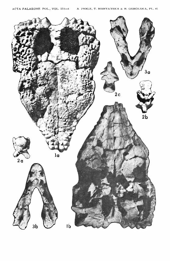
EXPLANATION OF THE PLATES 41-45
Plate 41
Goyocephale lat timorei gen. et sp. n .Upper Cretaceous, Boro Khovil , Gobi Desert, Mongolia GI SPS 100/1501
1. Skull roof ; a dorsal vi ew, b ventr al view: fragments of brain case and occiputvisible.
2. Basioccip it al-basisphenoid complex ; a po sterior view, b dorsal v iew, c ventralview.
3. Premaxillae and maxillae; a dorsal v iew, b ventral view.All Xl/2
lb, 2c, 3a, 3b ill uminated from below.
Plate 42
Goyocephale lat timorei gen. et sp. n.Upper Cr etaceous, Boro Khovil, Gobi Desert, Mongolia GI SPS 100/1501
1. Occiput lacking cen tr a l portion.2. Basioccipital-basisphenoid comp lex, r ight lateral view; sa me fragment as
pl. 41 : 2.3. Fragmentary right jugal , lateral view.4. Fragmentary left jugal, medial view .5. Premaxilla and fragmen tary maxilla wi th dentitions, left lateral view: note the
third canniniforme premaxillary tooth and diastem a; same fr agment as pl . 41: 3.6. Second le ft premaxillary tooth ; a lingual v iew: note the wear facet a long the
crown, b labial v iew.7. Se cond r ight premaxillary tooth ; a lingual v iew , b posterior view: note the
crenellate edge running centrally along the crown.8. A maxillary tooth; a lingual view : note wear facet occupying almost entire
surface of the crown, b labial view.

126 A . P ERLE , T . MARYANSKA & H . OSMOLSKA
9. F irst r ight mandibular tooth ; a lingual view, b same vi ew enlarged: note crenellation on posterior edge , c poster ior view, d same v iew enlarged: note crenellation.
10. A worn mandibular tooth; a lingual view, b labial v iew: wear facet outlined.11. An unworn mandibular too th , lingual v iew.12. Skull in dorsal vi ew, sli ght ly inclined forward and downward; compare w ith 13.
Homalocephale calathocercos Maryanska et Osm6lskaUpper Cr etaceous, Nemegt Formation, Nemegt, Gobi Desert, Mongolia
GI SPS 100/1201
13. Fr agmen tary sk ull in dorsa l v iew, sa me posi t ion as 12.1-5 X I/2, 9a, 9c X 2, 9b, 9d X 6, 12, 13Xl/4.
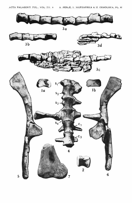
Plate 43
Goyocephale lat timorei gen. et sp. n .Upper Cretaceous, Boro Khovil, Gobi Deser t , Mongolia GI SPS 100/1501
1. Lower jaw ; a ventra l view, b left mandible, dorsal view , c sa me m andibl e,lateral view, d sa me mandi ble, med ial vi ew, e r igh t mandible, lateral vi ew,f sa me mandible, medial v iew.
2. Left ster nal bone , dorsal v iew .3. Right ster nal bone, ventral view.4. Left humerus; a dorsal view, b la teral view, c ven tral v iew, d medial vi ew.5. Left u lna, proximal pa r t; a lateral view, b ventral view.6. A thoracal r ib , lateral view of proxim al portion.7. A thorac al r ib , po sterior view .8. Fragments of two cauda l tendons.
All X 1I2lb illuminated from low er r igh t.
Pl a te 44
Goyocephale lattimorei gen. et sp. n.Upper Cretaceous, Boro Khovil , Gob i Desert, Mongolia GI SPS 100 /1501
1. Sacrum lacking an ter ior portion of 51 and neural arches, with two an ter iorca udals, fragmentary sa cral and ca uda l ribs attached ; dorsal view.
2. Left ilium, dorsal view.3. Right ilium, dor sal view.4. Le ft metatarsu s; a dorsal view of proximal portion, b dorsal view of di st al ends
of meta tarsals II , III, IV (from the left) , c ventral view of proximal portion ,d ve ntral v iew of dist a l ends of metatarsals II, III, IV (from the r ight), e proximal ar ticular sur face of t he metatarsu s.
5. Dist al tarsal s 1 and 2; a sur fa ce for prox im al ta rsals, b su rface for metata rsus.6. Left ph al anx II-I ; a dorsa l view, b ventral v iew.7. Le ft pe dal digit IV , lac ki ng pha lanx IV-4 and ungu al; a dorsa l view, b ventral
vi ew .

A NEW PACHYCEPHALOSAUR
8. Left peda l unguals II, III, IV; a dorsal v iew, b ventral vi ew.All X I /2
ill uminated fr om below , 6a, 7a, 8a - fro m right side.
Plate 45
127
Goyocephale lattimorei gen. et sp. n,Upper Cretaceous, Bora Khovil, Gobi Desert, Mongolia GI SPS 100/1501
1. Atlas intercentr um ; a dorsa l v iew, b ventral view.2. Centrum of a dorsal , left lateral vie w.3. Continuous seri es of caudals, left la teral v iew; a and b mo re anterior cauda Is,
tendons not preserved, c and d with tendons partly preserved.4. Sacrum lacking anter ior portion of Sl and neural a r ches , wi t h two an terior
cauda Is, fragmen tary sacral and ca udal ribs a t tached; ve ntral view.5. Right ilium, ven tr a l view.6. Left ilium, ven tral view.7. Distal porti on of left tibia, anterior view.
All X I /2
ACTA PALAEONT. P O L ., VOL. 27/1-4 A . PERLE, T . MARYANSK A & H . OSMOLSKA, PL. 41

ACT A PALAEONT. POL., V O L . 27/ 1- 4 A. PE:RL ~: , T . MARYA NSKA & 1I . O S M OLS K A , PL . 42
9a
Bb •.......•..\•.. BaII
JJ6a
lob

ACTA PALAEONT. POL., VOL. 27/1-4 A . P f: RLE , T . MARYA NSKA & H . OS MO L SKA, PL . 4:1
40 4b
7
4c
2
4d
3

ACTA PALAEONT. POL., VOL. 27/1- 4 A . PERLE, T . MARYANSKA & H . OSMOLSKA , PL. 44
5a
7a 8a
4b
8b 7b
6b
ACTA PALAEONT. POL.• VOL. 27/1-4 A . PERLE, T . MARYANSKA & H . OSMOLSKA , PL. 4~
30
3b