Embed Size (px)
Citation preview
ORIGINAL RESEARCH ARTICLEpublished: 01 December 2014
doi: 10.3389/fmicb.2014.00655
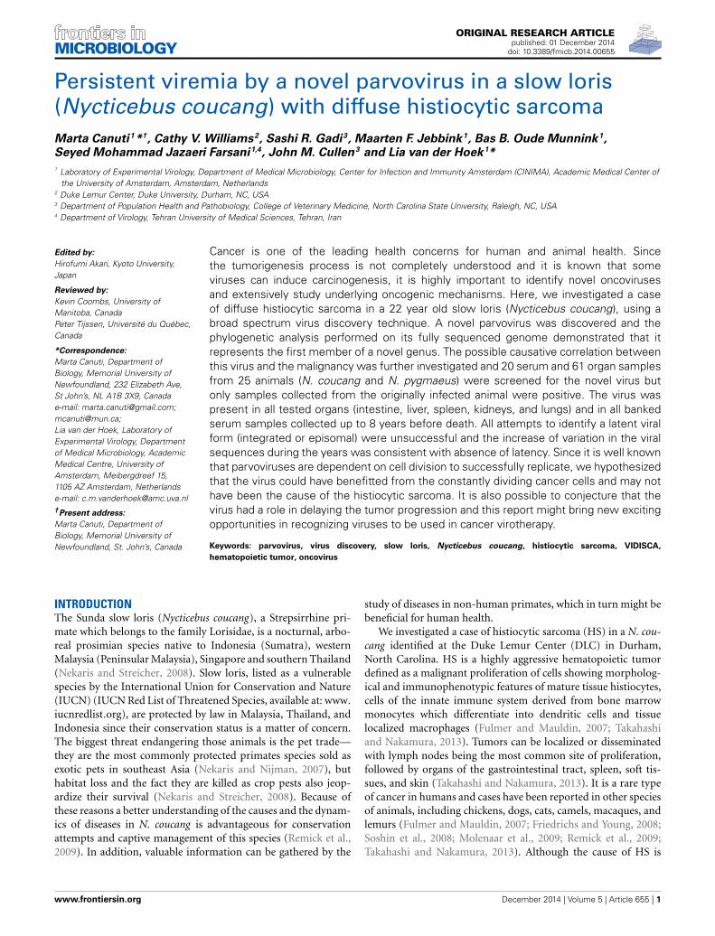
Persistent viremia by a novel parvovirus in a slow loris(Nycticebus coucang) with diffuse histiocytic sarcomaMarta Canuti1*†, Cathy V. Williams2, Sashi R. Gadi3, Maarten F. Jebbink1, Bas B. Oude Munnink1,
Seyed Mohammad Jazaeri Farsani1,4, John M. Cullen3 and Lia van der Hoek1*
1 Laboratory of Experimental Virology, Department of Medical Microbiology, Center for Infection and Immunity Amsterdam (CINIMA), Academic Medical Center ofthe University of Amsterdam, Amsterdam, Netherlands
2 Duke Lemur Center, Duke University, Durham, NC, USA3 Department of Population Health and Pathobiology, College of Veterinary Medicine, North Carolina State University, Raleigh, NC, USA4 Department of Virology, Tehran University of Medical Sciences, Tehran, Iran
Edited by:
Hirofumi Akari, Kyoto University,Japan
Reviewed by:
Kevin Coombs, University ofManitoba, CanadaPeter Tijssen, Université du Québec,Canada
*Correspondence:
Marta Canuti, Department ofBiology, Memorial University ofNewfoundland, 232 Elizabeth Ave,St John’s, NL A1B 3X9, Canadae-mail: [email protected];[email protected];Lia van der Hoek, Laboratory ofExperimental Virology, Departmentof Medical Microbiology, AcademicMedical Centre, University ofAmsterdam, Meibergdreef 15,1105 AZ Amsterdam, Netherlandse-mail: [email protected]†Present address:
Marta Canuti, Department ofBiology, Memorial University ofNewfoundland, St. John’s, Canada
Cancer is one of the leading health concerns for human and animal health. Sincethe tumorigenesis process is not completely understood and it is known that someviruses can induce carcinogenesis, it is highly important to identify novel oncovirusesand extensively study underlying oncogenic mechanisms. Here, we investigated a caseof diffuse histiocytic sarcoma in a 22 year old slow loris (Nycticebus coucang), using abroad spectrum virus discovery technique. A novel parvovirus was discovered and thephylogenetic analysis performed on its fully sequenced genome demonstrated that itrepresents the first member of a novel genus. The possible causative correlation betweenthis virus and the malignancy was further investigated and 20 serum and 61 organ samplesfrom 25 animals (N. coucang and N. pygmaeus) were screened for the novel virus butonly samples collected from the originally infected animal were positive. The virus waspresent in all tested organs (intestine, liver, spleen, kidneys, and lungs) and in all bankedserum samples collected up to 8 years before death. All attempts to identify a latent viralform (integrated or episomal) were unsuccessful and the increase of variation in the viralsequences during the years was consistent with absence of latency. Since it is well knownthat parvoviruses are dependent on cell division to successfully replicate, we hypothesizedthat the virus could have benefitted from the constantly dividing cancer cells and may nothave been the cause of the histiocytic sarcoma. It is also possible to conjecture that thevirus had a role in delaying the tumor progression and this report might bring new excitingopportunities in recognizing viruses to be used in cancer virotherapy.
Keywords: parvovirus, virus discovery, slow loris, Nycticebus coucang, histiocytic sarcoma, VIDISCA,
hematopoietic tumor, oncovirus
INTRODUCTIONThe Sunda slow loris (Nycticebus coucang), a Strepsirrhine pri-mate which belongs to the family Lorisidae, is a nocturnal, arbo-real prosimian species native to Indonesia (Sumatra), westernMalaysia (Peninsular Malaysia), Singapore and southern Thailand(Nekaris and Streicher, 2008). Slow loris, listed as a vulnerablespecies by the International Union for Conservation and Nature(IUCN) (IUCN Red List of Threatened Species, available at: www.
iucnredlist.org), are protected by law in Malaysia, Thailand, andIndonesia since their conservation status is a matter of concern.The biggest threat endangering those animals is the pet trade—they are the most commonly protected primates species sold asexotic pets in southeast Asia (Nekaris and Nijman, 2007), buthabitat loss and the fact they are killed as crop pests also jeop-ardize their survival (Nekaris and Streicher, 2008). Because ofthese reasons a better understanding of the causes and the dynam-ics of diseases in N. coucang is advantageous for conservationattempts and captive management of this species (Remick et al.,2009). In addition, valuable information can be gathered by the
study of diseases in non-human primates, which in turn might bebeneficial for human health.
We investigated a case of histiocytic sarcoma (HS) in a N. cou-cang identified at the Duke Lemur Center (DLC) in Durham,North Carolina. HS is a highly aggressive hematopoietic tumordefined as a malignant proliferation of cells showing morpholog-ical and immunophenotypic features of mature tissue histiocytes,cells of the innate immune system derived from bone marrowmonocytes which differentiate into dendritic cells and tissuelocalized macrophages (Fulmer and Mauldin, 2007; Takahashiand Nakamura, 2013). Tumors can be localized or disseminatedwith lymph nodes being the most common site of proliferation,followed by organs of the gastrointestinal tract, spleen, soft tis-sues, and skin (Takahashi and Nakamura, 2013). It is a rare typeof cancer in humans and cases have been reported in other speciesof animals, including chickens, dogs, cats, camels, macaques, andlemurs (Fulmer and Mauldin, 2007; Friedrichs and Young, 2008;Soshin et al., 2008; Molenaar et al., 2009; Remick et al., 2009;Takahashi and Nakamura, 2013). Although the cause of HS is
www.frontiersin.org December 2014 | Volume 5 | Article 655 | 1
Canuti et al. Novel slow loris parvovirus
largely unknown, a viral etiology can be postulated as the exis-tence of viruses involved in the development of hematopoieticcancers have been recognized, as in the case of Epstein Barrand human T-lymphotropic virus-induced human lymphomasand similar viruses in non-human primates (Miller et al., 1972;Donahue et al., 1992; Feichtinger et al., 1992; Vereide and Sugden,2009; Qayyum and Choi, 2014). Additionally it has been proventhat the subgroup J avian leukosis virus is associated with thedevelopment of histiocytic sarcomatosis in chickens (Pandiriet al., 2009) and an association between persistent Epstein Barrvirus infection and human HS has been reported (Kramer et al.,1985). In this study we applied a sequence independent virusdiscovery technique combined with high throughput sequenc-ing, VIDISCA-454 (De Vries et al., 2011), to investigate thepossible involvement of a previously unknown virus in the devel-opment of HS in a N. coucang. Several sequences with homol-ogy to parvoviral genes were identified indicating the presenceof a novel parvovirus, whose genome was subsequently fullysequenced.
Parvoviruses (viral family Parvoviridae) are small non-enveloped single stranded DNA viruses which are able to infecta wide range of species of vertebrates (subfamily Parvovirinae)and arthropods (subfamily Densovirinae). According to the lat-est ICTV classification (2013) 8 genera are recognized within thesubfamily Parvovirinae and 5 of them include viruses infectingprimates (Cotmore et al., 2013; Cotmore and Tattersall, 2014).Some parvoviruses classified within the genus Dependoparvovirusneed the presence of a helper virus for a productive infectionwhile viruses within other genera—the so called autonomousparvoviruses—are S phase–dependent: cells must undergo theS phase of growth for viral replication to occur (Berns,1990).
The spectrum of parvovirus induced diseases, which mainlyinvolve young individuals, is very wide and varies from moresevere forms [like severe enteritis with high mortality in youngdogs (Goddard and Leisewitz, 2010), erythema infectiosumor hydrops fetalis in children both caused by parvovirus B19(Heegaard and Brown, 2002, p. 19), or the Aleutian disease inminks (Best and Bloom, 2005)] to milder forms [like commonrespiratory and gastrointestinal diseases in humans (Jartti et al.,2012)]. Finally, parvovirus infections can also occur in asymp-tomatic individuals (Heegaard and Brown, 2002, p. 19; Lau et al.,2008; Clegg et al., 2012). Although there is no formal proofof the existence of oncoviruses within this family, parvoviruseshave been reported in literature to be associated with both solidand hematopoietic cancers (Fisgin et al., 2002; Li et al., 2012;Schildgen et al., 2013; Ibrahem et al., 2014). However, since theyrely on actively replicating cells, the increased presence of theseviruses in individuals with cancer might also derive from the per-missive nature of the tumor cells and some of these viruses haveeven proven to possess oncosuppressive effects on transformedcells (Berns, 1990; Nüesch et al., 2012).
Besides reporting the discovery and the molecular charac-terization of a novel parvovirus, the scope of this study wasto determine whether a possible correlation existed betweenthis virus and the presence of the histiocytic sarcoma in aN. coucang.
MATERIALS AND METHODSCLINICAL CASEThe virus was identified in a 22 year old, male Nycticebus coucang(slow loris) named Buddha, held in captivity for 22 years at theDuke University Primate Center and which had no prior signif-icant health issues. The individual was diagnosed with neoplasiaand euthanized due to poor condition, although a routine physi-cal inspection 8 months before death revealed an enlarged spleen.A complete postmortem examination was performed.
CLINICAL SAMPLESRepresentative tissues of Buddha were collected and fixed in 10%neutral formalin. Fixed tissue was processed routinely into paraf-fin blocks and sections were stained with hematoxylin and eosinand reviewed by a board of certified veterinary pathologist.
Twenty serum samples (belonging to 16 individuals: 11 N. cou-cang and 5 Nycticebus pygmaeus) and 61 organs collected atnecropsy (belonging to 17 individuals: 11 N. coucang—includingBuddha—and 6 N. pygmaeus) were screened for the presence ofthe virus. Organ samples included 17 livers, 6 spleens, 15 kid-neys, 10 lungs, 6 hearts, 7 intestines (5 small intestines and 2 largeintestines). Altogether these samples belonged to 25 individuals(18 N. coucang and 7 N. pygmaeus) with various types of disease.Liver, spleen, kidney, lung, and intestine samples from Buddhawere available as well as serum or whole blood collected on 4 dif-ferent time points: year 2000 (serum), year 2005 (serum), year2007 (whole blood), and year 2008 (serum).
Serum samples and organs were stored at −80◦C until virolog-ical examinations were performed.
Housing management and sample collection from the ani-mals in this report were approved by the appropriate federal andinstitutional regulatory authorities.
VIRUS DISCOVERYSequence independent virus discovery was performed on a serumsample collected from Buddha at necropsy (2008) with the previ-ously described VIDISCA-454 procedure (De Vries et al., 2011)with minor modifications. After a centrifugation and DNasetreatment to remove both intact cells and host DNA derived frombroken cells, viral nucleic acids were isolated from 100 μl of serumwith the QIAmp DNA mini kit (Qiagen). Subsequent to the lig-ation of adaptors containing specific Roche-454 primer bindingsequences, an amplicon size-selection was performed to preventthe amplification of DNA fragments smaller than 200 bp usingAgencourt AMPure XP beads (Beckman Coulter), followed by 30cycles of PCR amplification. The amplified library was subjectedto 2 consecutive purification rounds with Agencourt AMPure XPbeads to completely remove excess primers and short fragmentsand DNA concentration was measured on a Qubit Fluorometer(Quant-it ds DNA HS kit, Invitrogen). The library was pooledwith other samples, the average size of the whole library wasestimated with Agilent 2100Bioanalyzer (high sensitivity DNAkit, Agilent Technologies) and the final concentration (copies/μl)was calculated using the KAPA Library Quantification kit (KAPABiosystems). Samples were then diluted to a final concentrationof 1 million copies/μl and used as input for the emulsion PCR(LIB-A emPCR kit, Roche) and 454 pyrosequencing (Roche).
Frontiers in Microbiology | Virology December 2014 | Volume 5 | Article 655 | 2
Canuti et al. Novel slow loris parvovirus
After sequencing, primer sequences were trimmed from everyread and sequences were assembled with CodonCode Alignersoftware, version 3.5.6. The contigs (consensus sequences derivedfrom reads found multiple times) and unassembled sequenceswere compared to known nucleotide and protein sequences in theGenBank database using different standalone BLAST tools (blastnand tblastx) (Altschul et al., 1990). Blast results were visualizedusing the MEGAN software version 4.70.4 (Huson et al., 2011).
FULL GENOME SEQUENCINGSpecific primers were designed on the viral sequences identi-fied with VIDISCA-454 and PCRs using DreamTaq DNA poly-merase (Thermo Scientific) were performed to connect frag-ments. Sequencing reactions were carried out with nested primersdirectly on the amplified products using the Big Dye termina-tor chemistry (BigDye® Terminator v1.1 Cycle Sequencing Kit,Applied Biosystems).
The ends of the genome were determined using genomic frag-ments obtained by specific digestion with 2 different restrictionenzymes (MseI and CviAII from New England Biolabs) to obtainoverlapping fragments, to which VIDISCA adaptors were subse-quently ligated; semi specific PCRs were then performed with acombination of one primer annealing to the known viral sequenceand one to the adaptor. After sequencing the obtained ampli-cons, the novel sequence was used as a template for new primerdesign and the whole procedure was repeated until reaching theend of the genome. Specific PCRs were used as confirmation.All primers used for PCR and sequencing reactions are availableupon request.
VIRAL SCREENING AND DNA QUANTIFICATIONDNA isolations were performed from 100 μl of serum with theQIAmp DNA mini kit (Qiagen) and from 40 μl of blood or about20 mg of tissue (about 5 mg for spleen) with the DNeasy Bloodand Tissue kit (Qiagen). Absolute quantification of viral DNA(copies/ml of serum or blood and copies/g of tissue) was achievedusing plasmid-based standards: a portion of the NS1 ORF (posi-tion in the complete genome: nt 1129–1527) was amplified andcloned into TOPO® cloning vector according to the instruc-tions of the manufacturer (TOPO® TA cloning, Invitrogen),followed by plasmid purification (Plasmid DNA purificationNucleobond Xtra Midi, Macherey-Nagel) and quantification(NanoDrop 2000c, Thermo Scientific). Quantitative Real TimePCR assays were performed with IQ Supermix (BioRad) andusing 4 μl of DNA as input, with the following primersand probe: Buddha_RT_F, GCTAATCTGGTGGGAAGAAGG;Buddha_RT_R, CCTTTGCGATCTACCCTGAC; Buddha_RT_P,5′FAM—CCGCCAAGGAGAGCCTTAGCAC—TAMRA 3′. Real-time PCR reactions were performed with the Light Cycler 480system (Roche).
To detect possible differences between sequences obtainedfrom various organs or different years, all positive sampleswere subjected to a specific amplification of a 1231 nt longportion of the VP1 gene using DreamTaq DNA polymerase(Thermo Scientific) with primers Buddha_VP_5F—ATGTCTCCACTCATTCTGGTG—and Buddha_14_R—ACGATCTGGGTAGATGACTTC. Amplicons were diluted 1:10 and directly
sequenced employing the Big Dye terminator chemistry (BigDye®Terminator v1.1 Cycle Sequencing Kit, Applied Biosystems).
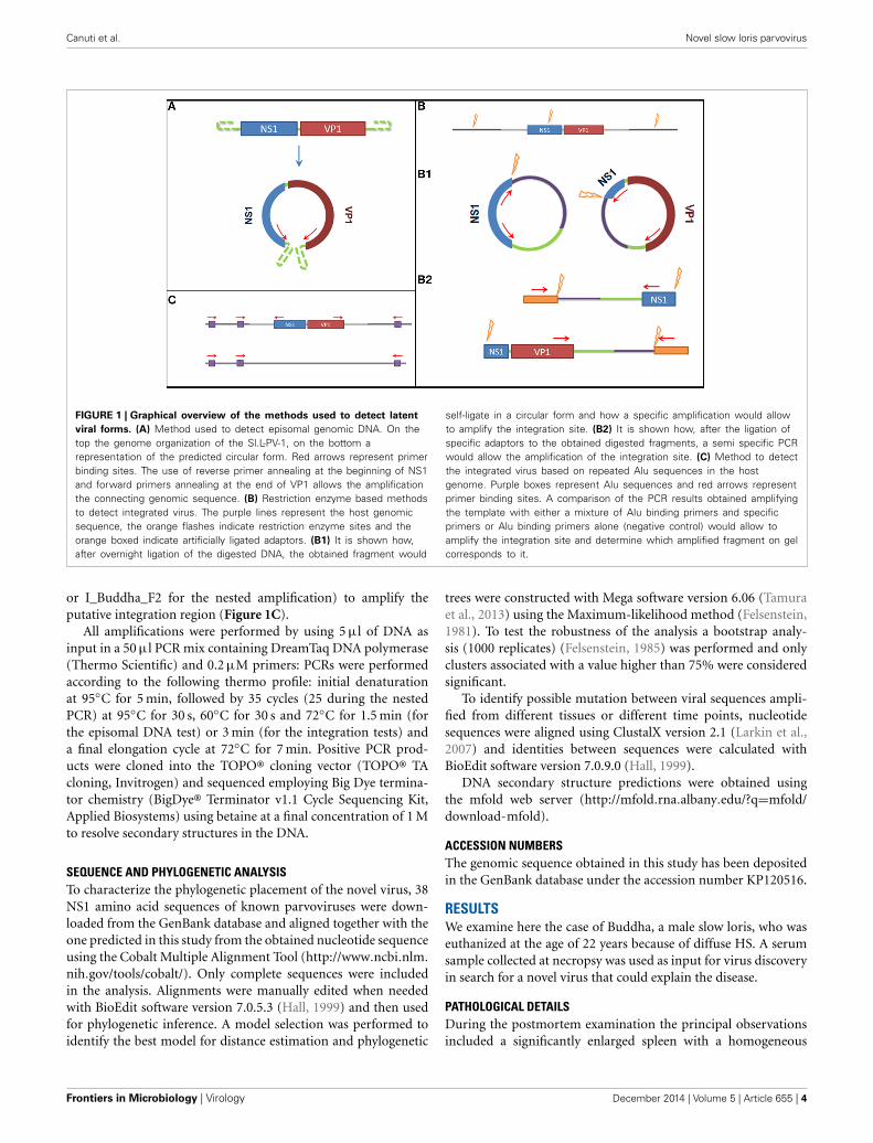
IDENTIFICATION OF INTEGRATED OR EPISOMAL VIRAL DNATo detect the presence of latent viral forms (extra-chromosomalcircular episomal DNA or integrated DNA) in the differenttissues, specific PCR-based assays were designed. A graphicaloverview of the methods used to detect latency is available inFigure 1. For this purpose the DNA isolated from all organs col-lected during the necropsy of Buddha (lung, liver, spleen, kidney,small, and large intestine) and from blood (collected one yearbefore the animal died) were used as input.
To detect the presence of circularized viral genome aninverted nested PCR was performed using primers annealingat the end of the two open reading frames (ORFs) (Figure 1A):I_Buddha_R1—CCTTGTTCGATCTGTCCAAAATAATTGC—and I_Buddha_F1—CCAGTAGTGGAGAAGTCATCTACCC—for the first step of amplification; I_Buddha_R2—GTAATTGCTTTAATGGCTTTGATTCCCAG—and I-Buddha_F2—TGGAAGTCGAGTAAAATACCGACACATG—for the nested amplifica-tion. As a control DNA isolated from serum samples (collected in2008 both with and without VIDISCA pre-treatment) was alsoincluded.
To detect chromosome-integrated viral forms 3 differentapproaches have been employed, of which 2 involved a spe-cific enzymatic digestion of the isolated DNA (Figure 1B): 7.5 μlDNA eluate was cut (restriction site at nucleotides 868–873) ina 20 μl reaction mix containing 5 U of NdeI restriction enzyme(New England Biolabs) during an incubation of 2 h at 37◦C.Prior to inactivation, 5 μl of cut DNA were collected and usedto perform a specific ligation of VIDISCA adaptors (De Vrieset al., 2011) to the digested fragments using 4 U of T4 DNAligase (life technologies). Afterwards 2 semi virus-specific nestedamplifications were performed using one primer complementaryto the virus (I_Buddha_R1 and I_Buddha_R2 for the 5 ′ side;I_Buddha_F1 and I_Buddha_F2 for the 3 ′ side) and one adaptorspecific primer. In parallel, 5 μl of the cut DNA was subjected toan inactivation step for 20 min at 65◦C, followed by an overnightincubation at room temperature with 4 U of T4 DNA ligase toallow self-ligation and consequent circularization of the digestedfragments (containing host sequences flanking the viral inte-grated DNA). Two specific inverted nested PCRs were used after-wards with primer pairs I_Buddha_F1/I_Buddha_R3 (TCCCGACACAAATAATCGGACAACC) and I_Buddha_R1/I_Buddha_F3(GACGTGCTTTGTTAGAATCTGTTCCTG) during the first stepand I_Buddha_F2/I_Buddha_R4 (GAAATTTGAATTTCCATCTTAGCTTGAGTC) and I_Buddha_R2/I_Buddha_F4 (CAAACCCTGCAATGGTGTGTAGATG) during the second amplificationstep.
The third approach for integrated DNA detection made use ofthe Alu elements, DNA sequences which are highly repeated inprimate genomes (Häsler and Strub, 2006). A primer (Ny_Alu:CCTCCCAGAGTGCTAGGATTGCAC) binding to one of theseregion was designed (according to GenBank sequence with acces-sion number DQ822059) and used alone (as a negative control)or in combination with viral specific primers (I_Buddha_R1 orI_Buddha_F1 for the first amplification step and I_Buddha_R2
www.frontiersin.org December 2014 | Volume 5 | Article 655 | 3
Canuti et al. Novel slow loris parvovirus
FIGURE 1 | Graphical overview of the methods used to detect latent
viral forms. (A) Method used to detect episomal genomic DNA. On thetop the genome organization of the Sl.L-PV-1, on the bottom arepresentation of the predicted circular form. Red arrows represent primerbinding sites. The use of reverse primer annealing at the beginning of NS1and forward primers annealing at the end of VP1 allows the amplificationthe connecting genomic sequence. (B) Restriction enzyme based methodsto detect integrated virus. The purple lines represent the host genomicsequence, the orange flashes indicate restriction enzyme sites and theorange boxed indicate artificially ligated adaptors. (B1) It is shown how,after overnight ligation of the digested DNA, the obtained fragment would
self-ligate in a circular form and how a specific amplification would allowto amplify the integration site. (B2) It is shown how, after the ligation ofspecific adaptors to the obtained digested fragments, a semi specific PCRwould allow the amplification of the integration site. (C) Method to detectthe integrated virus based on repeated Alu sequences in the hostgenome. Purple boxes represent Alu sequences and red arrows representprimer binding sites. A comparison of the PCR results obtained amplifyingthe template with either a mixture of Alu binding primers and specificprimers or Alu binding primers alone (negative control) would allow toamplify the integration site and determine which amplified fragment on gelcorresponds to it.
or I_Buddha_F2 for the nested amplification) to amplify theputative integration region (Figure 1C).
All amplifications were performed by using 5 μl of DNA asinput in a 50 μl PCR mix containing DreamTaq DNA polymerase(Thermo Scientific) and 0.2 μM primers: PCRs were performedaccording to the following thermo profile: initial denaturationat 95◦C for 5 min, followed by 35 cycles (25 during the nestedPCR) at 95◦C for 30 s, 60◦C for 30 s and 72◦C for 1.5 min (forthe episomal DNA test) or 3 min (for the integration tests) anda final elongation cycle at 72◦C for 7 min. Positive PCR prod-ucts were cloned into the TOPO® cloning vector (TOPO® TAcloning, Invitrogen) and sequenced employing Big Dye termina-tor chemistry (BigDye® Terminator v1.1 Cycle Sequencing Kit,Applied Biosystems) using betaine at a final concentration of 1 Mto resolve secondary structures in the DNA.
SEQUENCE AND PHYLOGENETIC ANALYSISTo characterize the phylogenetic placement of the novel virus, 38NS1 amino acid sequences of known parvoviruses were down-loaded from the GenBank database and aligned together with theone predicted in this study from the obtained nucleotide sequenceusing the Cobalt Multiple Alignment Tool (http://www.ncbi.nlm.
nih.gov/tools/cobalt/). Only complete sequences were includedin the analysis. Alignments were manually edited when neededwith BioEdit software version 7.0.5.3 (Hall, 1999) and then usedfor phylogenetic inference. A model selection was performed toidentify the best model for distance estimation and phylogenetic
trees were constructed with Mega software version 6.06 (Tamuraet al., 2013) using the Maximum-likelihood method (Felsenstein,1981). To test the robustness of the analysis a bootstrap analy-sis (1000 replicates) (Felsenstein, 1985) was performed and onlyclusters associated with a value higher than 75% were consideredsignificant.
To identify possible mutation between viral sequences ampli-fied from different tissues or different time points, nucleotidesequences were aligned using ClustalX version 2.1 (Larkin et al.,2007) and identities between sequences were calculated withBioEdit software version 7.0.9.0 (Hall, 1999).
DNA secondary structure predictions were obtained usingthe mfold web server (http://mfold.rna.albany.edu/?q=mfold/download-mfold).
ACCESSION NUMBERSThe genomic sequence obtained in this study has been depositedin the GenBank database under the accession number KP120516.
RESULTSWe examine here the case of Buddha, a male slow loris, who waseuthanized at the age of 22 years because of diffuse HS. A serumsample collected at necropsy was used as input for virus discoveryin search for a novel virus that could explain the disease.
PATHOLOGICAL DETAILSDuring the postmortem examination the principal observationsincluded a significantly enlarged spleen with a homogeneous
Frontiers in Microbiology | Virology December 2014 | Volume 5 | Article 655 | 4
Canuti et al. Novel slow loris parvovirus
cut surface and a mottled liver with irregular patches of pallor.Pancreatic and mediastinal lymph nodes were enlarged.
Incidental lesions included a cystic mass in the right mam-mary gland and several small ulcers in the fundus of the stomach.Histologic examination of the spleen revealed a loss of normalarchitecture and an intense infiltration of neoplastic round topolygonal cells with moderate variation in cell and nuclear sizes(anisocytosis and anisokaryosis). Mitotic figures were commonand multinucleate cells were occasionally seen. In the liver therewere similar neoplastic cell aggregates surrounding the portalveins and the central veins, expanding the associated connec-tive tissue. Ingested red blood cells (erythrophagocytosis) byneoplastic cells were also observed. Affected lymph nodes werecharacterized by a loss of normal architecture which was replacedby sheets of round to polygonal neoplastic cells similar to thosefound in the liver and spleen. These findings led to a diagnosis ofdisseminated histiocytic sarcoma.
Additional findings included evidence of an apocrine glandcyst causing the mass in the right mammary gland. In additionthere was evidence of inflammation and necrosis in the adrenalglands associated with a protozoal infection, interpreted to beToxoplasma gondii. In addition, large deeply basophilic intranu-clear inclusions were found in a minority of adrenal cortical cells.Renal tubules contained eosinophilic droplets in the epithelialcells, as is often seen in mice with histiocytic sarcoma, althoughthe cause is not known. Chronic interstitial nephritis was alsoevident.
IDENTIFICATION OF A NOVEL PARVOVIRUS AND COMPLETE GENOMESEQUENCINGTo explore the possibility of viral involvement in the etiopatho-genesis of the HS we used VIDISCA-454, a sequence independentvirus identification technique which is able to detect virtually anyDNA and RNA virus from various clinical samples (van der Hoeket al., 2004; Canuti et al., 2011; Jazaeri Farsani et al., 2013; Tanet al., 2013; Canuti et al., 2014; Oude Munnink et al., 2014; Parianiet al., 2014). A total of 8003 sequence reads were obtained fromthe serum sample of the slow loris with HS and, among these,5 sequences were identified with homology to different mem-bers of the Parvovirinae subfamily: 3 were identified with blastn(nucleotide identity: 62–70%) and 2 were only recognizable whenidentity search was performed at amino acid level (amino acididentity: 69–82%). Those recognized as viral fragments were usedas template for primer design, and a combination of specific PCRsand genome walking techniques allowed retrieval of the almostcomplete genomic sequence of the virus (only part of the termi-nal repeats is lacking), which was tentatively named Slow Lorisparvovirus 1 (Sl.L-PV-1). The genomic sequence allowed us toidentify 3 further viral sequences which were not identifiable viaidentity search. In total 35 reads (0.44% of the total) belonged tothe novel virus.
GENOME ORGANIZATIONThe rules set by the parvovirus study group in the latestICTV report mention that a novel parvovirus, which lackedisolation, can be classified within the Parvoviridae family onlyif the complete coding sequence is provided and the typical
parvoviral motif patterns can be recognized (Cotmore et al.,2013).
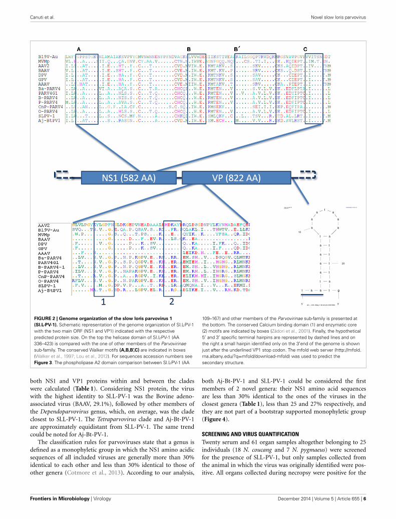
The genome of the novel parvovirus is 4844 nt andthe genomic organization reflects the other members of theParvovirinae subfamily with 2 large ORFs and terminal sequenceswhich form end loop structures important for viral replication,although the structure of the loops could not be entirely resolvedsince only partial sequences could be retrieved (Figure 2) (Berns,1990). The first ORF—located at the 5′ part of the genome(nt 146–1894)—putatively encodes for the non-structural pro-tein NS1 (582 AA). The second ORF—located at the 3′ part (nt2187–4655)—encodes for the capsid protein VP1 (822 AA).
Other molecular features typical of parvoviruses were alsoidentified (Figure 2). A phospholipase A2 domain of Sl.L-PV-1,that is conserved in the majority of parvoviruses and which isessential for viral genome transfer to the nucleus, is located atthe N-terminal part of the VP1 unique region and contains thetypical PLA2 catalytic domain (HDXXY, AA 140–144) and Ca++binding loop (YXGXG, AA 117–121) (Zádori et al., 2001).
Another molecular marker of parvoviruses is the presence ofconserved helicase sequence motifs in the NS1 protein (Walkeret al., 1997; Lou et al., 2012). Within the carboxy-terminal halfof the NS1 protein of Sl.L-PV-1 (AA 336-423) the typical ATPbinding loop or p-loop (Walker box A: GXXXXGK(T/S)) couldbe identified, immediately followed by the Mg++ binding WalkerB (hhhh(D/E)E) and B’ motifs. Finally, the C motif (a stretch ofhydrophobic residues usually followed by asparagine), commonto the helicases belonging to the superfamily type III, was alsopresent (Figure 2).
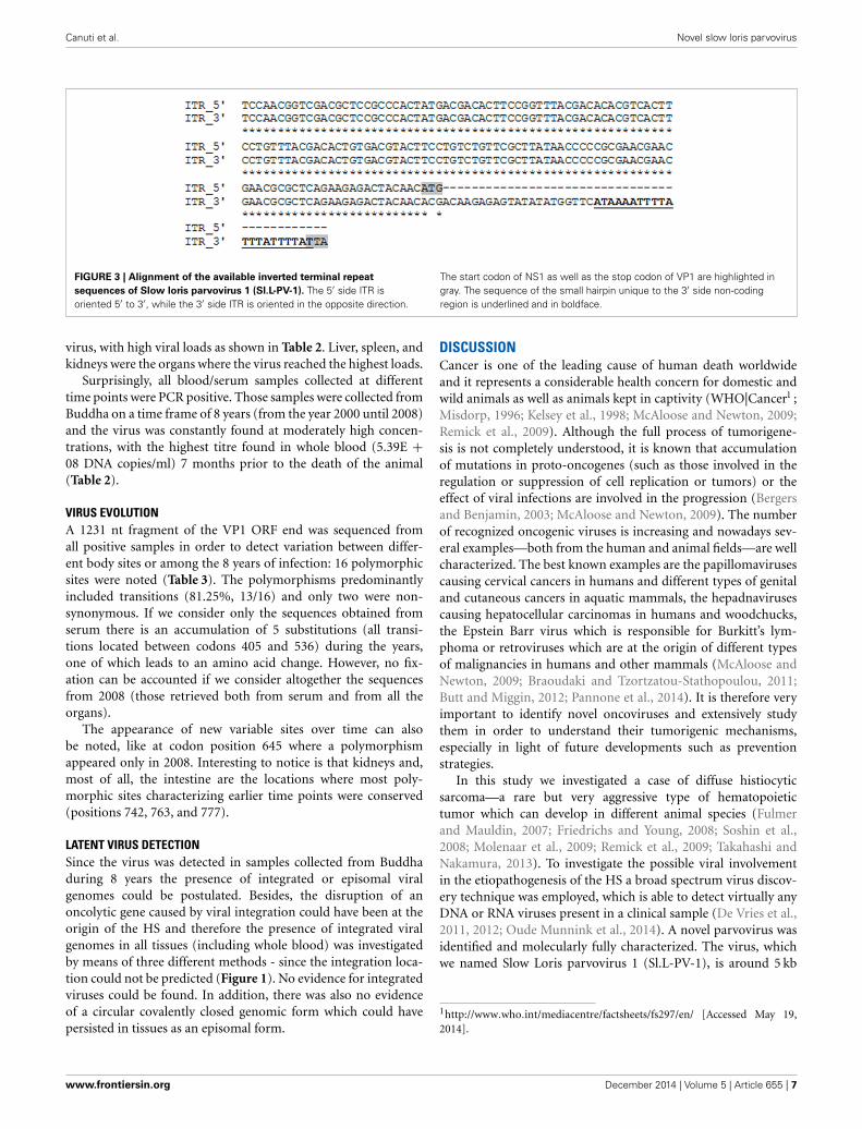
We were able to obtain only part of the sequences from theterminal non coding regions, 148 and 192 nt of the 5′ and 3′end respectively. An alignment of these 2 sequences shows thatthey are 100% identical (but oriented in the opposite direction)and start differentiating from the ATG start codon of NS1, whichbecomes ACG on the 3′ side terminal sequence (Figure 3). Thesedata suggest that Sl.L-PV-1 possesses identical inverted terminalrepeats. Besides, a 44 nt sequence was identified which is presentonly on the 3′ side of the genome, just after the TAA stop codonof VP1/VP2, that contains a 21 nt region that can fold into a smallhairpin (Figures 2, 3).
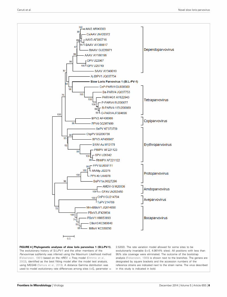
PHYLOGENETIC ANALYSIS AND PROPOSED CLASSIFICATIONPhylogenetic analysis of Sl.L-PV-1 was performed using the pre-dicted amino acid sequences of the NS1 protein belonging tomembers of the Parvovirinae subfamily (Figure 4). In the phylo-genetic tree the 8 Parvovirinae genera are indicated: each definedcluster was supported by a significant bootstrap value (between93 and 100). Only 2 viruses did not cluster in any of these 8clades: a parvovirus identified in Artibeus jamaicensis fruit bat(Aj-Bt-PV-1, which we previously reported as the first memberof a novel parvovirus genus, Canuti et al., 2011) and the Sl.L-PV-1. Both viruses are located between the Dependoparvovirus andthe Tetraparvovirus genera and do not significantly cluster withany other known parvovirus, possibly representing 2 independentstill undefined genera.
To better clarify the relationship between Sl.L-PV-1 and themost closely related parvoviruses, the amino acid identities of
www.frontiersin.org December 2014 | Volume 5 | Article 655 | 5
Canuti et al. Novel slow loris parvovirus
FIGURE 2 | Genome organization of the slow loris parvovirus 1
(Sl.L-PV-1). Schematic representation of the genome organization of Sl.L-PV-1with the two main ORF (NS1 and VP1) indicated with the respectivepredicted protein size. On the top the helicase domain of Sl.L-PV-1 (AA336–423) is compared with the one of other members of the Parvovirinaesub-family. The conserved Walker motifs (A,B,B’,C) are indicated in boxes(Walker et al., 1997; Lou et al., 2012). For sequences accession numbers seeFigure 3. The phospholipase A2 domain comparison between Sl.L-PV-1 (AA
109–167) and other members of the Parvovirinae sub-family is presented atthe bottom. The conserved Calcium binding domain (1) and enzymatic core(2) motifs are indicated by boxes (Zádori et al., 2001). Finally, the hypothetical5′ and 3′ specific terminal hairpins are represented by dashed lines and onthe right a small hairpin identified only on the 3′end of the genome is shownjust after the underlined VP1 stop codon. The mfold web server (http://mfold.
rna.albany.edu/?q=mfold/download-mfold) was used to predict thesecondary structure.
both NS1 and VP1 proteins within and between the cladeswere calculated (Table 1). Considering NS1 protein, the viruswith the highest identity to Sl.L-PV-1 was the Bovine adeno-associated virus (BAAV, 29.1%), followed by other members ofthe Dependoparvovirus genus, which, on average, was the cladeclosest to Sl.L-PV-1. The Tetraparvovirus clade and Aj-Bt-PV-1are approximately equidistant from Sl.L-PV-1. The same trendcould be noted for Aj-Bt-PV-1.
The classification rules for parvoviruses state that a genus isdefined as a monophyletic group in which the NS1 amino acidicsequences of all included viruses are generally more than 30%identical to each other and less than 30% identical to those ofother genera (Cotmore et al., 2013). According to our analysis,
both Aj-Bt-PV-1 and Sl.L-PV-1 could be considered the firstmembers of 2 novel genera: their NS1 amino acid sequencesare less than 30% identical to the ones of the viruses in theclosest genera (Table 1), less than 25 and 27% respectively, andthey are not part of a bootstrap supported monophyletic group(Figure 4).
SCREENING AND VIRUS QUANTIFICATIONTwenty serum and 61 organ samples altogether belonging to 25individuals (18 N. coucang and 7 N. pygmaeus) were screenedfor the presence of Sl.L-PV-1, but only samples collected fromthe animal in which the virus was originally identified were pos-itive. All organs collected during necropsy were positive for the
Frontiers in Microbiology | Virology December 2014 | Volume 5 | Article 655 | 6
Canuti et al. Novel slow loris parvovirus
FIGURE 3 | Alignment of the available inverted terminal repeat
sequences of Slow loris parvovirus 1 (Sl.L-PV-1). The 5′ side ITR isoriented 5′ to 3′, while the 3′ side ITR is oriented in the opposite direction.
The start codon of NS1 as well as the stop codon of VP1 are highlighted ingray. The sequence of the small hairpin unique to the 3′ side non-codingregion is underlined and in boldface.
virus, with high viral loads as shown in Table 2. Liver, spleen, andkidneys were the organs where the virus reached the highest loads.
Surprisingly, all blood/serum samples collected at differenttime points were PCR positive. Those samples were collected fromBuddha on a time frame of 8 years (from the year 2000 until 2008)and the virus was constantly found at moderately high concen-trations, with the highest titre found in whole blood (5.39E +08 DNA copies/ml) 7 months prior to the death of the animal(Table 2).
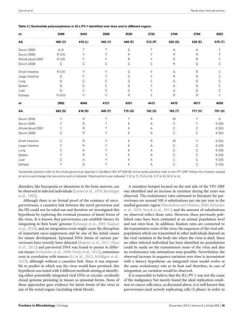
VIRUS EVOLUTIONA 1231 nt fragment of the VP1 ORF end was sequenced fromall positive samples in order to detect variation between differ-ent body sites or among the 8 years of infection: 16 polymorphicsites were noted (Table 3). The polymorphisms predominantlyincluded transitions (81.25%, 13/16) and only two were non-synonymous. If we consider only the sequences obtained fromserum there is an accumulation of 5 substitutions (all transi-tions located between codons 405 and 536) during the years,one of which leads to an amino acid change. However, no fix-ation can be accounted if we consider altogether the sequencesfrom 2008 (those retrieved both from serum and from all theorgans).
The appearance of new variable sites over time can alsobe noted, like at codon position 645 where a polymorphismappeared only in 2008. Interesting to notice is that kidneys and,most of all, the intestine are the locations where most poly-morphic sites characterizing earlier time points were conserved(positions 742, 763, and 777).
LATENT VIRUS DETECTIONSince the virus was detected in samples collected from Buddhaduring 8 years the presence of integrated or episomal viralgenomes could be postulated. Besides, the disruption of anoncolytic gene caused by viral integration could have been at theorigin of the HS and therefore the presence of integrated viralgenomes in all tissues (including whole blood) was investigatedby means of three different methods - since the integration loca-tion could not be predicted (Figure 1). No evidence for integratedviruses could be found. In addition, there was also no evidenceof a circular covalently closed genomic form which could havepersisted in tissues as an episomal form.
DISCUSSIONCancer is one of the leading cause of human death worldwideand it represents a considerable health concern for domestic andwild animals as well as animals kept in captivity (WHO|Cancer1 ;Misdorp, 1996; Kelsey et al., 1998; McAloose and Newton, 2009;Remick et al., 2009). Although the full process of tumorigene-sis is not completely understood, it is known that accumulationof mutations in proto-oncogenes (such as those involved in theregulation or suppression of cell replication or tumors) or theeffect of viral infections are involved in the progression (Bergersand Benjamin, 2003; McAloose and Newton, 2009). The numberof recognized oncogenic viruses is increasing and nowadays sev-eral examples—both from the human and animal fields—are wellcharacterized. The best known examples are the papillomavirusescausing cervical cancers in humans and different types of genitaland cutaneous cancers in aquatic mammals, the hepadnavirusescausing hepatocellular carcinomas in humans and woodchucks,the Epstein Barr virus which is responsible for Burkitt’s lym-phoma or retroviruses which are at the origin of different typesof malignancies in humans and other mammals (McAloose andNewton, 2009; Braoudaki and Tzortzatou-Stathopoulou, 2011;Butt and Miggin, 2012; Pannone et al., 2014). It is therefore veryimportant to identify novel oncoviruses and extensively studythem in order to understand their tumorigenic mechanisms,especially in light of future developments such as preventionstrategies.
In this study we investigated a case of diffuse histiocyticsarcoma—a rare but very aggressive type of hematopoietictumor which can develop in different animal species (Fulmerand Mauldin, 2007; Friedrichs and Young, 2008; Soshin et al.,2008; Molenaar et al., 2009; Remick et al., 2009; Takahashi andNakamura, 2013). To investigate the possible viral involvementin the etiopathogenesis of the HS a broad spectrum virus discov-ery technique was employed, which is able to detect virtually anyDNA or RNA viruses present in a clinical sample (De Vries et al.,2011, 2012; Oude Munnink et al., 2014). A novel parvovirus wasidentified and molecularly fully characterized. The virus, whichwe named Slow Loris parvovirus 1 (Sl.L-PV-1), is around 5 kb
1http://www.who.int/mediacentre/factsheets/fs297/en/ [Accessed May 19,2014].
www.frontiersin.org December 2014 | Volume 5 | Article 655 | 7
Canuti et al. Novel slow loris parvovirus
FIGURE 4 | Phylogenetic analyses of slow loris parvovirus 1 (Sl.L-PV-1).
The evolutionary history of Sl.L-PV-1 and the other members of theParvovirinae subfamily was inferred using the Maximum Likelihood method(Felsenstein, 1981) based on the rtREV + Freq model (Dimmic et al.,2002), identified as the best fitting model after the model test analysis,using MEGA6 (Tamura et al., 2013). A distance Gamma distribution wasused to model evolutionary rate differences among sites (+G, parameter =
2.5202). The rate variation model allowed for some sites to beevolutionarily invariable ([+I], 4.0614% sites). All positions with less than95% site coverage were eliminated. The outcome of the bootstrapanalysis (Felsenstein, 1985) is shown next to the branches. The genera aredesignated by square brackets and the accession numbers of thereference strains are indicated next to the strain name. The virus describedin this study is indicated in bold.
Frontiers in Microbiology | Virology December 2014 | Volume 5 | Article 655 | 8
Canuti et al. Novel slow loris parvovirus
Table 1 | Identities between and within the clades more closely related to Sl.L-PV-1.
Dependoparvovirus Tetraparvovirus Aj-Bt_PV-1 Sl.L-PV-1
Dependoparvovirus 81.87 (61–45) 17.21 (18.9–15.1) 21.01 (22.4–19.4) 23.4 (24.1–22.4)
43.27 (64.3–28)
Tetraparvovirus 50.14 (72.9–27.3) 18.8 (19.6–18.2) 20.4 (20.9–19.6)
21.18 (23.4–18.6) 46.45 (75.7–29.6)
Aj-Bt_PV-1 id 21.3
24.81 (26–22.4) 22.6 (23.1–22) id
Sl.L-PV-1 id
26.6 (29.1–24.5) 22.8 (25–19.9) 23.6 id
Values represent average percentage identity (upper limit—lower limit) calculated using representative members of each species in the clades. Values in bold
(bottom left) refers to NS1 amino acid sequence, while the other numbers (top right) refer to VP1 amino acid sequence. On the diagonal in italic are the identities
within each clade; id, identical. The extended analysis is available in the Supplementary Tables 1, 2.
Table 2 | Sl.L-PV-1 loads determined in different organs (collected at
necropsy in 2008) and from serum/blood samples collected during
various years.
Material Collection date Viral load (copies/g or
(mm/dd/yyyy) copies/ml)
Lung 06/03/2008 6.09E + 07
Small intestine 06/03/2008 4.82E + 07
Large intestine 06/03/2008 1.24E + 07
Liver 06/03/2008 1.87E + 08
Spleen 06/03/2008 1.79E + 08
Kidney 06/03/2008 1.43E + 09
Serum 03/21/2000 1.90E + 07
Serum 12/08/2005 4.20E + 07
Whole blood 11/08/2007 5.39E + 08
Serum* 06/03/2008 1.08E + 06
*After VIDISCA pre-treatment (centrifugation and DNase treatment prior to DNA
isolation)
in size and possesses all the molecular features typical of par-voviruses, including sequences coding for conserved enzymaticmotifs and the 2 main ORFs, flanked by non-coding termi-nal regions, which appear identical but inverted like describedfor other parvoviruses (Lusby et al., 1980; Berns, 1990; Deisset al., 1990). The virus is phylogenetically located between theDependoparvovirus and the Tetraparvovirus genera and, accord-ing to the classification rules defined by the ICTV (Cotmore et al.,2013), is possibly the first member of a new genus.
Thanks to the availability of a series of serum and organ sam-ples collected over multiple years from slow loris with variousdiseases, we were able to screen different samples from a total of25 individuals belonging to the N. coucang species and to the clos-est related species N. pygmaeus. No other animal was positive forthe virus but, as expected, all organs collected during the necropsywere positive, as also reported for other parvoviruses (Meunieret al., 1985; Canuti et al., 2011), and liver, spleen and kidneyswere the organs where the virus reached the highest loads. The
high viral concentrations found in these organs might reflect theirelevated content of blood and blood cells. Besides, during the his-tological investigation the presence of basophilic inclusions in therenal tissue could be observed and these might represent parvovi-ral accumulations, similar to other reports for parvoviruses bothin vivo (Hayes et al., 1979; Bestetti and Zwahlen, 1985; Porteret al., 1988; Decaro and Buonavoglia, 2012) and in vitro (Inabaet al., 1973; Oleksiewicz et al., 1996). Surprisingly, all blood orserum samples collected during various years were positive at arather constant load: we could detect viremia 8 years prior to thedeath of the animal. Given the involvement in the disease of whiteblood cells, a presumed condition of immunosuppression (causedeither by the virus, the tumor, or by a combination of these 2factors) can be postulated and might be a possible explanationfor the fact that the virus was not cleared in 8 years. The sup-posed presence of immunodeficiency in the loris is supported bythe detection of opportunistic infections, as the pathological testsreported the presence of Toxoplasma gondii infection.
Since HS is a fast and very aggressive type of cancer wecould have hypothesized that the virus was present in the Lorisbefore tumor development. Histiocytes can refer to cells ofeither the macrophage or dendritic cell lineage as both arisefrom a common precursor cell. Neoplasms of histiocytes canarise from macrophages or one of two types of dendritic cells,Langerhans cells or interstitial dendritic cells. Langerhans cellsare found within the epithelium and interstitial dendritic cellsoccupy a perivascular position in most tissues. Most forms ofHS in animals arise as a malignant proliferation of interstitialdendritic cells, although there is one form, the hemophagocyticvariant, that arises from macrophages (Fulmer and Mauldin,2007; Takahashi and Nakamura, 2013; Moore, 2014). There isno established classification scheme for HS in prosimians. Weinterpreted this tumor to have most likely arisen from intersti-tial dendritic cells. Some parvoviruses have proven tropism forcells of the hematopoietic system and the bone marrow pro-viding the perfect condition for parvoviral replication, since awide spectrum of cells at different dividing stages are presentand parvoviruses need actively dividing cells (S phase) to repli-cate (Berns, 1990; Segovia et al., 1991). In fact, hematopoietic
www.frontiersin.org December 2014 | Volume 5 | Article 655 | 9
Canuti et al. Novel slow loris parvovirus
Table 3 | Nucleotide polymorphisms in Sl.L-PV-1 identified over time and in different organs.
nt 3399 3443 3506 3530 3722 3746 3794 3923
AA 405 (V) 419 (L) 440 (Y) 448 (E) 512 (P) 520 (Q) 536 (E) 579 (T)
Serum 2000 A (I) T T G T A A Y
Serum 2005 R (V/I) Y Y R Y R R Y
Whole blood 2007 R (V/I) Y Y R Y R R Y
Serum 2008 G C C G C R G C
Small intestine R (V/I) Y Y G Y A R C
Large intestine G Y Y G Y R R C
Lung G C C G C A G C
Spleen G C C G Y A G C
Liver G C C G Y A G C
Kidneys R (IV/I) Y Y R Y R R Y
nt 3992 4040 4121 4331 4412 4475 4517 4558
AA 603 (S) 618 (R) 645 (F) 715 (G) 742 (S) 763 (T) 777 (V) 791 (S)
Serum 2000 Y R T T R M Y G
Serum 2005 Y R T K A C Y K (S/I)
Whole blood 2007 Y R T K A C C K (S/I)
Serum 2008 C R Y K A C C K (S/I)
Small intestine C R Y K R M Y K (S/I)
Large intestine Y R Y K A C C K (I/S)
Lung C A Y K A C C K (I/S)
Spleen Y R Y K A C C K (I/S)
Liver C A Y K A C C K (I/S)
kidneys Y A Y K A C C K (I/S)
Nucleotide positions refer to the whole genome as reported in GenBank (AN: KP120516); amino acids positions refer to the VP1 ORF. Where the mutation caused
an amino acid change the new amino acid is indicated. Polymorphisms are indicated: Y (C or T), R (A or G), K (T or G), M (C or A).
disorders, like leucopenia or alterations in the bone marrow, canbe observed in infected individuals (Larsen et al., 1976; Boosingeret al., 1982).
Although there is no formal proof of the existence of onco-parvoviruses, a causative link between the novel parvovirus andthe HS could not be ruled out and therefore we investigated thishypothesis by exploring the eventual presence of latent forms ofthe virus. It is known that parvoviruses can establish latency byintegrating in their hosts’ genome (Schnepp et al., 2005; Kapooret al., 2010), and an integration event might cause the disruptionof important onco-suppressors and be one of the initial causesfor tumor development. Episomal DNA forms of various par-voviruses have recently been detected (Kapoor et al., 2011; Zhaoet al., 2012) and parvoviral DNA was found to persist in differ-ent tissues (Schneider et al., 2008; Norja et al., 2012), sometimeseven in correlation with tumors (Li et al., 2012; Schildgen et al.,2013), although without a causative link. Since it was impossi-ble to predict in which way the virus would have persisted, thishypothesis was tested with 4 different methods aiming at identify-ing either potentially integrated viral DNA or circular covalentlyclosed genome persisting in tissues as episomal forms. None ofthese approaches gave evidence for latent forms of the virus inany of the tested organs (including whole blood).
A mutation hotspot located on the end side of the VP1 ORFwas identified and an increase in variation during the years wasobserved. The evolutionary rates estimated in literature for par-voviruses are around 10E-4 substitutions per site per year in thestudied genomic region (Shackelton and Holmes, 2006; Zehenderet al., 2010; Streck et al., 2011) and the amount of substitutionswe observed reflect those rates. However, these previously pub-lished rates have been estimated at an animal population leveland not inter-host. In addition, fixation rates are connected tothe transmission route of the virus: the sequences of the viral sub-population which are transmitted to other individuals depend onthe viral variation at the body site where the virus is shed. Sinceno other infected individual has been identified no postulationscould be made on the transmission route of the virus and alsono evolutionary rate estimations were possible. Nevertheless, theobserved increase in sequence variation over time is inconsistentwith a latency hypothesis: an integrated virus would evolve atthe same evolutionary rate as its host and therefore, in case ofintegration, no variation would be observed.
It is reasonable to believe that the Sl.L-PV-1 was not the causeof the malignancy but merely found the ideal replication condi-tion in cancer cells since, as discussed above, it is well known thatparvoviruses need actively replicating cells (S-phase) in order to
Frontiers in Microbiology | Virology December 2014 | Volume 5 | Article 655 | 10
Canuti et al. Novel slow loris parvovirus
proliferate (Berns, 1990). In fact it is known that certain viruses,called oncolytic viruses, have tropism for specific cancer cells andthey can even lead to “spontaneous regression” of malignancies(Butt and Miggin, 2012; Sze et al., 2013). This has been proven forthe adeno-associated dependoparvovirus, although in an indirectway by enhancing adenoviral replication, and for a rodent par-vovirus (Nüesch et al., 2012; Laborda et al., 2013). Future researchwill have to clarify whether this viral infection precedes tumordevelopment and has a role in the oncogenic process, whether thevirus simply benefits from the replication activity of the cancercells or if the persistent viremia has no correlation with the HS.An answer to this question will be obtained by identifying the celltypes where viral replication occurs after the detection of otherinfected loris and the constant monitoring of their physical con-dition and disease progression, and after the discovery of relatedviruses in other animals with similar malignancies. Althoughmore studies are required to provide a conclusive answer, theinfection with Sl.L-PV-1 might have delayed tumor progression.If this mechanism will be proven feasible new exciting possibilitiesmight open for oncolytic parvovirotherapy (Nüesch et al., 2012;Russell et al., 2012).
In conclusion we discovered and molecularly characterized anovel parvovirus, the first member of a not yet defined genusand the first described in prosimians. The virus was identified ina slow loris with HS but we found no evidence for a causativeinvolvement in the neoplastic disease and postulated that thevirus had a replication advantage derived from the constantlyreplicating cancer cells.
ACKNOWLEDGMENTSThe authors thank Dr. Rienk Jeeninga for appreciated advices ondesigning latency tests and Dr. Elena Herrera Carrillo for provid-ing the protocol for sequencing the terminal loop regions. Thisstudy was supported by funding from the European Community’sSeventh Framework Programme (FP7/2007–2013) under theproject EMPERIE, EC grant agreement number 223498.
SUPPLEMENTARY MATERIALThe Supplementary Material for this article can be foundonline at: http://www.frontiersin.org/journal/10.3389/fmicb.2014.00655/abstract
REFERENCESAltschul, S. F., Gish, W., Miller, W., Myers, E. W., and Lipman, D. J. (1990). Basic
local alignment search tool. J. Mol. Biol. 215, 403–410. doi: 10.1016/S0022-2836(05)80360-2
Bergers, G., and Benjamin, L. E. (2003). Tumorigenesis and the angiogenic switch.Nat. Rev. Cancer 3, 401–410. doi: 10.1038/nrc1093
Berns, K. I. (1990). Parvovirus replication. Microbiol. Rev. 54, 316–329.Best, S. M., and Bloom, M. E. (2005). Pathogenesis of aleutian mink disease par-
vovirus and similarities to b19 infection. J. Vet. Med. B Infect. Dis. Vet. PublicHealth 52, 331–334. doi: 10.1111/j.1439-0450.2005.00864.x
Bestetti, G., and Zwahlen, R. (1985). Generalized parvovirus infection withinclusion-body myocarditis in two kittens. J. Comp. Pathol. 95, 393–397. doi:10.1016/0021-9975(85)90043-X
Boosinger, T. R., Rebar, A. H., DeNicola, D. B., and Boon, G. D. (1982). Bonemarrow alterations associated with canine parvoviral enteritis. Vet. Pathol. 19,558–561.
Braoudaki, M., and Tzortzatou-Stathopoulou, F. (2011). Tumorigenesis related toretroviral infections. J. Infect. Dev. Ctries. 5, 751–758. doi: 10.3855/jidc.1773
Butt, A. Q., and Miggin, S. M. (2012). Cancer and viruses: a double-edged sword.Proteomics 12, 2127–2138. doi: 10.1002/pmic.201100526
Canuti, M., Deijs, M., Jazaeri Farsani, S. M., Holwerda, M., Jebbink, M. F., de Vries,M., et al. (2014). Metagenomic analysis of a sample from a patient with respira-tory tract infection reveals the presence of a γ-papillomavirus. Front. Microbiol5:347. doi: 10.3389/fmicb.2014.00347
Canuti, M., Eis-Huebinger, A. M., Deijs, M., de Vries, M., Drexler, J. F., Oppong, S.K., et al. (2011). Two novel parvoviruses in frugivorous new and old world bats.PLoS ONE 6:e29140. doi: 10.1371/journal.pone.0029140
Clegg, S. R., Coyne, K. P., Dawson, S., Spibey, N., Gaskell, R. M., and Radford, A. D.(2012). Canine parvovirus in asymptomatic feline carriers. Vet. Microbiol. 157,78–85. doi: 10.1016/j.vetmic.2011.12.024
Cotmore, S. F., Agbandje McKenna, M., Chiorini, J. A., Gatherer, D., Mukha, D. V.,Pintel, D. J., et al. (2013). Rationalization and Extension of the Taxonomy of theFamily Parvoviridae. Available online at: http://www.ictvonline.org/proposals/2013.001a-aaaV.A.v4.Parvoviridae.pdf
Cotmore, S. F., and Tattersall, P. (2014). Parvoviruses: small does not meansimple. Annu. Rev. Virol. 1, 517–537. doi: 10.1146/annurev-virology-031413-085444
De Vries, M., Deijs, M., Canuti, M., van Schaik, B. D. C., Faria, N. R., van de Garde,M. D. B., et al. (2011). A sensitive assay for virus discovery in respiratory clinicalsamples. PLoS ONE 6:e16118. doi: 10.1371/journal.pone.0016118
De Vries, M., Oude Munnink, B. B., Deijs, M., Canuti, M., Koekkoek, S.M., Molenkamp, R., et al. (2012). Performance of VIDISCA-454 in feces-suspensions and serum. Viruses 4, 1328–1334. doi: 10.3390/v4081328
Decaro, N., and Buonavoglia, C. (2012). Canine parvovirus–a review of epidemi-ological and diagnostic aspects, with emphasis on type 2c. Vet. Microbiol. 155,1–12. doi: 10.1016/j.vetmic.2011.09.007
Deiss, V., Tratschin, J. D., Weitz, M., and Siegl, G. (1990). Cloning of the human par-vovirus B19 genome and structural analysis of its palindromic termini. Virology175, 247–254.
Dimmic, M. W., Rest, J. S., Mindell, D. P., and Goldstein, R. A. (2002). rtREV: anamino acid substitution matrix for inference of retrovirus and reverse transcrip-tase phylogeny. J. Mol. Evol. 55, 65–73. doi: 10.1007/s00239-001-2304-y
Donahue, R. E., Kessler, S. W., Bodine, D., McDonagh, K., Dunbar, C., Goodman,S., et al. (1992). Helper virus induced T cell lymphoma in nonhuman primatesafter retroviral mediated gene transfer. J. Exp. Med. 176, 1125–1135.
Feichtinger, H., Li, S. L., Kaaya, E., Putkonen, P., Grünewald, K., Weyrer, K., et al.(1992). A monkey model for Epstein Barr virus-associated lymphomagenesis inhuman acquired immunodeficiency syndrome. J. Exp. Med. 176, 281–286.
Felsenstein, J. (1981). Evolutionary trees from DNA sequences: a maximum likeli-hood approach. J. Mol. Evol. 17, 368–376.
Felsenstein, J. (1985). Confidence limits on phylogenies: an approach using thebootstrap. Evolution 39, 783. doi: 10.2307/2408678
Fisgin, T., Yarali, N., Duru, F., and Kara, A. (2002). Parvovirus-B19 infectionpreceding acute myeloid leukemia with orbital granulocytic sarcoma. Leuk.Lymphoma 43, 2059–2061. doi: 10.1080/1042819021000016168
Friedrichs, K. R., and Young, K. M. (2008). Histiocytic sarcoma of macrophageorigin in a cat: case report with a literature review of feline histiocytic malig-nancies and comparison with canine hemophagocytic histiocytic sarcoma. Vet.Clin. Pathol. 37, 121–128. doi: 10.1111/j.1939-165X.2008.00005.x
Fulmer, A. K., and Mauldin, G. E. (2007). Canine histiocytic neoplasia: an overview.Can. Vet. J. 48, 1041–1050.
Goddard, A., and Leisewitz, A. L. (2010). Canine parvovirus. Vet. Clin. North Am.Small Anim. Pract. 40, 1041–1053. doi: 10.1016/j.cvsm.2010.07.007
Hall, T. (1999). BioEdit: a user-friendly biological sequence alignment editor andanalysis program for Windows 95/98/NT. Nucleic Acids Symp. Ser. 41, 95–98.
Häsler, J., and Strub, K. (2006). Alu elements as regulators of gene expression.Nucleic Acids Res. 34, 5491–5497. doi: 10.1093/nar/gkl706
Hayes, M. A., Russell, R. G., and Babiuk, L. A. (1979). Sudden death in young dogswith myocarditis caused by parvovirus. J. Am. Vet. Med. Assoc. 174, 1197–1203.
Heegaard, E. D., and Brown, K. E. (2002). Human Parvovirus B19. Clin. Microbiol.Rev. 15, 485–505. doi: 10.1128/CMR.15.3.485-505.2002
Huson, D. H., Mitra, S., Ruscheweyh, H.-J., Weber, N., and Schuster, S. C. (2011).Integrative analysis of environmental sequences using MEGAN4. Genome Res.21, 1552–1560. doi: 10.1101/gr.120618.111
Ibrahem, W. N., Hasony, H. J., and Hassan, J. G. (2014). Human parvovirus B19in childhood acute lymphoblastic leukaemia in Basrah. J. Pak. Med. Assoc.64, 9–12.
www.frontiersin.org December 2014 | Volume 5 | Article 655 | 11
Canuti et al. Novel slow loris parvovirus
Inaba, Y., Kurogi, H., Takahashi, E., Sato, K., Tanaka, Y., Goto, Y., et al.(1973). Isolation and properties of bovine parvovirus type 1 from Japanesecalves. Arch. Für Gesamte Virusforsch. 42, 54–66. doi: 10.1007/BF01250507
Jartti, T., Hedman, K., Jartti, L., Ruuskanen, O., Allander, T., and Söderlund-Venermo, M. (2012). Human bocavirus—the first 5 years. Rev. Med. Virol. 22,46–64. doi: 10.1002/rmv.720
Jazaeri Farsani, S. M., Jebbink, M. F., Deijs, M., Canuti, M., van Dort, K. A., Bakker,M., et al. (2013). Identification of a new genotype of Torque teno mini virus.Virol. J. 10, 323. doi: 10.1186/1743-422X-10-323
Kapoor, A., Hornig, M., Asokan, A., Williams, B., Henriquez, J. A., and Lipkin, W.I. (2011). Bocavirus episome in infected human tissue contains non-identicaltermini. PLoS ONE 6:e21362. doi: 10.1371/journal.pone.0021362
Kapoor, A., Simmonds, P., and Lipkin, W. I. (2010). Discovery and characteriza-tion of mammalian endogenous parvoviruses. J. Virol. 84, 12628–12635. doi:10.1128/JVI.01732-10
Kelsey, J. L., Moore, A. S., and Glickman, T. (1998). Epidemiologic studies of riskfactors for cancer in pet dogs. Epidemiol. Rev. 20, 204–217.
Kramer, P., Prins, M. E., Kapsenberg, J. G., Bornkamm, G. W., Bijnen, A. B.,Rohol, P. J., et al. (1985). Persistent Epstein-Barr virus infection and a histiocyticsarcoma in a renal transplant recipient. Cancer 55, 503–509.
Laborda, E., Puig-Saus, C., Cascalló, M., Chillón, M., and Alemany, R.(2013). Adeno-Associated virus enhances wild-type and oncolytic aden-ovirus spread. Hum. Gene Ther. Methods 24, 372–380. doi: 10.1089/hgtb.2013.124
Larkin, M. A., Blackshields, G., Brown, N. P., Chenna, R., McGettigan, P. A.,McWilliam, H., et al. (2007). Clustal W and Clustal X version 2.0. Bioinformatics23, 2947–2948. doi: 10.1093/bioinformatics/btm404
Larsen, S., Flagstad, A., and Aalbæk, B. (1976). Experimental feline pan-leucopenia in the conventional cat. Vet. Pathol. 13, 216–240. doi:10.1177/030098587601300306
Lau, S. K. P., Woo, P. C. Y., Tse, H., Fu, C. T. Y., Au, W.-K., Chen, X.-C., et al. (2008). Identification of novel porcine and bovine parvovirusesclosely related to human parvovirus 4. J. Gen. Virol. 89, 1840–1848. doi:10.1099/vir.0.2008/000380-0
Li, Y., Dong, Y., Jiang, J., Yang, Y., Liu, K., et al. (2012). High prevelance of humanparvovirus infection in patients with malignant tumors. Oncol. Lett. 3, 635–640.doi: 10.3892/ol.2012.548
Lou, S., Xu, B., Huang, Q., Zhi, N., Cheng, F., Wong, S., et al. (2012). Molecularcharacterization of the newly identified human parvovirus 4 in the familyParvoviridae. Virology 422, 59–69. doi: 10.1016/j.virol.2011.09.033
Lusby, E., Fife, K. H., and Berns, K. I. (1980). Nucleotide sequence of the invertedterminal repetition in adeno-associated virus DNA. J. Virol. 34, 402–409.
McAloose, D., and Newton, A. L. (2009). Wildlife cancer: a conservation perspec-tive. Nat. Rev. Cancer 9, 517–526. doi: 10.1038/nrc2665
Meunier, P. C., Cooper, B. J., Appel, M. J., Lanieu, M. E., and Slauson, D. O. (1985).Pathogenesis of canine parvovirus enteritis: sequential virus distribution andpassive immunization studies. Vet. Pathol. 22, 617–624.
Miller, G., Shope, T., Lisco, H., Stitt, D., and Lipman, M. (1972). Epstein-barr virus:transformation, cytopathic changes, and viral antigens in squirrel monkey andmarmoset leukocytes. Proc. Natl. Acad. Sci. U.S.A. 69, 383–387.
Misdorp, W. (1996). Veterinary cancer epidemiology. Vet. Q. 18, 32–36.Molenaar, F. M., Breed, A. C., Flach, E. J., McCandlish, I. A. P., Pocknell, A.
M., Strike, T., et al. (2009). Brain tumours in two Bactrian camels: a histio-cytic sarcoma and a meningioma. Vet. Rec. 164, 684–688. doi: 10.1136/vr.164.22.684
Moore, P. F. (2014). A review of histiocytic diseases of dogs and cats. Vet. Pathol. 51,167–184. doi: 10.1177/0300985813510413
Nekaris, A., and Streicher, U. (2008). “Nycticebus coucang,” in IUCN 2013.IUCN Red List of Threatened Species. Version 2013.2. Available online at: www.
iucnredlist.orgNekaris, K. A. I., and Nijman, V. (2007). CITES proposal highlights rarity of asian
nocturnal primates (Lorisidae: Nycticebus). Folia Primatol. (Basel) 78, 211–214.doi: 10.1159/000102316
Norja, P., Hedman, L., Kantola, K., Kemppainen, K., Suvilehto, J., Pitkäranta, A.,et al. (2012). Occurrence of human bocaviruses and parvovirus 4 in solid tissues.J. Med. Virol. 84, 1267–1273. doi: 10.1002/jmv.23335
Nüesch, J. P. F., Lacroix, J., Marchini, A., and Rommelaere, J. (2012). Molecularpathways: rodent parvoviruses—mechanisms of oncolysis and prospects for
clinical cancer treatment. Clin. Cancer Res. 18, 3516–3523. doi: 10.1158/1078-0432.CCR-11-2325
Oleksiewicz, M. B., Costello, F., Huhtanen, M., Wolfinbarger, J. B., Alexandersen, S.,and Bloom, M. E. (1996). Subcellular localization of aleutian mink disease par-vovirus proteins and DNA during permissive infection of crandell feline kidneycells. J. Virol. 70, 3242–3247.
Oude Munnink, B. B., Canuti, M., Deijs, M., de Vries, M., Jebbink, M. F., Rebers, S.,et al. (2014). Unexplained diarrhoea in HIV-1 infected individuals. BMC Infect.Dis. 14:22. doi: 10.1186/1471-2334-14-22
Pandiri, A. R., Gimeno, I. M., Reed, W. M., Fitzgerald, S. D., and Fadly, A. M.(2009). Subgroup J avian leukosis virus-induced histiocytic sarcomatosis occursonly in persistently viremic but not immunotolerized meat-type chickens. Vet.Pathol. 46, 282–287. doi: 10.1354/vp.46-2-282
Pannone, G., Zamparese, R., Pace, M., Pedicillo, M. C., Cagiano, S., Somma, P., et al.(2014). The role of EBV in the pathogenesis of Burkitt’s Lymphoma: an Italianhospital based survey. Infect. Agent. Cancer 9:34. doi: 10.1186/1750-9378-9-34
Pariani, E., Martinelli, M., Canuti, M., Jazaeri Farsani, S. M., Oude Munnink, B.B., Deijs, M., et al. (2014). Influenza and other respiratory viruses involvedin severe acute respiratory disease in northern Italy during the pandemicand postpandemic period (2009-2011). BioMed Res. Int. 2014:241298. doi:10.1155/2014/241298
Porter, H. J., Khong, T. Y., Evans, M. F., Chan, V. T., and Fleming, K. A. (1988).Parvovirus as a cause of hydrops fetalis: detection by in situ DNA hybridisation.J. Clin. Pathol. 41, 381–383.
Qayyum, S., and Choi, J. K. (2014). Adult T-cell leukemia/lymphoma. Arch. Pathol.Lab. Med. 138, 282–286. doi: 10.5858/arpa.2012-0379-RS
Remick, A. K., Wettere, A. J. V., and Williams, C. V. (2009). Neoplasia in prosimi-ans: case series from a captive prosimian population and literature review. Vet.Pathol. 46, 746–772. doi: 10.1354/vp.08-VP-0154-R-FL
Russell, S. J., Peng, K.-W., and Bell, J. C. (2012). Oncolytic virotherapy. Nat.Biotechnol. 30, 658–670. doi: 10.1038/nbt.2287
Schildgen, V., Malecki, M., Tillmann, R.-L., Brockmann, M., and Schildgen, O.(2013). The human bocavirus is associated with some lung and colorectalcancers and persists in solid tumors. PLoS ONE 8:e68020. doi: 10.1371/jour-nal.pone.0068020
Schneider, B., Fryer, J. F., Reber, U., Fischer, H.-P., Tolba, R. H., Baylis, S. A., et al.(2008). Persistence of novel human parvovirus PARV4 in liver tissue of adults.J. Med. Virol. 80, 345–351. doi: 10.1002/jmv.21069
Schnepp, B. C., Jensen, R. L., Chen, C.-L., Johnson, P. R., and Clark, K. R.(2005). Characterization of adeno-associated virus genomes isolated fromhuman tissues. J. Virol. 79, 14793–14803. doi: 10.1128/JVI.79.23.14793-14803.2005
Segovia, J. C., Real, A., Bueren, J. A., and Almendral, J. M. (1991). In vitromyelosuppressive effects of the parvovirus minute virus of mice (MVMi) onhematopoietic stem and committed progenitor cells. Blood 77, 980–988.
Shackelton, L. A., and Holmes, E. C. (2006). Phylogenetic evidence for therapid evolution of human B19 erythrovirus. J. Virol. 80, 3666–3669. doi:10.1128/JVI.80.7.3666-3669.2006
Soshin, T., Adachi, K., Suzuki, S., Kanisawa, K., Hayashi, S., Kato, A., et al. (2008).Histiocytic sarcoma in a cynomolgus macaque (Macaca fascicularis) fed with ahigh-fat diet. J. Toxicol. Pathol. 21, 69–72. doi: 10.1293/tox.21.69
Streck, A. F., Bonatto, S. L., Homeier, T., Souza, C. K., Gonçalves, K. R.,Gava, D., et al. (2011). High rate of viral evolution in the capsid proteinof porcine parvovirus. J. Gen. Virol. 92, 2628–2636. doi: 10.1099/vir.0.033662-0
Sze, D. Y., Reid, T. R., and Rose, S. C. (2013). Oncolytic virotherapy. J. Vasc. Interv.Radiol. 24, 1115–1122. doi: 10.1016/j.jvir.2013.05.040
Takahashi, E., and Nakamura, S. (2013). Histiocytic sarcoma: an updated literaturereview based on the 2008 WHO classification. J. Clin. Exp. Hematop. 53, 1–8.doi: 10.3960/jslrt.53.1
Tamura, K., Stecher, G., Peterson, D., Filipski, A., and Kumar, S. (2013). MEGA6:molecular evolutionary genetics analysis version 6.0. Mol. Biol. Evol. 30,2725–2729. doi: 10.1093/molbev/mst197
Tan, L. V., van Doorn, H. R., Nghia, H. D. T., Chau, T. T. H., Tu, L. T. P., de Vries,M., et al. (2013). Identification of a new cyclovirus in cerebrospinal fluid ofpatients with acute central nervous system infections. mBio 4, e00231–e00213.doi: 10.1128/mBio.00231-13
van der Hoek, L., Pyrc, K., Jebbink, M. F., Vermeulen-Oost, W.,Berkhout, R. J. M., Wolthers, K. C., et al. (2004). Identification of
Frontiers in Microbiology | Virology December 2014 | Volume 5 | Article 655 | 12
Canuti et al. Novel slow loris parvovirus
a new human coronavirus. Nat. Med. 10, 368–373. doi: 10.1038/nm1024
Vereide, D., and Sugden, B. (2009). Proof for EBV’s sustaining role in burkitt’slymphomas. Semin. Cancer Biol. 19, 389–393. doi: 10.1016/j.semcancer.2009.07.006
Walker, S. L., Wonderling, R. S., and Owens, R. A. (1997). Mutational analysis ofthe adeno-associated virus type 2 Rep68 protein helicase motifs. J. Virol. 71,6996–7004.
Zádori, Z., Szelei, J., Lacoste, M.-C., Li, Y., Gariépy, S., Raymond, P., et al. (2001).A Viral phospholipase a2 is required for parvovirus infectivity. Dev. Cell 1,291–302. doi: 10.1016/S1534-5807(01)00031-4
Zehender, G., De Maddalena, C., Canuti, M., Zappa, A., Amendola, A., Lai,A., et al. (2010). Rapid molecular evolution of human bocavirus revealedby Bayesian coalescent inference. Infect. Genet. Evol. 10, 215–220. doi:10.1016/j.meegid.2009.11.011
Zhao, H., Zhao, L., Sun, Y., Qian, Y., Liu, L., Jia, L., et al. (2012). Detectionof a bocavirus circular genome in fecal specimens from children with acutediarrhea in Beijing, China. PLoS ONE 7:e48980. doi: 10.1371/journal.pone.0048980
Conflict of Interest Statement: The authors declare that the research was con-ducted in the absence of any commercial or financial relationships that could beconstrued as a potential conflict of interest.
Received: 19 October 2014; paper pending published: 06 November 2014; accepted: 13November 2014; published online: 01 December 2014.Citation: Canuti M, Williams CV, Gadi SR, Jebbink MF, Oude Munnink BB, JazaeriFarsani SM, Cullen JM and van der Hoek L (2014) Persistent viremia by a novel par-vovirus in a slow loris (Nycticebus coucang) with diffuse histiocytic sarcoma. Front.Microbiol. 5:655. doi: 10.3389/fmicb.2014.00655This article was submitted to Virology, a section of the journal Frontiers inMicrobiology.Copyright © 2014 Canuti, Williams, Gadi, Jebbink, Oude Munnink, Jazaeri Farsani,Cullen and van der Hoek. This is an open-access article distributed under the terms ofthe Creative Commons Attribution License (CC BY). The use, distribution or repro-duction in other forums is permitted, provided the original author(s) or licensor arecredited and that the original publication in this journal is cited, in accordance withaccepted academic practice. No use, distribution or reproduction is permitted whichdoes not comply with these terms.
www.frontiersin.org December 2014 | Volume 5 | Article 655 | 13