Embed Size (px)
Citation preview
PGC-1s inhibit NF-B activity in muscle
The PGC-1 coactivators repress the transcriptional activity of NF-B in skeletal muscle cells*
Petra S. Eisele1,2
, Silvia Salatino1, Jens Sobek
3, Michael O. Hottiger
2,4, Christoph Handschin
1,2
1Biozentrum, Division of Pharmacology/Neurobiology, University of Basel, Basel, Switzerland,
2Zurich Center for Integrative Human Physiology, University of Zurich, Zurich,
Switzerland,3Functional Genomics Center Zurich, ETH Zurich/University of Zurich, Zurich,
Switzerland, 4Institute of Veterinary Biochemistry and Molecular Biology, University of Zurich,
Zurich, Switzerland
*Running title: PGC-1s inhibit NF-B activity in muscle
To whom correspondence should be addressed: Christoph Handschin, Biozentrum, University of
Basel, Klingelbergstrasse 50/70, CH-4056 Basel, Switzerland, Phone: +41 61 267 2378, Fax +41 61
267 2208, Email: [email protected]
Keywords: PGC-1; skeletal muscle; inflammation; NF-B
Background: PGC-1 and PGC-1 are
metabolic coactivators that are dysregulated in
muscle in many chronic diseases.
Results: PGC-1 and PGC-1 differentially
suppress expression of pro-inflammatory
cytokines induced by various stimuli.
Conclusion: In muscle cells, PGC-1 and PGC-
1 modulate the NF-B pathway thus
profoundly affecting inflammatory processes.
Significance: Targeting PGC-1 and PGC-1 in
chronic diseases might reduce inflammation and
thereby reverse disease progression.
SUMMARY
A persistent, low-grade inflammation
accompanies many chronic diseases that are
promoted by physical inactivity and improved
by exercise. The beneficial effects of exercise
are mediated in large part by PGC-1 while
its loss correlates with propagation of local
and systemic inflammatory markers. We
examined the influence of PGC-1 and the
related PGC-1 on inflammatory cytokines
upon stimulation of muscle cells with TNF,
toll-like receptor agonists and free fatty acids.
PGC-1s differentially repressed expression of
pro-inflammatory cytokines by targeting NF-
B signaling. Interestingly, PGC-1 and
PGC-1 both reduced phoshorylation of the
NF-B family member p65 and thereby its
transcriptional activation potential. Taken
together, the data presented here show that
the PGC-1 coactivators are able to constrain
inflammatory events in muscle cells and
provide a molecular link between metabolic
and immune pathways. The PGC-1s therefore
represent attractive targets to not only
improve metabolic health in diseases like type
2 diabetes but also to limit the detrimental,
low-grade inflammation in these patients.
A sedentary lifestyle is a strong and
independent risk factor for a large number of
chronic diseases including musculoskeletal,
metabolic, cardiovascular and neurological
disorders. These diseases have been linked to a
sterile, persistent, low-grade inflammation with
elevated levels of circulating cytokines like
interleukin 6 (IL-6), tumor necrosis factor
(TNF) and IL-1 that often worsen disease
progression (1-3). The nuclear factor B (NF-
B) pathway is a central regulator of
inflammatory processes: NF-B activation has
accordingly been associated with obesity and
insulin resistance in different organs (4-6). A
wide array of signals including cytokines like
TNF and toll-like receptor (TLR) agonists of
pathogenic or dietary origin (e.g. excess free
fatty acids as in obesity (7)) is able to boost NF-
B activity upon cell surface receptor binding.
These ligand-receptor interactions trigger the
recruitment of adaptor proteins and receptor-
proximal kinases ultimately culminating in the
activation of the inhibitor of NF-B (IB) kinase
(IKK) complex. IKK subsequently
phosphorylates IB which is then degraded by
the proteasome. Decreased levels of IB free
NF-B, thereby enabling cytosolic-nuclear
translocation and ultimately transcriptional
induction of a large amount of genes involved in
immune function (8). The NF-B family
comprises the 5 members RelA/p65, RelB, c-Rel,
p100/p52 and p105/p50, of which the
1
http://www.jbc.org/cgi/doi/10.1074/jbc.M112.375253The latest version is at JBC Papers in Press. Published on December 8, 2012 as Manuscript M112.375253
Copyright 2012 by The American Society for Biochemistry and Molecular Biology, Inc.
by guest on April 26, 2020
http://ww
w.jbc.org/
Dow
nloaded from
by guest on April 26, 2020
http://ww
w.jbc.org/
Dow
nloaded from
by guest on April 26, 2020
http://ww
w.jbc.org/
Dow
nloaded from
PGC-1s inhibit NF-B activity in muscle
heterodimer p65/p50 is the most common form
and the target of so-called “classical” NF-B
activation (8). The transcriptional activity of p65
is further modulated by post-translational
modification i.e. inducible phosphorylation
events that affect the binding affinity to
coactivators and corepressors without altering
the recruitment to DNA response elements
(9,10).
While physical inactivity clearly has a
negative impact on health favoring an inflamed
environment, regular, moderate exercise is
beneficial against systemic inflammation and
counteracts the development of chronic diseases
(11). Besides prevention, exercise also is an
effective therapeutic strategy to treat obesity,
type 2 diabetes, sarcopenia and
neurodegeneration (12-14).
At the molecular level, many of the
beneficial effects of exercise are mediated by the
transcriptional coactivator peroxisome
proliferator-activated receptor coactivator 1
(PGC-1) (15). PGC-1 is transiently induced
by a single bout of exercise and chronically
elevated in endurance trained muscle (16).
Activated PGC-1 then controls the expression
of genes encoding proteins involved in
mitochondrial biogenesis, oxidative
phosphorylation and other features of oxidative
muscle fibers (17). Accordingly, mice with
transgenic skeletal muscle-specific PGC-1
overexpression perform better in endurance tests
and display a switch towards oxidative type I and
type IIA fibers (18,19). The increased fitness of
healthy transgenic animals translates into
improvement of symptoms when PGC-1 is
expressed in the context of different muscle
wasting conditions as shown for Duchenne
muscular dystrophy, sarcopenia, a mitochondrial
myopathy and denervation- or lovastatin-induced
fiber atrophy (20-22).
Inversely, a skeletal muscle-specific
deletion of the PGC-1 gene facilitates a type
IIB and type IIX fiber type switch, reduces
exercise performance and promotes muscle fiber
damage (23). Furthermore, loss of PGC-1
results in elevated levels of pro-inflammatory
factors locally in muscle as well as systemically
(24). These findings suggest an anti-
inflammatory role for PGC-1: in fact, in
skeletal muscle of diabetic patients, PGC-1
levels negatively correlate with IL-6 or TNF
levels independent of BMI (24).
PGC-1, a close related member of the
PGC-1 gene family, also exhibits dysregulated
expression in skeletal muscle of diabetic patients
and thereby contributes to the mitochondrial
dysfunction observed in type 2 diabetes (25).
While both PGC-1s share the ability to boost
oxidative metabolism, PGC-1 is not regulated
by exercise and primarily drives the formation of
type IIX fibers (26). Interestingly, PGC-1 is
required for alternative activation of and
mitochondrial ROS production in macrophages
(27,28); an immunomodulatory role in skeletal
muscle has however not been described so far.
Based on these observations, we now
tested the hypothesis that the PGC-1 coactivators
exert anti-inflammatory effects in muscle. More
precisely, we explored if PGC-1 and PGC-1
are able to modify cytokine expression upon
exposure of muscle cells to different
inflammatory stimuli like TNF, TLR agonists
and free fatty acids (FFAs). We found that the
PGC-1s repress the transcriptional activity of
p65 and thereby modulate the NF-B signaling
pathway. These data represent a prime example
of crosstalk between metabolic and immune
pathways with important implications for
skeletal muscle function.
EXPERIMENTAL PROCEDURES
Cell culture and treatments: The mouse
skeletal muscle cell line C2C12 was maintained
below confluence in Dulbecco’s Modified Eagle
Medium (DMEM) supplemented with 10% fetal
calf serum and 1x Penicillin/Streptomycin
(Invitrogen). For differentiation into myotubes,
growth medium was exchanged for DMEM
supplemented with 2% horse serum (Invitrogen)
for at least 3 days. PGC-1, PGC-1 and GFP
were overexpressed from recombinant
adenoviral constructs 48 h prior to treatment.
Stimulation with TNF (Sigma-Aldrich) and
TLR agonists (Invivogen) in growth or
differentiation medium lasted for 2 h unless
otherwise stated. Concentrations were as
follows: TNF 10ng/ml, PAM3CSK4 1g/ml
(TLR1/2 agonist), HKLM 108 cells/ml (TLR2
agonist), poly(I:C) 25g/ml (TLR3 agonist),
E.coli K12 LPS 1g/ml (TLR4 agonist), S.
typhimurium flagellin 1g/ml (TLR5 agonist),
FSL1 1g/ml (TLR6/2 agonist), ssRNA40
1g/ml (TLR8 agonist) and ODN18266 5M
(TLR9 agonist). Free fatty acids (FFA, Sigma-
Aldrich) were dissolved in ethanol and further
diluted to 1mM final concentration in DMEM
containing 2% fatty acid- and endotoxin-free
bovine serum albumin (Sigma-Aldrich); FFA
treatment lasted for 16 h in serum-free medium.
2
by guest on April 26, 2020
http://ww
w.jbc.org/
Dow
nloaded from
PGC-1s inhibit NF-B activity in muscle
The protein phosphatase inhibitor okadaic acid
(OA, Sigma-Aldrich, 250nM) was present 30
minutes prior to and during treatment with TNF
where indicated, while control samples were
incubated with vehicle (DMSO, 0.04%) alone for
equal times. The PPAR inhibitor MK 886
(Tocris Bioscience, 20µM) was present 24 h
prior to and during treatment with TNF where
indicated, while control samples were incubated
with vehicle (DMSO, 0.02%) alone for equal
times.
Semiquantitative real-time PCR: RNA
was isolated from treated C2C12 cells using
Trizol (Invitrogen) and residual DNA
contamination was removed by DNase I
digestion (Invitrogen). 1g of RNA was reverse
transcribed with SuperScript II (Invitrogen) and
the resulting cDNA used as template for RT-
PCR. To detect relative expression levels, cDNA
was amplified with the SYBR Green Master mix
(Applied Biosystems) and analyzed on a
StepOnePlus RT-PCR System (Applied
Biosystems). The respective primer pairs are
listed in Supplemental Table 1. All values are
normalized to the expression of TATA-Box
binding protein (TBP) and expressed as fold
induction over the untreated control condition.
ELISA: To determine cytokine
concentrations in cell culture supernatants,
sandwich immunoassays against IL-6 were
performed according to the manufacturer’s
instructions (Quantikine, R&D Systems).
NF-B Customized Array analysis:
Differentiated C2C12 cells overexpressing PGC-
1, PGC-1 or GFP were treated with TNF for
2h and RNA was extracted (NucleoSpin RNA II,
Macherey-Nagel). The custom array analysis
was subsequently performed as previously
described (29). Briefly, RNA was processed with
the Amino Allyl MessageAMP II aRNA
Amplification Kit (Ambion) according to the
manufacturer’s instructions to yield aminoallyl-
modified aRNA that was further coupled to Cy3
or Cy5 dyes respectively. Biological duplicates
were labeled crosswise to control for dye effects.
Pairs of samples were hybridized on a total of 14
slides containing probes for 524 genes included
in the array as described elsewhere (29,
ArrayExpress A-MEXP-1502). Significantly
regulated genes as determined by ANOVA
(P≤0.01) were subject to cluster analysis. The 3
resulting clusters were the source of further
KEGG (Kyoto Encyclopedia of Genes and
Genomes) and GO (Gene Ontology) terms
enrichment analysis with FatiGO (30)
considering the adjusted p-value≤0.05 as
significant. To predict the relevant transcription
factors for the regulation of each cluster, motif
search within a region spanning +200bp to -
800bp around the transcription start site (TSS) of
each gene in the array was performed using
MotEvo (31) in combination with a non-
redundant set of position weight matrices mainly
derived from JASPAR and TRANSFAC.
Overrepresented motifs were retrieved by
comparing binding site occurrence in each
cluster to the occurrence in the whole array and
considered significant with a z score ≥2.
Microarray data were further analyzed with
MARA (Motif Activity Response Analysis (32))
to predict the most important transcription
factors that contribute to the changes in gene
expression in our set of samples (independent of
clustering).
Dual-luciferase reporter gene assays: 90
– 95% confluent C2C12 myoblasts were
transfected using Lipofectamin 2000 (Invitrogen)
with plasmids containing firefly luciferase under
the control of 3 NF-B sites (wildtype, wt) or a
construct with mutated sites as control in
combination with renilla luciferase as internal
control. Cells were then either treated with
TNF/ TLR agonists for 2h, or co-transfected
with p65, increasing amounts of PGC-1/ and
empty vector (pcDNA3.1) to keep total DNA
concentration constant. Cells were assayed 24h
post transfection with the Dual-Luciferase
Reporter Assay System (Promega) according to
the manufacturer’s protocol. Briefly, cells were
lysed and firefly and renilla luciferase activities
determined on a Centro LB 960 luminometer
(Berthold Technologies). Firefly-derived
luciferase values were normalized to renilla
activity and results expressed as ratio of wt to
mutated reporter with the control condition being
arbitrarily set to 1.
TransAM NF-B DNA binding assays:
Nuclear extracts from myotubes were prepared
by swelling of the cells in hypotonic buffer
(20mM Hepes, pH 7.5, 5mM NaF, 10M
Na2MoO4, 0.1mM EDTA) and breaking the
cytoplasmic membrane with Nonidet P-40
substitute (0.5%). After centrifugation, the
residual nuclear pellet was resuspended in
Complete Lysis Buffer to dismantle nuclear
membrane integrity and obtain the nuclear
extract as supernatant of the final, fast
centrifugation step. To determine the amount of
active NF-B in these nuclear extracts, TransAM
assays (Active Motif) were performed following
3
by guest on April 26, 2020
http://ww
w.jbc.org/
Dow
nloaded from
PGC-1s inhibit NF-B activity in muscle
the manufacturer’s instructions. In brief, 10 g
of nuclear extracts were added to wells coated
with oligonucleotides containing the NF-B
consensus sequence (5’-GGGACTTTCC-3’).
These oligonucleotides trap active transcription
factors, which then are detected in an ELISA-
like assay with specific primary antibodies for
the different NF-B subunits and a secondary
horseradish peroxidase (HRP)-conjugated
antibody. A colorimetric reaction accordingly
determines the amount of NFB-DNA binding.
Results are expressed in arbitrary units minus the
respective blank value, and normalized to Ctrl
conditions.
Western Blotting: After washing away
residual culture medium with PBS, cells were
scraped off the dish in lysis buffer (20mM Tris-
HCl pH7.5, 150mM NaCl, 1mM EDTA, 1mM
EGTA, 1% Triton X-100, 2.5mM Na4P2O7,1mM
-glycerophosphate, 1mM Na3VO4)
supplemented with protease- (complete tablets,
Roche) and phosphatase inhibitors (phosphatase
inhibitor cocktail 2, Sigma-Aldrich) and kept on
ice for 10 min with occasional vortexing.
Centrifugation at 10’000g yielded the protein
lysate, which was subjected to SDS-PAGE. The
resolved proteins were transferred to a
nitrocellulose membrane (Whatman) on a semi-
dry electroblotting system (Thermo Scientific)
with 1.5mA/cm2 and transfer confirmed by
Ponceau S staining (0.1% in 5% acetic acid).
Proteins were detected with the following
primary antibodies: anti-p65, anti-Pp65(ser536)
(both Cell Signaling Technology), anti-p105/p50
(abcam), anti-p100/p52, anti-RelB, anti-cRel,
anti-IB, anti-IBanti-IKK, anti-IKK,
anti--tubulin as loading control (all Cell
Signaling Technology) and an anti-rabbit-IgG
HRP-coupled secondary antibody (Dako). Bands
were finally visualized by autoradiography with
ECL substrate (Pierce) and density quantified
with ImageJ software.
Statistical analysis: Data were analyzed
with student’s t test using P≤0.05 as the
significance threshold.
RESULTS
PGC-1 and PGC- differentially
suppress pro-inflammatory cytokine expression.
and secretion To delineate the effect of PGC-1s
on inflammatory processes in muscle, myotubes
overexpressing either adenovirally encoded
PGC-1 or PGC-1 were compared to GFP-
expressing control cells treated with different
inflammatory stimuli. PGC-1 coactivator
overexpression was high and functional as
evident from the induction of the known target
gene medium-chain acyl-coenzyme A
dehydrogenase (MCAD, Suppl. Fig. 1A and 1B).
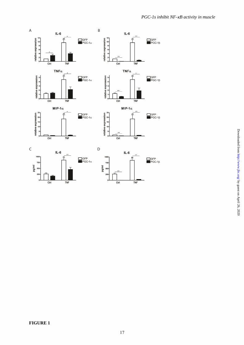
Ectopic, recombinant TNF strongly induced the
gene expression of the pro-inflammatory markers
IL-6, TNF and macrophage inflammatory
protein-1 (MIP-1/CCL3). While PGC-1 did
not negatively affect basal levels of these pro-
inflammatory cytokines, it diminished their
induction by TNF yielding lower levels than in
control cells (Fig. 1A). PGC-1 suppressed both
basal and TNF-induced expression of IL-6,
TNF and MIP-1 (Fig. 1B). As a consequence
of the PGC-1-mediated repression in gene
expression, lower levels of secreted IL-6 protein
were observed in culture media after TNF
treatment (Fig. 1C and 1D).
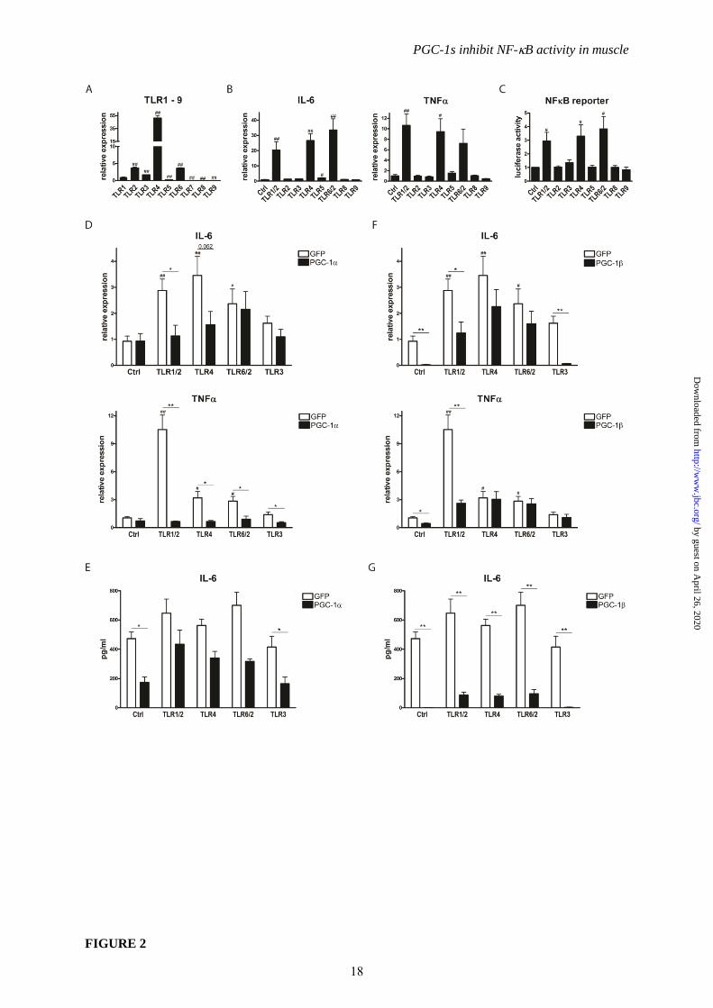
Next, we examined the effect of PGC-1s
on TLR stimulation in muscle cells. First, we
assessed the expression pattern of the TLRs in
our experimental system and found that TLR1,
TLR2, TLR3, TLR 4, TLR6, and at lower levels
TLR5 were detectable in the muscle cells (Fig.
2A). To study the activity of the TLRs, selective
agonists for TLR2, TLR3, TLR4, TLR5, TLR8,
TLR9, or the TLR1/2 and TLR6/2 heterodimers
were applied. Of those, only TLR1/2, TLR4 and
TLR6/2 activators consistently elevated
expression of IL-6 and TNF (Fig. 2B).
Moreover, the same compounds increased
luciferase reporter gene activity controlled by 3
repeats of a minimal NF-B DNA-response
element (Fig. 2C). For the subsequent
experiments, agonists for TLR1/2, TLR4 and
TLR6/2, the active TLRs in muscle cells, were
utilized and compared to the TLR3 activator that
was used as a negative, internal control. TLR1/2,
TLR4 and TLR6/2 agonists all induced
expression of IL-6 and TNF in control cells as
expected (Fig. 2D). Interestingly, PGC-1
effectively repressed TLR-mediated TNF
expression by all of the active agonists while IL-
6 expression was only reduced by PGC-1 in the
TLR1/2 and TLR4 agonist treatment (Fig. 2D).
PGC-1 furthermore lowered basal IL-6 protein
secretion, but not the elevated IL-6 secretion
caused by TLR agonist stimulation (Fig. 2E). In
contrast, PGC-1 significantly decreased IL-6
and TNF gene expression both in the basal state
and after activation of TLR1/2 while no effect
was observed in the TLR4 or TLR6/2 agonist
treated cells (Fig. 2F). Nevertheless however, IL-
6 secretion into culture medium was strongly
4
by guest on April 26, 2020
http://ww
w.jbc.org/
Dow
nloaded from
PGC-1s inhibit NF-B activity in muscle
repressed by PGC-1 in all of the different
experimental conditions (Fig. 2G).
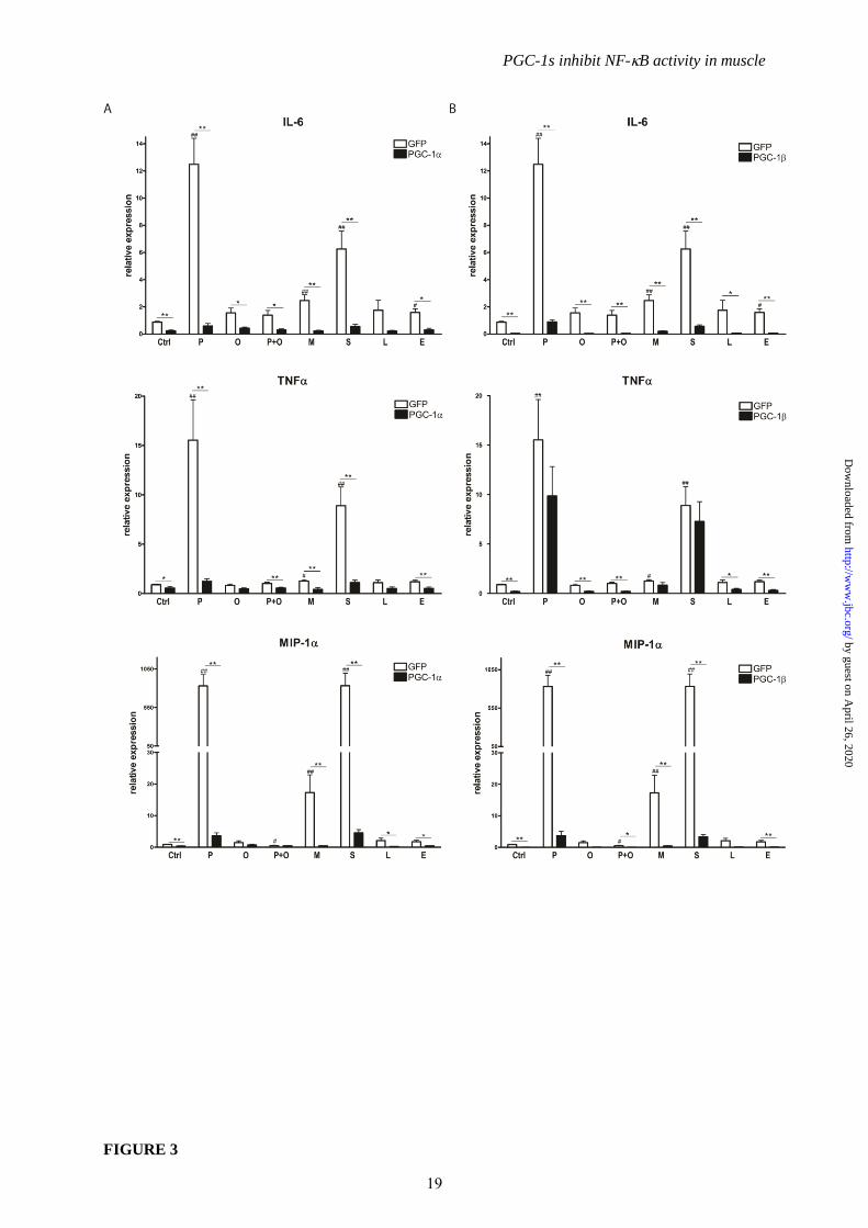
As a third inflammatory stimulus besides
TNF and TLR agonists, the free fatty acids
(FFA) palmitic acid (C16H32O2), oleic acid
(C18H34O2), myristic acid (C14H28O2), stearic acid
(C18H36O2), linoleic acid (C18H32O2) and elaidic
acid (C18H34O2) were administered to muscle
cells to mimick the lipid overload that is
associated with disease progression in the
metabolic syndrome. As expected, saturated fatty
acids, in particular stearic and palmitic acid and
to a lesser extent the shorter-chain species,
produced a strong pro-inflammatory response as
indicated by the transcriptional induction of the
IL-6, TNF and MIP-1 genes (Fig. 3A). In
contrast, mono- or polyunsaturated fatty acids
did not alter pro-inflammatory cytokine
expression (Fig. 3A); in fact, oleic acid was even
able to reverse the negative effects of palmitic
acid (Fig. 3A) as previously described (33).
Strikingly, PGC-1 potently inhibited the
increase in IL-6, TNF and MIP-1 gene
expression mediated by palmitic, myristic and
stearic acid (Fig 3A). Likewise, PGC-1 also
efficiently blocked the FFA-induced elevation of
IL-6 and MIP-1 transcript levels (Fig. 3B).
Interestingly however, transcriptional elevation
of TNF by palmitic, myristic and stearic acid
was unaffected by overexpression of PGC-1
(Fig 3B).
PGC-1 and PGC-1 target the NF-B
pathway to suppress inflammation. To examine
the mechanisms behind the repressive effect of
the PGC-1 coactivators on TNF-induced pro-
inflammatory cytokines, a customized array
designed to represent the most important
inflammatory genes and other NF-B targets
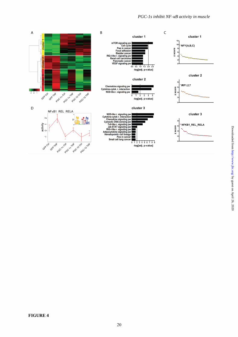
(29) was employed. Out of the 524 genes that are
present on the array, 55 genes were found to be
differentially regulated by TNF treatment
and/or PGC-1 overexpression (P <0.01) and thus
further analyzed. These 55 genes were grouped
into 3 clusters (Fig. 4A): cluster 1 contained 21
genes that were upregulated by PGC-1 and
PGC-1 including vascular endothelial growth
factor (VEGF), a known PGC-1 target (34).
The 13 and 21 genes in clusters 2 and 3,
respectively, were repressed by PGC-1 and
PGC-1. Importantly, genes in cluster 3 were
TNF-inducible while the expression of the
genes in cluster 2 was not modulated by TNF
treatment (Fig. 4A). To validate the results of the
microarray, three representative genes from each
cluster were chosen and their expression
analyzed by real-time PCR. Mitochondrial
translational initiation factor 2 (Mtif2), VEGF
and protein arginine methyltransferase 1 (Prmt1)
from cluster 1 were indeed induced (Suppl. Fig.
2A) while chemokine (C-X-C motif) ligand 12
(Cxcl12), complement component 2 (C2) and
E2F transcription factor 2 (E2f2) from cluster 2
(Suppl. Fig 2B) and chemokine (C-C motif)
ligand 2 (Ccl2), Ccl7 and Cxcl1 from cluster 3
(Suppl. Fig. 2C) were repressed by PGC-1
coactivators confirming the results of the
microarray. Furthermore, TNF-inducibility of
genes in cluster 3 was verified.
Functionally, the genes in cluster 1 were
enriched in only one Gene Ontology (GO) term
compared to 18 significant terms in cluster 2 and
96 terms in cluster 3 (of which only 46 terms
with a p-value≤0.01 are shown) (Suppl. Fig.
3). 12 of the GO categories in cluster 2 and 26
categories in cluster 3 are related to
inflammation and immunity. Importantly, the
inflammation-related GO terms comprise the top
10 and top 15 ranking categories in clusters 2
and 3, respectively (Suppl. Fig. 3). Likewise, all
3 KEGG pathways assigned to cluster 2 and the
top 9 KEGG pathways out of 11 of cluster 3 are
related to inflammatory signaling (Fig. 4B).
These results suggest that the PGC-1s are able to
repress inflammatory processes in muscle cells.
To predict which transcription factors
are involved in the regulation of each cluster,
promoter regions were analyzed in regard to their
motif composition. Motifs overrepresented in
each of the clusters are listed in Suppl. Table 2A.
As background for this analysis, all promoters of
the microarray were used, thereby eliminating
any putative bias that might have been
introduced with the specific choice of genes in
the customized microarray. The majority of
predicted binding sites were unique to one
cluster, i.e. 26 out of 33 motifs in cluster 1, 20
out of 26 motifs in cluster 2 and 21 out of 28
motifs in cluster 3 (Suppl. Table 2B).
Intriguingly, one distinct transcription factor
binding motif stood out as the clear top ranking
candidate in each of the three clusters based on z
score, namely NFY{A,B,C} in cluster 1 (z score
of 9.74 compared to the second ranking z score
of 4.9 for GFI1), IRF1,2,7 in cluster 2 (z score of
6.98 compared to 4.46 for FOX{F1,F2,J1}) and
NFB1_REL_RELA in cluster 3 (z score of 6.67
compared to 4.93 for AR) (Fig. 4C and Suppl.
Table 2A). These predictions indicate that the
NF-B pathway is a likely target for the PGC-1s
5
by guest on April 26, 2020
http://ww
w.jbc.org/
Dow
nloaded from
PGC-1s inhibit NF-B activity in muscle
to suppress TNF-inducible inflammatory gene
expression represented in cluster 3. Accordingly,
NF-B was also the highest scoring motif in the
Motif Activity Response Analysis (MARA) of
the whole array and thus the most likely
transcription factor to modulate TNF- and
PGC-1-dependent gene expression (Fig. 4D).
PGC-1 reduces p65 and p50 expression
levels. To experimentally validate the
biocomputational prediction of PGC-1-mediated
repression of NF-B signaling as the central
mechanism for the anti-inflammatory effect of
the PGC-1 coactivators on TNF-inducible
genes, reporter gene assays were performed with
a construct containing the luciferase gene under
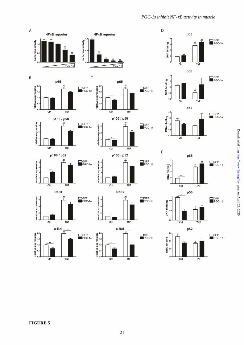
the control of 3 NF-B DNA response elements.
The activity of the reporter gene construct in
response to TNF treatment resembles
endogenous IL-6 and TNF gene expression
(Suppl. Fig. 1C and 1D). Increasing amounts of
co-transfected PGC-1 or PGC-1 progressively
inhibited transcription from a NFB-responsive
promoter that is activated by exogenous p65
(Fig. 5A).
To elucidate the mechanism by which
the PGC-1 coactivators repress NF-B activity
even on minimal NF-B response element-
driven gene expression, we first examined the
expression levels of the different NF-B family
members before and after TNF treatment and
PGC-1 overexpression, respectively (Fig. 5B and
5C). TNF led to a significant increase in
transcript levels of all 5 NF-B isoforms (p65,
p105/p50, p100/p52, RelB and c-Rel) in GFP-
infected control cells. PGC-1 did not change
the levels of the canonical isoforms p65 and
p105/p50 as well as RelB in the basal, vehicle-
treated cells while an induction of p100/p52 was
observed suggesting a switch towards the
noncanonical NF-B pathway. In contrast to the
first 4 NF-B isoforms, c-Rel expression was
clearly reduced by PGC-1 overexpression (Fig.
5B). After TNF treatment, the levels of all NF-
B isoforms tended to be lower in PGC-1
overexpressing cells, however only reaching
statistical significance in the case of c-Rel gene
expression (Fig 5B). In striking contrast to the
PGC-1-mediated effect, PGC-1 suppressed
p65 and p105/p50 as well as RelB and c-Rel
gene expression in the basal state (Fig. 5C). The
transcript levels of all 5 NF-B isoforms tended
to be lower in PGC-1 overexpressing cells after
TNF treatment; similar to PGC-1
overexpressing cells, this repression was
however only significant for c-Rel gene
expression (Fig. 5C). Therefore, while the basal
repression of p65 and p105/p50 could contribute
to the strong repression of inflammatory gene
expression by PGC-1 in non-stimulated cells, it
is unlikely that the small differences in
expression levels of the different NF-B
isoforms after TNF treatment underlie the
profound effects of PGC-1s on pro-inflammatory
cytokine expression in stimulated muscle cells.
Thus, to further elucidate the molecular
mechanism underlying this observation, we next
assessed the DNA binding capability of all NF-
B family members in the context of TNF
treatment and PGC-1 overexpression (Fig. 5D
and 5E). Of the 5 NF-B isoforms, RelB and c-
Rel were undetectable in nuclear extracts from
C2C12 myotubes in the TransAM assay and are
thus unlikely to play a major role in the
regulation of pro-inflammatory cytokine
expression in our experimental context of muscle
cells as hypothesized in other publications (35).
The DNA binding of p50 and p52 was detectable
though low (Fig. 5D and 5E). In contrast,
recruitment of p65 to DNA response elements
was substantial in non-stimulated cells and
further elevated after TNF treatment.
Interestingly however, DNA binding of p65 with
and without TNF treatment, respectively, was
not changed by PGC-1 (Fig. 5D) while PGC-1
strongly inhibited binding of p65 and p50 to
DNA in the basal, vehicle-treated muscle cells
(Fig. 5E). Comparable to PGC-1
overexpression, DNA binding of any of the NF-
B isoforms was not affected by ectopic PGC-1
in TNF-stimulated cells (Fig. 5E). It is thus
conceivable that both low p65/p50 expression as
well as reduced DNA binding account for the
PGC-1-mediated reduction in pro-inflammatory
cytokine expression compared to vehicle-treated
control cells. In contrast however, the diminished
levels of these cytokines upon TNF treatment
in both PGC-1- and PGC-1-overexpressing
cells can neither be attributed to changes in NF-
B expression nor to modulation of the NF-B
protein binding capability to DNA response
elements.
PGC-1 and PGC-1 diminish the
transcriptional activity of p65. Based on our data
implying alternative molecular mechanisms
distinct from transcriptional regulation or DNA
binding of NF-B to underlie the repressive
action of the PGC-1 coactivators on the activity
of this transcription factor, we next studied
upstream signaling and the post-translational
6
by guest on April 26, 2020
http://ww
w.jbc.org/
Dow
nloaded from
PGC-1s inhibit NF-B activity in muscle
modification of p65 that influence the
transcriptional activity of NF-B. First, we
examined the possibility that high levels of IBs
after TNF treatment could account for lowered
cytokine expression. However, the relative
amount of IB protein was not different
between conditions (Fig. 6A). IB, an NF-B
target gene, was accordingly increased on the
protein level by TNF treatment and reduced in
muscle cells overexpressing PGC-1 or PGC-1
(Fig. 6A and Suppl. Fig. 4A and 4B), similar to
other NF-B targets such as IL-6 and TNF
(Fig. 1) excluding the possibility of IB-
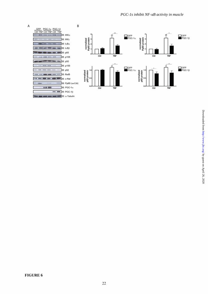
mediated repression. Subsequently, IKK protein
levels were assessed: IKK was not and IKK
was barely detectable (Fig. 6A). In contrast,
IKK was slightly increased by TNF
treatment; this effect was abrogated in cells
overexpressing PGC-1 or PGC-1 (Fig. 6A).
IKK is one of the protein kinases that is able to
phosphorylate p65 at serine 536. Importantly, the
phosphorylation status of p65 at serine 536
affects the transcriptional activity of NF-B even
when bound to DNA response elements (10). In
Western blot analyses of total and
phosphorylated p65 protein, a small but
significant increase in total p65 protein levels
was observed after TNF treatment in control
cells (Fig 6A and 6B). PGC-1 overexpression
did not affect basal levels of p65, while PGC-1
diminished total p65 protein expression in this
context, as expected based on the reduced
mRNA expression of p65 in PGC-1
overexpressing muscle cells (Fig. 5C). Strikingly
however, both PGC-1 and PGC-1 reduced
TNF-mediated phosphorylation of p65 by
about 50% (Fig. 6A and 6B). None of the other
NF-B family members underwent regulation by
TNF on the protein level. Interestingly, PGC-
1 overexpression resulted in elevated protein
levels of p105, p100, p52 and RelB in non-
stimulated muscle cells and, to a smaller extent,
of p105 and p100 in TNF-treated cells again
suggesting a PGC-1-dependent increase in the
non-canonical NF-B pathway (Fig. 6A and
Suppl. Fig. 4A). In contrast, PGC-1 does not
seem to elevate the non-canonical NF-B family
members like PGC-1. Thus, besides the strong
increase in c-Rel protein and the more moderate
elevation of p105 protein in non-stimulated cells,
PGC-1 significantly reduced RelB protein
levels (Fig. 6A and Suppl. Fig. 4B). The
discrepancy between the PGC-1-mediated
repression of c-Rel gene expression compared to
the elevation of c-Rel protein levels implies post-
transcriptional effects in the regulation of this
particular NF-B family member.
Dephosphorylation and transrepression
of p65 are potential molecular mechanisms for
diminished cytokine expression. To further
substantiate the findings implying that the PGC-
1 coactivators modulate NF-B activity by
preventing p65 phosphorylation, we examined
other upstream effectors for this phosphorylation
as well as downstream events affecting the
DNA-bound NF-B transcriptional complex.
The protein kinase Akt has been implicated in
the regulation of the NF-B signaling pathway
upstream of IKK (36) one of the protein
kinases to mediate p65 phosphorylation and of
which protein levels are reduced by PGC-1 and
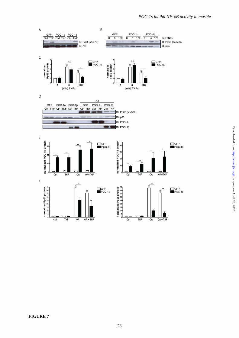
PGC-1 overexpression (Fig. 6A). As expected,
based on these findings, PGC-1 and PGC-1
diminished Akt activation as evident from
diminished phospho-Akt (ser473) levels
normalized to total Akt protein (Fig. 7A).
To obtain a more accurate picture of p65
phosphorylation, a time course experiment was
performed. It revealed that p65 is phosphorylated
at serine 536 after 5 minutes of TNF treatment
even in the presence of PGC-1 and PGC-1
(Fig. 7B and 7C) implying the possibility that not
only an altered kinase profile but also activity of
a protein phosphatase might be involved in the
PGC-1-mediated modulation of NF-B
phosphorylation. We therefore tested whether
pharmacological inhibition of protein
phosphatase 2A (PP2A) and PP1, two enzymes
that dephosphorylate p65 (37), by okadaic acid
abolishes the repression of p65 phosphorylation
mediated by PGC-1 and PGC-1 in muscle
cells. As expected, okadaic acid powerfully
stabilized phosphorylation of p65 at serine 536
(Fig. 7D and 7F). Strikingly however, PGC-1
and PGC-1 overexpression (Fig. 7D and 7E)
still reduced p65 phosphorylation, even in
okadaic acid-treated cells (Fig. 7D and 7F).
Thus, while PP1 and PP2A clearly affect the
serine 536 phosphorylation of p65 in our
experimental context, these two phosphatases are
most likely not involved in the modulation of
p65 phosphorylation by the PGC-1s.
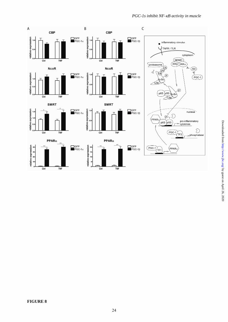
Since the phosphorylation status of p65
affects its affinity to cofactors (38), we also
determined the expression levels of the
coactivator CBP and corepressors NCoR and
SMRT. Of those, only SMRT transcript levels
were significantly increased by PGC-1 (Fig 8A
and 8B). A well-described mechanism of
7
by guest on April 26, 2020
http://ww
w.jbc.org/
Dow
nloaded from
PGC-1s inhibit NF-B activity in muscle
inhibition of inflammatory gene expression is
mediated by nuclear receptors and termed
transrepression (39). We evaluated the gene
expression of potential candidates for
transrepression and found PPAR to be strongly
induced by both PGC-1 and PGC-1 in non-
stimulated and TNF-treated muscle cells
similar to previous data (40) (Fig 8A and 8B).
PPAR therefore likely contributes to the PGC-
1-dependent reduction in NF-B transcriptional
activity by impairing the exchange of
corepressors for coactivators that is necessary to
effectively initiate transcription. In fact,
inhibition of PPAR recovered expression of IL-
6 and TNF in the presence PGC-1 (Suppl. Fig
5).
DISCUSSION
With an aging population and an
increasingly sedentary lifestyle, chronic diseases
are on the rise. Obesity and its comorbidities but
also some cancers and neurodegeneration have
been associated with local and systemic
inflammation that worsens disease progression,
while exercise has beneficial effects in many of
these disorders and even acts preventive (41).
PGC-1 is a major molecular mediator of
exercise in skeletal muscle and its loss not only
disturbs metabolic processes but also evokes a
local and systemic inflammation (24). In the
present report we thus tested the idea that PGC-1
coactivators have anti-inflammatory properties.
Indeed, we confirmed such properties as
PGC-1 and PGC-1 were able to diminish the
increase in pro-inflammatory cytokines elicited
by different inflammatory stimuli such as TNF,
TLR agonists and saturated FFAs. We identified
the NF-B pathway as main target of PGC-1-
dependent repression. These results are
complemented by in vivo findings of Brault and
coworkers who showed that NF-B reporter
activity decreases after electroporation of tibialis
anterior muscle with PGC-1 / PGC-1 in the
context of anti-atrophic effects of PGC-1s (42).
Similar conclusions were derived from
experiments in human aortic smooth muscle and
endothelial cells where PGC-1 suppressed
TNF-induced VCAM-1 and MCP-1 expression
that contribute to inflammation in atherosclerosis
(43).
Mechanistically, PGC-1 and PGC-1
lower phosphorylation of the NF-B family
member p65 which limits its transcriptional
activation potential. This is further substantiated
by data from muscle-specific PGC-1 transgenic
animals that also exhibit reduced p65
phosphorylation (44). Diametrically opposed to
this report, another recent publication states that
p65 phosphorylation is higher in PGC-1
transgenic muscle before as well as after
injection of TNF (45). The exact role of PGC-
1 on muscle inflammation in vivo thus remains
unresolved. In our cellular model, decreased p65
phosphorylation unequivocally corresponds to
the loss of IKK induction by TNF in PGC-1
overexpressing cells and, interestingly, to
diminished Akt activation. Akt-dependent
phophorylation of p65 by IKK is an important
mechanism to regulate p65 transactivation
potential (36,46) and thus a good candidate to
mediate the reduction observed (Fig. 8C). In
addition to modulation of kinase activity, the
involvement of a phosphatase is suggested by
sustained phosphorylation of p65 after TNF
treatment for 5 minutes even in the presence of
PGC-1 and PGC-1. As okadaic acid treatment
did not abrogate differences between conditions,
PP2A and PP1 are however unlikely to account
for this effect. Therefore, further experiments are
needed to determine the contribution of different
phosphatases in this setting.
The transactivation potential of p65 is
controlled by its phosphorylation status as it
defines the affinity for cofactors important in
suppressing or stimulating transcription of target
genes (38). Decreased phosphorylation thus
favors interaction with corepressors such as
SMRT that decreases p65/p50 transactivation
(47). Interestingly, SMRT levels are slightly
induced by PGC-1 and accordingly could
contribute to lower cytokine expression.
Stabilization of corepressor complexes on DNA-
bound p65 is also fostered by nuclear receptor-
mediated transrepression (39). For example,
PPAR is able to exert anti-inflammatory action
by ligand-dependent and –independent
transrepressive mechanisms (48,49). We found a
marked induction of PPAR by PGC-1 and
PGC-1, which presumably also contributes to
negative regulation of pro-inflammatory
cytokines by transrepression (Fig. 8C). This
claim is substantiated by the reversal of the
repressive PGC-1 effects on pro-inflammatory
cytokine expression when PPAR was inhibited.
PPAR regulates lipid metabolism and FFAs are
able to serve as ligands for PPAR (50). Thus,
the very pronounced suppressive effect of the
PGC-1s on inflammatory gene expression
observed after FFA treatment might reflect an
additional ligand-dependent activation of
8
by guest on April 26, 2020
http://ww
w.jbc.org/
Dow
nloaded from

PGC-1s inhibit NF-B activity in muscle
PPAR leading to an even stronger
transrepression in that experimental context.
Besides the reduction in p65
phosphorylation that is exerted by both PGC-1
and PGC-1, only the latter was further found to
repress p65 and p50 transcription in the basal
state and accordingly the ability of these proteins
to bind to DNA response elements. This offers
an attractive explanation for the very low
cytokine levels observed in the presence of PGC-
1. The NF-B family members RelB and c-Rel
were also suppressed transcriptionally in the
basal state which puts PGC-1 in the position of
a broader anti-inflammatory factor in skeletal
muscle. Such anti-inflammatory potential was
previously described only in macrophages,
where PGC-1 is essential in alternative
activation and ROS production (27,28). In
contrast, PGC-1 does not alter expression of the
classically activated / canonical NF-B isoforms
p65 and p50. However, PGC-1 overexpression
induces transcript and protein levels of the
alternative isoforms p100/p52 and RelB. This
indicates a switch towards noncanonical /
alternative NF-B signaling. A recent
publication outlined that alternative signaling via
IKK and RelB induces an oxidative phenotype
in muscle driven by PGC-1(51). Furthermore,
activation of c-Rel and p50 was suggested to
play a role in disuse atrophy (52). Finally,
canonical p65 activation was linked to
mitochondrial biogenesis in mouse embryonic
fibroblasts and liver cells (53,54). All of these
findings together with our data suggest a
reciprocal, functional link between NF-B
signaling and oxidative metabolism. A potential
induction of the alternative NF-B pathway by
PGC-1 in muscle therefore warrants further
investigations.
Strikingly, inflammatory gene
transcription was often selectively regulated by
the two PGC-1 coactivators depending on the
stimulus and the specific gene. These findings
argue against a general repressive effect of PGC-
1 and PGC-1 on tissue inflammation in
muscle but rather indicate a specific, fine-tuned
effect of these coactivators on NF-B target
genes. For example, while TLR1/2-induced
cytokine production was suppressed by both
PGC-1 and PGC-1, only PGC-1 was able to
block this production after treatment of the cells
with a TLR4-specific ligand. Moreover, TLR6/2-
induced TNF expression was also diminished
by PGC-1, which was however not the case for
TLR6/2-induced expression of IL-6. This
probably reflects the activation of pathways
other than NF-B downstream of TLRs that are
not subject to regulation by PGC-1 or targeted by
only one of the PGC-1 isoforms, respectively.
Interestingly, while neither PGC-1 nor PGC-1
affected expression of TNF receptor 1 (TNFR1)
(Suppl. Fig. 6A and 6B) both coactivators
lowered mRNA levels of TLR1, TLR4 and
TLR6 (Suppl. Fig. 6C and 6D). This
downregulation might presumably contribute to
the repressive effects observed with some TLR
agonists. The expression pattern of TLR1, TLR4
and TLR6 (Suppl. Fig. 6C and 6D) further
resembles genes in cluster 2 of the microarray,
confirming a broader influence of PGC-1s on
inflammatory genes. Moreover, the selectivity of
the PGC-1s most likely reflects the distinct
expression pattern as well as the distinct
functions of PGC-1 and PGC-1 in the
regulation of skeletal muscle physiology that will
have to be further dissected in future
experiments. In any case however, the concept
that PGC-1s selectively and specifically repress
inflammatory processes and thereby avoid the
harmful consequences of broad and general
cytokine suppression is compelling. Once
understood in greater detail, the therapeutic
potential of targeting PGC-1s to modulate
specific inflammatory responses in muscle would
be immense. Obviously, patients suffering from
obesity and type 2 diabetes would profit from
lower systemic inflammation mediated by
rectified gene expression of PGC-1 and PGC-
1 in skeletal muscle. Intriguingly however,
muscle disorders like cachexia, muscular
dystrophies, disuse atrophy or inflammatory
myopathy also involve an inflammatory
component with activation of the NF-B
pathway (55-58). In fact, chronic stimulation of
the classical NF-B pathway in muscle is
sufficient to induce muscle wasting (5).
Accordingly, mice heterozygous for p65 or
harboring a genetic ablation of the IKK gene in
the mdx background, a model for Duchenne
muscular dystrophy, have improved pathology
(59). Strikingly, ectopic elevation of PGC-1 in
animal models for some of these diseases
resulted in an amelioration of fiber damage and
muscle functionality, e.g. in Duchenne muscular
dystrophy, sarcopenia, a mitochondrial
myopathy and denervation-induced fiber atrophy
(20,21,44,60). Our data now suggest that at least
part of the therapeutic effect of PGC-1 in these
disease paradigms might stem from the anti-
9
by guest on April 26, 2020
http://ww
w.jbc.org/
Dow
nloaded from
PGC-1s inhibit NF-B activity in muscle
inflammatory effect. It is, thus, tempting to
speculate that elevating PGC-1 and / or PGC-
1 in muscle would also be beneficial in other
conditions of muscle wasting by limiting the
detrimental inflammatory component of the
disease. In fact, muscle adaptation to endurance
training that correlate with increased PGC-1
expression includes an increased resistance
against fiber damage, tissue inflammation and, as
a consequence, decreased exercise-induced
muscle soreness. In contrast to PGC-1, the
implications of the repressive effect of PGC-1
on inflammatory gene expression remain less
obvious until the physiological context of PGC-
1 regulation and the function of this coactivator
in muscle tissue have been more clearly
delineated. Nevertheless, by virtue of their
ability to reduce NF-B activation, the PGC-1
coactivators are promising targets to antagonize
inflammatory reactions in skeletal muscle
associated with a large number of diseases.
REFERENCES
1. Hotamisligil, G. S. (2006) Inflammation and metabolic disorders. Nature 444, 860-867
2. Haffner, S. M. (2006) The metabolic syndrome: inflammation, diabetes mellitus, and
cardiovascular disease. The American journal of cardiology 97, 3A-11A
3. Perry, V. H. (2004) The influence of systemic inflammation on inflammation in the brain:
implications for chronic neurodegenerative disease. Brain, behavior, and immunity 18, 407-
413
4. Arkan, M. C., Hevener, A. L., Greten, F. R., Maeda, S., Li, Z. W., Long, J. M., Wynshaw-
Boris, A., Poli, G., Olefsky, J., and Karin, M. (2005) IKK-beta links inflammation to obesity-
induced insulin resistance. Nature medicine 11, 191-198
5. Cai, D., Frantz, J. D., Tawa, N. E., Jr., Melendez, P. A., Oh, B. C., Lidov, H. G., Hasselgren,
P. O., Frontera, W. R., Lee, J., Glass, D. J., et al. (2004) IKKbeta/NF-kappaB activation
causes severe muscle wasting in mice. Cell 119, 285-298
6. Zhang, X., Zhang, G., Zhang, H., Karin, M., Bai, H., and Cai, D. (2008) Hypothalamic
IKKbeta/NF-kappaB and ER stress link overnutrition to energy imbalance and obesity. Cell
135, 61-73
7. Shi, H., Kokoeva, M. V., Inouye, K., Tzameli, I., Yin, H., and Flier, J. S. (2006) TLR4 links
innate immunity and fatty acid-induced insulin resistance. The Journal of clinical
investigation 116, 3015-3025
8. Ghosh, S., and Hayden, M. S. (2008) New regulators of NF-kappaB in inflammation. Nature
reviews 8, 837-848
9. Sakurai, H., Chiba, H., Miyoshi, H., Sugita, T., and Toriumi, W. (1999) IkappaB kinases
phosphorylate NF-kappaB p65 subunit on serine 536 in the transactivation domain. The
Journal of biological chemistry 274, 30353-30356
10. Buss, H., Dorrie, A., Schmitz, M. L., Hoffmann, E., Resch, K., and Kracht, M. (2004)
Constitutive and interleukin-1-inducible phosphorylation of p65 NF-{kappa}B at serine 536 is
mediated by multiple protein kinases including I{kappa}B kinase (IKK)-{alpha}, IKK{beta},
IKK{epsilon}, TRAF family member-associated (TANK)-binding kinase 1 (TBK1), and an
unknown kinase and couples p65 to TATA-binding protein-associated factor II31-mediated
interleukin-8 transcription. The Journal of biological chemistry 279, 55633-55643
11. Gleeson, M. (2007) Immune function in sport and exercise. J Appl Physiol 103, 693-699
12. Knowler, W. C., Barrett-Connor, E., Fowler, S. E., Hamman, R. F., Lachin, J. M., Walker, E.
A., and Nathan, D. M. (2002) Reduction in the incidence of type 2 diabetes with lifestyle
intervention or metformin. The New England journal of medicine 346, 393-403
13. Fiatarone, M. A., O'Neill, E. F., Ryan, N. D., Clements, K. M., Solares, G. R., Nelson, M. E.,
Roberts, S. B., Kehayias, J. J., Lipsitz, L. A., and Evans, W. J. (1994) Exercise training and
nutritional supplementation for physical frailty in very elderly people. The New England
journal of medicine 330, 1769-1775
10
by guest on April 26, 2020
http://ww
w.jbc.org/
Dow
nloaded from
PGC-1s inhibit NF-B activity in muscle
14. Tillerson, J. L., Caudle, W. M., Reveron, M. E., and Miller, G. W. (2003) Exercise induces
behavioral recovery and attenuates neurochemical deficits in rodent models of Parkinson's
disease. Neuroscience 119, 899-911
15. Puigserver, P., Wu, Z., Park, C. W., Graves, R., Wright, M., and Spiegelman, B. M. (1998) A
cold-inducible coactivator of nuclear receptors linked to adaptive thermogenesis. Cell 92, 829-
839
16. Pilegaard, H., Saltin, B., and Neufer, P. D. (2003) Exercise induces transient transcriptional
activation of the PGC-1alpha gene in human skeletal muscle. The Journal of physiology 546,
851-858
17. Puigserver, P., Adelmant, G., Wu, Z., Fan, M., Xu, J., O'Malley, B., and Spiegelman, B. M.
(1999) Activation of PPARgamma coactivator-1 through transcription factor docking. Science
(New York, N.Y 286, 1368-1371
18. Lin, J., Wu, H., Tarr, P. T., Zhang, C. Y., Wu, Z., Boss, O., Michael, L. F., Puigserver, P.,
Isotani, E., Olson, E. N., et al. (2002) Transcriptional co-activator PGC-1 alpha drives the
formation of slow-twitch muscle fibres. Nature 418, 797-801
19. Calvo, J. A., Daniels, T. G., Wang, X., Paul, A., Lin, J., Spiegelman, B. M., Stevenson, S. C.,
and Rangwala, S. M. (2008) Muscle-specific expression of PPARgamma coactivator-1alpha
improves exercise performance and increases peak oxygen uptake. J Appl Physiol 104, 1304-
1312
20. Handschin, C., Kobayashi, Y. M., Chin, S., Seale, P., Campbell, K. P., and Spiegelman, B. M.
(2007) PGC-1alpha regulates the neuromuscular junction program and ameliorates Duchenne
muscular dystrophy. Genes & development 21, 770-783
21. Sandri, M., Lin, J., Handschin, C., Yang, W., Arany, Z. P., Lecker, S. H., Goldberg, A. L., and
Spiegelman, B. M. (2006) PGC-1alpha protects skeletal muscle from atrophy by suppressing
FoxO3 action and atrophy-specific gene transcription. Proceedings of the National Academy
of Sciences of the United States of America 103, 16260-16265
22. Hanai, J., Cao, P., Tanksale, P., Imamura, S., Koshimizu, E., Zhao, J., Kishi, S., Yamashita,
M., Phillips, P. S., Sukhatme, V. P., et al. (2007) The muscle-specific ubiquitin ligase atrogin-
1/MAFbx mediates statin-induced muscle toxicity. The Journal of clinical investigation 117,
3940-3951
23. Handschin, C., Chin, S., Li, P., Liu, F., Maratos-Flier, E., Lebrasseur, N. K., Yan, Z., and
Spiegelman, B. M. (2007) Skeletal muscle fiber-type switching, exercise intolerance, and
myopathy in PGC-1alpha muscle-specific knock-out animals. The Journal of biological
chemistry 282, 30014-30021
24. Handschin, C., Choi, C. S., Chin, S., Kim, S., Kawamori, D., Kurpad, A. J., Neubauer, N., Hu,
J., Mootha, V. K., Kim, Y. B., et al. (2007) Abnormal glucose homeostasis in skeletal muscle-
specific PGC-1alpha knockout mice reveals skeletal muscle-pancreatic beta cell crosstalk. The
Journal of clinical investigation 117, 3463-3474
25. Patti, M. E., Butte, A. J., Crunkhorn, S., Cusi, K., Berria, R., Kashyap, S., Miyazaki, Y.,
Kohane, I., Costello, M., Saccone, R., et al. (2003) Coordinated reduction of genes of
oxidative metabolism in humans with insulin resistance and diabetes: Potential role of PGC1
and NRF1. Proceedings of the National Academy of Sciences of the United States of America
100, 8466-8471
26. Arany, Z., Lebrasseur, N., Morris, C., Smith, E., Yang, W., Ma, Y., Chin, S., and Spiegelman,
B. M. (2007) The transcriptional coactivator PGC-1beta drives the formation of oxidative type
IIX fibers in skeletal muscle. Cell metabolism 5, 35-46
27. Vats, D., Mukundan, L., Odegaard, J. I., Zhang, L., Smith, K. L., Morel, C. R., Wagner, R. A.,
Greaves, D. R., Murray, P. J., and Chawla, A. (2006) Oxidative metabolism and PGC-1beta
attenuate macrophage-mediated inflammation. Cell metabolism 4, 13-24
28. Sonoda, J., Laganiere, J., Mehl, I. R., Barish, G. D., Chong, L. W., Li, X., Scheffler, I. E.,
Mock, D. C., Bataille, A. R., Robert, F., et al. (2007) Nuclear receptor ERR alpha and
coactivator PGC-1 beta are effectors of IFN-gamma-induced host defense. Genes &
development 21, 1909-1920
11
by guest on April 26, 2020
http://ww
w.jbc.org/
Dow
nloaded from

PGC-1s inhibit NF-B activity in muscle
29. Jayne, S., Rothgiesser, K. M., and Hottiger, M. O. (2009) CARM1 but not its enzymatic
activity is required for transcriptional coactivation of NF-kappaB-dependent gene expression.
Journal of molecular biology 394, 485-495
30. Al-Shahrour, F., Diaz-Uriarte, R., and Dopazo, J. (2004) FatiGO: a web tool for finding
significant associations of Gene Ontology terms with groups of genes. Bioinformatics 20, 578-
580
31. Arnold, P., Erb, I., Pachkov, M., Molina, N., and van Nimwegen, E. (2012) MotEvo:
integrated Bayesian probabilistic methods for inferring regulatory sites and motifs on multiple
alignments of DNA sequences. Bioinformatics 28, 487-494
32. Suzuki, H., Forrest, A. R., van Nimwegen, E., Daub, C. O., Balwierz, P. J., Irvine, K. M.,
Lassmann, T., Ravasi, T., Hasegawa, Y., de Hoon, M. J., et al. (2009) The transcriptional
network that controls growth arrest and differentiation in a human myeloid leukemia cell line.
Nature genetics 41, 553-562
33. Coll, T., Eyre, E., Rodriguez-Calvo, R., Palomer, X., Sanchez, R. M., Merlos, M., Laguna, J.
C., and Vazquez-Carrera, M. (2008) Oleate reverses palmitate-induced insulin resistance and
inflammation in skeletal muscle cells. The Journal of biological chemistry 283, 11107-11116
34. Arany, Z., Foo, S. Y., Ma, Y., Ruas, J. L., Bommi-Reddy, A., Girnun, G., Cooper, M., Laznik,
D., Chinsomboon, J., Rangwala, S. M., et al. (2008) HIF-independent regulation of VEGF and
angiogenesis by the transcriptional coactivator PGC-1alpha. Nature 451, 1008-1012
35. Bhatnagar, S., Panguluri, S. K., Gupta, S. K., Dahiya, S., Lundy, R. F., and Kumar, A. (2010)
Tumor necrosis factor-alpha regulates distinct molecular pathways and gene networks in
cultured skeletal muscle cells. PloS one 5, e13262
36. Sizemore, N., Lerner, N., Dombrowski, N., Sakurai, H., and Stark, G. R. (2002) Distinct roles
of the Ikappa B kinase alpha and beta subunits in liberating nuclear factor kappa B (NF-kappa
B) from Ikappa B and in phosphorylating the p65 subunit of NF-kappa B. The Journal of
biological chemistry 277, 3863-3869
37. Yang, J., Fan, G. H., Wadzinski, B. E., Sakurai, H., and Richmond, A. (2001) Protein
phosphatase 2A interacts with and directly dephosphorylates RelA. The Journal of biological
chemistry 276, 47828-47833
38. Zhong, H., Voll, R. E., and Ghosh, S. (1998) Phosphorylation of NF-kappa B p65 by PKA
stimulates transcriptional activity by promoting a novel bivalent interaction with the
coactivator CBP/p300. Molecular cell 1, 661-671
39. Ghisletti, S., Huang, W., Ogawa, S., Pascual, G., Lin, M. E., Willson, T. M., Rosenfeld, M.
G., and Glass, C. K. (2007) Parallel SUMOylation-dependent pathways mediate gene- and
signal-specific transrepression by LXRs and PPARgamma. Molecular cell 25, 57-70
40. Huss, J. M., Torra, I. P., Staels, B., Giguere, V., and Kelly, D. P. (2004) Estrogen-related
receptor alpha directs peroxisome proliferator-activated receptor alpha signaling in the
transcriptional control of energy metabolism in cardiac and skeletal muscle. Molecular and
cellular biology 24, 9079-9091
41. Handschin, C., and Spiegelman, B. M. (2008) The role of exercise and PGC1alpha in
inflammation and chronic disease. Nature 454, 463-469
42. Brault, J. J., Jespersen, J. G., and Goldberg, A. L. (2010) Peroxisome proliferator-activated
receptor gamma coactivator 1alpha or 1beta overexpression inhibits muscle protein
degradation, induction of ubiquitin ligases, and disuse atrophy. The Journal of biological
chemistry 285, 19460-19471
43. Kim, H. J., Park, K. G., Yoo, E. K., Kim, Y. H., Kim, Y. N., Kim, H. S., Kim, H. T., Park, J.
Y., Lee, K. U., Jang, W. G., et al. (2007) Effects of PGC-1alpha on TNF-alpha-induced MCP-
1 and VCAM-1 expression and NF-kappaB activation in human aortic smooth muscle and
endothelial cells. Antioxidants & redox signaling 9, 301-307
44. Wenz, T., Rossi, S. G., Rotundo, R. L., Spiegelman, B. M., and Moraes, C. T. (2009)
Increased muscle PGC-1alpha expression protects from sarcopenia and metabolic disease
during aging. Proceedings of the National Academy of Sciences of the United States of
America 106, 20405-20410
12
by guest on April 26, 2020
http://ww
w.jbc.org/
Dow
nloaded from
PGC-1s inhibit NF-B activity in muscle
45. Olesen, J., Larsson, S., Iversen, N., Yousafzai, S., Hellsten, Y., and Pilegaard, H. Skeletal
Muscle PGC-1alpha Is Required for Maintaining an Acute LPS-Induced TNFalpha Response.
PloS one 7, e32222
46. Madrid, L. V., Mayo, M. W., Reuther, J. Y., and Baldwin, A. S., Jr. (2001) Akt stimulates the
transactivation potential of the RelA/p65 Subunit of NF-kappa B through utilization of the
Ikappa B kinase and activation of the mitogen-activated protein kinase p38. The Journal of
biological chemistry 276, 18934-18940
47. Lee, S. K., Kim, J. H., Lee, Y. C., Cheong, J., and Lee, J. W. (2000) Silencing mediator of
retinoic acid and thyroid hormone receptors, as a novel transcriptional corepressor molecule of
activating protein-1, nuclear factor-kappaB, and serum response factor. The Journal of
biological chemistry 275, 12470-12474
48. Staels, B., Koenig, W., Habib, A., Merval, R., Lebret, M., Torra, I. P., Delerive, P., Fadel, A.,
Chinetti, G., Fruchart, J. C., et al. (1998) Activation of human aortic smooth-muscle cells is
inhibited by PPARalpha but not by PPARgamma activators. Nature 393, 790-793
49. Blanquart, C., Mansouri, R., Paumelle, R., Fruchart, J. C., Staels, B., and Glineur, C. (2004)
The protein kinase C signaling pathway regulates a molecular switch between transactivation
and transrepression activity of the peroxisome proliferator-activated receptor alpha. Molecular
endocrinology (Baltimore, Md 18, 1906-1918
50. Kliewer, S. A., Sundseth, S. S., Jones, S. A., Brown, P. J., Wisely, G. B., Koble, C. S.,
Devchand, P., Wahli, W., Willson, T. M., Lenhard, J. M., et al. (1997) Fatty acids and
eicosanoids regulate gene expression through direct interactions with peroxisome proliferator-
activated receptors alpha and gamma. Proceedings of the National Academy of Sciences of the
United States of America 94, 4318-4323
51. Bakkar, N., Ladner, K., Canan, B. D., Liyanarachchi, S., Bal, N. C., Pant, M., Periasamy, M.,
Li, Q., Janssen, P. M., and Guttridge, D. C. (2012) IKKalpha and alternative NF-kappaB
regulate PGC-1beta to promote oxidative muscle metabolism. The Journal of cell biology 196,
497-511
52. Hunter, R. B., Stevenson, E., Koncarevic, A., Mitchell-Felton, H., Essig, D. A., and
Kandarian, S. C. (2002) Activation of an alternative NF-kappaB pathway in skeletal muscle
during disuse atrophy. Faseb J 16, 529-538
53. Mauro, C., Leow, S. C., Anso, E., Rocha, S., Thotakura, A. K., Tornatore, L., Moretti, M., De
Smaele, E., Beg, A. A., Tergaonkar, V., et al. (2011) NF-kappaB controls energy homeostasis
and metabolic adaptation by upregulating mitochondrial respiration. Nature cell biology 13,
1272-1279
54. Suliman, H. B., Sweeney, T. E., Withers, C. M., and Piantadosi, C. A. (2010) Co-regulation of
nuclear respiratory factor-1 by NFkappaB and CREB links LPS-induced inflammation to
mitochondrial biogenesis. Journal of cell science 123, 2565-2575
55. Tracey, K. J., Wei, H., Manogue, K. R., Fong, Y., Hesse, D. G., Nguyen, H. T., Kuo, G. C.,
Beutler, B., Cotran, R. S., Cerami, A., et al. (1988) Cachectin/tumor necrosis factor induces
cachexia, anemia, and inflammation. J Exp Med 167, 1211-1227
56. Spencer, M. J., Walsh, C. M., Dorshkind, K. A., Rodriguez, E. M., and Tidball, J. G. (1997)
Myonuclear apoptosis in dystrophic mdx muscle occurs by perforin-mediated cytotoxicity.
The Journal of clinical investigation 99, 2745-2751
57. Hunter, R. B., Stevenson, E., Koncarevic, A., Mitchell-Felton, H., Essig, D. A., and
Kandarian, S. C. (2002) Activation of an alternative NF-kappaB pathway in skeletal muscle
during disuse atrophy. FASEB journal : official publication of the Federation of American
Societies for Experimental Biology 16, 529-538
58. Creus, K. K., De Paepe, B., Werbrouck, B. F., Vervaet, V., Weis, J., and De Bleecker, J. L.
(2009) Distribution of the NF-kappaB complex in the inflammatory exudates characterizing
the idiopathic inflammatory myopathies. Ann N Y Acad Sci 1173, 370-377
59. Acharyya, S., Villalta, S. A., Bakkar, N., Bupha-Intr, T., Janssen, P. M., Carathers, M., Li, Z.
W., Beg, A. A., Ghosh, S., Sahenk, Z., et al. (2007) Interplay of IKK/NF-kappaB signaling in
macrophages and myofibers promotes muscle degeneration in Duchenne muscular dystrophy.
The Journal of clinical investigation 117, 889-901
13
by guest on April 26, 2020
http://ww
w.jbc.org/
Dow
nloaded from
PGC-1s inhibit NF-B activity in muscle
60. Wenz, T., Diaz, F., Spiegelman, B. M., and Moraes, C. T. (2008) Activation of the
PPAR/PGC-1alpha pathway prevents a bioenergetic deficit and effectively improves a
mitochondrial myopathy phenotype. Cell metabolism 8, 249-256
Acknowledgement We thank Dr. Matthias Altmeyer and Dr. Karin Rothgiesser for help with the
microarray experiment, Dr. Hubert Rehrauer for advice on statistical analysis of the microarray and
Markus Beer for excellent technical assistance.
FOOTNOTES
*This project was funded by the Swiss National Science Foundation, the Muscular Dystrophy
Association USA (MDA), the SwissLife ‘Jubiläumsstiftung für Volksgesundheit und medizinische
Forschung’, the Swiss Society for Research on Muscle Diseases (SSEM), the Swiss Diabetes
Association, the Roche Research Foundation, the United Mitochondrial Disease Foundation (UMDF),
the Association Française contre les Myopathies (AFM), the Gebert-Rüf Foundation “Rare Diseases”
Program and the University of Basel. 1 To whom correspondence may be addressed: Biozentrum, Division of Pharmacology/Neurobiology,
University of Basel, Klingelbergstrasse 50/70, CH-4056 Basel, Switzerland, Phone: +41 61 267 2378,
Fax +41 61 267 2208, Email: [email protected]
3Functional Genomics Center Zurich, University and ETH Zurich, Winterthurerstr. 190, CH-8057
Zurich, Switzerland 4Institute of Veterinary Biochemistry and Molecular Biology, University of Zurich, Winterthurerstr.
190, CH-8057 Zurich, Switzerland 5The abbreviations used are: PGC-1/, peroxisome proliferator-activated receptor coactivator 1/;
NF-B, nuclear factor B; IL-6, interleukin 6; TNF, tumor necrosis factor ; TLR, toll-like receptor;
FFAs, free fatty acids; IB, inhibitor of NF-B; IKK, IB kinase; MIP-1 (CCL3), macrophage
inflammatory protein-1; BMI, body mass index; ROS, reactive oxygen species; VEGF, vascular
endothelial growth factor; CBP, cyclic AMP-responsive element-binding protein(CREB)-binding
protein; NcoR, nuclear receptor corepressor 1; SMRT, silencing mediator of retinoic acid and thyroid
hormone receptor; PPAR, peroxisome proliferator-activated receptor ; OA, okadaic acid
Accession Numbers: Microarray data have been deposited in the ArrayExpress under accession
number A-MEXP-1502 (array design) and E-MEXP-3676 (experimental data).
FIGURE LEGENDS
FIGURE 1. PGC-1and PGC-1 suppress TNF-induced pro-inflammatory cytokines. A-D,
Differentiated C2C12 myotubes overexpressing PGC-1 and GFP (Panels A and C) or PGC-1 and
GFP (Panels B and D) were treated with TNF for 2h. Expression of pro-inflammatory cytokines was
determined by real-time PCR (Panels A and B) and release of IL-6 into the medium quantified by
ELISA (Panels C and D). Values represent the mean of at least 3 independent experiments +SEM. ##
P≤0.01 GFP TNF versus GFP Ctrl, * P≤0.05, ** P≤0.01 PGC-1/ versus GFP.
FIGURE 2. PGC-1and PGC-1 differentially suppress TLR agonist-induced pro-inflammatory
cytokines. A, Differentiated C2C12 myotubes were tested for mRNA expression of TLR1-9.
Expression values are arbitrarily normalized to those of TLR1. B, C2C12 cells were differentiated and
treated with different TLR agonists for 2h. Expression of IL-6 and TNF was assessed by real-time
PCR. C, C2C12 cells were transfected with an NFB reporter construct (or a mutated reporter
construct as control) and treated with different TLR agonists for 2h. Luciferase activity was
determined and is expressed as ratio of wt to mutated reporter gene values. D-G, Differentiated C2C12
myotubes overexpressing PGC-1 and GFP (Panels D and E) or PGC-1 and GFP (Panels F and G)
14
by guest on April 26, 2020
http://ww
w.jbc.org/
Dow
nloaded from
PGC-1s inhibit NF-B activity in muscle
were treated with agonists for TLR1/2, TLR4, TLR6/2 and TLR3 for 2h. Expression of pro-
inflammatory cytokines was determined by real-time PCR (Panels D and F) and release of IL-6 into
the medium quantified by ELISA (Panels E and G). Values represent the mean of at least 3
independent experiments +SEM. # P≤0.05, ## P≤0.01 TLR agonist treated versus Ctrl (A, B, D-G) or
TLR expression versus TLR1 (C), * P≤0.05, ** P≤0.01 PGC-1/ versus GFP.
FIGURE 3. PGC-1and PGC-1 differentially suppress fatty acid-induced pro-inflammatory
cytokines. A, B, Differentiated C2C12 myotubes overexpressing PGC-1 and GFP (Panel A), or PGC-
1 and GFP (Panel B) were treated with different fatty acids (P=palmitic acid, O=oleic acid,
M=myristic acid, S=stearic acid, L=linoleic acid, E=elaidic acid) for 16h. Expression of pro-
inflammatory cytokines was determined by real-time PCR. Values represent the mean of at least 3
independent experiments +SEM. # P≤0.05, ## P≤0.01 GFP FFA versus GFP Ctrl, * P≤0.05, ** P≤0.01
PGC-1/ versus GFP.
FIGURE 4. Inflammatory pathways and NF-B activity are suppressed by PGC-1s. Differentiated
C2C12 myotubes overexpressing PGC-1, PGC-1 and GFP were treated with TNF for 2h and
subsequently subjected to a customized microarray analysis. A, Significantly regulated genes (P≤0.01)
were clustered and are depicted as heat map. B, KEGG pathways enriched within each cluster
(adjusted p-value≤0.05) are shown, the dotted line indicates an adjusted p-value of 0.01.
(Abbreviations: cytok. = cytokine, pw = pathway, r. = receptor). C, Motifs overrepresented in the
promoters of each cluster were identified and their z score distribution plotted. For a complete list see
Suppl. Table 2A. D, Activity plot of NF-B (top scoring transcription factor motif) over different
conditions as predicted by MARA (Motif Activity Response Analysis) and the corresponding
sequence logo of the position weight matrix.
FIGURE 5. PGC-1s suppress NF-B transcription activation potential without changing DNA
binding or affecting NF-B expression levels. A, C2C12 cells were transfected with a wildtype and a
mutated NFB reporter construct, p65 and increasing amounts of PGC-1 or PGC-1. Luciferase
activity was determined after 24h and is expressed as ratio of wt reporter to mutated reporter gene
expression. ## P< 0.01 PGC-1 versus Ctrl. B-E, Differentiated C2C12 myotubes overexpressing PGC-
1 and GFP (Panels B and D) or PGC-1 and GFP (Panels C and E) were treated with TNF for 2h.
B, C, Expression of NF-B family members was determined by real-time PCR. D, E, DNA binding of
NFB family members in nuclear extracts was measured by TransAM. Values represent the mean of
at least 3 independent experiments +SEM. # P≤0.05, ## P≤0.01 GFP TNF versus GFP Ctrl, * P≤0.05,
** P≤0.01 PGC-1/ versus GFP.
FIGURE 6. PGC-1s diminish p65 phosphorylation at serine 536. Differentiated C2C12 myotubes
overexpressing PGC-1, PGC-1 and GFP were treated with TNF for 2h. A, Protein abundance of
NFB family members, pathway components, phospho-p65 (ser536), PGC-1, PGC-1 and -
Tubulin was assessed by immunoblotting. B, Quantification of phospho-p65 (ser536) and total p65
protein levels, normalized to -Tubulin. Values represent the mean of at least 3 independent
experiments +SEM. # P≤0.05, ## P≤0.01 GFP TNF versus GFP Ctrl, * P≤0.05, ** P≤0.01 PGC-1/ versus GFP.
FIGURE 7. Dephosphorylation of p65 is a potential molecular mechanism for diminished cytokine
expression. A, Differentiated C2C12 myotubes overexpressing PGC-1, PGC-1 and GFP were
15
by guest on April 26, 2020
http://ww
w.jbc.org/
Dow
nloaded from
PGC-1s inhibit NF-B activity in muscle
treated with TNF for 2h. Protein levels of Akt and phospho-Akt (ser473) were determined by
immunoblotting. B, Differentiated C2C12 myotubes overexpressing PGC-1, PGC-1 and GFP were
treated with TNF for 5 minutes and 2h, respectively. Protein abundance of Phospho-p65 (ser536) and
total p65 was assessed by immunoblotting. C, Quantification of Phospho-p65 (ser536) levels from B,
normalized to -Tubulin. D, Differentiated C2C12 myotubes overexpressing PGC-1, PGC-1 and
GFP were treated with TNF for 2h in the presence or absence of okadaic acid (OA). Protein
abundance of phospho-p65 (ser536), total p65, PGC-1 and PGC-1 was assessed by immunoblotting
E, F, Quantification of PGC-1 and PGC-1 protein levels from D, normalized to -Tubulin (Panel
E). Quantification of Phospho-p65 (ser536) protein levels from D, normalized to -Tubulin (Panel F).
Values represent the mean of at least 3 independent experiments +SEM. # P≤0.05, ## P≤0.01 GFP OA
versus GFP Ctrl, * P≤0.05, ** P≤0.01 PGC-1/ versus GFP.
FIGURE 8. Transrepression of p65 is a potential molecular mechanism for diminished cytokine
expression. A, B, Differentiated C2C12 myotubes overexpressing PGC-1 and GFP (Panel A) or
PGC-1 and GFP (Panel B) were treated with TNF for 2h. Expression of coactivator and corepressor
proteins, and of nuclear receptor PPAR was determined by real-time PCR. * P≤0.05, ** P≤0.01
PGC-1/ versus GFP. C, Proposed model of PGC-1/ interference with NFB signaling. Gene-,
activator- and PGC-1-specific repression of NFB target genes is mediated by reduced
phosphorylation of p65, transcriptional induction of PPAR and subsequent transrepression as well as
activation of an unknown protein phosphatase by both PGC-1s. Furthermore, PGC-1 specifically
inhibits p65 and p50 expression while PGC-1 elevates members of the NFB family involved in the
alternative, non-canonical activation (not depicted).
16
by guest on April 26, 2020
http://ww
w.jbc.org/
Dow
nloaded from
PGC-1s inhibit NF-B activity in muscle
FIGURE 1
17
by guest on April 26, 2020
http://ww
w.jbc.org/
Dow
nloaded from
PGC-1s inhibit NF-B activity in muscle
FIGURE 2
18
by guest on April 26, 2020
http://ww
w.jbc.org/
Dow
nloaded from
PGC-1s inhibit NF-B activity in muscle
FIGURE 3
19
by guest on April 26, 2020
http://ww
w.jbc.org/
Dow
nloaded from
PGC-1s inhibit NF-B activity in muscle
FIGURE 4
20
by guest on April 26, 2020
http://ww
w.jbc.org/
Dow
nloaded from
PGC-1s inhibit NF-B activity in muscle
FIGURE 5
21
by guest on April 26, 2020
http://ww
w.jbc.org/
Dow
nloaded from
PGC-1s inhibit NF-B activity in muscle
FIGURE 6
22
by guest on April 26, 2020
http://ww
w.jbc.org/
Dow
nloaded from
PGC-1s inhibit NF-B activity in muscle
FIGURE 7
23
by guest on April 26, 2020
http://ww
w.jbc.org/
Dow
nloaded from
PGC-1s inhibit NF-B activity in muscle
FIGURE 8
24
by guest on April 26, 2020
http://ww
w.jbc.org/
Dow
nloaded from
HandschinPetra S. Eisele, Silvia Salatino, Jens Sobek, Michael O. Hottiger and Christoph
muscle cellsB in skeletalκThe PGC-1 coactivators repress the transcriptional activity of NF-
published online December 8, 2012J. Biol. Chem.
10.1074/jbc.M112.375253Access the most updated version of this article at doi:
Alerts:
When a correction for this article is posted•
When this article is cited•
to choose from all of JBC's e-mail alertsClick here
Supplemental material:
http://www.jbc.org/content/suppl/2012/12/08/M112.375253.DC1
by guest on April 26, 2020
http://ww
w.jbc.org/
Dow
nloaded from
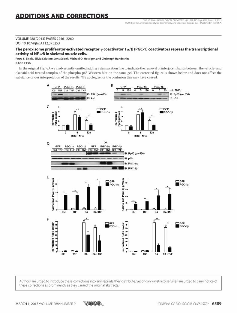
VOLUME 288 (2013) PAGES 2246 –2260DOI 10.1074/jbc.A112.375253
The peroxisome proliferator-activated receptor � coactivator 1�/� (PGC-1) coactivators repress the transcriptionalactivity of NF-�B in skeletal muscle cells.Petra S. Eisele, Silvia Salatino, Jens Sobek, Michael O. Hottiger, and Christoph Handschin
PAGE 2256:
In the original Fig. 7D, we inadvertently omitted adding a demarcation line to indicate the removal of interjacent bands between the vehicle- andokadaid acid-treated samples of the phospho-p65 Western blot on the same gel. The corrected figure is shown below and does not affect thesubstance or our interpretation of the results. We apologize for the confusion this may have caused.
THE JOURNAL OF BIOLOGICAL CHEMISTRY VOL. 288, NO. 9, p. 6589, March 1, 2013© 2013 by The American Society for Biochemistry and Molecular Biology, Inc. Published in the U.S.A.
MARCH 1, 2013 • VOLUME 288 • NUMBER 9 JOURNAL OF BIOLOGICAL CHEMISTRY 6589
ADDITIONS AND CORRECTIONS
Authors are urged to introduce these corrections into any reprints they distribute. Secondary (abstract) services are urged to carry notice ofthese corrections as prominently as they carried the original abstracts.