Embed Size (px)
Citation preview
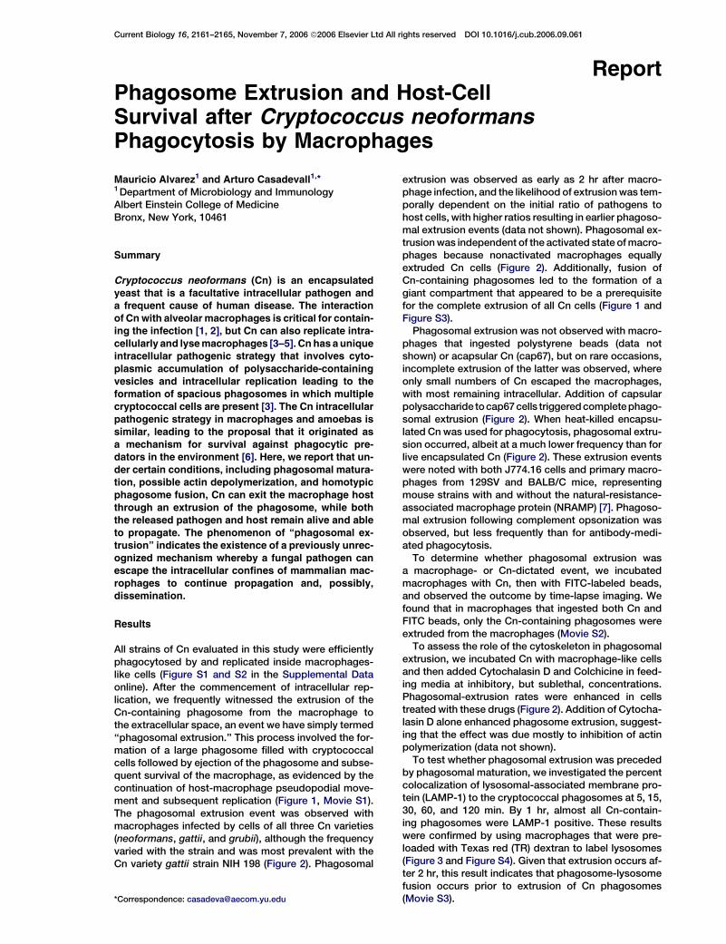
Current Biology 16, 2161–2165, November 7, 2006 ª2006 Elsevier Ltd All rights reserved DOI 10.1016/j.cub.2006.09.061
ReportPhagosome Extrusion and Host-CellSurvival after Cryptococcus neoformansPhagocytosis by Macrophages
Mauricio Alvarez1 and Arturo Casadevall1,*1Department of Microbiology and ImmunologyAlbert Einstein College of MedicineBronx, New York, 10461
Summary
Cryptococcus neoformans (Cn) is an encapsulated
yeast that is a facultative intracellular pathogen anda frequent cause of human disease. The interaction
of Cn with alveolar macrophages is critical for contain-ing the infection [1, 2], but Cn can also replicate intra-
cellularly and lyse macrophages [3–5]. Cn has a uniqueintracellular pathogenic strategy that involves cyto-
plasmic accumulation of polysaccharide-containingvesicles and intracellular replication leading to the
formation of spacious phagosomes in which multiplecryptococcal cells are present [3]. The Cn intracellular
pathogenic strategy in macrophages and amoebas issimilar, leading to the proposal that it originated as
a mechanism for survival against phagocytic pre-dators in the environment [6]. Here, we report that un-
der certain conditions, including phagosomal matura-tion, possible actin depolymerization, and homotypic
phagosome fusion, Cn can exit the macrophage host
through an extrusion of the phagosome, while boththe released pathogen and host remain alive and able
to propagate. The phenomenon of ‘‘phagosomal ex-trusion’’ indicates the existence of a previously unrec-
ognized mechanism whereby a fungal pathogen canescape the intracellular confines of mammalian mac-
rophages to continue propagation and, possibly,dissemination.
Results
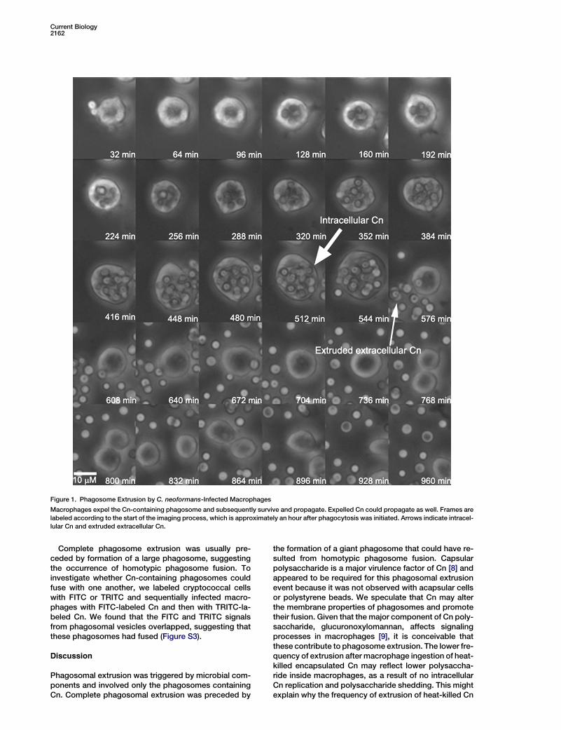
All strains of Cn evaluated in this study were efficientlyphagocytosed by and replicated inside macrophages-like cells (Figure S1 and S2 in the Supplemental Dataonline). After the commencement of intracellular rep-lication, we frequently witnessed the extrusion of theCn-containing phagosome from the macrophage tothe extracellular space, an event we have simply termed‘‘phagosomal extrusion.’’ This process involved the for-mation of a large phagosome filled with cryptococcalcells followed by ejection of the phagosome and subse-quent survival of the macrophage, as evidenced by thecontinuation of host-macrophage pseudopodial move-ment and subsequent replication (Figure 1, Movie S1).The phagosomal extrusion event was observed withmacrophages infected by cells of all three Cn varieties(neoformans, gattii, and grubii), although the frequencyvaried with the strain and was most prevalent with theCn variety gattii strain NIH 198 (Figure 2). Phagosomal
*Correspondence: [email protected]
extrusion was observed as early as 2 hr after macro-phage infection, and the likelihood of extrusion was tem-porally dependent on the initial ratio of pathogens tohost cells, with higher ratios resulting in earlier phagoso-mal extrusion events (data not shown). Phagosomal ex-trusion was independent of the activated state of macro-phages because nonactivated macrophages equallyextruded Cn cells (Figure 2). Additionally, fusion ofCn-containing phagosomes led to the formation of agiant compartment that appeared to be a prerequisitefor the complete extrusion of all Cn cells (Figure 1 andFigure S3).
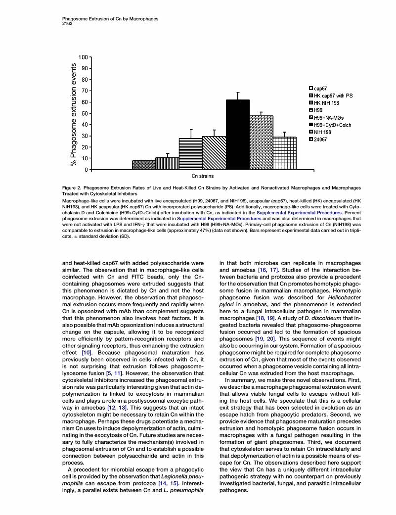
Phagosomal extrusion was not observed with macro-phages that ingested polystyrene beads (data notshown) or acapsular Cn (cap67), but on rare occasions,incomplete extrusion of the latter was observed, whereonly small numbers of Cn escaped the macrophages,with most remaining intracellular. Addition of capsularpolysaccharide to cap67 cells triggered complete phago-somal extrusion (Figure 2). When heat-killed encapsu-lated Cn was used for phagocytosis, phagosomal extru-sion occurred, albeit at a much lower frequency than forlive encapsulated Cn (Figure 2). These extrusion eventswere noted with both J774.16 cells and primary macro-phages from 129SV and BALB/C mice, representingmouse strains with and without the natural-resistance-associated macrophage protein (NRAMP) [7]. Phagoso-mal extrusion following complement opsonization wasobserved, but less frequently than for antibody-medi-ated phagocytosis.
To determine whether phagosomal extrusion wasa macrophage- or Cn-dictated event, we incubatedmacrophages with Cn, then with FITC-labeled beads,and observed the outcome by time-lapse imaging. Wefound that in macrophages that ingested both Cn andFITC beads, only the Cn-containing phagosomes wereextruded from the macrophages (Movie S2).
To assess the role of the cytoskeleton in phagosomalextrusion, we incubated Cn with macrophage-like cellsand then added Cytochalasin D and Colchicine in feed-ing media at inhibitory, but sublethal, concentrations.Phagosomal-extrusion rates were enhanced in cellstreated with these drugs (Figure 2). Addition of Cytocha-lasin D alone enhanced phagosome extrusion, suggest-ing that the effect was due mostly to inhibition of actinpolymerization (data not shown).
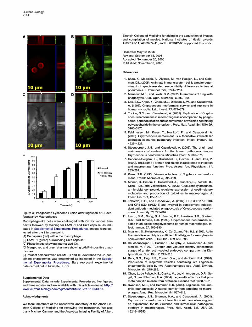
To test whether phagosomal extrusion was precededby phagosomal maturation, we investigated the percentcolocalization of lysosomal-associated membrane pro-tein (LAMP-1) to the cryptococcal phagosomes at 5, 15,30, 60, and 120 min. By 1 hr, almost all Cn-contain-ing phagosomes were LAMP-1 positive. These resultswere confirmed by using macrophages that were pre-loaded with Texas red (TR) dextran to label lysosomes(Figure 3 and Figure S4). Given that extrusion occurs af-ter 2 hr, this result indicates that phagosome-lysosomefusion occurs prior to extrusion of Cn phagosomes(Movie S3).
Current Biology2162
Figure 1. Phagosome Extrusion by C. neoformans-Infected Macrophages
Macrophages expel the Cn-containing phagosome and subsequently survive and propagate. Expelled Cn could propagate as well. Frames are
labeled according to the start of the imaging process, which is approximately an hour after phagocytosis was initiated. Arrows indicate intracel-
lular Cn and extruded extracellular Cn.
Complete phagosome extrusion was usually pre-ceded by formation of a large phagosome, suggestingthe occurrence of homotypic phagosome fusion. Toinvestigate whether Cn-containing phagosomes couldfuse with one another, we labeled cryptococcal cellswith FITC or TRITC and sequentially infected macro-phages with FITC-labeled Cn and then with TRITC-la-beled Cn. We found that the FITC and TRITC signalsfrom phagosomal vesicles overlapped, suggesting thatthese phagosomes had fused (Figure S3).
Discussion
Phagosomal extrusion was triggered by microbial com-ponents and involved only the phagosomes containingCn. Complete phagosomal extrusion was preceded by
the formation of a giant phagosome that could have re-sulted from homotypic phagosome fusion. Capsularpolysaccharide is a major virulence factor of Cn [8] andappeared to be required for this phagosomal extrusionevent because it was not observed with acapsular cellsor polystyrene beads. We speculate that Cn may alterthe membrane properties of phagosomes and promotetheir fusion. Given that the major component of Cn poly-saccharide, glucuronoxylomannan, affects signalingprocesses in macrophages [9], it is conceivable thatthese contribute to phagosome extrusion. The lower fre-quency of extrusion after macrophage ingestion of heat-killed encapsulated Cn may reflect lower polysaccha-ride inside macrophages, as a result of no intracellularCn replication and polysaccharide shedding. This mightexplain why the frequency of extrusion of heat-killed Cn
Phagosome Extrusion of Cn by Macrophages2163
Figure 2. Phagosome Extrusion Rates of Live and Heat-Killed Cn Strains by Activated and Nonactivated Macrophages and Macrophages
Treated with Cytoskeletal Inhibitors
Macrophage-like cells were incubated with live encapsulated (H99, 24067, and NIH198), acapsular (cap67), heat-killed (HK) encapsulated (HK
NIH198), and HK acapsular (HK cap67) Cn with incorporated polysaccharide (PS). Additionally, macrophage-like cells were treated with Cyto-
chalasin D and Colchicine (H99+CytD+Colch) after incubation with Cn, as indicated in the Supplemental Experimental Procedures. Percent
phagosome extrusion was determined as indicated in Supplemental Experimental Procedures and was also determined in macrophages that
were not activated with LPS and IFN-g that were incubated with H99 (H99+NA-MØs). Primary-cell phagosome extrusion of Cn (NIH198) was
comparable to extrusion in macrophage-like cells (approximately 47%) (data not shown). Bars represent experimental data carried out in tripli-
cate, 6 standard deviation (SD).
and heat-killed cap67 with added polysaccharide weresimilar. The observation that in macrophage-like cellscoinfected with Cn and FITC beads, only the Cn-containing phagosomes were extruded suggests thatthis phenomenon is dictated by Cn and not the hostmacrophage. However, the observation that phagoso-mal extrusion occurs more frequently and rapidly whenCn is opsonized with mAb than complement suggeststhat this phenomenon also involves host factors. It isalso possible that mAb opsonization induces a structuralchange on the capsule, allowing it to be recognizedmore efficiently by pattern-recognition receptors andother signaling receptors, thus enhancing the extrusioneffect [10]. Because phagosomal maturation haspreviously been observed in cells infected with Cn, itis not surprising that extrusion follows phagosome-lysosome fusion [5, 11]. However, the observation thatcytoskeletal inhibitors increased the phagosomal extru-sion rate was particularly interesting given that actin de-polymerization is linked to exocytosis in mammaliancells and plays a role in a postlysosomal exocytic path-way in amoebas [12, 13]. This suggests that an intactcytoskeleton might be necessary to retain Cn within themacrophage. Perhaps these drugs potentiate a mecha-nism Cn uses to induce depolymerization of actin, culmi-nating in the exocytosis of Cn. Future studies are neces-sary to fully characterize the mechanism(s) involved inphagosomal extrusion of Cn and to establish a possibleconnection between polysaccharide and actin in thisprocess.
A precedent for microbial escape from a phagocyticcell is provided by the observation that Legionella pneu-mophila can escape from protozoa [14, 15]. Interest-ingly, a parallel exists between Cn and L. pneumophila
in that both microbes can replicate in macrophagesand amoebas [16, 17]. Studies of the interaction be-tween bacteria and protozoa also provide a precedentfor the observation that Cn promotes homotypic phago-some fusion in mammalian macrophages. Homotypicphagosome fusion was described for Helicobacterpylori in amoebas, and the phenomenon is extendedhere to a fungal intracellular pathogen in mammalianmacrophages [18, 19]. A study of D. discoideum that in-gested bacteria revealed that phagosome-phagosomefusion occurred and led to the formation of spaciousphagosomes [19, 20]. This sequence of events mightalso be occurring in our system. Formation of a spaciousphagosome might be required for complete phagosomeextrusion of Cn, given that most of the events observedoccurred when a phagosome vesicle containing all intra-cellular Cn was extruded from the host macrophage.
In summary, we make three novel observations. First,we describe a macrophage phagosomal extrusion eventthat allows viable fungal cells to escape without kill-ing the host cells. We speculate that this is a cellularexit strategy that has been selected in evolution as anescape hatch from phagocytic predators. Second, weprovide evidence that phagosome maturation precedesextrusion and homotypic phagosome fusion occurs inmacrophages with a fungal pathogen resulting in theformation of giant phagosomes. Third, we documentthat cytoskeleton serves to retain Cn intracellularly andthat depolymerization of actin is a possible means of es-cape for Cn. The observations described here supportthe view that Cn has a uniquely different intracellularpathogenic strategy with no counterpart on previouslyinvestigated bacterial, fungal, and parasitic intracellularpathogens.
Current Biology2164
Supplemental Data
Supplemental Data include Experimental Procedures, five figures,
and three movies and are available with this article online at: http://
www.current-biology.com/cgi/content/full/16/21/2161/DC1/.
Acknowledgments
We thank members of the Casadevall laboratory of the Albert Ein-
stein College of Medicine for reviewing the manuscript. We also
thank Michael Cammer and the Analytical Imaging Facility of Albert
Figure 3. Phagosome-Lysosome Fusion after Ingestion of C. neo-
formans by Macrophages
Macrophage-like cells were challenged with Cn for various time
points followed by staining for LAMP-1 and Cn’s capsule, as indi-
cated in Supplemental Experimental Procedures. Images were col-
lected after the 1 hr time point.
(A) Cn capsule (red) within the macrophage.
(B) LAMP-1 (green) surrounding Cn’s capsule.
(C) Phase image showing internalized Cn.
(D) Merged red and green channels showing LAMP-1-positive phag-
osomes.
(E) Percent colocalization of LAMP-1 and TR-dextran to the Cn-con-
taining phagogomes was determined as indicated in the Supple-
mental Experimental Procedures. Bars represent experimental
data carried out in triplicate, 6 SD.
Einstein College of Medicine for aiding in the acquisition of images
and compilation of movies. National Institutes of Health awards
AI033142-11, AI033774-11, and HL059842-08 supported this work.
Received: May 19, 2006
Revised: September 18, 2006
Accepted: September 20, 2006
Published: November 6, 2006
References
1. Shao, X., Mednick, A., Alvarez, M., van Rooijen, N., and Gold-
man, D.L. (2005). An innate immune system cell is a major deter-
minant of species-related susceptibility differences to fungal
pneumonia. J. Immunol. 175, 3244–3251.
2. Mansour, M.K., and Levitz, S.M. (2002). Interactions of fungi with
phagocytes. Curr. Opin. Microbiol. 5, 359–365.
3. Lee, S.C., Kress, Y., Zhao, M.L., Dickson, D.W., and Casadevall,
A. (1995). Cryptococcus neoformans survive and replicate in
human microglia. Lab. Invest. 73, 871–879.
4. Tucker, S.C., and Casadevall, A. (2002). Replication of Crypto-
coccus neoformans in macrophages is accompanied by phago-
somal permeabilization and accumulation of vesicles containing
polysaccharide in the cytoplasm. Proc. Natl. Acad. Sci. USA 99,
3165–3170.
5. Feldmesser, M., Kress, Y., Novikoff, P., and Casadevall, A.
(2000). Cryptococcus neoformans is a facultative intracellular
pathogen in murine pulmonary infection. Infect. Immun. 68,
4225–4237.
6. Steenbergen, J.N., and Casadevall, A. (2003). The origin and
maintenance of virulence for the human pathogenic fungus
Cryptococcus neoformans. Microbes Infect. 5, 667–675.
7. Canonne-Hergaux, F., Gruenheid, S., Govoni, G., and Gros, P.
(1999). The Nramp1 protein and its role in resistance to infection
and macrophage function. Proc. Assoc. Am. Physicians 111,
283–289.
8. Kozel, T.R. (1995). Virulence factors of Cryptococcus neofor-
mans. Trends Microbiol. 3, 295–299.
9. Monari, C., Bistoni, F., Casadevall, A., Pericolini, E., Pietrella, D.,
Kozel, T.R., and Vecchiarelli, A. (2005). Glucuronoxylomannan,
a microbial compound, regulates expression of costimulatory
molecules and production of cytokines in macrophages. J.
Infect. Dis. 191, 127–137.
10. Taborda, C.P., and Casadevall, A. (2002). CR3 (CD11b/CD18)
and CR4 (CD11c/CD18) are involved in complement-indepen-
dent antibody-mediated phagocytosis of Cryptococcus neofor-
mans. Immunity 16, 791–802.
11. Levitz, S.M., Nong, S.H., Seetoo, K.F., Harrison, T.S., Speizer,
R.A., and Simons, E.R. (1999). Cryptococcus neoformans re-
sides in an acidic phagolysosome of human macrophages. In-
fect. Immun. 67, 885–890.
12. Muallem, S., Kwiatkowska, K., Xu, X., and Yin, H.L. (1995). Actin
filament disassembly is a sufficient final trigger for exocytosis in
nonexcitable cells. J. Cell Biol. 128, 589–598.
13. Rauchenberger, R., Hacker, U., Murphy, J., Niewohner, J., and
Maniak, M. (1997). Coronin and vacuolin identify consecutive
stages of a late, actin-coated endocytic compartment in Dic-
tyostelium. Curr. Biol. 7, 215–218.
14. Berk, S.G., Ting, R.S., Turner, G.W., and Ashburn, R.J. (1998).
Production of respirable vesicles containing live Legionella
pneumophila cells by two Acanthamoeba spp. Appl. Environ.
Microbiol. 64, 279–286.
15. Chen, J., de Felipe, K.S., Clarke, M., Lu, H., Anderson, O.R., Se-
gal, G., and Shuman, H.A. (2004). Legionella effectors that pro-
mote nonlytic release from protozoa. Science 303, 1358–1361.
16. Swanson, M.S., and Hammer, B.K. (2000). Legionella pneumo-
phila pathogesesis: A fateful journey from amoebae to macro-
phages. Annu. Rev. Microbiol. 54, 567–613.
17. Steenbergen, J.N., Shuman, H.A., and Casadevall, A. (2001).
Cryptococcus neoformans interactions with amoebae suggest
an explanation for its virulence and intracellular pathogenic
strategy in macrophages. Proc. Natl. Acad. Sci. USA 98,
15245–15250.
Phagosome Extrusion of Cn by Macrophages2165
18. Rittig, M.G., Shaw, B., Letley, D.P., Thomas, R.J., Argent, R.H.,
and Atherton, J.C. (2003). Helicobacter pylori-induced homo-
typic phagosome fusion in human monocytes is independent
of the bacterial vacA and cag status. Cell. Microbiol. 5, 887–899.
19. Rupper, A.C., Rodriguez-Paris, J.M., Grove, B.D., and Cardelli,
J.A. (2001). p110-related PI 3-kinases regulate phagosome-
phagosome fusion and phagosomal pH through a PKB/Akt de-
pendent pathway in Dictyostelium. J. Cell Sci. 114, 1283–1295.
20. Duhon, D., and Cardelli, J. (2002). The regulation of phagosome
maturation in Dictyostelium. J. Muscle Res. Cell Motil. 23, 803–
808.