Embed Size (px)
Citation preview
Thermochimica Acta, 193 (1991) 195-214 Elsevier Science Publishers B.V., Amsterdam
195
Pharmaceutical calorimetry: a selective review
G. Buckton a, S.J. Russell b and A.E. Beezer b
a The School of Pharmacy, University of London, 29/39 Brunswick Square, London WClN IAX (UK) b Chemical Laboratory, The University, Canterbury, Kent CT2 7NH (UK)
(Received 28 December 1990)
Abstract
Calorimetric investigations of systems of pharmaceutical interest are selectively reviewed. Attention is drawn to the need for more systematic and quantitative investigations of bioactivity, especially of synergic and antagonistic drug combinations. Formulation affects biological response (BR) and hence attention is given in this review to some of the factors that may be of biopharmaceutical importance.
INTRODUCTION
Calorimetry has the capacity, in principle, to investigate all chemical and physical changes. It is unsurprising, therefore, that the process and progress of metabolism of microbial species should have received serious attention (see other contributions in this issue). This necessarily, therefore, raises immediately the possibility of investigating added components which mod- ify metabolism. Such metabolic modifiers can be stimulants (trace materi- als, vitamins, etc.) or inhibitors. In this latter category fall such materials as heavy metals, toxic organic compounds and, specifically, drugs. The pur- pose of this contribution is to provide, not a comprehensive review, but to indicate the interesting areas of research and enquiry, based on previous work, which are now under active study. The original literature should be consulted for detailed accounts of methods, experimental design and analy- sis of results. This review will simply address some important new direc- tions in the calorimetric study of drug substances, formulated drugs and their interactions with sensitive target organisms.
BACKGROUND
One of the most prominent features of the microbial growth process is the production of heat. This fact alone ensures that as long as close attention is paid to careful experimental design much useful information, both qualitative and quantitative, may be gained by using calorimetry.
0040-6031/91/$03.50 0 1991 - Elsevier Science Publishers B.V. All rights reserved
196
Power (WI ee j
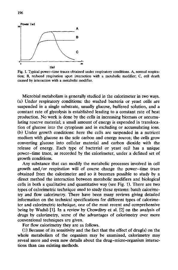
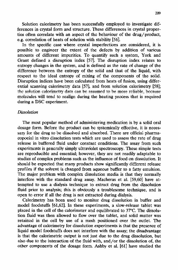
Fig. 1. Typical power-time traces obtained under respiratory conditions. A, normal respira- tion; B, reduced respiration upon interaction with a metabolic modifier; C, cell death caused by interaction with a metabolic modifier.
Microbial metabolism is generally studied in the calorimeter in two ways. (a) Under respiratory conditions: the washed bacteria or yeast cells are suspended in a single substrate, usually glucose, buffered solution, and a constant rate of glycolysis is established leading to a constant rate of heat production. No work is done by the cells in increasing biomass or accumu- lating reserve material; a small amount of energy is expended in transloca- tion of glucose into the cytoplasm and in excluding or accumulating ions. (b) Under growth conditions: here the cells are suspended in a nutrient medium with glucose as the sole carbon and energy source; the cells grow converting glucose into cellular material and carbon dioxide with the release of energy. Each type of bacterial or yeast cell has a unique power-time trace, as recorded by the calorimeter, under a defined set of growth conditions.
Any substance that can modify the metabolic processes involved in cell growth and/or respiration will of course change the power-time trace obtained from the calorimeter and so it becomes possible to study by a direct method the interaction between metabolic modifiers and biological cells in both a qualitative and quantitative way (see Fig. 1). There are two types of calorimetric technique used to study these systems: batch calorime- try and flow calorimetry. There have been many reviews giving detailed information on the technical specifications for different types of calorime- ter and calorimetric technique, one of the most recent and comprehensive being by Wads6 [l]. In a review by Chowdhry et al. [2] on the analysis of drugs by calorimetry, some of the advantages of calorimetry over more conventional techniques are given.
For flow calorimetry they are as follows. (1) Because of its sensitivity and the fact that the effect of drug(s) on the
whole metabolism of the organism may be examined, calorimetry may reveal more and even new details about the drug-micro-organism interac- tions than can existing methods.
197
(2) The calorimetric method requires only an observable difference between the power production in the treated and untreated (control) incubations, and unlike many other procedures does not require a trans- parent solution. Coloured or turbid solutions or even suspensions can be put through the calorimeter, This is particularly important when studying some pharmaceutical formulations.
(3) The system can be automated. (4) The kinetics of the action of anti-microbial agents (or other cytotoxic
agents) can be studied conveniently by means of their effects on the power-time trace.
(5) Both ant’ f I- ungal and anti-bacterial drugs can be studied, as indeed can be tissue cells.
(6) The flow calorimeter is connected to an external fermenter in which the culture grows. All manipulations are performed in the fermenter; thus the thermal equilibrium of the calorimeter is not disturbed. Simultaneous measurements on the culture may be made, i.e. pH, substrate consumption and viable cell counts, as the volume of the flow lines is small compared to the culture volume.
(7) The flow system is more akin to the in vivo state than are, for example, the systems used in diffusion assays.
(8) The flow system represents the effect of the drug on the total metabolism of the micro-organism. This again approximates more closely the in vivo state than do techniques which measure a single parameter of cell metabolism for analytical purposes.
(9) The appearance of cytoplasmic cell components, due to interaction of cells with drug(s), may be monitored to elucidate the mechanism of action of a drug or provide insight into the nature of the power-time trace.
Of course any technique must have its disadvantages and for flow calorimetry they are as follows.
(1) Cell concentrations must be in the range 104-lo5 cells ml-‘. (2) There is a lag time of approximately 3 min between initiation and
detection in the flow-through chamber of a calorimeter and, in this mode, very fast reactions cannot be studied. However, utilisation of a flow-mix chamber does allow events from reaction initiation to be followed.
(3) It is, as yet, very seldom that the signals from thermopile conduction calorimeters are corrected to give true kinetic curves. However, biological processes are often so slow that the distortion is negligible.
Descriptions of classical microbiological assay procedures, such as tur- bidimetric, agar diffusion, respirometric and serial dilution methods, are to be found in the manual published by Grove and Randall [3], and the treatises edited by Kavanagh [4,5]; the official methods are to be found in the various pharmacopoeias and in the publication by Arret et al. [6].
The disadvantages of classical microbiological assay methods have been examined by Newell [7] and Cosgrove [8]. They are time consuming (4-18
198
h), often have low reproducibility and precision, and are open to subjective errors. Furthermore, they are subject to the vagaries and variations inher- ent in classical microbiological assays. Microbiological methods require large numbers of observations to yield statistically satisfactory results; they are labour intensive and little advantage is gained through automation.
Each of the classical assay methods show disadvantages particular to themselves. Photometric methods require that the metabolic modifier of interest, e.g. an anti-fungal or antibiotic drug, does not interfere in the measurement at the wavelength at which turbidity is measured. Tube dilution techniques depend upon the growth of a micro-organism in a control tube compared to growth in assay tubes containing various dilutions of drug [4]. The time at which the tubes are read may affect the results of the test. The theory of agar diffusion methods is not well understood from a molecular point of view [4]. For example, if an antibiotic preparation is not homogeneous, or if the nature of the antibiotic causes “tailing” or partial inhibition, then difficulties may be encountered in assessing the results of assays. Synergistic or antagonistic effects of combinations of drugs against micro-organisms can also be difficult to assess by agar diffusion tube and other classical methods.
QUALITATIVE WORK
Much of the earlier work studying the interaction of metabolic modifiers with micro-organisms using calorimetry was of a qualitative nature [9]. This represented a quite natural progression from classical techniques, for example different bacterial cells might be grown up overnight for use in the calorimeter the next day. The power-time traces obtained in any form of experiment would certainly vary from batch to batch due to the fact that it is impossible to recreate the same “history” for the cells from different batches. Any work involving the use of non-chemically defined media must fall into the category of being only qualitative. For example the power-time traces for organisms growing in complex media (brain-heart infusion, Columbia broth, Oxoid nutrient broth, blood culture broth, etc.) often with added vitamins, are more complex than those recorded during growth in defined medium with a growth-limiting substrate [lo-151. Detailed inter- pretation in terms of changes in metabolic processes is difficult, if not impossible, because there is no known growth-limiting substrate, and there is minimal control of environmental conditions such as pH and little or no information on associated growth parameters.
An extensive review by Chowdhry et al. [2] covers in detail the qualita- tive effects of anti-bacterial agents on the power-time traces of different micro-organisms. For example the induction period for action of penicillin on E. coli (initial concentration 6 X lo6 cells ml-‘) was found [16] to depend on the phase of growth at which the antibiotic was added to the
199
culture. At 25”C, the power-time trace for E. co&penicillin interaction indicated that the induction period decreased with increase in the time spent by the cells in the logarithmic phase of growth before the drug was added. James [17] describes how metabolic inhibitors (sodium azide, 2,4-d& nitrophenol (DNP) and carboxy-cyanid-p-trifluoromethyl-phenylhydrazone (FCCP)) all h a d a pronounced effect on the power-time traces of cells of K aerogenes growing in glucose-limited medium; that of azide was very different from those of DNP or FCCP [18].
Exposure of non-P-lactamase-producing E. cob and S. aureus to benzyl penicillin and ampicillin gave rise to a “paradoxical zone phenomenon” in which the inhibitory effect on bacterial metabolism, judged on the heat generated, was smaller when the concentration was 5-10 X MIC (minimum inhibitory concentration) than at 2 x MIC and at 100-10000 X MIC [19].
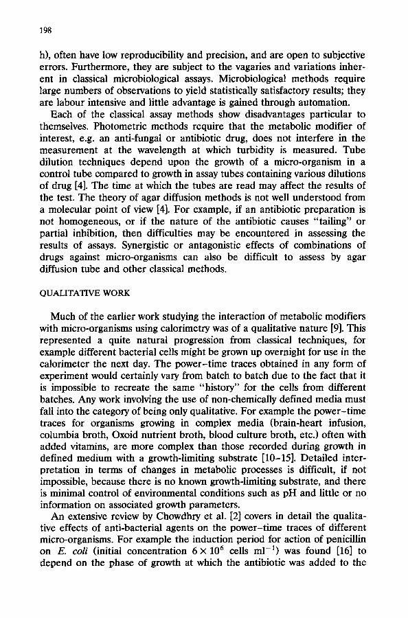
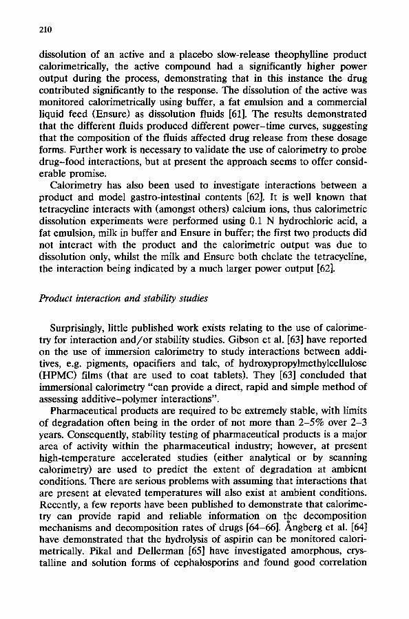
When either amoxycillin (AMX 40 pg cme3) or clavulinic acid (CA 10 kg cmU3> were added separately to AMP-resistant E. coli cultures (in the exponential growth phase), the power-time and growth curves were the same as for the control. When AMX (20 pg cme3) and CA (10 pg cmm3), i.e. Augmentin@, were added together to the growing-resistant culture, the power-time trace showed a marked reduction in heat production and the growth curve showed bacterial lysis [20,21] (see Fig. 2). CA had no effect on the power-time trace of AMP-sensitive E. coli, but AMX caused a marked decrease in heat production and cell lysis; the extent of both depends upon the concentration of AMX. Exposure of the sensitive strain to a combina- tion of AMX and CA (at concentrations which individually had no effect) produced a considerable inhibition of heat production and bacterial lysis. The morphological changes undergone by AMP-resistant strains in the presence of AMX and CA were qualitatively similar to those produced in the sensitive strain by AMX alone. The effect of CA is due to its inhibition of p-lactamase. CA alone caused a reduction in the heat production and the turbidity of a p-lactamase-producing strain of S. aureus; the inhibitory effect was even more pronounced when AMX was also present, i.e. a synergistic effect.
It is also possible to investigate in a qualitative way the interaction of structurally related compounds with micro-organisms. For example, M%rdh et al. [22] have examined the biological activity of the tetracyclines minocy- cline, doxycycline, tetracycline and oxytetracycline on interaction with E. coli. The effect of the antibiotics on cells in the lag phase was examined with a batch calorimeter. A flow calorimeter was used to study cell-drug interactions in the logarithmic phase of cell growth under aerated condi- tions. The viable cell count and MIC for the cell cultures were also recorded. The addition of half the MIC of minocycline and tetracycline (0.8 hg cmm3) prolonged the lag phase of the cells by 8 h. All four antibiotics decreased the power output immediately they were added to cells in the logarithmic growth phase but the extent and duration of the effect varied
POWERll7lW 0.6 -
0.6 -- . . ______ ---. __ _.__.. ,’
OA-- ,,,’ ,/ 01
TIME/h POWCWIIIW
0.6
0.6 I ,---- 0.4 (iii)
POWEWmW 0.6 -
0.6 - __._.. _-- -
i-
0.4 I
(ii)
,’
TIME/h
0.6 -
(iv1 0.1
0 a0 0 6.0
TIME/h TIME/h
Fig. 2. Power-time traces for the growth of E. coli. JT39 @lactamase-producing strain) in Columbia broth in the presence of anti-bacterial agents (after Seminitz and Casey [20,21])): ---9 growth curve); (i) control; (ii) clavulinic acid, 10 pg cme3; (iii) amoxycillin, 40 pg cmV3; (iv) amoxycillin 20 pg cmm3 +clavulinic acid 10 pg cm-3.
with the biological activity of the drugs. Although the antibiotics had the same MIC values, at levels above and below this they showed differences in their ability to suppress the metabolism of the test organisms, the order being minocycline > doxycycline > oxytetracycline > tetracycline both for lag phase and logarithmic phase cells. It was suggested that the extent and duration of the inhibitory effect on the overall metabolism of E. coli could be combined with pharmacokinetic data to establish dose intervals during antibiotic therapy.
Most recently, Hoffner et al. [23,24] have studied the synergistic effects of combinations of anti-mycobacterial drugs on Mycobacterium avium complex (MAC). Pronounced synergy was seen for several drug combina- tions where ethambutol was found to be the key drug in the synergistic potentiation. Batch reaction calorimetry was used to study the energetics of the initial interaction between anti-bacterial drugs and the mycobacterial cell surface. The drug-cell interaction process is very complex and so far it has not been possible to identify the processes, i.e. dissolution of the drug, drug binding, membrane disturbances, transport, etc., that dominate and lead to exo- or endothermicity. The endo- and exothermic signals observed did show that there were substantial differences in the modes of action.
201
When single drugs were tested, ethambutol induced the most pro- nounced thermal response indicating a strong, rapid, physico-chemical interaction with the MAC cell envelope. When MAC were simultaneously exposed to ethambutol and streptomycin, the thermal effects were similar to that of exposure to ethambutol alone. Studies of drug interactions with MAC pre-incubated with anti-mycobacterial drugs in sub-inhibitory con- centrations revealed information on changes in cell wall properties, caused by the drug. It was shown that there was a marked change in the initial thermal reponse to a subsequent exposure of MAC cells to streptomycin when the MAC cells were pre-exposed to ethambutol. It had previously been shown [25] that streptomycin can penetrate the cell wall of MAC strains exposed at the same time to ethambutol and, hence, can inhibit the growth of the otherwise streptomycin-resistant strains. The explanation proposed for these observations was that interference of ethambutol with the outer cell envelope increases the intracellular influx of other anti- mycobacterial agents [23].
TISSUE CELLS
Another area where much qualitative work has been done is in the study of tissue cells [26]. Animal cells are not as easy to work with as microbes; they are more fragile and generally more sticky. In addition, they do not usually grow and divide in suspension; because of this, progress in this field has been slower than in others. To give just one example in this area, Pate1 et al. [27] succeeded in maintaining long-term cultures of isolated human skin fibroplasts in a Bioflux differential calorimeter. They seeded human foreskin fibroplasts onto plastic foil. Time was allowed for cell attachment before the foil was inserted into the calorimetric vessel, together with growth medium containing 15% (v/v> calf serum. A culture of 3 x lo6 cells gave a power-time trace with an initial maximum over the first 3 h, followed by slightly increasing heat production for lo-20 h. Power then decreased to a lower value which was maintained for several days. The first peak was ascribed to spreading and growth of the cells, and the increasing heat evolution that followed was thought to be due to cell multiplication. Under the particular conditions of this study, the cells became confluent after 2-3 divisions and heat production declined to a constant value, supposedly because metabolism was “restricted to processes of mainte- nance”. For more detailed accounts of the study of tissue cells, reference should be made to other contributions to this volume and to the review by Kemp [26].
QUANTITATIVE WORK
In order to obtain quantitative experimental results, it is necessary to have reproducible power-time traces for a given organism under a given
202
set of experimental conditions. Precautions must be taken to control as many of the experimental variables as possible. These include (i) a chemi- cally defined medium, preferably with a growth-limiting substrate and of sufficient buffer capacity to prevent change of pH during growth or respiration; (ii) use of a standard inoculum. An ampoule of cryogenically stored cells ensures a fixed number of viable cells in the inoculum [28]; (iii) constant conditions of stirring and aeration (or maintenance of an anaero- bic atmosphere if necessary) to ensure a reproducible and constant gas environment in the calorimeter cell and in the fermenter when the flow cell assembly is used; (iv) for flow calorimetry; (a) the maintenance of an identical and constant temperature in both the fermenter and calorimeter with the tubes joining them thermostated and kept as short as possible; (b) a constant pump rate through the calorimeter cell, with as high a pump rate as possible consistent with the attainment of thermal equilibrium in the calorimeter (the highest pump rate ensures the control of aerobiosis/ anaerobiosis during passage of the culture through the closed loop ef the calorimetric “plumbing”).
When reported, the confidence limits at the 95% level for the heat evolved or enthalpy change for cells growing under carbon limitation are generally in the range + 1% to +4%.
Quantitative information was gained by calorimetry in a comprehensive study of the interaction of the polyene antibiotics nystatin, filipin, pimarcin, amphotericin B, candicin and lucensomycin, and the synthetic anti-fungal imidazole drug clotrimazole with Saccharomyces cerevisiae NCYC239, by Beezer et al. [29-321. A flow calorimeter was used in all experiments, which were conducted under anaerobic conditions.
A linear relationship was found between the logarithm of the nystatin dose and the time required for the power-time trace to rise and fall from the first calorimetric response to some arbitrary level (X%‘o> above the baseline. Calorimetric investigations of the action of nystatin on cell preparations of different incubation age before liquid nitrogen freezing indicated that yeast cells were more susceptible to nystatin as the prepara- tion age of the cells decreased. In another study employing identical experimental conditions, except for a change in the temperature of the assay from 25 to 3O”C, various dose-response relationships were obtained between the antibiotic concentration and instrument response, the nature of the relationship depending on the identity and concentration of the anti-microbial agent [30]. This method showed distinct advantages over the agar diffusion test, in terms of the lowest determinable concentration. The order of bioactivity of the polyene antibiotics against respiring yeast cells was found to be nystatin > filipin > pimarcin > candicin > amphotericin B.
It was noted that the concentration dependence of the interaction of lucensomycin with respiring yeast cells fell between the two extremes of the observed calorimetric response. Lucensomycin differs, in structural terms,
203
from pimarcin only in an increase in length of an alkyl side chain R, where R = CH,(CH,),- for lucensomycin and R = -CH, for pimarcin. These two compounds show completely different modes of action towards yeast cells in the concentration range 5 X 10m6 to lo-’ mol lm3. The experiments indicated that lucensomycin can act as both a fungistatic and fungicidal antibiotic, depending on its concentration, whereas the other anti-fungals are either only fungicidal (nystatin, candicin, filipin, amphotericin B) or only fungistatic (pimarcin, clotrimazole). Thus the power-time traces re- veal some of the differences in the mode of action of the antibiotics. This series of experiments also included study of the dosage forms of ampho- tericin B. Dosage forms are, of course, of major importance in controlling dissolution rate, locus of uptake and ultimately, therefore, bioavailability (see below). It is notable that the dosage forms did not appear to interfere with the direct determination of the bioactivity of the antifungal present in such complex formulations. Interestingly, a combined drug preparation (of amphotericin B plus tetracycline) could be assayed for the antifungal content alone (amphotericin B) without separation of the active, anti- bacterial, component nor indeed from the excipients.
DRUG COMBINATIONS
Calorimetry, especially flow calorimetry, is therefore, a powerful tool for the study of the effect of mixtures of drugs on microbial metabolism. Classical microbiological assay techniques cannot deal adequately with drug combinations. Until recently, there has been only one example de- scribed [33] of the calorimetric investigation of the consequences of com- bined antibiotics-cell interaction. This study showed that clotrimazole reduced the effectiveness of amphotericin B against S. cerevisiae. This antagonism was shown to result from preferential uptake of clotrimazole by the yeast, thus effectively reducing the uptake of amphotericin B.
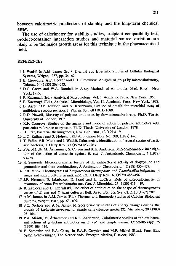
Reference has already been made to the synergic interaction of ethamb- utol with other drugs upon MAC [23,24]. Other recent work published in this area is by Joslin Kjeldsen et al. [34]. The system studied was the combination of polymixin B sulphate (PolB), neomycin sulphate (Neo) and zinc bacitracin (ZnB), which constitute a post-surgery spray powder prepa- ration (Trisep @, ICI Pharmaceutical Division, Alderly Park, Macclesfield, Cheshire, UK). The test organisms for each antibiotic in conventional antibiotic assays are: PolB, BordeteZZu bronchisepticu (NCTC 8344); Neo, Bacillus pumiZu.s (NCTC 8241); ZnB, Micrococcus Zuteus (NCTC 7743). All three antibiotics were also tested using a single test organism Escherichia coli (NCTC 10418). For all the test organisms, calorimetric assay proce- dures were developed, exhibiting improvements in time, reproducibility and sensitivity compared with conventional microbiological assays.
204
(a) 4. PCWCR (ARIITRARY UWITO)
r
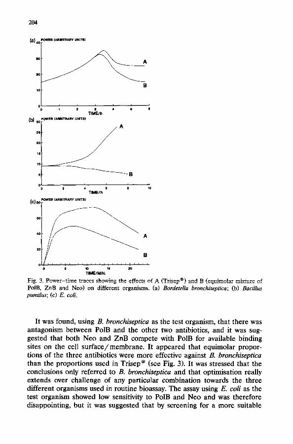
Fig. 3. Power-time traces showing the effects of A (Trisep@) and B (equimolar mixture of PolB, ZnB and Neo) on different organisms. (a) Bordetella bronchiseptica; (b) Bacillus pumilus; (c) E. coli.
It was found, using B. bronchiseptica as the test organism, that there was antagonism between PolB and the other two antibiotics, and it was sug- gested that both Neo and ZnB compete with PolB for available binding sites on the cell surface/membrane. It appeared that equimolar propor- tions of the three antibiotics were more effective against B. bronchisepticu than the proportions used in Trisep@ (see Fig. 3). It was stressed that the conclusions only referred to B. bronchisepticu and that optimisation really extends over challenge of any particular combination towards the three different organisms used in routine bioassay. The assay using E. coli as the test organism showed low sensitivity to PolB and Neo and was therefore disappointing, but it was suggested that by screening for a more suitable
205
test organism a more sensitive and rapid assay could be developed for several antibiotics using only one or a small number of bacterial strains.
An attempt to establish a synergy index for the drugs Trimethoprim and Sulfamethoxazole upon interaction with E. cob has been reported by Shafiq [35]. Development of such an index proved to be a difficult task because of the different kinetic data presented in the power-time traces for the two drugs when administered separately and in combination. This highlights both the advantage of using calorimetry, i.e. access to kinetic information, and the disadvantage, i.e. there is no basic theory to relate da/dt to the organisms response to a drug; it requires, as yet, an empirical solution.
QUANTITATIVE STRUCTURE ACTIVITY RELATIONSHIPS
This review has described above the quantitative bioassay of both anti- bacterial and anti-fungal drugs. The quantitative aspects of calorimetry have revealed its reproducibility, sensitivity and its capacity to discriminate between the bioactivities of drugs of closely related chemical structures. A major objective for quantitative bioactivity determinations is the establish- ment of quantitative structure activity relationships (QSAR).
The conventional view is that the basis of QSAR is the relationship between partition coefficients (P) and biological response (BR). This is correlated, generally, through some form of the Hansch equation [36,37]. The partition coefficient is, however determined for drug transfer between water and a bulk, non-aqueous solvent, usually octan-l-01.
To examine the basis of this assignment of octan-l-01 as a mimic of a biological cell, measurements have been made [38,39] of the enthalpies of transfer of a series of alkoxyphenols from water to octan-l-01, heptane and propylene carbonate; also the enthalpies of transfer of these same solutes from isotonic aqueous medium to E. cob suspended in isotonic aqueous medium have been determined. It is quite apparent from these results that none of the solvents studied mimics, as far as AHCtrans) is concerned, the biological cells as a solvent. However the correlation between BR and P is still explored vigorously [37].
A more interesting and rewarding possibility is to explore the direct relationship between BR and chemical structure. In the study of an homologous series of alkoxyphenols, it was noted that the intercept in the linear plot of log dose versus calorimetric response [40] was related to the carbon number in the alkyl side chain (for y11-, p- and o- derivatives). The intercept was labelled log(dose),, , i.e. the maximum dose of a drug that could be added without soliciting a calorimetric response. It appears, therefore, that log(dose),, versus carbon number constitutes a linear free energy relationship (LFER). The conclusion is that the BR can be factored into group additivity terms, e.g. the slope of the plot of log(dose),, versus
206
carbon number is the contribution to log(dose),, per methylene group and the intercept that to the remainder of the molecule, i.e. the parent grouping. Thus for the first time, chemical structure appears to have been related directly to BR.
As noted above, in respiration experiments two limiting forms of interac- tion can be identified that indicate different modes of action, i.e. a “kill” or decline of the power-time trace to the baseline value or a simple reduction in the control signal to some new, lower, value (this may constitute a reduction in overall metabolic activity or a reduction in total cell numbers through selective killing of various cell (age) populations). To examine, therefore, the importance of specific steps in the overall mechanism of action of drugs on interaction with sensitive target organisms, the relation- ship between log(dose),, and log P for a series of non-homologously related compounds has been investigated [41]. The compounds studied were a variety of substituted derivatives of cardanol (a derivative of cashew nut component shell liquid). It was demonstrated that a linear relationship existed between log(dose),, and log P for these compounds on interac- tion with Saccharomyces cerevisiae, i.e. an LFER exists. The derived data did not show any correlation with Hamrnett u parameters and this implies that partitioning, not some feature of electronic density distribution, is the leading process in determining the bioactivity of these compounds.
Thus, not only can calorimetry reveal details of mechanisms, allowing the development of bioassay procedures but it may, in addition, allow the introduction of LFER relationships which could guide the synthesis and design of new bioactive molecules through a biologically based directly determined group additivity parameter.
DRUG FORMULATIONS
As noted above, biological response can be followed calorimetrically. Numerous formulation design factors will influence drug bioavailability and hence BR; these include wetting (as a first step towards dissolution), dissolution, product stability, etc.
It is possible to investigate BR directly from a prepared dosage form, and indeed to consider the individual processes that affect its performance. Limited published calorimetric data exist, however.
Wetting
It has always been acknowledged that wettability is important in the preparation, storage and use of pharmaceuticals. Typical examples of the importance of wetting include the dispersion of powders to form suspen- sions, the addition of binders during the process of wet granulation (often a necessary step in tablet manufacture), the adhesion of polymer films to
207
tablets (to control release rate of drugs or to modify appearance, mask tastes, aid swallowing, etc.) and the dissolution rate of solid dosage forms in the gastrointestinal tract. Recently, the role of surface energies and polarities has been investigated as a method of predicting interactions between components of a formulation, thus aiding formulation optimisa- tion (see, for example, ref. 42).
Wettability is usually assessed by means of a contact angle, but for powdered systems this can be problematic [43]; thus alternatives are required. An obvious alternative to contact angle measurements is the use of calorimetry to quantify the interaction between the wetting agent and the powder.
Most workers have attempted the use of breaking ampoule calorimetry in order to determine the enthalpy of immersion of a solid in a liquid (see, for example, refs. 44-46). In general, the enthalpy is obtained calorimetri- cally, and the Gibbs function is derived from contact angle data [44,45]. The difficulties here are associated with problems of measuring contact angles for powders [43]. Immersional approaches can be successful, for example in the papers quoted above, Hansford et al. [44] were able to demonstrate a change in the thermodynamic functions of immersion for different samples of a hydrophobic drug (Griseofulvin), which had been milled by different techniques; Storey [45] considered the effect of various substituents at different positions on an imidazole ring structure; and Hollenbeck et al. [46] measured enthalpies of immersion of samples of microcrystalline cellulose which had been equilibrated with different water contents. Hollenbeck et al. [46] noted that immersional techniques can be suitable for hydrophilic powders, which are particularly difficult to study by contact angle methods; however, the difficulties associated with the use of immersional calorimetry were not addressed.
Many drug powders are hydrophobic, and incomplete immersion (in water) can result; although this issue has been addressed (see, for example, refs. 47-49), uncertainties about the surface coverage of the powder prior to immersion, i.e. the extent of water sorption, and the degree of flotation, lead to the need for alternative approaches.
Flow calorimetry has been used, where vapours of different humidities have been passed over a powder [50]; however, this approach has not been given serious critical examination in the pharmaceutical field.
The use of vapour sorption onto a vacuum-cleaned surface offers some unique advantages to this field of study. Although it is acknowledged that immersion is often the process of interest, e.g. it is the first step required for a product to dissolve in the gastro-intestinal tract, on a fundamental basis, it is the adsorption of the first layers of water molecules onto a powder surface that is most indicative of the interaction between that powder and the liquid (in line with the BET approach). Further adsorption after these initial molecular layers, will tend to be equivalent to condensa-
208
tion, and immersion of this vapour-laden surface will result in a reordering of the molecules of condensed liquid (usually water) to be accommodated in the structure of the bulk liquid. The work of Hollenbeck et al. [46] demonstrates this point, as the enthalpy of immersion falls exponentially as a function of the moisture content of the powder. The practical difficulty with the approach of Hollenbeck et al. [46] is that the powder is equili- brated in humidity-controlled chambers, then generally removed into ambi- ent conditions before sealing; this leads to at least some uncertainty about the exact water content. A more disturbing situation is the insistence of some workers on using the powder in the “as received” form, which will mean that the water content will vary from day to day (depending upon the shape of the adsorption isotherm(s), and on the humidity/temperature profiles in the laboratory), and results should show similar variability. The argument offered to attempt to justify the use of undefined powder surfaces is a need to model the “practical” situation of product use.
Calorimetric vapour sorption studies have been linked with parallel experiments in a vacuum microbalance [Xl. It is possible to obtain the Gibbs function from an equilibrium constant using the vacuum microbal- ante, and then to combine the calorimetric power output with the knowl- edge of the mass adsorbed to obtain the enthalpy of adsorption. This approach has been used to study hydrophobic drugs [51], small differences in surface properties of the same drug (induced by different milling techniques) [52], and the complex sorption processes that make up the interaction between microcrystalline celluloses and starches with water [53]. The use of the combination of a vacuum microbalance and the sorption calorimeter offers advantages over other systems, especially for hydropho- bic powders. Firstly, it is possible to obtain the thermodynamic functions without the need to assess the contact angle. Secondly, the thermodynamic parameters, and the kinetics, can allow a molecular mechanism of the interaction between the vapour and the powder to be postulated [51,53]. Thirdly, the use of the calorimeter is more accurate and more rapid than the isosteric approaches to this problem.
Crystal properties
Changes in the crystalline nature of a solid, e.g. polymorphic form, crystal habit and crystal lattice imperfections, result in different physical properties. The changes in properties can alter the processability of the powder, e.g. flow, compression, etc, and the behaviour, e.g. dissolution rate, and thus the bioavailability (or therapeutic benefit) of the product. The prevalence of lattice imperfections is influenced by many processes including crystallisation technique (solvent, stirring rate, crystallisation rate etc., see York [54]), drying, milling and compression [55].
Solution calorimetry has been successfully employed to investigate dif- ferences in crystal form and structure. These differences in crystal proper- ties often correlate with an aspect of the behaviour of the drug/product, e.g. correlation of heats of solution with stability [56].
In the specific case where crystal imperfections are considered, it is possible to engineer the extent of the defects by addition of various amounts of different impurities. To quantify such a system, York and Grant defined a disruption index [57]. The disruption index relates to entropy changes in the system, and is defined as the rate of change of the difference between the entropy of the solid and that of the liquid, with respect to the ideal entropy of mixing of the components of the solid. Disruption indices have been calculated from heats of fusion, using differ- ential scanning calorimetry data [57], and from solution calorimetry [58]; the solution calorimetry data can be assumed to be more reliable, because molecules will tend to realign during the heating process that is required during a DSC experiment.
Dissolution
The most popular method of administering medication is by a solid oral dosage form. Before the product can be systemically effective, it is neces- sary for the drug to be dissolved and absorbed. There are official pharma- copoeial in vitro dissolution tests which are used to assess the rate of drug release in buffered fluid under constant conditions. The assay from such experiments is generally simply ultraviolet spectroscopy. These simple tests are reproducible and essential; however, they are not readily adaptable to studies of complex problems such as the influence of food on dissolution. It should be expected that many products show significantly different release profiles if the solvent is changed from aqueous buffer to a fatty emulsion. The major problem with complex dissolution media is that they normally interfere with the standard drug assay. Macheras et al. [59,60] have at- tempted to use a dialysis technique to extract drug from the dissolution fluid prior to analysis; this is obviously a troublesome technique, and is open to error if all the drug is not extracted during dialysis.
Calorimetry has been used to monitor drug dissolution in buffer and model foodstuffs [61,62]. In these experiments, a slow-release tablet was placed in the cell of the calorimeter and equilibrated to 37°C. The dissolu- tion fluid was then allowed to flow over the tablet, and solid matter was retained in the cell by use of a mesh positioned over the outlet. The advantage of calorimetry for dissolution experiments is that the presence of liquid model foodstuffs does not interfere with the assay; the disadvantage is that the calorimetric output is not just due to the drug dissolution, but also due to the interaction of the fluid with, and/or the dissolution of, the other components of the dosage form. Ashby et al. [61] have studied the
210
dissolution of an active and a placebo slow-release theophylline product calorimetrically, the active compound had a significantly higher power output during the process, demonstrating that in this instance the drug contributed significantly to the response. The dissolution of the active was monitored calorimetrically using buffer, a fat emulsion and a commercial liquid feed (Ensure) as dissolution fluids [61]. The results demonstrated that the different fluids produced different power-time curves, suggesting that the composition of the fluids affected drug release from these dosage forms. Further work is necessary to validate the use of calorimetry to probe drug-food interactions, but at present the approach seems to offer consid- erable promise.
Calorimetry has also been used to investigate interactions between a product and model gastro-intestinal contents [62]. It is well known that tetracycline interacts with (amongst others) calcium ions, thus calorimetric dissolution experiments were performed using 0.1 N hydrochloric acid, a fat emulsion, milk in buffer and Ensure in buffer; the first two products did not interact with the product and the calorimetric output was due to dissolution only, whilst the milk and Ensure both chelate the tetracycline, the interaction being indicated by a much larger power output [62].
Product interaction and stability studies
Surprisingly, little published work exists relating to the use of calorime- try for interaction and/or stability studies. Gibson et al. [63] have reported on the use of immersion calorimetry to study interactions between addi- tives, e.g. pigments, opacifiers and talc, of hydroxypropylmethylcellulose (HPMC) films (that are used to coat tablets). They [63] concluded that immersional calorimetry “can provide a direct, rapid and simple method of assessing additive-polymer interactions”.
Pharmaceutical products are required to be extremely stable, with limits of degradation often being in the order of not more than 2-5% over 2-3 years. Consequently, stability testing of pharmaceutical products is a major area of activity within the pharmaceutical industry; however, at present high-temperature accelerated studies (either analytical or by scanning calorimetry) are used to predict the extent of degradation at ambient conditions. There are serious problems with assuming that interactions that are present at elevated temperatures will also exist at ambient conditions. Recently, a few reports have been published to demonstrate that calorime- try can provide rapid and reliable information on the decomposition mechanisms and decomposition rates of drugs [64-661. Angberg et al. [64] have demonstrated that the hydrolysis of aspirin can be monitored calori- metrically. Pikal and Dellerman [65] have investigated amorphous, crys- talline and solution forms of cephalosporins and found good correlation
211
between calorimetric predictions of stability and the long-term chemical assay.
The use of calorimetry for stability studies, excipient compatibility test, product-container interaction studies and material source variation are likely to be the major growth areas for this technique in the pharmaceutical field.
REFERENCES
1 I. Wad&i in A.M. James (Ed.), Thermal and Energetic Studies of Cellular Biological Systems, Wright, 1987, pp. 34-67.
2 B. Chowdhry, A.E. Beezer and E.J. Greenhow, Analysis of drugs by microcalorimetry, Talanta, 30 (1983) 208-243.
3 D.C. Grove and W.A. Randall, in Assay Methods of Antibiotics, Med. Encyl., New York, 1955.
4 F. Kavanagh (Ed.), Analytical Microbiology, Vol. I, Academic Press, New York, 1963. 5 F. Kavanagh (Ed.), Analytical Microbiology, Vol. II, Academic Press, New York, 1972. 6 B. Arret, D.P. Johnson and A. Krishbaum, Outline of details for microbial assay of
antibiotics: second revision, J. Pharm. Sci., 60 (1971) 1689. 7
8
9 10 11
12
13
14
15
16
17
18
19
20
R.D. Newell, Bioassay of polyene antibiotics by flow microcalorimetry, Ph.D. Thesis, University of London, 1975. R.F. Cosgrove, Studies on the analysis and mode of action of polyene antibiotics with particular reference to nystatin, Ph.D. Thesis, University of London, 1978. H. Prat, Bacterial thermogenesis, Rev. Can. Biol., 12 (1953) 19. L.O. Kallings and S. Hofner, LKB Application Note No. 309, (1977) l-6. T. Fujita, P.R. Monk and I. Wads& Calorimetric identification of several strains of lactic acid bacteria, J. Dairy Res., 45 (1978) 457-463. P.A. Mardh, M. &hammer, S. Coleen and ICE. Anderson, Microcalorimetric investiga- tion of the action of cinoxacin against E. coli, J. Antimicrob. Chemother., 4 (1978) 73-78. E. Semenitz, Microcalorimetric testing of the antibacterial activity of doxycycline and gentamicin and their combinations, J. Antimicrob. Chemother., 4 (1978) 455-457. P.R. Monk, Thermograms of Streptococcus thermophilus and Lactobacillus bulgaricus in single and mixed culture in milk medium, J. Dairy Res., 46 (1979) 485-496. J.P. Herman, E. Jakubczak, D. Izard and H. LeClerc, Role of microcalorimetry in taxonomy of some Enterobacteriaceae, Can. J. Microbial., 26 (1980) 413-419. B. Zablocki and E. Czerniaski, The effect of antibiotics on the shape of thermogenesis curves of E. coli and S. typhi cultures, Bull. Acad. Pol. Sci. Ser. Cl. 2, 10 (1962) 209. A.M. James, in A.M. James (Ed.), Thermal and Energetic Studies of Cellular Biological Systems, Wright, 1987, pp. 68-105. S.C. Nichols and A.M. James, Microcalorimetry studies of energy changes during the growth of Klebhella aerogenes in simple salts/glucose media (2), Microbios, 29 (1980) 95-104. P.A. MLrdh, M. &hammer and K.E. Andersson, Calorimetric studies of the antibacte- rial actions of p-lactam antibiotics on E. coli and Staph. aureus, Chemotherapy, 25 (1979) 106-116. E. Semenitz and P.A. Casey, in E.A.P. Croyden and M.F. Michel (Eds.), Proc. Eur. Symp. Scheveningen, The Netherlands. Excerpta Medica, Elsevier, 1983.
212
21
22
23
24
25
26
21
28
29
30
31
32
33
34
35 36
37
E. Semenitz, P.A. Casey, W. Pfaller and G. Gstraunthaler, Microcalorimetric, turbidi- metric, phase contrast microscopic, and electron microscopic investigations of the actions of Amoxicilin clavulinic acid and Augmentin on AMX sensitive and AMX resistant strains of E. coli, Chemotherapy, 29 (1983) 192-207. P.A. Mlrdh, P. Ripa, K.E. Anderson and I. Wadso, Kinetics of action of tetracyclins on E. Coli as studied by microcalorimetry, Antimicrob. Agents Chemother., 10 (1976) 604. S. Hofner, G. Kallenius, A.E. Beezer and S.B. Svenson, Studies on the mechanisms of the synergystic effects of ethambutol and other antimicrobial drugs on a Mycobacterium avium complex, Acta Leprologica, 7 (1989) 195-199. S. Hofner, S. Svenson and A.E. Beezer, Microcalorimetric studies of the initial interac- tion between antimycobacterium drugs and Mycobacterium avium, J. Antimicrob. Chemother., 25 (1990) 353-359. S. Hofner, S. Svenson and G. Kallenius, Synergistic effects of antimycobacterial drug combinations on Mycobacterium avium complex determined radiometrically in liquid medium, Eur. J. Clin. Microbial., 6 (1987) 530-535. R.B. Kemp, in A.M. James (Ed.), Thermal and Energetic Studies of Cellular Biological Systems, Wright, 1987, pp. 147-166. M. PItel, U. Reichart, B. Schaarschmidt and I. Lamprecht, in W. Hemminger (Ed.), Thermal Analysis, Vol. II, Birkhguser, Basel, 1980, pp. 559-564. S. Nichols, A.M. James and F.E. Prichard, Microcalorimetry studies of energy changes during the growth of Klebisella aerogenes in simple salts/glucose media (11, Microbios, 25 (1979) 187-203 A.E. Beezer, R.D. Newell and H.J.V. Tyrrell, Bioassay of mystatin bulk material by flow microcalorimetry, Anal. Chem., 49 (1977) 34. A.E. Beezer, B.Z. Chowdhry, R.D. Newell and H.J.V. Tyrrell, Bioassay of antifungal antibiotics by flow microcalorimetry. Anal. Chem., 49 (1977) 1781. A.E. Beezer and B.Z. Chowdhry, Combined microassay and determination of bioactivity of iV-acetylnystatin by flow microcalorimetry, Talanta, 27 (1980) l-6. A.E. Beezer and B.Z. Chowdhry, Flow microcalorimeteric bioassay of polyene antibi- otics: interaction with growing Saccharomyces cerevisiae, Experientia, 37 (1981) 828-830. R.F. Cosgrove, A.E. Beezer and R.J. Miles, In vitro studies of amphotericin B in combination with the imidazole antifungal compounds clotrimazole and miconazole, J. Infect. Dis., 138 (19781681-685. N. Joslin Kjeldsen, A.E. Beezer, R.J. Miles and H. Sodha, Flow microcalorimetric assay of antibiotics. IV. Polymyxin B sulphate, neomycin sulphate, zinc bacitracin and their combinations with E. coli suspended in buffer plus glucose medium, J. Pharm. Biomed. Anal., 7 (1989) 851-875. M. Shafiq, Ph.D. Thesis, University of London, 1991, in preparation. A. Leo, C. Hansch and D. Elkins, Partition coefficients and their uses, Chem. Rev., 71 (1971) 525-618. J.C. Dearden (Ed.), Quantitative Approaches to Drug Design, Elsevier, Amsterdam, 1983.
38 A.E. Beezer, P.L.O. Volpe, R.J. Miles and W.H. Hunter, Microcalorimetric measure- ment of the enthalpies of transfer of a series of m-alkoxyphenols from isotonic aqueous solution to E. coli, J. Chem. Sot. Faraday Trans. 1, 82 (1986) 2929-2932.
39 A.E. Beezer, M.C.P. Lima, G.G. Fox, P. Arriaga, W.H. Hunter and B.V. Smith, Microcalorimetric measurement of the enthalpies of transfer of a series of o- and p-alkoxyphenols from water to octan-l-01 and from isotonic solution to E. coli, J. Chem. Sot. Faraday Trans. 1, 83 (1987a) 2705-2707.
40 A.E. Beezer, CA. Gooch, W.H. Hunter, M.C.P. Lima and B.V. Smith, Quantitative structure-activity relationships-a group additivity scheme for biological response of E. coli to the action of o-, m-, and p-alkoxyphenol, Int. J. Pharm., 38 (1987b) 251-254.
213
41 S.M. de Morais, A.E. Beezer, L.J. Ashby and R. Bolton, Biologically based QSARs: study of cardanol derivatives on interaction with Saccharomyces cerevisiae, Int. J. Pharm., 66 (1990) 107-110.
42 R.C. Rowe, Binder-substrate interactions in tablets: a theoretical approach based on surface free energy and polarity, Int. J. Pharm., 52 (1989) 149.
43 G. Buckton, Contact angle, adsorption and wettability-a review with respect to pow- ders, Powder Technol., 61 (1990) 237.
44 D.T. Hansford, D.J.W. Grant and J.M. Newton, Surface energetics of the wetting of a hydrophobic powder, J. Chem. Sot. Faraday Trans. 1, 76 (1980) 2417.
45 D.E. Storey, Thermodynamics of the aqueous immersion of a series of imidazoles, J. Pharm. Pharmacol., 37 (1985) 25P.
46 R.G. Hollenbeck, G.E. Peck and D.O. Kildsig, Application of immersional calorimetry to investigation of solid-liquid interactions: microcrystalline cellulose-water system, J. Pharm. Sci., 67 (1978) 1599.
47 D.H. Everett, A.G. Langdon and P. Maher, Developments in immersion calorimetry: design and testing of an improved seal-breaking technique, J. Chem. Thermodyn., 16 (1984) 981.
48 G. Buckton, Assessment of the wettability of powders, Ph.D. Thesis, University of London, 1985.
49 G. Buckton, The assessment, and pharmaceutical importance, of the solid/liquid and the solid/vapour interface: a review with respect to powders, Int. J. Pharm., 44 (1988) 1.
50 B. Bhatt and M.H. Rubinstein, The use of flow calorimetry to characterise powder surfaces, Drug Dev. Ind. Pharm., 9 (1983) 215.
51 G. Buckton and A.E. Beezer, A microcalorimetric study of powder surface energetics, Int. J. Pharm., 41 (1988) 139.
52 G. Buckton, A. Choularton, A.E. Beezer and S.M. Chatham, The effect of comminution technique on the surface energy of a powder, Int. J. Pharm., 47 (1988) 139.
53 T.C. Blair, G. Buckton, A.E. Beezer and S.F. Bloomfield, The interaction of various types of microcrystalline cellulose and starch with water, Int. J. Pharm., 63 (1990) 251.
54 P. York, Solid-state properties of powders in the formulation and processing of solid dosage forms, Int. J. Pharm., 14 (1983) 1.
55 R. Hiittenrauch, Molekulargalenicals Grundlage modemer Arzneiformung, Acta Pharm. Technol., APV Informationsdienst Suppl., 6 (1968) 55.
56 M.J. Pikal, A.L. Lukes, J.E. Lang and K. Gaines, Quantitative crystallinity determina- tions for beta-lactam antibiotics by solution calorimetry: correlations with stability, J. Pharm. Sci., 67 (1978) 767.
57 P. York and D.J.W. Grant, A disruption index for quantifying the solid state disorder induced by additives or impurities. I. Definition of evaluation from heat of fusion, Int. J. Pharm., 25 (1985) 57.
58 D.J.W. Grant and P. York, A disruption index for quantifying the solid state disorder induced by impurities. II. Evaluation from heat of solution, Int. J. Pharm., 28 (1986) 103.
59 P. Macheras, M. Koupparis and E. Apostolelli, Dissolution of four controlled release theophylline formulations in milk, Int. J. Pharm., 36 (1987) 73.
60 P. Macheras, M. Koupparis and S. Antimisiaris, An in vitro model of exploring CR theophylline-milk fat interactions, Int. J. Pharm., 54 (1989) 123.
61 L.J. Ashby, A.E. Beezer and G. Buckton, In vitro dissolution testing of oral controlled release preparations in the presence of artificial foodstuffs. I. Exploration of alternative methodology: microcalorimetry, Int. J. Pharm., 51 (1989) 245.
62 G. Buckton, A.E. Beezer, S.M. Chatham and K.K. Patel, In vitro dissolution testing of oral controlled release preparations in the presence of artifical foodstuffs. II. Probing drug/food interactions using microcalorimetry, Int. J. Pharm., 56 (1989) 151.
214
63 S.H.M. Gibson, R.C. Rowe and E.F.T. White, Quantitative assessment of additive-poly- mer interactions in pigmented hydroxypropyl methyl cellulose formulations using immer- sion calorimetry, Int. J. Pharm., 48 (1988) 113.
64 M. &gberg, C. Nystriim and S. Castensson, Evaluation of heat-conduction micro- calorimetry in pharmaceutical stability studies, Acta Pharm. Suet., 25 (1988) 307.
65 M.J. Pikal and K.M. Dellerman, Stability testing of pharmaceuticals by high sensitivity isothermal calorimetry at 25 ’ C: cephalosporins in the solid and aqueous states, Int. J. Pharm., 50 (1989) 233.
66 L.D. Hansen, E.A. Lewis, D.J. Eatough, R.G. Bergstrom and D. DeGraft-Johnson, Kinetics of drug decomposition by heat conduction calorimetry, Pharm. Res., 6 (1989) 20.