Embed Size (px)
Citation preview
PHARMACOGNOSTICAL STUDY
The microscopic method allows more detailed examination of a drug which can be used to identify the organized drugs by their known histological characters. It is mostly used for qualitative evaluation of organized crude drugs in the entire and powdered form of the drugs.
Chapter 4 Pharmacognostical Study
Pharmacognostical and pharmacological evaluation of some medicinal plants Page 36
CHAPTER 4
PHARMACOGNOSY: MORPHOLOGICAL, ANATOMICAL AND
PROXIMATE ANALYSIS OF LEAF AND STEM OF
AEGLE MARMELOS CORREA, MORINGA OLEIFERA LAM, PAEDERIA FOETIDA LINN
AND MELASTOMA MALABATHRICUM LINN
4.1 Introduction
The microscopic method allows more detailed examination of a drug which can be used to identify the
organized drugs by their known histological characters. It is mostly used for qualitative evaluation of
organized crude drugs in the entire and powdered form of the drugs. Ash values and extractive values
are used for the study of physical properties.
Arrangement of plant in groups and subgroups is commonly spoken as classification. The
foundation of taxonomy is mainly laid down by the International Code of Botanical Nomenclature,
binomial nomenclature mainly deals with designing a plant in terms of its genus and species name.
A large number of plant families have certain distinguishing characteristics that permits crude
drug from these families to be studied at one time. It is a scientific way of naming, describing and
arranging the plants in an orderly manner.
4.2 Pharmacognostical studies of Aegle marmelos (Corr.)
4.2.1 Materials and methods
Material and method procedure for the Aegle marmelos (Corr.), Moringa oleifera (Lam.), Paederia
foetida (Linn.) and Melastoma malabathricum (Linn.).
4.2.2 Collection of plant material
Aegle marmelos (Corr.) and Moringa oleifera (Lam.) were collected from Herbal garden, Department of
pharmaceutical sciences, Faculty of Health Sciences, SHIATS, Allahabad. Paederia foetida (Linn.) and
Melastoma malabathricum (Linn.) were collected from the Herbal garden, Department of Life Sciences,
Dibrugarh University, Dibrugarh, Assam.
4.2.3 Authentication of the plant materials
Aegle marmelos (Corr.) (SIP/HD/054/17) and Moringa oleifera (Lam.) (SIP/HD/054/18) were
authenticated by Dr. Imran kazmi, Pharmacognosit, Siddhartha Institute of Pharmacy, Dehradun, India
and a specimen voucher has been deposited in the herbarium of the department for further reference.
Paederia foetida (Linn.) and Melastoma malabathricum (Linn.) were collected from the Herbal garden,
Department of Life Sciences, Dibrugarh university, Dibrugarh, Assam and authenticated by Botanical
survey of India, Shillong, India and a specimen voucher was submitted has been deposited in the
department for further references.
Chapter 4 Pharmacognostical Study
Pharmacognostical and pharmacological evaluation of some medicinal plants Page 37
4.2.4 Evaluation of drugs
Evaluation means of confirmation of the purity and identify the quality. The main basis of evaluation of
drugs is to identify quality and determine the purity. The identity of the drug is established by actual
collection of the drugs from a plant or animal which has been correctly identified for which “drug
gardens” are often established for the authenticity of the plants. Another method of identification is to
compare the drug sample with a published description of drug and with authentic drug sample (Mitra et
al., 2004).
The evaluation of drugs includes the following methods:
1. Organoleptic
2. Microscopic
3. Biological
4. Chemical
5. Physical
4.2.4.1 Macroscopic/ Organoleptic Evaluation of the drugs
4.2.4.1.1 Organoleptic studies
Organoleptic (i. e., impression on the organs) refers to evaluation by means of the organs of sense and
includes the microscopic appearance and the odor, taste etc. of the drugs. The macroscopic
characteristics of the drug include,
Size and shapes,
Colour and external marking,
Fracture and internal colour,
Odour and taste.
4.2.4.1.2 Microscopic Studies
The uses of the microscope in the Pharmacognosy have been made since 1847. The microscope is not
only essential in the study of adulterants in powdered plants and animal drugs, but it is also
indispensable in the identification of pure powder drug.
Organoleptic studies of the many drug plants, the microscopic characters are very prominent. In
order to study these characters (also in order to differentiate it from the common adulterants) a good
laboratory technique is essential. Arrangement must be made to prepare the drug for section cutting.
Dried drugs must be softened by exposing them in moist atmosphere or by soaking or boiling them in
water. The following aims should be kept in mind:
The determination of the size, shape and relative position of the different cells and tissues.
The determination of the chemical nature of the cell walls.
Chapter 4 Pharmacognostical Study
Pharmacognostical and pharmacological evaluation of some medicinal plants Page 38
The determination of the form and chemical nature of the cell walls.
In the general idea of distribution of tissues can be obtained by the examination of transverse, radial and
tangential longitudinal sections. First, sections should be mounted in water or dilute glycerin. Then the
section should be cleaned by chloral hydrate or 50% KOH (either aqueous or alcoholic) which dissolve
starch, proteins, chlorophylls, resins, volatile oils etc and causes shrunken cells to expand. After
cleaning, defatting or bleaching, the sections are stained with suitable reagents.
4.2.5 Anatomical studies of plant materials
4.2.5.1 Specimens preparation
Leaves and stem of the Aegle marmelos (Corr.), Moringa oleifera (Lam.), Paederia foetida (Linn.) and
Melastoma malabathricum (Linn.) obtained from a living specimen of the plant were fixed in FAE
(1:1:18) (formalin – 5ml + Acetic acid -5ml + 70% ethyl alcohol-90 ml) for 48-72 h. The specimens of
the plant material were dehydrated with a graded series of tertiary butyl alcohol (Sass JE, 1940).
Infiltration of the specimens was carried by gradual addition of paraffin wax (melting point 58°- 60°C)
until TBA solution attains super saturation. The specimens were cast into paraffin blocks.
4.2.5.2 Sectioning
All types of plant material can be sectioned by hand with the use of the correct razor. Hand sectioning is
a quick method of obtaining a few sections or for identification of specimens (Purvis et al., 1969). A
wedge shaped or planoconcave razor is required to cut hand sections of timber and these sections must
consist only of a very small portion of the surface. The material should be held firmly between the
fingers and thumbs of one hand, with the end of material supported by the fourth finger. The stem of the
razor should be held between the thumb and the first two fingers, with the handle at right angles to the
blade, gripped between the second and third fingers. In this way both the material and the razor are held
firmly. When cutting sections of any types of material, both the specimen and the razor must be kept
wet. If cutting preserved material, the razor must be kept flooded with 70% alcohol, but if cutting fresh
material water is substituted. On no account should the specimen be allowed to dry out. The sections can
be removed from the razor with a finger or a soft brush and placed in either 70% alcohol or water. Pin
dishes and specimen tubes containing 70% alcohol are useful for holding and storing sections. Soft
haired brushes, size 3 and 5, or section lifters are useful to transfer sections from one liquid to another.
4.2.5.3 Photomicrographs
Microscopic descriptions of tissues are supplemented with micrographs wherever necessary.
Photographs of different magnifications were taken with Canon Power shot digital camera (S-80) and
Leica microscope (Photographic Attachment). For normal observations bright field was used. For the
study of crystals, starch grains and lignified cells, polarized light was employed. Since these structures
Chapter 4 Pharmacognostical Study
Pharmacognostical and pharmacological evaluation of some medicinal plants Page 39
have bi-refringent property, under polarized light they appear bright against a dark background.
Magnifications of the figures are indicated by the scale- bars. Descriptive terms of the anatomical
features are as given in the standard plant anatomy textbooks.
4.2.6 Determination of proximate analysis
The purpose of standardization of medicinal plant products is to ensure therapeutic efficacy.
Standardization is essentially a measure for ensuring the quality control of the herbal drugs. Quantitative
standards are a number of standards numerical in nature, which can be applied to the evaluation of crude
drugs either in the whole or the powdered conditions. These are standards for identity, purity and quality
of drugs. Purity depends upon the absence of foreign matter, while quality refers essentially to the
concentration of the active constituents in the drugs that make it valuable to medicine.
4.2.6.1 Determination of ash value
The residue remaining after incineration of the crude drug at 450oC is designated as ash. The residue
obtained after the incineration of crude drug is following types. One is physiological type which is
obtained directly from the plant and other one non physiological obtained from the sand, silica and
extraporeneous matters. It may also include inorganic matter added for the purpose of adulteration.
Hence, an ash value determination furnishes the basis for judging the identity and cleanliness of any
drug and gives information relative to its adulteration/contamination with inorganic matter, thus ash
values are helpful in determining the quality and purity of the drug. The ash remaining following
ignition of medicinal plant materials is determined as total ash, acid insoluble ash, water soluble ash and
sulphated ash.
4.2.6.2 Determination of total ash
Accurately weighed 2g of plant powder was incinerated in crucible at a temperature not exceeding
450oC in a muffle furnace, until ash free from carbon was obtained. It was then cooled in desiccators,
weighed and percentage of ash was calculated with reference to the air-dried drug (Trease and Evans,
1983; Kokate, 1994; Lala, 1981; Khandelwal, 2004).
4.2.6.3 Determination of acid insoluble ash
The ash obtained by the above procedure was boiled for 5 minutes with 25 ml of dilute hydrochloric
acid and filtered using an ash less filter paper to collect insoluble matter. The ash obtained was washed
with hot water and filter paper was burnt to a constant weight. The percentage of acid insoluble ash was
calculated with reference to the air-dried drug (Trease and Evans, 1983; Kokate, 1994; Lala, 1981;
Khandelwal, 2004).
Chapter 4 Pharmacognostical Study
Pharmacognostical and pharmacological evaluation of some medicinal plants Page 40
4.2.6.4 Determination of extractive values
Extractive values of crude drugs are useful for their evaluation, especially when the constituent of a drug
cannot be rapidly estimated by any other means. Further, these values indicate the nature of constituents
present in a crude drug. Extractive values are-
Useful for the evaluation of crude drug.
Gives idea about the nature of the chemical constituents present in a Crude drug.
Useful for the estimation of specific constituents soluble in that particular solvent used for extraction.
This method determines the amount of active constituents extracted with solvents from a given amount
of medicinal plant material. It is employed for material for which as yet no suitable chemical or
biological assay exists. The extraction of any crude drug with a particular solvent yields a solution
containing different phyto–constituent. The composition of these phyto-constituents in that particular
solvent depends upon the nature of the drug and solvent used. The use of a single solvent can be the
means of providing preliminary information on the quality of a particular drug sample.
4.2.6.5 Determination of alcohol soluble extractive
The air-dried, each powdered drug (5g) was macerated with 100ml of alcohol in a closed flask for 24
hours, shaking frequently at an interval of six hours. It was then allowed to stand for 18 hours and
filtered rapidly. To prevent any loss during evaporation, 25 ml of the filtrate was evaporated to dryness
in a porcelain dish and weighed. The percentage of alcohol soluble extractive was calculated with
reference to the air-dried drug (Trease and Evans, 1983; Kokate, 1994; Lala, 1981; Khandelwal, 2004).
4.2.6.6 Determination of water-soluble extractive
5g of the each air-dried, powdered drug was macerated with 100ml of distilled water in a closed flask
for 24 hours with shaking in a closed flask at an interval of 6 hr. It was then allowed to stand for 18
hours and filtered rapidly. To prevent any loss during evaporation, 25 ml of the filtrate was evaporated
to dryness in a porcelain dish and dried at 105�C and weighed. The percentage of water-soluble
extractive was calculated with reference to the air-dried drug (Treaseand Evans, 1983; Kokate, 1994;
Lala, 1981; Khandelwal, 2004).
4.2.6.7 Determination of loss on drying
The percentages of the active chemical constituents in crude drugs are mentioned on air-dried basis.
Hence the moisture content of a drug should be determined and should also be controlled. The moisture
content of a drug should be minimized in order to prevent decomposition of crude drugs either due to
chemical changes or by microbial contamination. About 1.5 g each of the coarsely powdered material
was accurately weighed into a previously weighed flat and thin porcelain dish. Dry in the oven at 100°C
Chapter 4 Pharmacognostical Study
Pharmacognostical and pharmacological evaluation of some medicinal plants Page 41
or 105°C. Cool in a desiccator. The loss in weight is usually recorded as a reduction of moisture content
(Kokate, 1994).
4.2.7 Result and discussion
4.2.7.1 Morphological characters
The morphological character of the aegle marmelos (Corr.) is a stem bark gray in color more warty and
less number of cracks and fissures. The stem is having thickness 4-8mm and cork zone showing 5-8
stratification. It is pleasantly aromatic. The length of the plant is about 6-10 meter. The leaves of the
plant are green in color, length about 10-20 cm and flower generally greenish white in color, sweet in
taste, about 2.5 cm across, 2-sexual, in short axillary panicles. The fruit of the plant is 5-18 cm in
diameter externally green in color when young and yellowish brown when ripe. The outer surface of the
fruit is about 1.5-3 mm thick hard and woody (Figure 2.1).
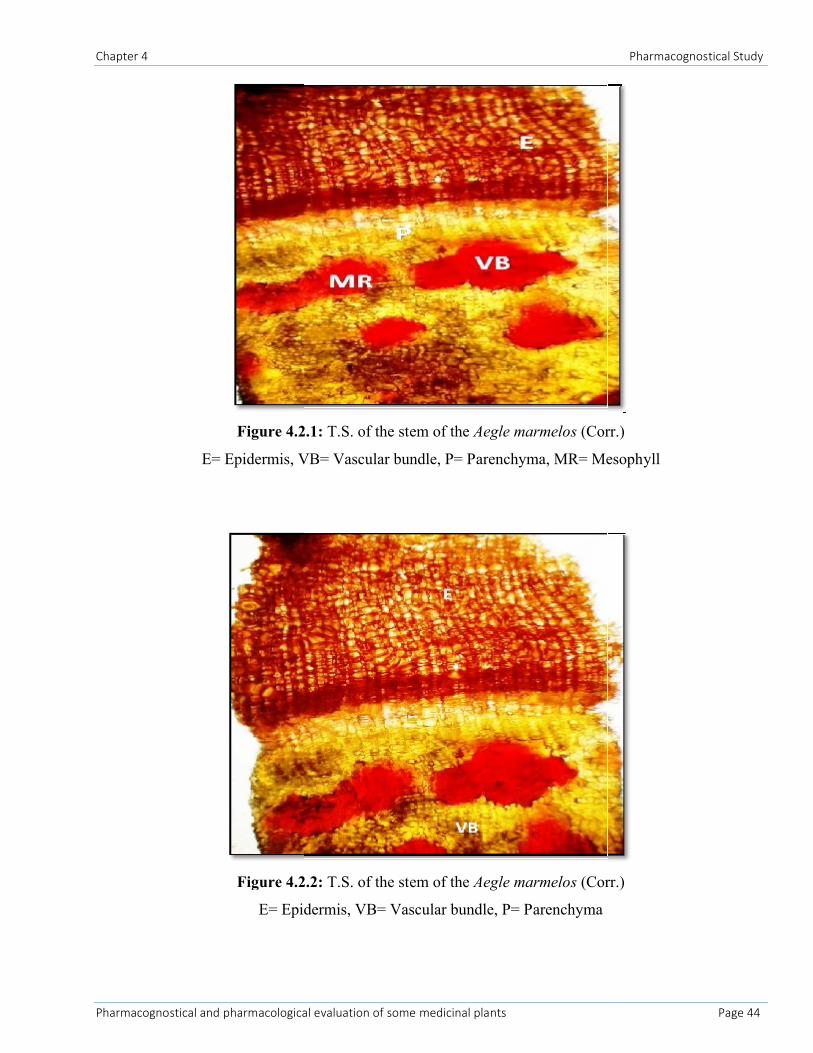
4.2.7.2 Anatomy of Aegle marmelos (Corr.) stem
A thin section of the aegle marmelos (Corr.) stem showed small, thick-walled sclerenchyma on the
inside of the epidermis. The epidermis layer consists of the single layer of living cells, thickened and
covered with waterproof layer called as the cuticle. The layers of the epidermis are closely packed. The
layer of the epidermis followed by large thin-walled parenchyma cells which having the intracellular sir
spaces. Just below the epidermis there are 2-3 layer of collenchymatous cells with thickened cell walls
present. Blow the collenchymas cells are a few layers of parenchyma present with intracellular space
(figure 4.2.1). The vascular bundles are situated in a ring on the inside of the plant. The t.s. of the stem
contain the xylem, phloem and cambium. The xylem found in the center part of the vascular bundle and
phloem found in the outside of the vascular bundle. The cambium separates the xylem and phloem.
Xylem of vessels with simple end-walls, partially developed. The smaller vessels constituting the
protoxylem and the bigger ones constituting the metaxylem lie away from the center (figure 4.2.2). The
protoxylem consists of annular, spiral and scalariform vessels, and the metaxylem of reticulate and
pitted vessels. Cambium consists of 2-3 layers of thin walled and rectangular, and is arranged in radial
rows. Fewer layers of fairly big polygonal or readily elongated parenchymatous cell packed with
yellow-brown masses (pigments).
4.2.7.3 Proximate analysis
4.2.7.3.1 Ash value
The ash value such as total ash, acid-insoluble and water soluble ash of the stem of Aegle marmelos
(Corr.), has been reported in Table 4.2.1. Here the value obtained from the stem of Aegle
marmelos (Corr.) is around 12.5% as total ash. The acid insoluble ash determines 1.75% present in the
drug material and water soluble ash 2.76% present in the plant material. The ash of the any organic
Chapter 4 Pharmacognostical Study
Pharmacognostical and pharmacological evaluation of some medicinal plants Page 42
material is composed of their non-volatile inorganic components. Controlled incineration of crude drugs
completely removes the organic material in the form of CO2 leaving behind an inorganic components in
ash residue. Ash determines the inorganic material such as carbonate, silicate oxalates and phosphates.
With the help of the ash value we can easily detect the adulteration on the crude material.
4.2.7.3.2 Extractive value
The ethanol and water soluble extractive value of the stem of Aegle marmelos (Corr.) is 32.25% and
14.32% respectively (table 4.2.1). The extraction of any crude drug with particular solvent yields a
solution containing different phyto-constituent present in the plant material. The composition of these
phyto-constituents in that particular solvent depends upon the nature of the drug and solvent used. The
use of a single solvent can be the means of providing preliminary information on the quality of a
particular drug sample. The extractive value of the crude drug determines the quality as well as the
purity of the drug material.
4.2.7.3.3 Loss on drying
The values of loss on drying of stem powder of Aegle marmelos (Corr.) is reported in table 4.2.1. The
loss on the drying of the stem of aegle marmelos (Corr.) was 3.25%. The result suggests that the plant
material containing the considerable amount of the moisture constantly present in the plant material. The
present of the moisture content in the plant material should be determined and also be controlled to
make the solution of definite strength. The moisture content of the plant material should be less in the
plant material due to prevent the chemical changes and microbial contamination. The main objective to
determine the moisture content in the fresh plant material is fix their constituents, i.e. to check
enzymatic or hydrolytic reactions that might alter the chemical composition of the crude drug and to
reduce their weight and quality. The insufficient drying of the plant material favors the spoilage of the
material by mould and bacteria and enzymatic destructions. Therefore, in general, drying should be
accomplished as rapidly as is possible with good practices.
Chapter 4 Pharmacognostical Study
Pharmacognostical and pharmacological evaluation of some medicinal plants Page 43
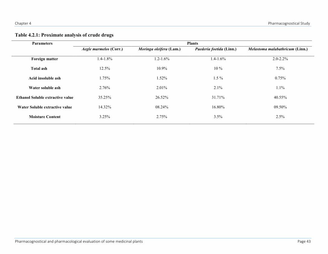
Table 4.2.1: Proximate analysis of crude drugs
Parameters PlantsAegle marmelos (Corr.) Moringa oleifera (Lam.) Paederia foetida (Linn.) Melastoma malabathricum (Linn.)
Foreign matter 1.4-1.8% 1.2-1.6% 1.4-1.6% 2.0-2.2%
Total ash 12.5% 10.9% 10 % 7.5%
Acid insoluble ash 1.75% 1.52% 1.5 % 0.75%
Water soluble ash 2.76% 2.01% 2.1% 1.1%
Ethanol Soluble extractive value 35.25% 26.52% 31.71% 40.55%
Water Soluble extractive value 14.32% 08.24% 16.80% 09.50%
Moisture Content 3.25% 2.75% 3.5% 2.5%
Chapter 4
Pharmacognostical and pharmacological evaluation of some medicinal plants
Figure 4.2.1:
E= Epidermis, VB= Vascular bundle, P= Parenchyma
Figure 4.2.2:
E= Epidermis, VB= Vascular bundle, P= Parenchyma
Chapter 4
harmacological evaluation of some medicinal plants
Figure 4.2.1: T.S. of the stem of the Aegle marmelos (Corr.
E= Epidermis, VB= Vascular bundle, P= Parenchyma, MR= Mesophyll
Figure 4.2.2: T.S. of the stem of the Aegle marmelos (Corr.
E= Epidermis, VB= Vascular bundle, P= Parenchyma
Pharmacognostical Study
Page 44
Corr.)
, MR= Mesophyll
Corr.)
Chapter 4 Pharmacognostical Study
Pharmacognostical and pharmacological evaluation of some medicinal plants Page 45
4.3 Pharmacognostical studies of Moringa oleifera (Lam.)
4.3.1 Material and method
Material and method mention in the section 4.2
4.3.2 Result and discussion
4.3.2.1 Morphological characters
Moringa oleifera (Lam.) is a fast growing, perennial tree with an average height of 7-12 m. The stem of
the Moringa oleifera (Lam.) straight. The branches of the plant grow disorganized manner and the cover
is umbrella shaped. The leaves of the plant are alternate, twice or thrice pinnate leaves grow mostly at
the tips of the branches. The leaves of the plant are grayish downy when young, 20-70 cm long with
long petiole with 8-10 pairs of pinnae, each bearing two pairs of opposite, elliptic or obovate leaflets and
one at the apex (Morton, 1991). The flowers of the plant are cream and yellow-dotted color and having
pleasantly fragrant. The fruit of the plant is three lobed pods 20-60 cm in length and hang down from the
branches. The fruit of the plant contain 12-35 seeds and it open into 3 parts (Figure 2.2).
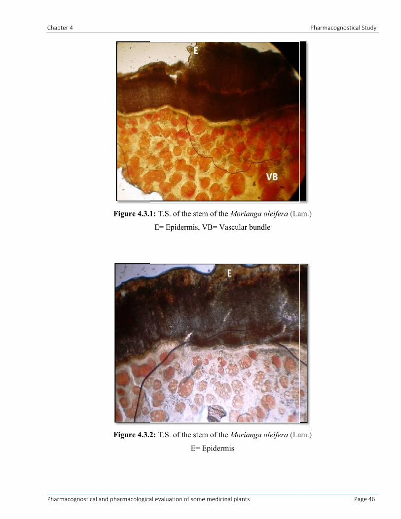
4.3.2.2 Anatomy of Moringa oleifera (Lam.) Stem
A thin section of the Moringa oleifera (Lam.) stem showed the small thicked walled sclerenchyma on
the inside of the epidermis section. Epidermis layer is closely packed and consists of the single layer of
the thickened and covered waterproof layer of the cuticle (figure 4.3.1). The layer of the collenchyma
found below the epidermis layer. Below the collenchyma layer there is 4-5 layer of the parenchyma
found in intracellular space. The vascular bundle found on the inside of the ring. The ts of the plant
contain the xylem, phloem and cambium. The xylem found in the center part and phloem found in the
outside in the vascular bundle. The cambium found in the separation part of the xylem and phloem.
Xylem of vessels with simple end-walls, partially developed. The smaller vessels constituting the
protoxylem and the bigger ones constituting the metaxylem lie away from the center (figure 4.3.2). The
protoxylem consists of annular, spiral and scalariform vessels, and the metaxylem of reticulate and
pitted vessels. Cambium consists of 2-3 layers of thin walled and rectangular, and is arranged in radial
rows. Fewer layers of fairly big polygonal or readily elongated parenchymatous cell packed with
yellow-brown masses (pigments).
Chapter 4
Pharmacognostical and pharmacological evaluation of some medicinal plants
Figure 4.3.1:
Figure 4.3.2:
Chapter 4
harmacological evaluation of some medicinal plants
Figure 4.3.1: T.S. of the stem of the Morianga oleifera (Lam.)
E= Epidermis, VB= Vascular bundle
Figure 4.3.2: T.S. of the stem of the Morianga oleifera (Lam.)
E= Epidermis
Pharmacognostical Study
Page 46
Lam.)
Lam.)
Chapter 4 Pharmacognostical Study
Pharmacognostical and pharmacological evaluation of some medicinal plants Page 47
4.3.2.3 Proximate analysis
4.3.2.3.1 Ash values
The ash value such as total ash, acid-insoluble and water soluble ash of the stem of Moringa oleifera
(Lam.), has been reported in table 4.2.1. Here the ash value obtained from the stem of Moringa
oleifera Lam is around 10.9% as total ash, acid insoluble ash 1.52% and water soluble 2.01% present in
the drug material (table 4.2.1). The ash of the any organic material is composed of their non-volatile
inorganic components. Controlled incineration of crude drugs completely removes the organic material
in the form of CO2 leaving behind an inorganic components in ash residue. Ash determines the inorganic
material such as carbonate, silicate oxalates and phosphates (Kumar V et al, 2009; Kumar V et al, 2011).
4.3.2.3.2 Extractive value
The extractive value of the crude drug determines the quality as well as the purity of the drug material.
The ethanol and water soluble extractive value of the stem of moringa oleifera (Lam.) is 26.52% and
8.24% respectively (table 4.2.1). The extraction of any crude drug with particular solvent yields a
solution containing different phyto-constituent present in the plant material. The composition of these
phyto-constituents in that particular solvent depends upon the nature of the drug and solvent used. The
use of a single solvent can be the means of providing preliminary information on the quality of a
particular drug sample (Kumar V et al, 2009; Kumar V et al, 2011).
4.3.2.3.3 Loss on drying
The values of moisture content present in the stem powder of moringa oleifera (Lam.) were 2.75%
(table 4.2.1). The present of the moisture content in the plant material should be determined and also be
controlled to make the solution of definite strength. The main objective to determine the moisture
content in the fresh plant material is fix their constituents, i.e. to check enzymatic or hydrolytic reactions
that might alter the chemical composition of the crude drug and to reduce their weight and quality. The
moisture content of the plant material should be less in the plant material due to prevent the chemical
changes and microbial contamination. The insufficient drying of the plant material favors the spoilage of
the material by mould and bacteria and enzymatic destructions. Therefore, in general, drying should be
accomplished as rapidly as is possible with good practices (Kumar et al., 2009; Kumar et al., 2011). The
result suggests that the plant material containing the considerable amount of the moisture constantly
present in the plant material.
Chapter 4 Pharmacognostical Study
Pharmacognostical and pharmacological evaluation of some medicinal plants Page 48
4.4 Pharmacognostical studies of Paederia foetida (Linn.)
4.4.1 Material and method
Material and method mention in the section 4.2
4.4.2 Result and Discussion
4.4.2.1 Morphological characters
Paederia foetida (Linn.) is a slender, perennial herb. The branches of the plant are stinking and twining
about 1.5-7 m long. The new stem of the plant is purplish or reddish brown, almost hairless to densely
hairy and older stem is yellowish brown to grayish in color with smooth and shiny surface. The leaves of
the plant about 2-21 cm long and 0.7-9 cm width with simple, broadly egg-shaped surface. The base of
the leaves is heart shaped, rounded or sometimes hastate and the apex of the leaves is acute to
acuminate. The flower of the plant is pinkish to white color, bisexual, usually 5- merous and the petal of
the flower is cylindrical to bell shaped. The fruit of the plant is a drupe at 4-6 mm in diameter with thin
dry wall (Figure 2.3).
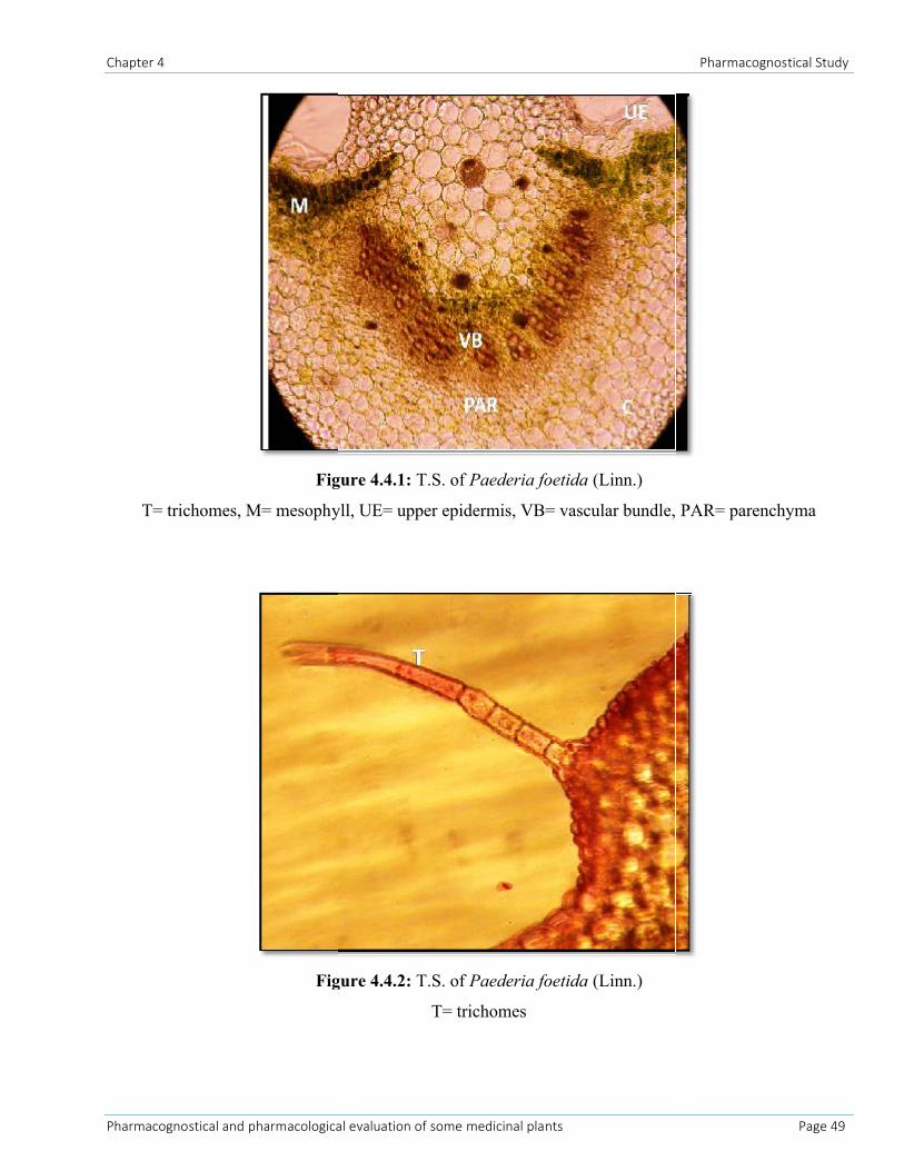
4.4.2.2 Anatomy of leaf
A thin section of the leaves of the paederia foetida (Linn.) is particularly dorsi-ventral with prominent
midrib and lamina (figure 4.4.1). The microscopy of the leaves of the paederia foetida (Linn.) showed
the 3-4 layers of the lower and the upper epidermis covered by the uniseriate straight trichomes (figure
4.4.2). The mesophyll composed of single layered palisade cells and 3-4 layered spongy tissue. The
epidermis composed of the 2-5 layer of the collenchyma towards the upper and lower side and the rest of
the part covered by the parenchyma. The leaves contain the 3-4 layer of the collenchyma. The lamina
part of the leaves showed the dorsiventral structure, epidermis single layered covered externally with
striated cuticles. The center part of the leave microscopy contains the crescent shaped vascular bundle
consisting usual elements with xylem (lignified cells, present at ventral surface) towards upper side and
the lower side phloem (non lignified cells present at dorsal surface) are present (figure 4.4.1). Other cell
content such as starch grain, oil globules, calcium oxalate are found near the vascular bundle.
Chapter 4
Pharmacognostical and pharmacological evaluation of some medicinal plants
Figure 4.4.1:
T= trichomes, M= mesophyll, UE= upper epidermis, VB= vascular bundle, PAR= parenchyma
Figure 4.4.2:
Chapter 4
harmacological evaluation of some medicinal plants
Figure 4.4.1: T.S. of Paederia foetida (Linn.)
T= trichomes, M= mesophyll, UE= upper epidermis, VB= vascular bundle, PAR= parenchyma
Figure 4.4.2: T.S. of Paederia foetida (Linn.)
T= trichomes
Pharmacognostical Study
Page 49
T= trichomes, M= mesophyll, UE= upper epidermis, VB= vascular bundle, PAR= parenchyma
Chapter 4 Pharmacognostical Study
Pharmacognostical and pharmacological evaluation of some medicinal plants Page 50
4.4.2.3 Proximate analysis
4.4.2.3.1 Ash values
The ash value obtained from the leaves of paederia foetida (Linn.) is around 10% as total ash,
acid insoluble ash 1.5% and water soluble 2.1% present in the drug material (table 4.3.1). The ash value
of the leaves of the paederia foetida (Linn.) is around 10% as total ash. The ash of the any organic
material is composed of their non-volatile inorganic components. Controlled incineration of crude drugs
completely removes the organic material in the form of CO2 leaving behind an inorganic components in
ash residue. Ash determines the inorganic material such as carbonate, silicate oxalates and phosphates
(Kumar et al., 2009; Kumar et al., 2011).
4.4.2.3.2 Extractive value
The ethanol and water soluble extractive value of the leaves of Paederia foetida (Linn.) is 26.52% and
8.24% respectively (table 4.3.1). The extractive value of the crude drug determines the quality as well as
the purity of the drug material. The extraction of any crude drug with particular solvent yields a solution
containing different phyto-constituent present in the plant material. The composition of these phyto-
constituents in that particular solvent depends upon the nature of the drug and solvent used. The use of a
single solvent can be the means of providing preliminary information on the quality of a particular drug
sample (Kumar et al., 2009; Kumar et al., 2011).
4.4.2.3.3 Loss on drying
The values of moisture content present in the leaf powder of Paederia foetida (Linn.) were 3.5% (table
4.3.1). The result suggests that the plant material containing the considerable amount of the moisture
constantly present in the plant material. The present of the moisture content in the plant material should
be determined and also be controlled to make the solution of definite strength. The main objective to
determine the moisture content in the fresh plant material is fix their constituents, i.e. to check
enzymatic or hydrolytic reactions that might alter the chemical composition of the crude drug and to
reduce their weight and quality. The moisture content of the plant material should be less in the plant
material due to prevent the chemical changes and microbial contamination. The insufficient drying of
the plant material favors the spoilage of the material by mould and bacteria and enzymatic destructions.
Therefore, in general, drying should be accomplished as rapidly as is possible with good practices
(Kumar et al., 2009; Kumar et al., 2011).
Chapter 4 Pharmacognostical Study
Pharmacognostical and pharmacological evaluation of some medicinal plants Page 51
4.5 Pharmacognostical studies of Melastoma malabathricum (Linn.)
4.5.1 Material and method
Material and method mention in the section 4.2
4.5.2 Result and discussion
4.5.2.1 Morphological characters
Melastoma malabathricum (Linn.) plant stems having the 4-sided to substrate, generally bristly, covered
with small rough scales, reddish in color. Young branches, petioles and nerve of the leaves beneath
clothed with apprised flat lanceolate acuminate paleaceous hairs. The leaves of the m. malabathricum
(Linn.) approx 4-14x1.7-3.5 cm an axially densely string, puberulous, adaxially densely strigose,
secondary veins 2-3 on each side of the mid - vein and the petiole is approx 0.5-1.9 cm in size (Joffry
SM et al, 2012). The flower of the m. malabathricum (Linn.) short lived and grows in 5-10 clusters and
having 5 petals. The flower having 10 stamens of two different kinds: 5 large one with yellow filaments
and another 5 smaller with purple straight filaments and yellow anthers (Zakaria and Mohd, 1994). The
seeds of the m. malabathricum (Linn.) are dimorphic with or without embryos. Fertile seed folded or
spiral, triangular to d-shaped in outline, 0.45-0.8 mm long, 0.35-0.6 mm wide, 0.17-0.3 mm thick, with
light yellow and dark cream colored taste. But the non fertile seed smaller in size, thick, appears
collapsed, dented or wrinkled, with completely reddish or black text in color (Koay, 2008).
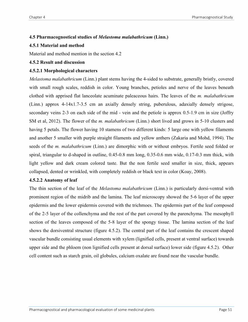
4.5.2.2 Anatomy of leaf
The thin section of the leaf of the Melastoma malabathricum (Linn.) is particularly dorsi-ventral with
prominent region of the midrib and the lamina. The leaf microscopy showed the 5-6 layer of the upper
epidermis and the lower epidermis covered with the trichmoes. The epidermis part of the leaf composed
of the 2-5 layer of the collenchyma and the rest of the part covered by the parenchyma. The mesophyll
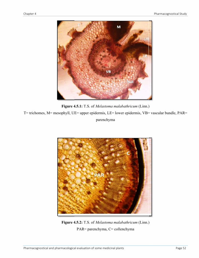
section of the leaves composed of the 5-8 layer of the spongy tissue. The lamina section of the leaf
shows the dorsiventral structure (figure 4.5.2). The central part of the leaf contains the crescent shaped
vascular bundle consisting usual elements with xylem (lignified cells, present at ventral surface) towards
upper side and the phloem (non lignified cells present at dorsal surface) lower side (figure 4.5.2). Other
cell content such as starch grain, oil globules, calcium oxalate are found near the vascular bundle.
Chapter 4
Pharmacognostical and pharmacological evaluation of some medicinal plants
Figure 4.5.1:
T= trichomes, M= mesophyll, UE= upper epidermis, LE= lower epidermis, VB= vascular bundle, PAR=
Figure 4.5.2:
Chapter 4
harmacological evaluation of some medicinal plants
Figure 4.5.1: T.S. of Melastoma malabathricum (Linn.)
T= trichomes, M= mesophyll, UE= upper epidermis, LE= lower epidermis, VB= vascular bundle, PAR=
parenchyma
Figure 4.5.2: T.S. of Melastoma malabathricum (Linn.)
PAR= parenchyma, C= collenchyma
Pharmacognostical Study
Page 52
Linn.)
T= trichomes, M= mesophyll, UE= upper epidermis, LE= lower epidermis, VB= vascular bundle, PAR=
Linn.)
Chapter 4 Pharmacognostical Study
Pharmacognostical and pharmacological evaluation of some medicinal plants Page 53
4.5.2.3 Proximate analysis
4.5.2.3.1 Ash values
The ash value of the leaves of Melastoma malabathricum (Linn.) is around 7.5% as total ash, acid
insoluble ash 0.75% and water soluble 1.1% present in the drug material (table 4.3.1). The ash of the any
organic material is composed of their non-volatile inorganic components. Controlled incineration of
crude drugs completely removes the organic material in the form of CO2 leaving behind an inorganic
components in ash residue. Ash determines the inorganic material such as carbonate, silicate oxalates
and phosphates (Kumar et al., 2009; Kumar et al., 2011).
4.5.2.3.2 Extractive value
The ethanol and water soluble extractive value of the leaves of melastoma malabathricum (Linn.) is
40.55% and 9.50% respectively (table 4.3.1). The extractive value of the crude drug determines the
quality as well as the purity of the drug material. The extraction of any crude drug with particular
solvent yields a solution containing different phyto-constituent present in the plant material. The
composition of these phyto-constituents in that particular solvent depends upon the nature of the drug
and solvent used. The use of a single solvent can be the means of providing preliminary information on
the quality of a particular drug sample (Kumar et al., 2009; Kumar et al., 2011).
4.5.2.3.3 Loss on drying
The values of moisture content present in the leaves of melastoma malabathricum (Linn.) were 2.5%
(table 4.3.1). The result suggests that the plant material containing the considerable amount of the
moisture constantly present in the plant material. The present of the moisture content in the plant
material should be determined and also be controlled to make the solution of definite strength. The main
objective to determine the moisture content in the fresh plant material is fix their constituents, i.e. to
check enzymatic or hydrolytic reactions that might alter the chemical composition of the crude drug and
to reduce their weight and quality. The moisture content of the plant material should be less in the plant
material due to prevent the chemical changes and microbial contamination. The insufficient drying of
the plant material favors the spoilage of the material by mould and bacteria and enzymatic destructions.
Therefore, in general, drying should be accomplished as rapidly as is possible with good practices
(Kumar et al., 2009; Kumar et al., 2011).